

Protecting
Pets, People & Planet
Join a group of veterinarians leveraging the latest technologies to deliver excellent care to their patients while being a responsible and positive force for their local and global communities.
100% secure. We do not share your information


Recent Articles
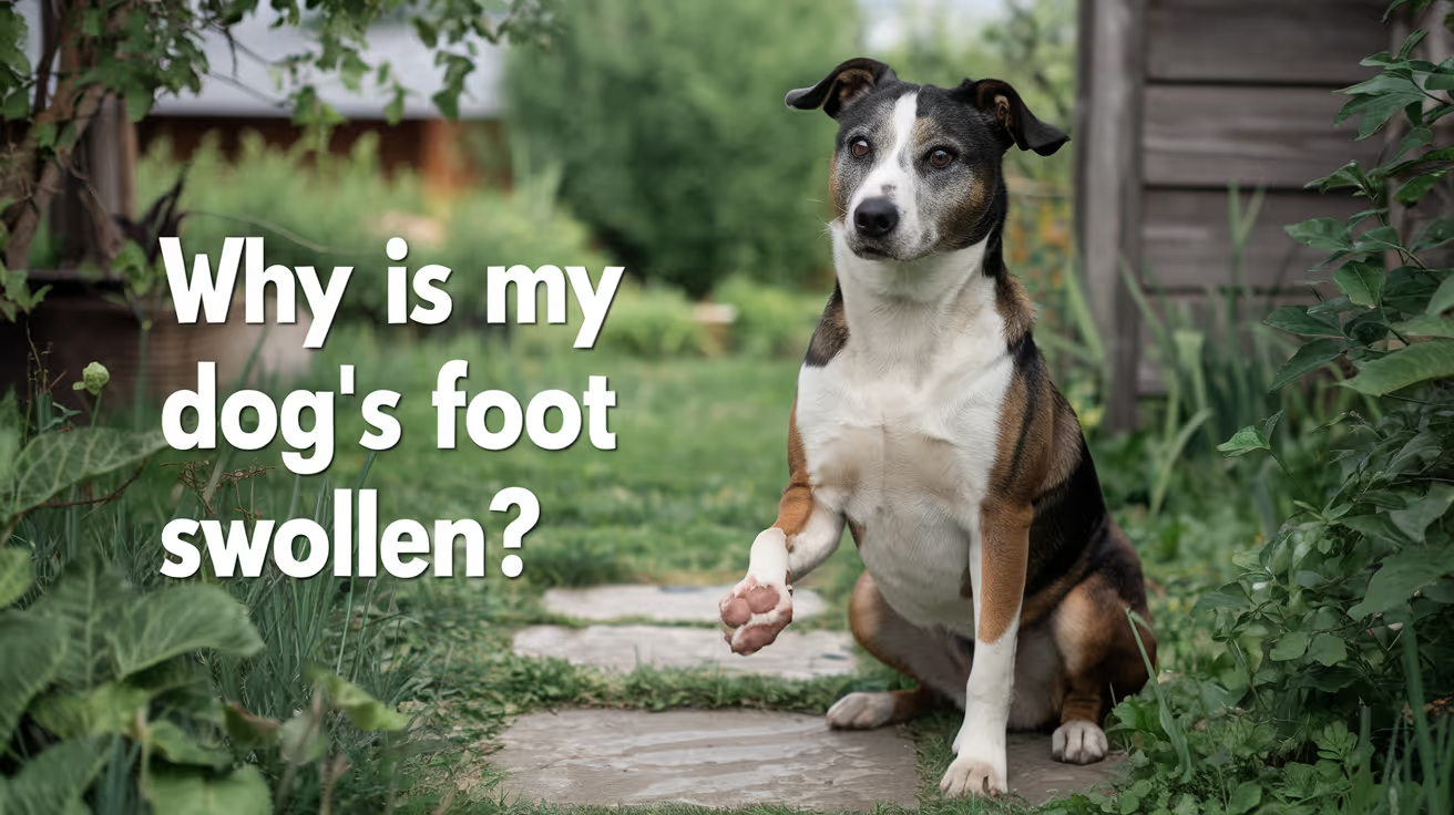
Why Is My Dog's Foot Swollen?
Discover why your dog's foot is swollen, common causes, treatments, and when to see a vet for proper care.
A swollen dog foot is one of the most common reasons owners call their vet. It can be as simple as a bee sting or as serious as a deep infection or bone tumor. The location, speed of onset, and other symptoms all help narrow down the cause.
This guide covers every common cause, how to assess severity at home, and the exact signs that mean your dog needs same-day care.
Quick answer: A swollen dog foot usually has a local cause: injury, insect sting, foreign object, or infection. Warm, increasing, or discharging swelling needs same-day vet care. Multiple swollen paws suggest allergies.
Key takeaways
- Single-paw swelling almost always has a local cause: injury, sting, foreign body, or infection
- Interdigital furunculosis (infected hair follicle cysts between toes) is among the most misidentified causes of recurring paw swelling
- Foreign objects like foxtails and glass splinters can migrate deep into tissue if left untreated
- Pododermatitis is the clinical term for inflamed paw skin; allergies are the most common underlying driver
- Swelling spreading up the leg or paired with face or neck swelling is an emergency requiring immediate care
- Most mild swelling from a sting or minor sprain improves within 24 to 48 hours with rest and monitoring
How to assess your dog's swollen foot before calling the vet
Before looking up causes, do a quick home assessment. This takes two minutes and tells you how urgent the situation is.
Step 1: Which paw?One paw = likely local cause. All four paws = likely allergic or systemic.
Step 2: Feel for heat.Warm or hot tissue means active inflammation or infection.
Step 3: Look between every toe.Redness, swelling between the digits, or visible nodules between the toes points to interdigital furunculosis or a foreign body.
Step 4: Check the pads.Burns appear as red, blistered, or peeling pad tissue. Cuts and punctures are often visible on the pad surface.
Step 5: Look at each nail.A broken nail or swollen nail bed causes localized pain and swelling at the toe tip.
VCA Animal Hospitals: "A deeper infection, like an abscess, will appear as a warm, soft to mildly firm swelling under the skin."
8 common causes of a swollen foot in dogs
1. Injury or trauma
Sprains, cuts, fractured toes, torn nails, and pad burns are the most frequent causes of sudden single-paw swelling. Dogs running on rough, hot, or icy terrain are especially prone.
PetMD notes that pad burns are particularly common in summer: check the pads after any walk on hot pavement. If your palm cannot hold against the pavement for five seconds, it is too hot for your dog's feet.
Signs: sudden lameness after activity, localized swelling, visible wound or bruising.
2. Foreign object
Thorns, glass, foxtails, and grass awns lodge between the toes or penetrate the pad. The body mounts an inflammatory response around the object, causing swelling that worsens over days.
SpectrumCare: "Foxtails and grass awns can lodge between the toes or in the pad and trigger pain, swelling, and infection."
Foxtails are particularly dangerous because they are barbed and can migrate deeper into tissue over days, eventually requiring surgical removal. If you cannot see and safely remove the object, do not probe. See a vet.
3. Insect sting or bite
Bees, wasps, fire ants, and spiders cause rapid single-paw swelling that appears within minutes of the sting. The paw may look puffy and your dog may lick or hold it up.
Dyer Animal Clinic advises watching for anaphylaxis signs: hives, difficulty breathing, excessive swelling spreading beyond the paw. These require emergency care immediately.
Localized sting swelling that stays in the paw and is not worsening can be monitored at home for 24 to 48 hours.
4. Infection: bacterial and fungal
Bacterial or fungal infections enter through cuts, puncture wounds, or damaged skin. Infected paws are typically warm, red, swollen, and often have an odor or discharge. VCA lists the visual signs: "Skin infections on the feet may result in red, moist lesions between the toes."
Pododermatitis (inflammation of the paw skin) is the umbrella clinical term. Common causes include:
- Secondary bacterial infection from chronic licking
- Yeast overgrowth in skin folds between toes
- Fungal infections (ringworm can affect paws)
- Demodectic mange (mite overgrowth)
Pododermatitis does not resolve without treatment. Antibiotics, antifungal medication, or both are typically required.
5. Interdigital furunculosis
This is one of the most commonly missed causes of recurring paw swelling. Interdigital furunculosis occurs when hair follicles between the toes become infected, forming painful reddish-purple nodules that may rupture and drain.
AKC notes that the condition is especially prevalent in short-coated, heavyset breeds: Bulldogs, Labrador Retrievers, and Chinese Shar-Pei are among the most commonly affected. Chronic licking due to allergies is a major driver.
Signs: swollen, painful nodules between the toes, draining tracts, recurrent swelling in the same location.
For how furuncles between the toes connect to abscess formation, see abscess as a cause of limb swelling.
6. Allergic reaction and pododermatitis
Environmental allergens (pollen, grass, lawn chemicals, road salt) and food allergies frequently cause paw inflammation. Dogs with allergies lick their feet chronically, creating secondary infection on top of the allergic reaction.
Wakefield Pet Vet: "Allergic reactions typically cause itching, redness, and sometimes blistering between the toes or on paw pads."
Allergic paw swelling usually affects multiple paws. It tends to recur seasonally or after contact with the trigger substance. Dogs with seasonal allergies often have their worst paw symptoms in spring and fall.
7. Bursitis
Bursae are small fluid-filled sacs that cushion joints. Repeated pressure on bony prominences, especially the elbow and hock, can cause bursitis. Affected joints may appear as soft, fluctuant swellings near a joint.
For how bursitis specifically produces foot and joint swelling, see bursitis as a common cause of foot swelling.
8. Cysts, tumors, and nail bed disease
Cysts, mast cell tumors, and subungual (under-nail) tumors can all present as localized swelling on or near the foot. Petcube notes that toenail tumors are more prevalent in large black-coated breeds such as Standard Poodles, Gordon Setters, and Schnauzers.
A lump that is growing, firm, or pigmented should always be evaluated by a vet rather than monitored at home.
For an overview of lumps that cause swelling on the legs, see lumps that can cause swelling.
Severity triage: what to do right now
Home care for mild cases
For minor swelling without infection signs:
- Keep the dog calm and limit walking
- Soak the paw in warm (not hot) water with Epsom salts for 10 minutes. PetMD recommends this as an excellent short-term measure regardless of cause.
- Gently clean any visible wound with mild soap and warm water
- Apply a cool damp cloth for 10 to 15 minutes to reduce swelling from sprains or stings
- Do not apply antibiotic ointments without vet guidance some formulations are toxic to dogs if licked
For pressure-related paw and leg swelling in dogs that rest in one position for extended periods, see pressure-related swelling in dogs.
Frequently asked questions
How do I treat my dog's swollen paw at home?
For mild swelling with no wound or discharge, rest the dog and soak the paw in warm Epsom salt water for 10 minutes. Check carefully for a foreign object or insect stinger. Do not use human antibiotic creams without vet guidance. If swelling has not reduced within 24 to 48 hours or is getting worse, contact your vet.
When should I be worried about my dog's swollen paw?
Be concerned immediately if the swelling is warm, increasing, or has discharge or odor. Call the vet the same day if your dog refuses to bear weight, if the swelling is spreading up the leg, or if there is any sign of an allergic reaction such as facial swelling or breathing difficulty.
What can I give my dog for a swollen paw?
Do not give human pain medications (ibuprofen, acetaminophen, aspirin) to dogs; many are toxic. For minor swelling, warm Epsom salt soaks are safe and effective for short-term relief. Your vet can prescribe appropriate anti-inflammatory medication if needed after examining the paw.
My dog's foot was normal this morning and is swollen now. What happened?
Sudden single-paw swelling most often means an insect sting, a foreign object picked up during activity, or a minor sprain. Check each toe carefully for a stinger, thorn, or visible wound. If swelling is mild and localized, monitor for 24 hours. If increasing or painful, call the vet the same day.
Can swelling in a dog's foot go away on its own?
Minor swelling from a bee sting or small sprain typically resolves within 24 to 48 hours with rest. Infections, foreign objects, interdigital furunculosis, and bursitis do not resolve without treatment. If swelling has not improved within 48 hours, veterinary assessment is needed.
My dog's foot smells bad and is swollen. Is that serious?
Yes. Odor from a swollen paw strongly indicates infection. A foul smell means bacteria are actively present. Common sources include interdigital furunculosis, a nail bed infection, or an abscess. This requires prompt veterinary treatment and should not be left to resolve on its own.
Resources
- PetMD. Dogs Swollen Paws: Causes and Treatments. petmd.com
- VCA Animal Hospitals. First Aid for Limping Dogs. vcahospitals.com
- AKC. Pododermatitis on Dog Paw: Causes, Symptoms and Treatment. akc.org
- SpectrumCare. Paw Swelling in Dogs. spectrumcare.pet
- Dyer Animal Clinic. Reasons Your Dog's Paw is Swollen. dyeranimalclinic.com
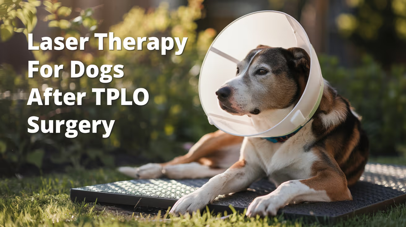
Laser Therapy for Dogs After TPLO Surgery
Learn how laser therapy helps dogs recover faster and with less pain after TPLO surgery for cruciate ligament repair.
Laser therapy also called photobiomodulation (PBMT) or low-level laser therapy (LLLT) is one of the most commonly offered adjunct treatments during TPLO recovery. Many specialist and rehabilitation centres include it routinely.
But the evidence for its benefits is more nuanced than the marketing suggests, and owners deserve an honest picture of what it does and does not reliably achieve.
Quick answer: Laser therapy after TPLO uses specific light wavelengths to reduce inflammation and support tissue healing. Evidence for early pain reduction is moderately supported; evidence for improved radiographic bone healing is weak. It is a safe adjunct but should not replace rehabilitation exercises, pain medication, or activity restriction.
Key takeaways
- Laser therapy reduces postoperative inflammation and may improve gait scores: a TPLO study found better hindlimb function at 8 weeks in treated dogs
- Evidence for improving radiographic bone healing is weak: three controlled studies found no statistically significant difference in healing time
- The 2024 AVMA randomized trial found no significant difference in CRP, weight bearing, pain scores, or SSI rates between PBMT and sham groups
- Sessions typically begin within the first few days of surgery and continue through the rehabilitation phase
- Laser therapy is safe with few contraindications: avoid eyes and active tumour sites; safe over the TPLO incision once closed
- It works best as part of a multimodal plan: exercise therapy and pain medication carry stronger evidence than laser alone
What laser therapy does
Laser therapy for dogs, also known as photobiomodulation, involves using specific wavelengths of light to penetrate tissues and promote cellular regeneration and healing. The laser light stimulates the production of ATP (adenosine triphosphate), enhancing cell repair and growth, reducing inflammation, and increasing blood circulation.
Photobiomodulation therapy has been shown to decrease inflammation, and increase analgesia, vascularization, and tissue healing after musculoskeletal injury or surgery.
The mechanism is photochemical: light energy at specific wavelengths (typically 630 to 980 nm) is absorbed by mitochondria.
This increases ATP production, modulates reactive oxygen species, and influences gene expression related to inflammation and healing.
The effects are local confined to the tissue depth the light reaches.
For post-TPLO use, the targets are: the surgical incision, the osteotomy site in the proximal tibia, and the surrounding periarticular soft tissues.
What the clinical evidence shows
Pain and function
Research following TPLO surgery showed that dogs receiving LLLT had better hindlimb function and gait scores at 8 weeks compared to controls. This is especially valuable in orthopedic recovery, where early weight-bearing can prevent muscle atrophy and joint stiffness.
In a controlled veterinary study, dogs with surgical incisions treated with laser therapy exhibited significantly less inflammatory cell infiltration and tissue necrosis within the first week post-op compared to untreated controls.
The 2024 randomized trial
54 client-owned dogs with CCL rupture undergoing unilateral TPLO surgery were enrolled. The study population was randomly assigned to either a treatment group receiving PBMT (24 dogs) or a control group (30 dogs). PBMT was performed immediately after induction, and at 6 hours, 24 hours, 48 hours, and 8 weeks postoperatively. Evaluation of CRP, pain scores, evidence of SSI, and percentage weight bearing were assessed at all time points.
The trial found the therapy showed promise but no statistically significant difference between groups on any primary outcome measure.
Bone healing
Three studies compared LLLT to a control and concluded that LLLT treatment did not make a significant difference in improving radiographic bone healing. The studies collectively provide weak evidence for this outcome.
This is an important distinction: laser therapy may support soft tissue healing, pain, and early function but it does not appear to accelerate the osteotomy healing visible on radiographs.
When to start and how often
Laser therapy uses focused light energy on the surgical site to support healing.
Most rehabilitation programmes begin laser therapy within the first 1 to 3 days after TPLO surgery, often at the surgical centre before discharge or at the first rehabilitation visit.
Typical post-TPLO laser protocol:
- Frequency: 3 to 5 sessions per week in the first 2 to 3 weeks
- Frequency: 1 to 2 sessions per week from weeks 3 to 8
- Session duration: 5 to 15 minutes depending on the laser system and dosing protocol
- Total sessions: typically 6 to 12 in the first 8-week recovery phase
The protocol varies by laser system, power output, and the individual patient's response.
Realistic expectations
Laser therapy is a useful adjunct in TPLO recovery. It is not a substitute for the treatments with stronger evidence: pain medication, activity restriction, and structured rehabilitation exercises.
Laser therapy could be particularly helpful for dogs with weight-bearing and gait issues while recovering from TPLO surgery after a cruciate injury.
Dogs with significant early swelling, wound sensitivity, or slow initial weight-bearing progress may benefit most. Dogs recovering well with standard multimodal analgesia and rehabilitation may show less measurable difference.
For the bone healing timeline that laser therapy supports during recovery, see TPLO bone healing time in dogs explained.
For the full recovery plan that laser therapy fits into, see 10 essential TPLO recovery tips for pet owners.
For the physical therapy that is the primary evidence-based adjunct, see when to start physical therapy after TPLO surgery.
For swelling management in the recovery period, see how long does swelling last after TPLO surgery.
Frequently asked questions
Is laser therapy safe over the TPLO incision?
Yes, once the incision is closed. Laser therapy is safe over sutured incisions and can be applied at the surgical site from the first post-operative day in most protocols.
Avoid direct application over open wounds or actively infected tissue.
How many laser sessions does a dog need after TPLO?
Typically 6 to 12 sessions across the first 8 weeks.
Start at 3 to 5 sessions per week for the first 2 to 3 weeks, tapering to 1 to 2 per week through the rehabilitation phase.
The exact protocol depends on the laser system and rehabilitation plan.
Does laser therapy replace pain medication after TPLO?
No. Laser therapy is an adjunct to pharmaceutical pain management, not a replacement. Post-TPLO pain management requires NSAIDs, and often gabapentin or other analgesics.
Laser therapy may reduce the pain burden and support earlier mobility but does not provide sufficient analgesia on its own.
Can I do laser therapy at home with a consumer device?
Consumer-grade red light therapy devices exist but operate at lower power densities than veterinary therapeutic lasers. The clinical evidence discussed in this article relates to veterinary-grade PBMT devices.
Home devices may offer some benefit but cannot replicate the dosing of professional equipment. Discuss with your rehabilitation veterinarian before purchasing.
Will laser therapy prevent my dog from needing more medication?
Possibly. If laser therapy reduces post-operative inflammation and pain, some dogs may need lower doses of pain medication or taper off sooner.
This should be guided by your veterinarian based on your dog's individual recovery trajectory.
Resources
- AVMA Journal. Photobiomodulation Therapy in Dogs Undergoing TPLO After Cranial Cruciate Ligament Rupture. avmajournals.avma.org
- Veterinary Evidence. Does LLLT Improve Radiographic Healing for Dogs with CCL Rupture Undergoing TPLO Surgery? veterinaryevidence.org
- AKC. Laser Therapy For Dogs: Uses, Side Effects, and Alternatives. akc.org
- Erchonia. Laser Therapy for Post-Surgical Recovery in Pets. erchonia.com
All Articles

How Much Does TPLO Surgery Cost?
Discover the cost of TPLO surgery, key pricing factors, and hidden fees. Learn what affects the price and explore payment options in this detailed guide
TPLO (Tibial Plateau Leveling Osteotomy) is a special surgery used to treat cranial cruciate ligament (CCL) injuries in dogs. By adjusting the tibial plateau angle, TPLO stabilizes the knee joint, allowing for better weight distribution, reducing pain, and improving mobility. It is considered one of the most effective treatments for CCL tears, especially in large or active dogs.
The cost of TPLO surgery can vary widely, usually ranging from $3,500 to $7,000 (or even more). Several factors affect the price, including:
- the location of the veterinary clinic
- the surgeon's expertise
- preoperative diagnostics
- hospitalization
- anesthesia
- post-surgical care
Additional costs may include medications, follow-up visits, rehabilitation, and potential complications.
Specialist hospitals and board-certified surgeons often charge more due to their advanced training and high success rates. Additionally, regional differences in veterinary care costs can affect pricing, making TPLO surgery more expensive in urban areas than in smaller towns.
Factors That Influence TPLO Surgery Costs
Dog’s Size and Weight
Larger dogs need stronger, more durable implants, which raises material costs. They also require higher doses of anesthesia, longer surgeries, and more monitoring during recovery. The complexity of handling a large dog's knee joint makes the surgery more technically challenging, leading to higher fees.
Smaller dogs, while still needing precise surgery, usually have lower implant costs and reduced medication expenses. Veterinary clinics adjust their pricing based on the dog's weight, as it directly impacts the surgery's difficulty, recovery time, and post-operative care, causing variations in overall TPLO surgery costs.
Geographic Location
TPLO surgery costs can vary greatly depending on the clinic's location. In urban areas, prices are usually higher because of increased overhead costs, like facility expenses, staff salaries, and the demand for specialized services. Rural clinics might offer lower prices, but they may not have access to board-certified surgeons or advanced surgical equipment.
Costs also vary between states and countries, affected by regional economic conditions, veterinary licensing fees, and medical supply costs. Pet owners in areas with a high cost of living should expect to pay more, while those in smaller towns might find more affordable options.
Veterinary Clinic’s Pricing Strategy
The type of veterinary clinic also affects TPLO surgery costs. Corporate-owned clinics often have set prices, which may be higher due to overhead and administrative expenses. Private practices might offer more flexible pricing but can also charge higher rates if they have a strong reputation.
Clinics with high demand, advanced equipment, and top-notch facilities usually charge more. Some hospitals include follow-up care in their pricing, while others list these costs separately, impacting the total amount pet owners need to pay. Choosing between different clinic types can significantly influence the final surgery bill.
Severity of the Injury
The extent of ligament damage directly impacts TPLO surgery costs. A partial tear might need less surgical work and result in lower costs, while a complete rupture usually requires more complex reconstruction.
Additional issues like meniscus tears, osteoarthritis, or joint inflammation make the surgery more difficult and increase post-operative care, raising overall expenses.
In severe cases, longer hospital stays, extra imaging, and specialized rehabilitation may be necessary, further increasing costs. Veterinary surgeons evaluate each dog's condition individually, deciding on the level of intervention needed and adjusting the treatment plan as necessary.
Necessary Implants and Materials Used
The type and quality of implants, plates, and screws used in TPLO surgery can greatly affect costs. High-grade titanium or stainless steel implants are more expensive but offer better durability and a lower risk of complications. Customized implants, made for larger or more active dogs, also increase costs due to their specialized design.
Some clinics might use generic implants to keep costs down, while others choose premium, brand-name materials for better long-term results. The choice of implants impacts not only the price but also post-surgery stability, healing time, and overall success rates.
Surgeon’s Expertise and Qualifications
Board-certified veterinary surgeons usually charge more than general veterinarians because of their specialized training and higher success rates. These experts have a lot of experience in orthopedic procedures, which leads to better surgical results.
While general veterinarians can perform TPLO surgery at a lower cost, they might not have the same level of expertise, which could affect recovery and complication rates.
Some pet owners choose experienced surgeons despite the higher cost, focusing on safety, precision, and long-term joint health for their dogs. Selecting a skilled surgeon often results in faster recovery and fewer post-surgical problems.
Regional Cost Comparison
The cost of TPLO surgery varies significantly by country and location due to differences in veterinary fees, cost of living, and availability of specialized surgeons. Below is a general comparison of TPLO surgery costs in different regions:
| Region | Average Cost Range (Per Knee) | Key Factors Affecting Cost |
|---|---|---|
| United States | $3,500 – $10,000 | Higher costs in major cities like New York and Los Angeles due to demand and overhead; rural clinics may charge less. |
| Canada | $3,500 – $8,000 | Urban centers like Toronto and Vancouver have higher fees; fewer specialists in remote areas may drive up costs. |
| United Kingdom | £3,000 – £6,500 ($3,800 – $8,200) | London and other metropolitan areas charge more; the availability of pet insurance can impact affordability. |
| Australia | AUD 4,000 – AUD 9,000 ($2,600 – $5,900) | Higher in cities like Sydney and Melbourne; regional clinics may offer lower rates but limited specialist availability. |
Urban clinics usually charge more because of higher operating costs and greater demand for specialist services. In contrast, rural clinics often have lower prices but may not have board-certified surgeons.
Additionally, pet insurance can greatly affect out-of-pocket costs, as some policies cover part or all of the TPLO surgery expenses, depending on the region and the plan.
Cost Variations by Dog Size
The cost of TPLO surgery varies based on the size of the dog, mainly because of differences in implant needs, anesthesia dosage, and surgical complexity. Here's a breakdown of approximate costs by weight category:
- Small Dogs (<40 lbs) – Approximately $4,450
Smaller dogs need smaller implants and less anesthesia, making their surgery a bit less complex. However, the cost is similar to larger dogs because specialized skills and equipment are still necessary. - Medium Dogs (40–70 lbs) – Approximately $4,450
Dogs in this weight range usually need standard-sized TPLO plates, keeping costs similar to small dogs. The surgical method and recovery care are also similar. - Large Dogs (70–100 lbs) – Varies Based on Plate Size
Costs increase for larger dogs due to the need for stronger, larger TPLO plates. Some clinics charge extra for custom implants that offer better durability and long-term joint support. - Extra-Large Dogs (>100 lbs) – $4,450 to $5,950
Giant breeds require reinforced implants, more anesthesia, and longer surgery time, which raises the overall cost. Post-surgical care and rehabilitation may also be more intensive, further affecting expenses.
Larger dogs often have higher long-term care costs, including pain management and physical therapy, making post-operative planning crucial for their recovery.
What’s Included in the TPLO Surgery Cost?
The total cost of TPLO surgery usually includes several components to ensure a safe procedure, proper recovery, and post-operative care. Here is a breakdown of what is typically included:
- Pre-Surgical Bloodwork: Before surgery, veterinarians perform blood tests to check the dog’s overall health, making sure they can safely handle anesthesia and recover without issues.
- X-Rays for Diagnosis and Surgical Planning: Preoperative X-rays are needed to confirm the severity of the CCL tear and determine the exact tibial angle for surgical correction. Post-surgical X-rays may also be taken to ensure the implant is properly placed.
- Anesthesia and Surgical Monitoring: TPLO surgery requires general anesthesia, which is carefully given and monitored throughout the procedure. The cost includes oxygen support, IV fluids, and continuous tracking of vital signs to ensure the dog's safety.
- Actual TPLO Surgical Procedure: This includes the orthopedic surgery itself, where the surgeon makes a precise bone cut, repositions the tibial plateau, and secures it with plates and screws.
- Post-Operative Pain Medications: Pain management is crucial for recovery. The cost typically includes anti-inflammatory drugs, antibiotics, and pain relievers to prevent infection and discomfort.
- Follow-Up Visits and Check-Ups: Most clinics include at least one or two post-surgery appointments to monitor healing and assess the dog's progress. Additional visits may be needed if there are complications.
- Physical Therapy or Rehabilitation Sessions (If Included): Some veterinary clinics offer rehabilitation programs like hydrotherapy or therapeutic exercises to help improve recovery. However, not all clinics include these services in the base surgery cost.
Each clinic has its own pricing structure, so it's important to check what is included to avoid unexpected costs during recovery.
Hidden or Unexpected Costs of TPLO Surgery
While TPLO surgery is highly effective, some additional expenses may arise depending on the dog's recovery process and the clinic’s pricing structure.
- Emergency Care for Complications: Although rare, complications such as implant loosening, poor bone healing, or excessive swelling may require extra treatment. Even with lower infection risks using Simini Protect Lavage, issues like implant irritation or allergic reactions might still occur, needing additional veterinary care.
- Additional Pain Management Medications: While most TPLO surgery packages include standard pain medications, some dogs might need stronger or longer-term pain relief, especially if they have pre-existing joint issues or heal slowly.
- Post-Surgery X-Rays for Recovery Monitoring: Some veterinary clinics charge separately for follow-up X-rays at 6-8 weeks post-surgery to confirm proper bone healing. If not included, these can cost $150 to $300 per session.
- Rehabilitation and Therapy (If Not Included in Base Cost): Hydrotherapy, laser therapy, or guided rehabilitation exercises can enhance recovery but may add $50 to $100 per session.
With advanced infection control measures like Simini Protect Lavage, unexpected costs related to post-operative infections are minimized, making TPLO recovery more predictable and reducing financial uncertainties.
Payment and Financing Options for TPLO Surgery
TPLO surgery is a significant financial commitment, but various payment and financing options can help manage the cost. Pet owners should explore different ways to reduce financial strain while ensuring their dog gets the necessary treatment.
- Flexible Payment Plans Offered by Some Clinics: Many veterinary clinics offer in-house payment plans, allowing owners to pay in installments instead of a lump sum. These plans typically require a deposit upfront, with the remaining balance spread over several months.
- Discounts for Upfront Payments: Some clinics provide discounts for paying in full before the surgery. This option can help save money, as interest fees from financing plans are avoided. It's worth asking if a prepayment discount is available.
- Third-Party Financing or Personal Loans: Veterinary financing companies like CareCredit and Scratchpay offer low-interest or interest-free short-term loans for pet medical expenses. Personal loans or credit cards can also be an option, but they may come with higher interest rates.
- Pet Insurance Coverage Considerations: Not all pet insurance plans cover TPLO surgery. Policies that include orthopedic conditions or CCL injuries may reimburse a portion of the cost. Companies like Trupanion, Healthy Paws, and Embrace often cover TPLO surgery, but pre-existing conditions are typically excluded. Pet owners should check waiting periods and coverage limits before proceeding.
Discussing these options with the veterinary clinic can help find the best financial solution for your dog’s surgery.
Additional Considerations Before Committing to TPLO
Before proceeding with TPLO surgery, it’s important to consider potential risks, unexpected costs, and pricing differences between clinics to ensure you are fully prepared for the financial and medical aspects of the procedure.
Risks of Unexpected Complications and Added Cost
While TPLO surgery has a high success rate, complications such as implant failure, delayed bone healing, or improper post-operative care can lead to additional expenses. Some dogs may require extended rehabilitation, extra pain management, or revision surgery, increasing costs beyond the initial estimate.
While infection risks are lower when using Simini Protect Lavage, other factors like poor post-op care or underlying health conditions may still affect recovery.
Comparing Quotes from Different Veterinary Clinics
Prices for TPLO surgery can vary significantly between clinics based on location, surgeon expertise, and included services. It’s recommended to get multiple quotes, ensuring you compare what’s included, such as pre-op tests, X-rays, follow-up visits, and rehabilitation. Some clinics offer payment plans or discounts, making it essential to explore all financial options before committing.
Taking the time to understand potential costs and evaluate different providers can help ensure the best possible care for your dog while managing expenses effectively.
Is TPLO Worth the Cost? Long-Term Financial Benefits
While TPLO surgery is expensive, it is often a cost-effective investment in a dog’s long-term health. Choosing not to proceed with TPLO can lead to chronic pain, progressive arthritis, and long-term medical expenses that may exceed the cost of surgery.
Cost of Not Doing TPLO
Without surgical intervention, a torn cranial cruciate ligament (CCL) leads to joint instability, inflammation, and arthritis progression. Managing these issues requires lifelong pain medications, anti-inflammatory drugs, physical therapy, and joint supplements—costs that add up over time. Many dogs that do not undergo TPLO eventually experience injury in the opposite knee, further increasing medical expenses.
TPLO as an Investment in Mobility and Health
TPLO surgery restores knee stability, reduces pain, and allows for full weight-bearing movement, enabling dogs to return to normal activity levels. Unlike conservative management, TPLO prevents further joint deterioration, reducing the need for ongoing medical intervention.
How TPLO Saves Money in the Long Run
While conservative treatments like bracing, pain management, and therapy may seem cheaper initially, they often require years of continuous care.
TPLO, on the other hand, is a one-time surgical cost that provides a permanent solution, minimizing future expenses and improving a dog’s quality of life.
Read more about:
TPLO Surgery for Older Dogs: Is It a Worthwhile Option?
Post-Surgery Recovery After TPLO in Dogs
Common Myths About TPLO Surgery Explained
Long-Term Outcomes of TPLO Surgery
What is Bilateral TPLO Surgery?
Conclusion
TPLO surgery is a major financial commitment, with costs usually ranging from $3,500 to $10,000 per knee. This price covers pre-surgery diagnostics, anesthesia, the surgery itself, post-operative care, and follow-up visits. However, there may be extra costs for rehabilitation, post-surgery X-rays, and emergency care for any complications.
Before deciding on TPLO, pet owners should think about their dog's size, the severity of the injury, the surgeon's expertise, and their location, as these factors significantly affect the total cost. Comparing quotes from different clinics, checking pet insurance coverage, and looking into financing options can help manage expenses.
Although the initial cost is high, TPLO offers a long-term solution, lowering the risk of arthritis and removing the need for lifelong pain management. In the long run, it is often more cost-effective than conservative treatments. Investing in TPLO means better mobility, pain relief, and an improved quality of life for your dog.
FAQs
How much does TPLO surgery cost on average?
The average cost of TPLO surgery ranges from $3,500 to $10,000 per knee, depending on clinic location, surgeon expertise, diagnostic tests, anesthesia, and post-operative care. Urban clinics and specialty hospitals tend to charge more. Additional costs may arise from follow-up visits, rehabilitation, and pain management, which are sometimes not included in the base price.
How much does it cost to remove a TPLO plate?
The cost of TPLO plate removal varies depending on factors such as the veterinary clinic, location, and the complexity of the procedure. On average, the cost ranges from $800 to $2,500. This typically includes anesthesia, surgical fees, and post-operative care. Plate removal is usually recommended only if complications arise, such as infection, irritation, or persistent pain. Some clinics may offer a lower cost if the removal is performed at the same facility where the TPLO surgery was originally done. Always consult your veterinarian for an accurate estimate based on your dog's specific case.
Why is TPLO surgery so expensive?
TPLO surgery is costly due to its complex nature and specialized equipment. It requires advanced surgical skills, high-quality implants, general anesthesia, and extensive post-operative care. Additionally, board-certified veterinary surgeons charge more due to their expertise and success rates. Factors like pre-surgical diagnostics, follow-up X-rays, and rehabilitation therapy further increase the total cost.
Can pet insurance cover TPLO surgery?
Yes, some pet insurance plans cover TPLO surgery, but coverage depends on the policy terms and pre-existing condition exclusions. Companies like Trupanion, Healthy Paws, and Embrace often reimburse part of the cost if the injury was not diagnosed before the policy started. Owners should check waiting periods, deductibles, and reimbursement limits before relying on insurance.
Are there cheaper alternatives to TPLO surgery?
Alternatives include lateral suture stabilization (Extracapsular Repair) and TTA (Tibial Tuberosity Advancement), which may be cheaper but are less effective for larger, active dogs. Conservative management with medications, weight control, and bracing can temporarily relieve symptoms but does not fully restore joint function and may lead to long-term arthritis.
What are the hidden costs of TPLO?
Hidden costs may include post-surgical X-rays ($150–$300 per session), extended pain management, rehabilitation therapy ($50–$100 per session), and emergency care for complications. While infection risks are lower with Simini Protect Lavage, some dogs may still require additional medications or follow-up procedures, adding to the overall cost.
How can I save money on TPLO surgery?
Pet owners can save money by comparing quotes from different clinics, asking about payment plans, checking pet insurance coverage, and looking for upfront payment discounts. Some veterinary schools offer lower-cost surgeries performed by supervised students. Financing options like CareCredit and Scratchpay also help spread out payments.
Does the cost of TPLO include physical therapy?
Not always. Some veterinary clinics bundle rehabilitation sessions in the total cost, but many charge separately. Physical therapy, including hydrotherapy, laser therapy, and therapeutic exercises, can cost $50 to $100 per session. It is essential to confirm with the clinic whether post-operative rehab is included.
Is TPLO surgery a one-time cost, or will my dog need additional procedures?
TPLO surgery is generally a one-time procedure, but additional costs may arise for post-surgery check-ups, pain management, and rehabilitation. In rare cases, complications like implant failure or infections may require revision surgery, adding to the expenses. However, with proper post-operative care, most dogs recover well without further surgical intervention.
What is TPLO Surgery in Dogs?
What is TPLO in dogs? A surgery to fix CCL injuries, restore mobility, and prevent arthritis. Learn the procedure, benefits, risks, and recovery timeline.
TPLO (tibial plateau leveling osteotomy) is the most widely performed surgery for cranial cruciate ligament (CCL) rupture in dogs.
It is the procedure most commonly recommended by board-certified veterinary surgeons for medium and large dogs, and it has the most extensive published evidence base of any CCL repair technique.
Understanding what TPLO is, why it works, and what recovery involves helps owners make informed decisions and prepare for the process.
Quick answer: TPLO (tibial plateau leveling osteotomy) corrects CCL rupture by changing tibial plateau geometry to eliminate the shear force making the CCL unnecessary. A curved cut is made through the tibia, rotated to 5 degrees, and secured with a locking plate while the bone heals. Success rate is 90 to 95%.
Key takeaways
- TPLO stands for tibial plateau leveling osteotomy: it levels the tibial plateau through a bone cut to stabilize the stifle without relying on the damaged CCL
- TPLO corrects CCL rupture by eliminating the shear force the CCL resists: it changes the tibial plateau geometry so the joint is stable without a CCL
- The procedure achieves a post-operative tibial plateau angle of approximately 5 degrees: VCA confirms the tibial plateau is rotated until appropriately level (ideally 5 degrees), eliminating cranial tibial thrust
- TPLO has a 90 to 95% success rate: RCVS confirms 90 to 95% of dogs regain complete or near-complete function; 93% of owners report satisfaction at 12 months
- Recovery requires 8 to 12 weeks of activity restriction for bone healing; progressive return to full activity follows; muscle recovery takes up to 6 months
- TPLO is the most commonly recommended procedure for CCL rupture by ACVS surgeons in dogs over approximately 15 to 20 kg
The CCL and why it ruptures
The cranial cruciate ligament (CCL) is the equivalent of the human ACL in dogs.
It connects the femur (thigh bone) to the tibia (shin bone) inside the stifle (knee) joint and serves three primary functions:
- Preventing the tibia from sliding forward relative to the femur during weight bearing
- Limiting internal rotation of the tibia
- Preventing hyperextension of the stifle
Unlike most human ACL tears (which are acute traumatic events), CCL rupture in dogs is typically degenerative. The fibers weaken progressively over months to years before the ligament fails completely.
By the time complete rupture occurs, most of the fibers have already been compromised.
This is why the contralateral (opposite) CCL is also at risk -- the same degenerative process typically affects both stifles.
When the CCL ruptures, the tibial plateau's natural slope creates an unconstrained cranial shear force during weight bearing.
The femur slides down the tibial plateau, the tibia shifts forward, and the dog is in pain and unable to use the leg normally.
CCL rupture is one of the most common orthopedic injuries in dogs. High-risk breeds include Labrador Retrievers, Rottweilers, Staffordshire Terriers, Mastiffs, Newfoundlands, and German Shepherds.
Neutered dogs have a higher risk than intact dogs of the same breed.
How TPLO works: the biomechanical principle
The normal canine tibial plateau slopes downward from front to back at approximately 23 to 29 degrees (the tibial plateau angle, or TPA).
When the dog bears weight, this slope creates a cranial shear force -- the tibia tries to slide forward relative to the femur. In a normal stifle, the CCL resists this force.
When the CCL is absent, the force is unopposed and the joint is unstable.
TPLO does not attempt to restore the CCL. Instead, it eliminates the shear force entirely.
By rotating the tibial plateau to approximately 5 degrees, the joint surface becomes level enough that the patellar tendon -- rather than the CCL -- provides the stabilizing force during weight bearing.
The joint is mechanically stable without any ligament. The need for the CCL is eliminated by changing the geometry of the tibial plateau.
Dispomed confirms: TPLO addresses CCL rupture by altering the biomechanics of the stifle. The tibial plateau normally slopes downward; TPLO creates a flat or level plateau, decreasing sliding motion at this joint.
The TPLO procedure: step by step
Pre-operative planning
Before surgery, the dog is sedated and lateral stifle radiographs are taken.
The surgeon measures the TPA from these images and calculates the amount of rotation required to achieve the target post-operative TPA of approximately 5 degrees.
The surgeon also selects the appropriate saw blade radius and implant size based on these measurements and the dog's body weight.
Anesthesia and preparation
The dog is placed under general anesthesia. The surgical site is clipped and prepared with antiseptic solution. IV antibiotics (typically cefazolin) are given before the incision.
Joint inspection and meniscal assessment
The surgeon makes an incision over the medial aspect of the proximal tibia and performs a small arthrotomy (opening into the joint) to inspect the menisci.
Damaged meniscal tissue is removed at this stage.
Chewy confirms: during surgery, the joint is examined and the surgeon might remove damaged tissue before proceeding with the osteotomy.
The osteotomy
Using a specialized oscillating saw blade, the surgeon makes a curved (arcuate) cut through the proximal tibia at the level of the tibial plateau.
The radius of this cut is determined by the pre-operative planning.
Rotation
The cut bone segment (containing the tibial plateau) is rotated backward (caudally) by the calculated amount to achieve the target TPA.
VCA Animal Hospitals confirms: the top section of the tibia is rotated backward until deemed appropriately level -- typically 2 to 14 degrees, with 5 degrees as the ideal angle.
Plate fixation
A TPLO plate (a contoured locking plate) is secured to the tibia across the osteotomy using locking screws. The plate holds the rotated tibial plateau in position while the bone heals.
Closure
The joint capsule, fascia, subcutaneous tissue, and skin are closed in layers. The incision is bandaged.
Success rate and outcomes
RCVS Canine Cruciate Registry confirms: TPLO has a 90 to 95% success rate, with 90 to 95% of dogs regaining complete or near-complete function; 93% of owners reporting satisfaction at 12 months.
A 2013 study found that TPLO patients achieved 93% restoration of limb function at 1 year.
TPLO produces better long-term outcomes than lateral suture stabilization in most studies of large dogs, and similar long-term outcomes to TTA in comparable patient populations.
RCVS confirms: all dogs with CCL rupture are expected to develop at least some osteoarthritis, but this is reduced or delayed in dogs that have had surgical stabilization.
Recovery overview
- Weeks 0 to 2: crate rest, 5-minute bathroom leash walks only, e-collar on at all times
- Weeks 2 to 6: graduated leash walk increase, passive range-of-motion exercises
- Week 6: radiographic assessment of bone healing; activity increase if healing is progressing
- Weeks 8 to 12: final radiographic confirmation of bone healing; activity restrictions lifted if healing is confirmed
- Months 3 to 6: progressive return to full activity; muscle recovery continues
For the full recovery guide, see what to expect after TPLO surgery in dogs. For the surgery comparisons, see TPLO vs CBLO vs TTA: a guide to cruciate surgery options.
For preparation, see preparing for your dog's TPLO surgery. For the signs that TPLO may be needed, see 13 signs your dog may need TPLO surgery.
Frequently asked questions
Is TPLO the only surgery for CCL rupture in dogs?
No. The main alternatives are lateral suture stabilization (extracapsular repair), TTA (tibial tuberosity advancement), and CBLO (CORA-based leveling osteotomy). Lateral suture achieves good outcomes in dogs under approximately 35 pounds.
TTA produces similar long-term outcomes to TPLO in appropriately selected dogs. CBLO is used mainly for immature dogs and revision surgery cases.
Can a dog live without surgery for CCL rupture?
Small dogs (under 10 to 15 kg) sometimes stabilize adequately with conservative management (strict rest, weight management, joint support).
For most dogs over 15 kg, conservative management does not restore normal joint stability and the dog remains in pain with progressive arthritis.
Surgery is strongly recommended for most medium and large dogs.
How long does TPLO surgery take?
The procedure typically takes 1.5 to 2 hours. Bilateral simultaneous TPLO takes 3 to 4 hours.
Does the TPLO plate stay in permanently?
Yes, in most cases. The plate is not routinely removed after the osteotomy heals. It is removed if it causes complications (infection, implant irritation, failure).
What is the cost of TPLO surgery?
TPLO typically costs $3,500 to $6,000 per stifle depending on the facility, surgeon, geographic location, and the dog's size.
This typically includes the surgical procedure, anesthesia, hospitalization, implants, and the immediate post-operative period.
Resources
- RCVS Canine Cruciate Registry. Tibial Plateau Levelling Osteotomy (TPLO). rcvsknowledge.org
- VCA Animal Hospitals. TPLO. vcahospitals.com
- Dispomed. TPLO Surgery in Dogs: Success Rates and Alternatives. dispomed.com
- ACVS. Cranial Cruciate Ligament Disease. acvs.org

When to Refer for Orthopedic Surgery: Identifying Surgical vs. Non-Surgical Cases
Guide for identifying surgical vs. non-surgical veterinary orthopedics, enhancing patient outcomes with timely, appropriate referrals
Orthopedic decisions in dogs come in two layers: is surgery needed, and if so, should it be performed by a general practitioner or a specialist? Getting both questions right is what gives a dog the best outcome.
This guide covers which orthopedic conditions typically require surgery, which are managed conservatively, and when specialist referral is the appropriate recommendation.
Quick answer: Most CCL ruptures, comminuted or articular fractures, severe hip dysplasia, and spinal cord compressions require referral to a board-certified orthopedic surgeon. Conservative management suits mild OA, early hip dysplasia, and stable incomplete fractures in large adult dogs.
Key takeaways
- CCL rupture is the most common orthopedic referral reason: TPLO or TTA required for long-term stifle stability
- Fractures require specialist referral when comminuted, articular, open, or involving toy breed radius and ulna
- Conservative management works for mild presentations of hip dysplasia, elbow dysplasia, and osteoarthritis when functional impairment is limited
- Cases not improving with conservative care after 4 to 6 weeks should trigger a referral conversation
- Cost transparency matters: owners need to understand the financial impact of surgical vs. non-surgical paths
- Teleconsultation with a specialist is available before formal referral when the diagnosis or management path is uncertain
The referral decision framework
Two separate questions must be answered:
1. Does this dog need surgery at all?Severity of pain, degree of functional impairment, rate of deterioration, the dog's age and size, and the owner's capacity for the post-operative recovery commitment all factor in.
2. If surgery is needed, who should perform it?Procedure complexity, required implants, access to specialized equipment, and the surgeon's case volume for this specific procedure determine whether a general practitioner or a DACVS specialist is the appropriate operator.
SustainableVet: "The cost implications of specialist surgery, the timeline for diagnostics, surgery, and recovery, and post-operative care responsibilities should all be discussed with owners before making a referral recommendation."
Conditions that typically require surgical referral
Cranial cruciate ligament (CCL) rupture
The most common reason dogs are referred to veterinary orthopedic surgeons. SustainableVet: "Cranial cruciate ligament (CCL) ruptures cause severe joint instability, leading to secondary osteoarthritis. Surgical stabilization, such as tibial plateau leveling osteotomy (TPLO) or tibial tuberosity advancement (TTA), is recommended for long-term function."
Veterinary Partner (VIN): "TPLO has emerged as the most reliable technique offered by veterinary orthopedic surgeons for the management of CCL disease in dogs."
TPLO and TTA require specialized equipment (radiolucent table, surgical templates, specific implant systems) and significant case volume to perform reliably. Coldwater Animal Hospital: "A torn CCL in dogs requires precise surgical technique, proper post-operative planning, and orthopedic follow-up. When you suspect your dog has a knee injury, you should request a referral to an orthopedic specialist as soon as possible."
SustainableVet: "TPLO for CCL rupture ranges from $3,000 to $6,000 and covers surgery, hospitalization, and post-op care."
Comminuted and articular fractures
SustainableVet (fracture management article): "Signs include joint involvement, multiple bone fractures, open wounds, unstable or comminuted breaks, or no healing progress after a few weeks. If proper imaging or surgical tools are not available, referral ensures better care."
Articular fractures (those entering a joint surface) require anatomical reduction and rigid fixation to prevent joint surface incongruity and secondary arthritis. These are specialist cases.
Open (compound) fractures require urgent management to reduce infection risk often an emergency referral.
Toy breed radius and ulna fractures
SustainableVet: "Toy breeds have a high risk of nonunion, especially in distal radius and ulna fractures. Their small bones have less blood supply, which slows healing. Even simple fractures may not heal with splints alone. Surgery is often recommended early to avoid long-term lameness or permanent damage."
Spinal cord compression
Intervertebral disc disease (IVDD) with neurological deficits paresis or paralysis is a neurosurgical emergency. Lumbosacral disease, spinal instability, and vertebral fractures with cord involvement require specialist management. VCA: "Spinal injuries and herniated discs are problems that are also commonly referred to board-certified surgeons."
Total hip replacement
Total hip replacement (THR) for severe hip dysplasia or femoral head necrosis. VCA: "THR procedures replacement of a dog's diseased hip joints with prosthetic ones cost between $5,000 and $7,000." The procedure requires specialized implant systems and is exclusively a specialist procedure.
Complex reconstructive surgery
Large skin defects, major wound reconstruction after trauma or tumor removal, and rotational flap procedures require specialist training and experience beyond general practice scope.
Conditions that may be managed conservatively
Mild to moderate hip dysplasia (young dogs)
Early-stage hip dysplasia in dogs under 18 months may respond to conservative management: weight control, controlled low-impact exercise, NSAIDs, joint supplements, and physical rehabilitation. Surgical intervention (triple pelvic osteotomy, total hip replacement) is considered when conservative management fails or for severe bilateral disease.
Elbow dysplasia (mild-to-moderate)
Mild elbow dysplasia with limited functional impairment can be managed with anti-inflammatory therapy, weight management, and rehabilitation. More severe presentations or those with fragmented coronoid process, osteochondrosis, or ununited anconeal process require arthroscopic or open surgery.
SustainableVet: "Referral to a veterinary orthopedic specialist is often the best step when elbow dysplasia becomes hard to manage or diagnose."
Incomplete or stable fractures in appropriate candidates
Greenstick fractures in large-breed adult dogs, some metaphyseal fractures in stable patients, and certain stable pelvic fractures may be managed with strict rest and splinting in appropriate candidates. The key qualifier: appropriate imaging must confirm stability, and the patient must be able to comply with strict rest.
Osteoarthritis (mild to moderate)
Early and moderate OA is managed medically: NSAIDs, weight management, physical rehabilitation, and joint-supportive nutrition. Surgery is considered when medical management fails to provide adequate pain control or when the underlying structural cause (CCL rupture, hip dysplasia) requires correction.
When conservative management should trigger referral
SustainableVet: "Cases not improving with conservative care: If your dog keeps limping, feels pain, or avoids movement after weeks of non-surgical treatments like rest, medication, and therapy, it might be time to refer to a specialist."
Specific triggers:
- Lameness not improving after 4 to 6 weeks of appropriate conservative management
- Progressive lameness despite treatment
- Neurological signs developing or worsening
- Severe lameness or joint deformity detected on examination
- Diagnosis unclear and advanced imaging needed
Today's Veterinary Practice (post-TPLO evaluation guide): "Mild routine complications may be handled by the general practitioner; however, with regard to more severe complications, it is important to know when a patient should be referred back to the orthopedic surgeon."
Teleconsultation before formal referral
Today's Veterinary Practice: "A team approach between the referral surgeon and primary veterinarian is optimal in order to achieve success in managing patients after an orthopedic procedure."
Many orthopedic specialists offer teleconsultation for referring vets considering a referral: sharing radiographs and case details digitally to get specialist input before formally sending the owner. This is appropriate when:
- The diagnosis is uncertain (is this a CCL rupture or not?)
- The conservative vs. surgical decision is unclear
- The referring vet wants specialist input on management before referral
SustainableVet: "Teleconsulting tools, including sending images digitally or having a quick virtual meeting, can facilitate veterinarian-to-specialist communication when distance or schedules are an issue."
For the post-surgical care once a referred dog returns from orthopedic surgery, see post-surgical complications in referred pets. For what to expect during recovery from a referred procedure, see orthopedic surgery home care guide. For a complete overview of the referral process, see what is veterinary referral surgery.
Frequently asked questions
My dog has a cruciate tear. Can my regular vet do the surgery?
Some GPs perform the lateral suture technique for small dogs. TPLO and TTA require specialized equipment and are performed by specialists. Ask your vet which technique they perform and their annual case volume.
How long can I wait before deciding on surgery for a CCL rupture?
Delay without stabilization allows progressive joint damage and meniscal injury. Most orthopedic surgeons recommend surgical evaluation within 2 to 4 weeks of diagnosis. Very prolonged delay (months) makes surgery more complex and recovery longer.
Is conservative management ever appropriate for CCL rupture?
In dogs under 10 kg, strict rest and rehabilitation has a reasonable success rate. In medium and large dogs, non-surgical management typically causes chronic instability and progressive osteoarthritis. Most specialists recommend surgery for medium and large breeds.
What does a specialist referral for orthopedic surgery actually involve?
The specialist performs a consultation examination, reviews imaging, discusses surgical options and recovery expectations, provides a written estimate, and schedules surgery. Your general vet receives a discharge summary and manages follow-up. See the full referral process guide for details.
My dog has both hips affected. Should both be done at the same time?
Generally no. Bilateral procedures are staged to allow one limb to recover before operating on the other. The interval is typically 8 to 12 weeks. Discuss staging with the specialist at the consultation.
How do I prepare my dog for an orthopedic specialist consultation?
Bring all prior radiographs, recent bloodwork, the full medication list, and records from prior treatments. A written summary of when lameness began and how it has progressed helps the specialist assess the timeline efficiently.
Resources
- SustainableVet. When to Refer: Surgical vs. Non-Surgical Orthopedic Cases. sustainablevet.org
- Today's Veterinary Practice. Evaluation After TPLO: A Guide for the General Practitioner. todaysveterinarypractice.com
- Veterinary Partner (VIN). TPLO (Tibial Plateau Leveling Osteotomy) Surgery. veterinarypartner.vin.com
- VCA Hospitals. Surgery. vcahospitals.com

Imaging and Diagnostics Before Referral: What’s Essential and When?
Before referring a patient, ensure you have the right imaging and diagnostics. Learn which tests are essential for accurate veterinary referrals and when to use them
A well-prepared referral saves the specialist time, prevents duplicate testing at the owner's expense, and results in a more productive first consultation.
A poorly prepared referral missing radiographs, incorrect views, or no clinical summary forces the specialist to start from scratch and may delay diagnosis and treatment.
Quick answer: Before orthopedic referral, take minimum two orthogonal radiographic views under sedation, run baseline blood work for any patient likely to need surgery, and write a clear referral summary. CT and MRI are not routinely required before referral; most specialists prefer to perform these in-house to their own protocols.
Key takeaways
- Two orthogonal radiographic views are the minimum for any orthopedic referral: lateral and craniocaudal/mediolateral single views are inadequate
- Sedation improves radiograph quality: precise positioning detects subtle lesions; unsedated films are frequently non-diagnostic
- Send DICOM files, not printed films: digital files allow the specialist to adjust contrast and zoom; printed films are inferior
- CT and MRI are usually performed at the specialist centre: refer before CT/MRI unless the specialist specifically requests pre-referral imaging
- Baseline blood work is essential for any patient expected to need surgical anesthesia: CBC and biochemistry at minimum
- A clear written referral summary with gait findings, lameness duration, and prior treatment adds more value than extra imaging alone
Why pre-referral imaging matters
The most important part of the orthopedic exam and probably the most important part of the workup is attempting to localize the cause of lameness to a single joint or single bone.
Radiographs at the primary care practice serve several purposes: confirming the anatomical location, ruling out differentials, giving the specialist baseline images, and identifying concurrent findings the owner needs to know about.
Poor imaging quality, missing essential baseline tests, and unclear communication with specialists can cause delayed diagnoses and ineffective treatment plans.
Radiography: the essential pre-referral modality
Two orthogonal views minimum
Always take at least two orthogonal views (e.g., lateral and craniocaudal/mediolateral). Use stress or oblique views for joint instability (e.g., CCL rupture, elbow dysplasia). Ensure proper sedation for accurate positioning, especially for pelvic or spinal radiographs.
A single lateral view is not adequate for referral. Fractures, joint mice, and osteophyte patterns require two planes to characterize. Subtle findings visible on one view may be absent on another.
Sedation for positioning: unsedated radiographs in painful patients are often rotated, foreshortened, or underexposed. These films reduce diagnostic value and may force repeat imaging at the specialist centre a cost the owner bears twice. Sedation is appropriate and usually brief for orthopedic positioning.
Specific views by condition
Sending digital files
Send DICOM format where possible. Digital files allow the specialist to adjust window and level settings, zoom into areas of interest, and use measuring tools.
Printed films or JPEG photographs of films are significantly inferior and should be avoided.
When to perform CT before referral
CT is significantly more sensitive than radiography for:
- Medial coronoid process disease (elbow dysplasia)
- Complex periarticular fractures
- Spinal cord compression assessment
- Nasal cavity and dental pathology
Thoracic radiographs provide metastatic and pre-anesthetic screening. No additional radiographs are necessary if the patient will be referred for advanced imaging.
Most specialist centres prefer to perform CT in-house to their own protocols. Referring a patient with CT already done can be counterproductive if the protocol does not meet the specialist's requirements.
Unless the specialist has specifically requested pre-referral CT, radiographs alone are appropriate.
Exception: if CT is readily available at your practice, in good quality, and the specialist has confirmed they will accept your images, pre-referral CT for elbow dysplasia or complex fractures is valuable.
MRI before referral
MRI is superior to CT for soft tissue detail: spinal cord, brain, tendons, and ligaments. It is almost never required before referral.
MRI requires general anesthesia and significant scan time. Specialist centres perform it to their own protocols and prefer to interpret their own studies.
Referring a patient with MRI images from another centre creates interpretation challenges that can slow, not speed, diagnosis.
Laboratory diagnostics
Run basic lab tests (CBC, chemistry, joint fluid analysis if needed) before referring the case.
Pre-referral blood work:
- CBC: detects anaemia, leukocytosis (infection/inflammation), thrombocytopenia
- Biochemistry panel: kidney and liver function essential before any anesthetic
- Relevant for: any patient likely to need surgical anaesthesia at the specialist centre
When joint fluid analysis adds value:
- Suspected immune-mediated polyarthritis
- Joint effusion without clear traumatic or degenerative cause
- Suspected septic arthritis
In chronic orthopedic cases, laboratory diagnostics help differentiate degenerative diseases from immune-mediated or infectious conditions, providing a more complete clinical picture. C-reactive protein levels indicate active inflammation, commonly seen in immune-mediated arthritis. Synovial fluid analysis helps assess infection, immune-mediated polyarthritis, or degenerative joint disease.
The referral summary
A well-written referral letter adds value that no imaging can replace:
Include:
- Signalment and body weight
- Duration and progression of the lameness
- Which limb(s) and which joints on palpation examination
- Gait assessment findings (grade of lameness, pattern)
- Orthopaedic test results (cranial drawer, tibial compression, Ortolani)
- Differential diagnoses considered
- Previous treatments and response
- Reason for referral and what you are asking the specialist to do
Keep it concise. A one-page summary covering the above is far more useful than three pages of unformatted notes.
For the communication framework with the specialist, see how to optimize communication between general practitioners and orthopedic surgeons.
For what happens at the specialist consultation, see what to expect when your patient sees an orthopedic specialist.
For the referral decision criteria, see when to refer for orthopedic surgery: surgical vs non-surgical cases.
Frequently asked questions
Do I need to sedate the dog for pre-referral radiographs?
Yes, for most orthopedic cases. Sedation improves positioning accuracy significantly. Unsedated films in painful patients are frequently rotated or foreshortened and may not provide diagnostic information.
Brief sedation for positioning is appropriate and is standard at most specialist centres for their own imaging.
The specialist centre is far away. Should I try to do a CT first?
Only if the specialist has specifically requested pre-referral CT and you can achieve the required protocol quality. Otherwise, radiographs are sufficient for referral.
CT performed without the specialist's protocol guidance may not meet their imaging requirements and could result in the owner paying for repeat CT at the specialist centre.
What format should I send radiographs in?
DICOM format wherever possible. If DICOM is not available, the highest-resolution uncompressed images you can export.
Avoid photographing films with a mobile phone these are not diagnostically useful and create a poor impression of the case preparation.
Is blood work always required before referral?
Not in an emergency. For elective or semi-urgent referrals where surgical anaesthesia is anticipated, blood work provides essential safety information.
It is best performed at the primary care practice to allow results interpretation by someone who knows the patient's history.
My radiographs show only mild changes. Should I still refer?
Yes, if the clinical signs warrant it. Radiographic severity does not always correlate with clinical severity. A dog with mild radiographic changes but significant lameness affecting quality of life merits specialist assessment.
Conversely, severe radiographic OA in a dog with good function may not need referral urgently.
Resources
- SustainableVet. Essential Imaging and Diagnostics Before Vet Referrals. sustainablevet.org
- MSPCA-Angell. Radiographic Approach to Bone Imaging. mspca.org
- Today's Veterinary Practice. Advanced Imaging: Its Place in General Practice. todaysveterinarypractice.com
- SustainableVet. When to Refer: Surgical vs Non-Surgical Orthopedic Cases. sustainablevet.org
How to Optimize Communication Between General Practitioners and Orthopedic Surgeons
Learn how to improve communication between GP and veterinary orthopedic surgeon for improved referrals, case management, and patient outcomes
Effective collaboration between general practitioners and veterinary orthopedic surgeons produces better outcomes for patients, smoother processes for both practices, and more satisfied clients.
Most breakdowns in the referral pathway trace back to a small number of communication failures that are straightforward to address.
Quick answer: Optimize GP-to-specialist communication with a structured referral letter covering examination findings and imaging in DICOM format; call directly for urgent cases; contact the specialist on the day of surgery to confirm findings and aftercare; and reinforce discharge instructions to owners at every follow-up visit.
Key takeaways
- A structured referral letter covering signalment, lameness history, examination findings, and specific referral question is the single most impactful improvement
- Send imaging in DICOM format: digital files allow contrast adjustment and measurement; printed films are not appropriate
- Call for urgent cases: email is too slow for fractures, spinal emergencies, and complete CCL rupture in active large-breed dogs
- Contact the specialist on surgery day: a brief call confirms findings and aftercare requirements
- Proactively seek information if specialist communication is delayed: the referring vet is the patient's advocate and can push for clarity
- Clear referrals build the relationship: complete, well-prepared referrals make you a specialist's preferred referring partner
Why communication fails
Miscommunication, incomplete referrals, and unclear follow-up responsibilities can lead to delays, frustration, and less effective care. If referral notes lack high-quality imaging or joint stability test results, the surgeon may need additional diagnostics, which can delay surgery. If post-op instructions are unclear, the GP may find it difficult to manage rehabilitation and pain.
The most common communication failures in the GP-to-specialist referral pathway are:
- Referral letters that are too brief or lack clinical findings
- Radiographs missing essential views or sent as low-resolution photographs
- No direct call for urgent cases relying on email or fax alone
- No contact after surgery to confirm findings and aftercare
- Discharge instructions not reinforced at follow-up visits
- Assuming the specialist's written summary will arrive before the patient needs assessment
The referral letter
A well-structured referral letter is the foundation of effective communication. It takes 10 minutes to write and saves the specialist 30 minutes of information-gathering at the consultation.
Elements of a strong referral letter:
- Signalment and body weight: breed, age, sex, neuter status, weight
- Chief complaint and duration: which limb, how long, acute or insidious onset
- Progression: improving, stable, or worsening
- Gait assessment: lameness grade (0 to 5), which limb, pattern
- Orthopedic examination findings: joints palpated, pain responses, positive tests (cranial drawer, tibial compression, Ortolani)
- Differential diagnoses considered
- Prior treatment and response: medications, duration, outcome
- Imaging provided: views taken, quality note, format (DICOM)
- Specific referral question: what are you asking the specialist to do?
Everyone's goal is to provide patients with the best care possible. A successful referral includes a patient with a resolved medical or surgical condition, a positive relationship between the general practitioner and the specialty institution, and an informed and satisfied client who will return to the referring veterinarian for continued care and follow-up. Good communication is the key to making all this possible.
Imaging communication
Send imaging in DICOM format wherever possible. DICOM files allow the specialist to manipulate window settings, measure angles, and compare views in ways that are not possible with printed films or photographs.
Make referrals clear and complete: provide a concise referral summary, attach high-quality imaging and lab results, and clearly state the reason for the referral to avoid delays. Perform essential pre-referral diagnostics: take proper X-rays (multiple views, high quality) and run basic lab tests before sending the case.
If DICOM is not available, export the highest resolution uncompressed image format your system supports. Contact the specialist centre to confirm their preferred file transfer method before sending large files.
When to call vs. when to email
Call the specialist for:
- Acute fractures requiring same-day or next-day surgical assessment
- Spinal emergencies (acute paralysis, suspected disc herniation)
- Complete CCL rupture in a working or very active large-breed dog
- Any case where timing of surgery materially affects outcome
- Post-operative complications requiring specialist guidance
Email or secure portal for:
- Elective orthopedic referrals (OA, patellar luxation, elbow dysplasia)
- Sending imaging files
- Requesting a non-urgent second opinion
- Routine post-operative updates
Teleconsulting tools, including sending images digitally or having a quick virtual meeting, can facilitate veterinarian-to-specialist communication when distance or schedules are an issue.
Communication on the day of surgery
This is the most frequently missed communication step in the GP-to-specialist pathway.
The GP's office should contact the orthopedic surgeon to get the discharge instructions. Moving forward, call the specialty hospital on the day of surgery to get a briefing on the outcome and expected aftercare. This highlights the need for proactive communication: do not assume you will automatically receive all the information. Sometimes you must actively seek it to ensure smooth post-surgical care.
A brief call on the day of surgery confirms:
- What was found at surgery (was the diagnosis confirmed?)
- Any intraoperative findings that change the post-operative plan
- Which medications were dispensed and for how long
- The recheck schedule and what is required at each visit
- Any specific complications to monitor for this individual patient
This call takes 5 minutes and prevents the most common post-operative care failure: the referring vet making management decisions without knowing what the surgeon found.
Communicating with owners
The referring vet's communication with the owner is as important as communication with the specialist.
Owners return to the primary care practice after surgery and will ask questions the GP needs to answer accurately.
By referring early, general practitioners can give patients the best chance at a smooth, effective recovery and help clients understand the value of structured physical therapy in long-term orthopedic success.
When discussing referral with owners:
- Explain why a specialist is the best option the specific expertise or procedure the GP cannot provide
- Set realistic expectations about the consultation process, timeline, and cost
- Clarify that the referring vet will continue to be involved in follow-up care after surgery
- Provide a written summary of what the owner needs to bring to the specialist appointment
When communication breaks down
If a communication breakdown occurs for example, you are not hearing back about a critical case do not hesitate to escalate appropriately. You might contact an alternate specialist at the practice or even a practice manager if needed. As a referring vet, you are the advocate for your patient and client; it is okay to politely push for information or clarity when the pet's well-being is on the line.
The specialist has a moral obligation to stay in good communication with the referring clinician, keeping them apprised of case development.
A breakdown in communication from the specialist side is uncommon but does occur.
The referring vet's appropriate response is to contact the specialist centre directly, not to make management decisions without the information needed to do so safely.
For the imaging and diagnostics preparation that makes referrals stronger, see imaging and diagnostics before referral: what's essential and when.
For the post-surgical follow-up protocol, see post-surgical follow-up: what referring veterinarians need to know. For the specialist consultation overview, see what to expect when your patient sees an orthopedic specialist.
Frequently asked questions
How long should a referral letter be?
One page is ideal. Longer letters risk the most important findings being buried.
A structured format (bullet points or a standard template) covering signalment, chief complaint, examination findings, imaging, and referral question is more useful than paragraphs of narrative text.
Should I send all my radiographs or just the relevant ones?
Send all views you have taken of the affected region, plus any prior films for comparison. Include thoracic radiographs if taken for metastatic screening.
Do not curate or selectively send images let the specialist review everything.
What if I disagree with the specialist's recommendation?
Contact the specialist and discuss it directly. There may be clinical context the specialist did not have, or a valid clinical reason for the recommendation you were not aware of.
Disagreements are resolved through dialogue, not by managing the case in a direction that contradicts the specialist's assessment without informing them.
How quickly should I expect a written referral report back?
Most specialist centres send a written consultation summary within 24 to 48 hours of the appointment. Surgical discharge letters typically accompany the patient on discharge or are sent the same day.
If you have not received a report within 3 to 5 working days, a follow-up call is appropriate.
Can I teleconsult with a specialist before deciding to refer?
Yes. Many specialist centres offer brief telephone or video teleconsultations to discuss whether referral is appropriate for a specific case.
This is a good option for borderline cases and for seeking guidance on pre-referral diagnostics.
Resources
- SustainableVet. Optimizing Communication Between Vets and Orthopedic Surgeons. sustainablevet.org
- DVM360. Practical Matters: Setting the Stage for Successful Referrals. dvm360.com
- PMC. The Ethics of Referral. pmc.ncbi.nlm.nih.gov
- PetVet Magazine. 5 Ways to Streamline Referrals for Successful Collaborative Care. petvetmagazine.com
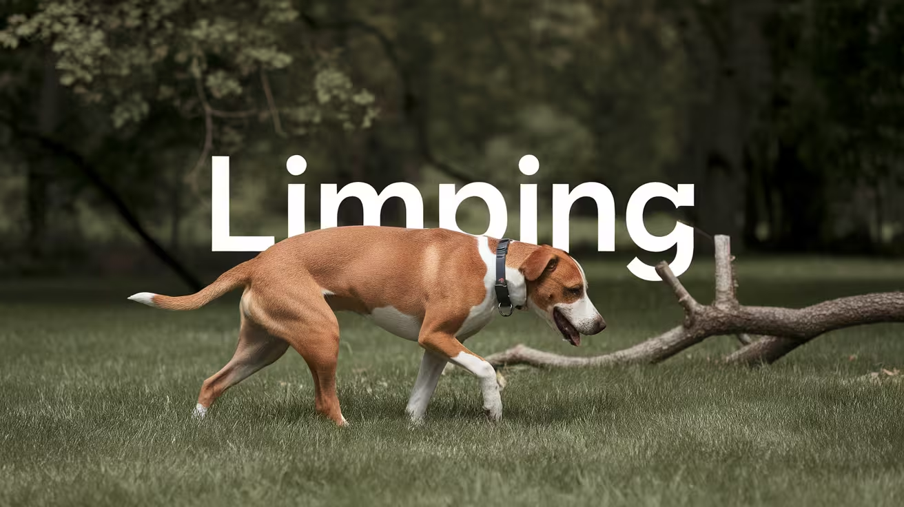
Common Causes of Limping in Dogs Post-TPLO Surgery
Discover common causes of dog limping post-TPLO surgery and explore solutions for regaining your dog's comfort and mobility a year later
Some degree of lameness after TPLO surgery is expected. The surgery involves a bone cut and plate fixation, and the dog's leg needs weeks to heal.
The clinical challenge for owners is distinguishing between normal post-surgical lameness (which requires patience) and lameness that indicates a complication (which requires prompt veterinary attention).
The answer depends heavily on timing, trajectory, and accompanying signs.
Quick answer: Normal post-TPLO lameness should show consistent week-over-week improvement. The most common pathological causes are: surgical site infection, late meniscal tear, implant loosening, muscle atrophy, delayed bone healing, and progressive osteoarthritis. The key signal is trajectory reversal -- lameness worsening after improvement or failing to improve for 2 or more weeks.
Key takeaways
- Some lameness is normal throughout the first 8 to 12 weeks of TPLO recovery: mild limping that improves gradually over 6 to 12 weeks is typical; the key variable is trajectory
- Trajectory reversal is the key warning sign: lameness that worsens after improvement, or fails to improve for 2 or more consecutive weeks, warrants veterinary evaluation before the next appointment
- Late meniscal tear is a recognized cause of sudden lameness regression: recurrence of lameness after initial resolution can indicate meniscal injury; it occurs in 2 to 22% of TPLO cases
- Muscle atrophy is a commonly underappreciated cause of persistent lameness: the affected leg loses muscle mass during restriction; inadequate rehabilitation leaves an asymmetric gait
- Progressive osteoarthritis is the most common cause of chronic mild lameness starting months after surgery; TPLO slows but does not stop this process
- Implant failure and infection must be ruled out for lameness that is worsening or not responding to conservative management; radiographs and culture are required for diagnosis
Normal post-surgical lameness: what to expect
Week 1 to 2
Most dogs are significantly lame in the first 1 to 2 weeks. They may toe-touch only or hold the leg off the ground entirely.
This is the acute post-surgical inflammatory phase combined with the effects of anesthesia wearing off.
SustainableVet.org confirms: some lameness is expected after TPLO surgery, especially in the first few weeks; normal recovery lameness is mild limping that improves gradually over 6 to 12 weeks.
Week 2 to 6
Consistent improvement should be visible week over week. By week 4, most dogs should be bearing significant weight on the operated leg during slow walking.
Three-legged walking during activity is still common but should be decreasing.
Week 6 to 12
Most dogs should be consistently weight-bearing on the operated leg. By week 12, most are nearly normal for household activities.
Radiographic confirmation of bone healing at weeks 6 to 8 and 12 guides when activity can increase.
Pathological causes of post-TPLO lameness
1. Surgical site infection (SSI)
Timing: most commonly weeks 1 to 4, but can occur at any stage (including months later for deep implant-associated infections).
Mechanism: bacterial contamination of the incision or implant causes inflammation and pain at the plate site, which produces or worsens lameness.
Signs accompanying lameness: redness, warmth, swelling, or discharge at the incision or plate site.
Action required: same-day veterinary contact for any infection signs alongside lameness.
2. Late meniscal tear
Timing: typically weeks 4 to 12 post-surgery, though it can occur months later.
Mechanism: the meniscus, which may have been structurally weakened before surgery or had a small tear not detected at surgery, fails under load during the recovery period.
Signs accompanying lameness: audible or palpable meniscal click; pain on stifle manipulation; sudden lameness regression in a dog that had been improving.
Animal Outpatient Surgery confirms: if lameness resolves after TPLO and recurs at a later time, this can be an indication of meniscal injury.
Action required: veterinary evaluation within 24 to 48 hours; arthroscopy or arthrotomy for diagnosis and partial meniscectomy for treatment.
3. Implant loosening or failure
Timing: most common in the first 6 to 12 weeks when the osteotomy is still healing and the plate bears the full load. Can also occur later from chronic infection or late trauma.
Mechanism: excessive activity before the osteotomy heals applies forces that exceed the plate's tolerance; screws pull out or the plate bends.
Signs accompanying lameness: sudden severe lameness; pain at the plate site; sometimes audible clicking or grinding; radiographic evidence of screw loosening or plate migration.
SustainableVet.org confirms: signs of implant failure include ongoing limping, pain, swelling around the implant site, and unusual clicking or grinding sounds from the joint.
Action required: same-day emergency veterinary contact for sudden severe lameness; radiographs urgently required.
4. Muscle atrophy and weakness
Timing: most prominent from weeks 4 to 12; persists for months if rehabilitation is inadequate.
Mechanism: during activity restriction, the operated leg muscles (quadriceps, hamstrings, hip extensors) atrophy rapidly. Asymmetric muscle mass leads to asymmetric weight distribution and abnormal gait patterns that present as persistent limping.
Vetplayas confirms: limited mobility during recovery leads to muscle loss and decreased strength. This imbalance results in abnormal gait patterns and subsequent limping.
Signs: visible muscle asymmetry (operated thigh smaller than opposite); lameness that worsens with activity and improves with rest; no accompanying warmth, redness, or plate-site changes.
Action required: rehabilitation assessment; physiotherapy to rebuild muscle mass.
5. Delayed bone healing
Timing: identified at the 6 to 8 week radiographic assessment.
Mechanism: the osteotomy is not showing expected callus formation and cortical bridging. Common causes include infection, excessive activity, poor blood supply, or systemic factors (NSAIDs at high dose for extended periods, nutritional deficiencies).
Signs: persistent lameness at a level that does not match the expected trajectory; confirmed radiographically.
Action required: veterinary investigation for underlying cause; extended activity restriction; sometimes additional diagnostics (culture to rule out occult infection).
6. Delayed union or non-union
Timing: identified at 12+ weeks when bone healing should be complete.
Mechanism: the osteotomy fails to heal. The plate is subject to continued cyclic loading without the protection of consolidated bone, increasing implant failure risk.
Action required: specialist reassessment; may require bone grafting, implant revision, or extended medical management.
7. Progressive osteoarthritis
Timing: typically becomes clinically apparent after 6 to 12 months.
Mechanism: CCL disease initiates an irreversible arthritis process in the stifle. TPLO reduces but does not eliminate arthritis progression. Over months and years, arthritis produces chronic mild lameness, particularly after rest and exercise.
Signs: stiffness that improves with movement; lameness worse in cold weather or after long periods of rest; gradual worsening over months; bilateral involvement common.
SustainableVet.org (failure symptoms article) confirms: limping 2 years post-TPLO may result from arthritis, implant irritation, meniscal damage, or muscle atrophy.
Action required: long-term management (joint supplements, weight management, NSAIDs for flares, hydrotherapy, physiotherapy).
8. Contralateral CCL rupture
Timing: any time during or after recovery from the first TPLO.
Mechanism: the contralateral CCL, subject to the same degenerative process and bearing extra load during recovery, may rupture. The dog appears to start limping on the opposite leg.
Signs: what appears to be recovery from one side suddenly accompanied by new lameness on the opposite side; the dog may be lame on a different leg or bilateral.
Action required: veterinary evaluation; bilateral CCL disease may require staged or simultaneous bilateral TPLO.
How to assess the trajectory at home
Create a simple weekly log: note the leg-use pattern (full bearing, toe-touching, three-legged) and the dog's willingness to walk at each prescribed walk time.
Compare week 4 to week 3, week 6 to week 5. If the pattern is consistently improving, continue the protocol. If it is static or worsening, contact your vet.
For the lameness guide, see lameness after TPLO surgery in dogs. For the TPLO failure signs guide, see signs of TPLO failure in dogs and when to see a vet.
For the meniscal tear guide, see dog meniscus tear after TPLO surgery. For the infection guide, see earliest signs of TPLO infection.
Frequently asked questions
My dog was progressing well at week 4 but is limping more at week 5. What do I do?
A single week of regression after a period of progress is not necessarily pathological -- increased activity, weather changes, or an off day can cause temporary setbacks.
But if the worsening persists for 2 weeks, or is accompanied by any infection signs, contact your vet.
Monitor closely and reduce activity to the week 2 level for 24 to 48 hours to see if the leg settles.
My dog is still limping 4 months after TPLO surgery. Is this normal?
Mild residual lameness can persist to 4 months, particularly in older dogs or those with significant muscle atrophy.
However, lameness at 4 months that is not showing week-over-week improvement warrants a veterinary assessment.
A radiograph will confirm bone healing status; physical examination will assess muscle mass and stifle range of motion.
My dog is limping on the opposite leg during TPLO recovery. What does that mean?
This raises concern for contralateral CCL disease. The opposite leg is bearing extra load during recovery and may have already been compromised.
A new limp on the opposite leg during recovery warrants veterinary evaluation, including assessment and radiographs of the contralateral stifle.
Can physiotherapy reduce long-term limping?
Yes, significantly. Rebuilding the atrophied quadriceps and hip extensor muscles through controlled hydrotherapy, cavaletti exercises, and progressive strength work reduces the compensatory gait patterns that produce chronic post-TPLO lameness.
Weight management is equally important -- every additional kilogram of body weight increases stifle load.
When is limping after TPLO an emergency?
Sudden complete non-weight bearing (holding the leg entirely off the ground) after a period of normal improvement is an emergency call. Same-day emergency veterinary contact is appropriate for this presentation.
Gradual worsening that does not resolve within 24 to 48 hours is a next-day call.
Resources
- SustainableVet. Common Causes of Limping in Dogs Post-TPLO Surgery. sustainablevet.org
- Animal Outpatient Surgery. What Are the Most Common Complications of TPLO Surgery in Dogs? animaloutpatientsurgery.com
- SustainableVet. Lameness After TPLO Surgery in Dogs. sustainablevet.org
- Laveen Vet Center. Dog Limping 1 Year After TPLO Surgery. laveenvetcenter.com

Long-Term Outcomes of TPLO Surgery
Explore the long-term outcomes of TPLO surgery, its effectiveness, potential risks, and benefits for maintaining canine mobility and quality of life
TPLO produces excellent long-term outcomes for most dogs with CCL rupture.
The published evidence covers follow-up periods from 1 year to 6.8 years post-surgery and consistently shows high rates of functional recovery alongside moderate but ongoing arthritis progression.
Understanding what the data shows helps owners set realistic expectations about what TPLO achieves and what ongoing management is required.
Quick answer: Published long-term TPLO outcomes: 90 to 95% of dogs regain complete or near-complete limb function; good to excellent results in 90.4% of patients at up to 6.8 years; 93% owner satisfaction at 12 months (RCVS). Arthritis progresses moderately in all CCL-affected stifles but TPLO produces less progression than TTA.
Key takeaways
- 90 to 95% of dogs regain complete or near-complete limb function: RCVS Canine Cruciate Registry confirms this range; 93% owner satisfaction at 12 months
- Good to excellent results in 90.4% of patients up to 6.8 years post-surgery: TPLO Info cites this long-term retrospective study, one of the longest published TPLO follow-up datasets
- Arthritis progresses in all CCL-affected joints regardless of surgical technique: TPLO slows progression compared to TTA and lateral suture but does not stop it
- TPLO produces better long-term outcomes than TTA in comparative studies: OA progressed more after TTA; TPLO dogs had less pain, better walking, less morning stiffness, and better quality of life at 3+ years per the 94-dog study
- TPLO produces better owner satisfaction than lateral suture: 93% vs. 75% for lateral fabellar suture per TPLO Info; kinematic gait analysis also favored TPLO
- Meniscal management at the time of TPLO affects long-term outcomes: dogs with partial meniscectomy had higher rates of excellent long-term results and less OA progression per TPLO Info
Functional outcomes: what the data shows
At 1 year post-surgery
A 2013 comparative study of TPLO vs. TTA found TPLO patients achieved 93% restoration of limb function at 1 year, as assessed by objective force plate analysis.
This is the most frequently cited 1-year outcome figure.
RCVS Canine Cruciate Registry confirms: 90 to 95% of dogs regain complete or near-complete function; 93% of owners report satisfaction at 12 months.
At 2 years post-surgery
A PMC long-term functional outcome study found that most dogs with CCL repair -- regardless of technique -- showed improvement in limb function at 2 years compared to pre-operative status.
Objective gait analysis at 2 years showed that the operated leg approached the contralateral leg in force plate measurements.
At 3+ years post-surgery
The comparative TPLO vs. TTA study with follow-up at 3+ years provides the clearest long-term comparative data:
- OA progressed more in TTA than in TPLO dogs
- TPLO dogs scored better on the Canine Brief Pain Inventory across all domains (average pain, walking interference, morning stiffness, jumping, climbing)
- TPLO dogs scored better on the Canine Orthopedic Index (quality of life)
- The study concluded TPLO provides better long-term radiographic and functional outcome than TTA
At up to 6.8 years post-surgery
TPLO Info cites a long-term retrospective study with follow-up extending to 6.8 years: good to excellent results were obtained in 90.4% of patients at this timeframe.
This is the longest published follow-up in a TPLO outcome dataset and confirms that the 90%+ functional recovery rate is durable over years rather than only at 12 months.
Arthritis: the universal long-term consideration
All dogs with CCL disease develop progressive osteoarthritis in the affected stifle. This is true regardless of whether surgical or conservative management is chosen.
TPLO reduces the rate of arthritis progression and the severity of clinical signs compared to lateral suture stabilization and TTA, but arthritis progression continues.
TPLO Info confirms: in the long term, there was a moderate but significant progression of OA following TPLO.
SustainableVet.org confirms: while TPLO stabilizes the knee and slows down arthritis progression, it does not completely eliminate the risk; mild arthritis is expected, even in successful cases.
Managing long-term arthritis after TPLO
Weight management: every additional kilogram of body weight increases stifle joint loading. Maintaining lean body condition is the single most impactful owner-controlled variable for arthritis management.
Joint supplements: glucosamine, chondroitin, and omega-3 fatty acids (fish oil) are commonly recommended and have evidence supporting modest benefit for joint health and comfort.
Low-impact exercise: regular, controlled exercise (walking, swimming, hydrotherapy) maintains muscle mass and joint lubrication without the impact forces of running and jumping. Consistent low-impact exercise is more beneficial than sporadic high-intensity sessions.
NSAIDs for flares: carprofen, meloxicam, or other veterinary NSAIDs are used for periods of increased arthritis pain. Long-term NSAID use requires periodic monitoring of renal and hepatic function.
Rehabilitation: an ongoing physiotherapy relationship with a certified canine rehabilitation practitioner helps maintain muscle mass and joint mobility as the dog ages.
Factors that affect long-term outcomes
Age at surgery: younger dogs typically achieve better long-term function than older dogs. Older dogs heal more slowly and have a higher baseline level of arthritis-related joint change before surgery.
Body weight: heavier dogs show more arthritis progression and more persistent lameness than lighter dogs at equivalent follow-up periods.
Meniscal management: TPLO Info confirms partial meniscectomy (when a meniscal tear is present) is associated with significantly higher rates of excellent long-term results and less OA progression compared to meniscal release or leaving the meniscus intact when abnormal.
Surgical timing: earlier intervention (before severe arthritis develops) produces better long-term outcomes. Dogs treated early in the disease process have less existing joint damage at the time of surgery.
Rehabilitation compliance: dogs that complete structured rehabilitation programs and achieve good muscle mass restoration have better long-term function than those managed with restricted activity alone.
Bilateral CCL disease: TPLO Info confirms OA progresses more in dogs with bilateral stifle surgery than unilateral. This may reflect the more severe underlying degenerative process in these dogs.
TPLO vs. lateral suture: long-term comparison
TPLO Info confirms owner satisfaction of 93% for TPLO vs. 75% for lateral fabellar suture; kinematic gait analysis also favored TPLO outcomes.
This comparison is most relevant for medium-sized dogs where both procedures are technically appropriate. For large dogs over 35 to 40 kg, TPLO consistently outperforms lateral suture in long-term outcomes.
For small dogs under 15 kg, lateral suture achieves comparable results at substantially lower cost.
TPLO vs. TTA: long-term comparison
SustainableVet.org confirms: when comparing TTA to TPLO, research suggests similar long-term arthritis outcomes in some studies, but TPLO patients usually recover faster and regain function sooner.
The 3+ year follow-up comparative study is the most comprehensive published comparison and clearly favors TPLO in both OA progression and functional/quality-of-life outcomes.
For the TPLO overview, see what is TPLO surgery in dogs?. For the long-term effects list, see 13 long-term effects of TPLO surgery on dogs.
For surgery comparisons, see TPLO vs CBLO vs TTA: a guide to cruciate surgery options. For arthritis management, see arthritis after TPLO surgery in dogs.
Frequently asked questions
Will my dog be completely normal after TPLO?
Most dogs return to their pre-injury activity level after full recovery.
RCVS and multiple studies confirm 90 to 95% regain complete or near-complete function.
"Completely normal" is achievable for most dogs, with the caveat that some mild arthritis develops over time in virtually all cases.
Dogs may show stiffness on cold days or after long rest periods as they age.
Does TPLO stop arthritis?
No -- it reduces and slows arthritis progression compared to conservative management and other surgical techniques, but does not stop it. All CCL-affected stifles develop progressive OA.
The goal of TPLO is to stabilize the joint and minimize the rate of arthritis development, not eliminate it.
How long does the TPLO plate last?
The plate is designed to be permanent.
Good to excellent results at 6.8 years (the longest published TPLO follow-up) with the plate in place demonstrates the durability of the implant in the overwhelming majority of cases.
Plate complications requiring removal occur in 3.5 to 7.5% of cases.
Is my dog at risk for the CCL on the other side?
Yes. RCVS confirms approximately 50% of dogs will develop CCL disease in the other hind limb.
This is not a consequence of TPLO -- it reflects the degenerative nature of CCL disease that typically affects both stifles. Monitoring the contralateral stifle is appropriate in all TPLO dogs.
Does the long-term outcome depend on the surgeon's experience?
Yes, to some degree. Surgeons with more TPLO experience have lower complication rates (including SSI and delayed union), and complications are associated with worse long-term outcomes.
Using a board-certified surgeon or an experienced general practitioner with dedicated TPLO training reduces risk. The published SSI studies confirm surgeon experience as an independent risk factor for post-TPLO complications.
Resources
- TPLO Info. TPLO Literature and Studies. tploinfo.com
- RCVS Canine Cruciate Registry. Tibial Plateau Levelling Osteotomy (TPLO). rcvsknowledge.org
- PMC. Long-Term Functional Outcome After Surgical Repair of Cranial Cruciate Ligament Disease in Dogs. ncbi.nlm.nih.gov
- SustainableVet. Long-Term Outcomes of TPLO Surgery. sustainablevet.org
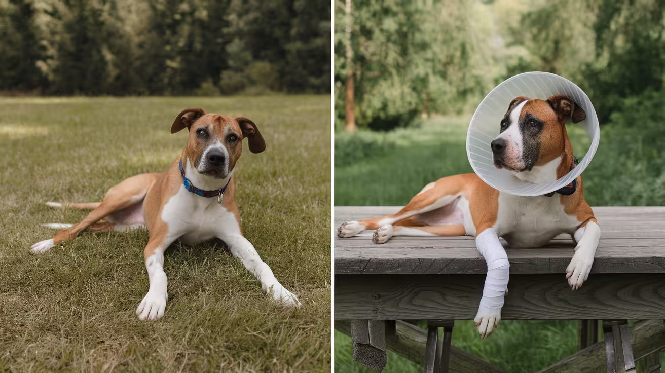
Before and After TPLO Surgery: Dog Health and Recovery Timeline
Discover how TPLO surgery restores mobility in dogs with torn CCL. Learn about recovery timelines, post-op care, and expected outcomes for your pet
TPLO surgery is a major orthopedic procedure that requires careful preparation before surgery and structured management after.
The recovery period -- 8 to 12 weeks of graduated activity restriction, multiple vet visits, and daily wound monitoring -- is as important as the surgery itself.
Most owners who struggle with recovery do so because they underestimated what the post-operative period involves. This guide walks through every phase.
Quick answer: Before TPLO surgery: complete pre-surgical bloodwork, fast 8 to 12 hours, prepare a crate and non-slip mats, fill prescriptions. After TPLO: crate rest weeks 1 to 2, progressive walking weeks 2 to 6, radiographic assessment weeks 6 to 8, activity increase after healing. Muscle recovery takes 4 to 6 months.
Key takeaways
- The home environment must be set up before surgery day: crate, non-slip mats, baby gates, low bedding, and supplies must be in place before the dog arrives home
- Fasting is required before surgery: no food for 8 to 12 hours before general anesthesia; water rules vary by facility
- The first 2 weeks are the most critical and restrictive: no running, jumping, stairs, or off-leash activity; bathroom trips on a short leash only; incision checked daily
- Radiographic assessment at 6 to 8 weeks is the gate that controls activity increase: without imaging confirmation, activity must not increase; radiographs are the only reliable confirmation
- Food intake should be reduced by approximately 30% during recovery: activity restriction rapidly leads to weight gain; extra weight stresses the healing osteotomy
- Full muscle recovery takes 4 to 6 months after bone healing is confirmed: bone healing at 12 weeks and full functional recovery are different endpoints
Before surgery: preparation checklist
Medical preparation (weeks before surgery)
Pre-surgical examination and bloodwork: your vet will perform a full physical examination and order a complete blood count and blood chemistry panel. These confirm the dog can safely handle anesthesia and healing. Senior dogs or those with health conditions may need additional testing.
Medication review: tell your vet all current medications and supplements. NSAIDs, certain supplements, and some other drugs need to be stopped before surgery. Never discontinue a prescription medication without vet instruction.
Weight management: if your dog is overweight, weight reduction before surgery reduces anesthetic and surgical risk. Even a 5 to 10% body weight reduction before surgery is beneficial.
Pre-surgical bathing: bathe 1 to 2 days before surgery if you wish; post-surgical bathing is restricted for 10 to 14 days.
Home preparation (day before surgery)
The recovery space:
- Crate (correctly sized: stand, turn, lie comfortably, not pace) in the main family living area
- Non-slip mats or yoga mats over all hard flooring in the recovery area
- Soft, washable bedding in the crate
- Water and food bowls at floor level
- Baby gates on stairs
- All furniture inaccessible to prevent jumping
Supplies to have ready:
- E-collar (confirm with the surgical facility whether one will be provided)
- Ice pack or bag of frozen peas for cold therapy
- Sling or lifting harness for larger dogs
- Short leash (4 to 6 foot, not retractable)
- Medications filled at the pharmacy before surgery day
SustainableVet.org confirms: before your dog comes home, prepare a safe and quiet recovery space with a crate or small room, soft bedding, good airflow, and away from stairs or slippery floors.
Fasting
No food for 8 to 12 hours before surgery. Water rules vary by facility -- confirm with your vet. Give no food the morning of surgery even if the dog appears hungry.
Surgery day: what to expect
Drop-off: typically early morning. The dog is admitted, weighed, examined, and an IV catheter is placed. IV antibiotics are given before the incision.
The procedure: the surgeon makes a medial incision over the proximal tibia, performs a small arthrotomy to assess the meniscus, makes the curved osteotomy cut, rotates the tibial plateau to approximately 5 degrees, and secures the plate and screws. The wound is closed in layers.
Duration: typically 1.5 to 2 hours.
Post-surgical care: the dog spends several hours in recovery before discharge (same day at some facilities; overnight at others).
Discharge: written instructions for medications, wound care, activity restrictions, and follow-up appointments are provided.
After surgery: week-by-week recovery timeline
Weeks 1 to 2: acute post-surgical phase
Activity: crate rest at all times except bathroom trips. Leash walks of 5 minutes maximum, 3 to 5 times daily. No stairs, no furniture, no off-leash time.
Weight bearing: most dogs begin toe-touching within the first few days. Some do not bear weight for the full 2 weeks -- both presentations are within normal range.
Wound care: e-collar on at all times except supervised eating. Inspect the incision daily at the same time. No bathing or wet incision exposure.
Cold therapy: 10 to 15 minutes of ice pack (wrapped in cloth) applied to the incision, 3 to 4 times daily for the first 3 to 5 days.
Medications: NSAIDs and gabapentin on the exact prescribed schedule. Give NSAIDs with food.
SustainableVet.org confirms: the first two weeks focus on rest and healing; the dog stays confined; short controlled leash walks are for bathroom breaks; check the incision daily for redness, swelling, or discharge.
2-week recheck: incision assessment, suture or staple removal if healing is confirmed, early mobility and pain management review.
Weeks 3 to 6: graduated activity phase
Activity: leash walk duration increases progressively. TPLO Info recommends increasing by up to 5 minutes per week. No off-leash activity. No stairs without guidance. No playing with other pets.
Weight bearing: most dogs should be bearing increasing weight through the operated leg. Three-legged walking during activity is still common but should be decreasing.
Passive range-of-motion exercises: gentle flexion and extension of the stifle, 5 to 10 repetitions, 2 to 3 times daily if the vet has approved.
Veterinary Healthcare Associates confirms: weeks 3 to 4, swelling should decrease; gradual increase in leash walking; passive range-of-motion exercises may begin.
Food management: Medcovet confirms reducing food intake by approximately 30% during the restriction period to prevent weight gain from inactivity.
Weeks 6 to 8: radiographic checkpoint
6-week radiograph (critical): images confirm whether the osteotomy is healing as expected. If healing is progressing, activity can be increased. If healing is delayed, restrictions continue.
If healing is confirmed: leash walks increase to 15 to 20 minutes, physiotherapy begins, short controlled hill walking may begin.
If healing is delayed: investigate for infection or non-union; extend restrictions; consider additional diagnostics.
Veterinary Healthcare Associates confirms: weeks 5 to 6, recheck X-rays may be taken to evaluate healing; improvement in weight-bearing and stability should be evident.
Weeks 8 to 12: consolidation and return to activity
8 to 10 week radiograph (in many protocols): confirms continued healing progression.
12-week radiograph (final major milestone): confirms bone union before lifting restrictions. After this confirmation:
- Running and off-leash play can gradually resume
- Stairs without support
- Normal household access
Post-12-week activity return: progressive reintroduction of activity over the following 4 to 6 weeks rather than an immediate full return.
Animal Outpatient Surgery confirms: in the final phase, your dog can return to normal activity levels, including running, jumping, and playing; continue monitoring for discomfort and maintain a balanced exercise routine.
Long-term: months 3 to 6
Full muscle recovery takes 4 to 6 months after bone healing. During this period:
- Progressive strengthening continues
- Hydrotherapy, balance work, and controlled running rebuild the atrophied operated leg
- Joint supplements (omega-3 fatty acids, glucosamine) are typically started or continued
- Weight management remains critical
Most dogs reach 90 to 95% of pre-injury function by 6 months with a compliant recovery.
For the recovery tips guide, see 10 essential TPLO recovery tips for pet owners. For confinement, see how to confine your dog after TPLO surgery.
For physical therapy, see when to start physical therapy after TPLO surgery. For long-term outcomes, see long-term outcomes of TPLO surgery.
Frequently asked questions
How strict does the activity restriction need to be in the first 2 weeks?
Very strict. No exceptions for stairs, sofa access, running, jumping, or unsupervised time without confinement.
A single running or jumping episode in the first 2 weeks can displace the plate before the osteotomy has any mechanical strength.
The restriction is designed to protect a bone that cannot yet protect itself.
My dog is eating less and seems depressed after surgery. Is this normal?
Yes, in the first 3 to 5 days. Anesthesia, post-surgical pain, and opioid medications can reduce appetite and alter behavior.
If the dog is not eating at all by day 3 or shows signs of significant distress, contact your vet.
Most dogs gradually return to normal eating and behavior within the first week.
When can my dog go swimming after TPLO?
Controlled hydrotherapy (underwater treadmill) can begin after the incision is confirmed healed at the 2-week recheck. Open water swimming (lakes, rivers) is typically not recommended until after the 12-week radiographic clearance.
Confirm with your vet in both cases.
Should I put my dog on a diet during recovery?
Reduce caloric intake rather than switching to a formal diet. Medcovet recommends approximately 30% reduction in food intake during the restriction period to prevent weight gain from inactivity.
If your dog was already at an ideal weight, a modest reduction (10 to 20%) is appropriate. Discuss with your vet.
My dog seems completely fine at week 4 and is pulling on the leash. Should I increase activity?
No. Continue the restriction protocol until the 6-week radiograph confirms bone healing progression.
The dog feeling well at week 4 is a positive sign, but bone healing lags significantly behind the dog's perceived comfort.
Dogs routinely feel ready to run before the osteotomy is safe to run on.
Resources
- SustainableVet. Before and After TPLO Surgery. sustainablevet.org
- Veterinary Healthcare Associates. TPLO Surgery in Dogs: Before and After. vhavets.com
- Medcovet. Essential Guide to Dog Recovery TPLO Surgery. medcovet.com
- TPLO Info. Preparing for Your Dog's TPLO Surgery. tploinfo.com

13 Long-Term Effects of TPLO Surgery on Dogs
Discover the 13 long-term effects of TPLO surgery in dogs, including benefits, risks, and care tips for optimal recovery and joint health
TPLO produces predominantly positive long-term effects in most dogs, but it also produces a predictable set of changes that owners should understand and manage proactively.
Most of these effects are manageable with appropriate care. Knowing what to expect prevents owners from being surprised by normal post-TPLO findings and helps them recognize when a change requires veterinary attention.
Quick answer: The 13 key long-term effects of TPLO surgery: improved joint stability, progressive osteoarthritis, muscle atrophy, morning stiffness, cold weather sensitivity, scar tissue formation, patellar tendon thickening, permanent implant in situ, contralateral CCL rupture risk, late implant complications, behavioral changes from chronic pain, weight management challenges, and long-term monitoring requirements.
Key takeaways
- Improved joint stability is the intended and most common long-term effect: for 90 to 95% of dogs, TPLO eliminates CCL-related instability and restores near-normal function
- Progressive osteoarthritis develops in all CCL-affected stifles regardless of surgical technique: TPLO slows OA progression but does not stop it; long-term OA management is a universal requirement
- Muscle atrophy is common in the operated leg throughout recovery; the thigh may remain visibly smaller than the contralateral side for months if rehabilitation is inadequate
- Contralateral CCL rupture risk is high: SustainableVet.org confirms up to a 50% chance of tearing the opposite CCL within a few years; the same degenerative process affects both stifles
- The TPLO plate and screws are permanent in the vast majority of cases: removal is only performed when complications arise; routine plate removal is not standard practice
- Weight management is the most impactful long-term owner-controlled variable: excess weight accelerates OA progression; maintaining ideal body condition is the primary long-term prevention strategy
The 13 long-term effects
1. Improved joint stability and mobility
The primary intended effect. The osteotomy and plate fixation permanently change the tibial plateau geometry, eliminating the cranial tibial thrust that caused instability.
For 90 to 95% of dogs, this produces lasting functional improvement: return to normal gait, ability to run and play, and freedom from the pain of unstable joint loading.
SustainableVet.org confirms: over 90% of dogs regain full limb function within a year; with proper care, many dogs remain active for 8 to 10 years after surgery.
2. Progressive osteoarthritis
The most universal long-term challenge. CCL disease initiates an arthritis process in the stifle that continues regardless of surgical outcome.
TPLO reduces the rate of progression and the severity of clinical signs compared to other treatment options, but OA continues.
Clinical signs of progressing OA emerge gradually over months and years: stiffness after rest, reluctance to run, difficulty climbing, and behavioral changes associated with chronic low-grade pain.
Management: weight management, omega-3 fatty acids, glucosamine and chondroitin, NSAIDs for flares, hydrotherapy, physiotherapy.
3. Muscle atrophy and asymmetry
The operated leg loses muscle mass rapidly during the restriction period and slowly rebuilds during rehabilitation. Full recovery of muscle symmetry takes 4 to 6 months in most dogs.
In some dogs -- particularly those with inadequate rehabilitation or ongoing joint pain -- visible thigh circumference asymmetry persists long-term.
Vetplayas confirms: due to reduced use during recovery and altered movement patterns post-surgery, muscles around the operated leg may weaken and shrink.
4. Morning stiffness
Morning stiffness -- the dog rises from sleep stiffly and walks awkwardly for the first few minutes before loosening up -- is one of the most common long-term effects of post-TPLO OA.
It typically develops months to years after surgery and gradually worsens as the dog ages.
This stiffness should not be confused with acute lameness. It consistently improves within 5 to 10 minutes of movement.
If it does not improve, or if the dog refuses to bear weight on waking, contact your vet.
5. Cold weather sensitivity
Dogs with OA in the operated stifle commonly show increased stiffness and lameness during cold weather or when barometric pressure changes.
This reflects the sensitivity of arthritic joints to temperature and pressure changes -- the same phenomenon seen in human arthritis.
Management: NSAIDs or joint supplements maintained year-round; warming the dog's sleeping area; a brief gentle walk before expecting full function in cold conditions.
6. Scar tissue formation
The surgical dissection and bone healing produce scar tissue in and around the stifle. This fibrous tissue generally has a beneficial stabilizing effect, contributing to long-term joint stability.
In some dogs, however, periarticular fibrosis restricts range of motion and causes stiffness that persists long after bone healing is complete.
Regular low-impact exercise and physiotherapy help maintain range of motion and prevent excessive fibrous tissue restriction.
7. Patellar tendon thickening
Fibrous thickening of the patellar tendon is a recognized long-term change after TPLO. It is typically subclinical -- detected on palpation at follow-up examinations but not producing clinical lameness.
In a small number of dogs, it contributes to reduced range of motion.
8. Permanent implant in situ
The TPLO plate and screws are designed to remain in place for life. In the vast majority of dogs, the implant is never removed and causes no long-term problems.
SustainableVet.org confirms: these implants are meant to stay in place permanently. In rare cases, some dogs may develop infections around the implant or experience loosening of the hardware over time.
The plate is palpable under the skin on the medial tibial surface. This is normal and not a complication.
9. Contralateral CCL rupture
The degenerative process that caused one CCL to rupture typically affects both stifles.
SustainableVet.org confirms dogs have up to a 50% chance of tearing the CCL in the opposite knee within a few years.
RCVS confirms approximately 50% of dogs develop CCL disease in the other hind limb.
This is not a complication of TPLO -- it is the natural history of bilateral CCL disease.
Monitoring the contralateral stifle for early signs of instability, effusion, or lameness is appropriate in all TPLO dogs.
10. Late implant-associated complications
Implant-related complications -- loosening, late infection from hematogenous seeding, or implant irritation -- can occur years after the original surgery. These are uncommon but recognized long-term effects.
Signs: new lameness focused at the plate site, warmth or swelling over the plate, a new draining tract in previously healed skin. Any of these warrants prompt veterinary assessment.
11. Behavioral changes
Chronic joint pain from progressive OA, late meniscal injury, or implant irritation can produce subtle behavioral changes: reduced engagement in play, reluctance to walk, increased irritability, or reduced stair use.
These changes are often attributed to aging but may reflect treatable pain.
Annual or semi-annual pain assessments, including use of validated canine pain scales (Canine Brief Pain Inventory, Helsinki Chronic Pain Index), help detect behavioral pain signs before they become severe.
12. Weight management challenges
Activity restriction during recovery predisposes many dogs to weight gain. Excess weight gained during the 8 to 12 week restriction period is then carried on the healing joint for the following months.
Long-term obesity accelerates OA progression and is the most modifiable risk factor for long-term joint health.
Medcovet confirms: because activity is restricted, veterinary teams may recommend reducing food intake by about 30% during recovery to prevent weight gain and extra strain on the knee joint.
13. Long-term monitoring and supplement requirements
TPLO dogs benefit from long-term monitoring (every 6 to 12 months after full recovery) to assess:
- Arthritis progression radiographically and clinically
- Body condition score and weight
- Contralateral stifle stability
- Implant integrity (radiographs every 1 to 2 years in some protocols)
Joint supplements (omega-3 fatty acids at anti-inflammatory doses; glucosamine and chondroitin) and ongoing weight management are the primary preventive interventions for the long-term joint changes that TPLO does not prevent.
For the long-term outcomes guide, see long-term outcomes of TPLO surgery. For arthritis management, see arthritis after TPLO surgery in dogs.
For the signs that require attention, see signs of TPLO failure in dogs. For the failure rate overview, see TPLO failure rate in dogs.
Frequently asked questions
Will my dog develop arthritis even if the TPLO was successful?
Yes. All CCL-affected stifles develop progressive osteoarthritis regardless of surgical technique or outcome.
TPLO reduces the rate of progression and the severity of clinical signs compared to non-surgical management and lateral suture repair.
The goal of TPLO is not to prevent OA but to minimize it while restoring function.
How long will the TPLO plate last?
The plate is designed to be permanent. Published long-term follow-up data at 6.8 years shows 90.4% of dogs with good to excellent results -- with the plate in place.
Plate complications requiring removal occur in 3.5 to 7.5% of cases.
My dog had TPLO 2 years ago and is now limping. Is this related to the surgery?
Possibly. Two-year post-TPLO lameness most commonly results from progressive OA, late meniscal tear, late implant-associated infection (hematogenous seeding), or contralateral CCL disease.
A veterinary examination and radiographs of both stifles will identify the cause and guide treatment.
Does the opposite leg always need TPLO as well?
Not necessarily and not always at the same time. RCVS confirms approximately 50% of dogs develop CCL disease in the opposite hind limb.
When and whether the second stifle requires surgery depends on whether the second CCL has ruptured or become significantly unstable. Annual monitoring of the contralateral stifle is appropriate.
Can TPLO dogs still live active lives long-term?
Yes. Most do. SustainableVet.org confirms that with proper care, many dogs remain active for 8 to 10 years after surgery.
The keys are weight management, regular low-impact exercise, joint supplements, and addressing pain or complications promptly.
Resources
- SustainableVet. 13 Long-Term Effects of TPLO Surgery on Dogs. sustainablevet.org
- RCVS Canine Cruciate Registry. Tibial Plateau Levelling Osteotomy. rcvsknowledge.org
- TPLO Info. TPLO Literature and Studies. tploinfo.com
- SustainableVet. Long-Term Outcomes of TPLO Surgery. sustainablevet.org

Common Myths About TPLO Surgery Explained
Discover the truth about TPLO surgery for dogs as we debunk common myths and provide evidence-based insights for informed pet care decisions
TPLO is the most commonly performed surgery for CCL rupture in dogs and the most commonly misunderstood.
Misconceptions about TPLO cause unnecessary hesitation, lead some owners to choose inferior alternatives, and create unrealistic expectations about recovery.
This guide addresses the 10 most common myths directly -- with evidence-based clarification for each.
Quick answer: The most persistent TPLO myths: that dogs can manage without surgery, that TPLO is only for large breeds, that recovery takes a few weeks, that the plate must be removed after healing, and that TPLO prevents arthritis. All are false. TPLO has a 90 to 95% success rate.
Key takeaways
- Myth: "My dog can heal without surgery": dog knee anatomy differs fundamentally from human; without surgery, severe OA, meniscal tearing, and chronic lameness are expected in most dogs over 15 kg
- Myth: "TPLO is only for large dogs": SustainableVet.org confirms TPLO is just as effective for small and toy breeds; the biomechanical correction is appropriate regardless of size
- Myth: "Recovery only takes a few weeks": bone healing takes 8 to 12 weeks; full muscle recovery takes 4 to 6 months; 8 to 12 weeks is the minimum restriction period
- Myth: "The plate needs to be removed after healing": the TPLO plate is designed to be permanent; removal is only performed when complications arise
- Myth: "TPLO prevents arthritis": all CCL-affected stifles develop progressive OA regardless of surgical technique; TPLO reduces OA progression but does not prevent it
- Myth: "Dogs are in severe pain after TPLO": SustainableVet.org confirms this is a misconception; modern multi-modal pain management controls post-surgical pain effectively
Myth 1: "My dog can heal without surgery"
The truth: dogs and humans have fundamentally different knee anatomy. In humans, the flat tibial plateau and strong surrounding musculature allow some CCL tears to be managed conservatively with physical therapy and bracing. In dogs, the tibial plateau slopes steeply downward, generating a cranial shear force that the absent CCL cannot resist. Without surgical correction of this force, the joint remains mechanically unstable with every weight-bearing step.
TPLO Info confirms: dogs and humans respond very differently to CCL tears. Without surgery, severe OA can develop, meniscal tearing can occur, and other side effects may be present.
Conservative management (strict rest, weight management, joint supplements, physical therapy) can provide temporary improvement, particularly in small dogs and partial tears.
For most dogs over 15 kg with complete CCL rupture, conservative management does not restore normal joint stability, and progressive lameness, OA, and joint deterioration are the expected outcome.
Who can be managed conservatively?
- Very small dogs (under 10 to 15 kg) with partial tears and low activity levels
- Dogs with significant comorbidities where anesthetic risk outweighs surgical benefit
- Owners who decline surgery after being fully informed of expected outcomes
For all other dogs -- medium and large breeds, young dogs, active dogs, complete ruptures -- surgery produces substantially better outcomes than conservative management.
Myth 2: "TPLO is only for large dogs"
The truth: TPLO was originally developed for large and giant breeds because they were the most severely affected by CCL disease and the most poorly served by lateral suture stabilization. But TPLO corrects the underlying biomechanical problem (excessive tibial plateau angle) in any dog, regardless of size.
SustainableVet.org confirms: TPLO was initially developed for large dogs due to biomechanical challenges, but has been found just as effective for small and toy breeds.
For small dogs, both TPLO and lateral suture stabilization can achieve good outcomes.
The decision for a small dog should be based on the dog's TPA, activity level, and individual assessment -- not a blanket assumption that TPLO is unnecessary.
Myth 3: "TPLO recovery only takes a few weeks"
The truth: bone healing takes 8 to 12 weeks. This is the period of activity restriction. But bone healing and functional recovery are not the same thing. Full muscle rebuilding and joint function restoration takes 4 to 6 months from surgery.
Canada Vet Express confirms: recovery is gradual; most dogs begin walking within a few days and return to normal activity within 3 to 4 months with proper care.
The 8 to 12 week bone healing timeline means:
- Activity restrictions are in place for 8 to 12 weeks minimum
- Radiographic confirmation of healing is required before restrictions are lifted
- Progressive return to full activity follows the radiographic clearance
After restrictions are lifted, the operated leg continues to rebuild strength and coordination for months. For sport dogs and working dogs, the return-to-sport protocol extends to 7 to 9 months from surgery.
Myth 4: "The TPLO plate needs to be removed after the bone heals"
The truth: the plate is designed to remain in place permanently. It does not need to be removed when the osteotomy heals. Routine plate removal is not standard practice in veterinary orthopedics.
SustainableVet.org confirms: these implants are meant to stay in place permanently, but in rare cases they can cause problems; removal is only performed when complications arise.
When plate removal is performed:
- Deep implant-associated infection requiring source removal
- Implant failure (breakage, loosening causing instability)
- Persistent implant irritation producing clinical lameness specifically attributable to the plate
In the vast majority of dogs, the plate is never removed. It remains in place for life with no adverse effects.
Myth 5: "TPLO prevents arthritis"
The truth: TPLO reduces and slows arthritis progression. It does not prevent it. All CCL-affected stifles develop progressive OA regardless of surgical technique or outcome.
SustainableVet.org confirms: mild arthritis is expected even in successful cases.
Published long-term data (TPLO Info, citing long-term retrospective studies) confirms moderate but significant OA progression in all TPLO cohorts with follow-up extending to 6.8 years.
TPLO produces substantially less OA progression than TTA or lateral suture stabilization, but OA progression is universal.
The appropriate expectation: TPLO minimizes OA development and clinical impact. Long-term management (weight management, exercise modification, joint supplements, and periodic NSAID use for flares) is required in all TPLO dogs.
Myth 6: "Dogs are in severe pain after TPLO"
The truth: modern veterinary anesthesia and pain management protocols produce well-controlled post-operative pain in TPLO dogs. Dogs typically begin bearing some weight within 24 to 48 hours. Pain that is not controlled is a clinical problem requiring medication adjustment -- not an expected feature of TPLO recovery.
SustainableVet.org confirms: it is a misconception that dogs experience severe pain after TPLO surgery; modern multi-modal pain management controls post-surgical pain effectively.
Post-operative pain management typically includes: NSAIDs (carprofen, meloxicam), gabapentin, opioids during hospitalization, local anesthetic nerve blocks at surgery.
Dogs in adequate pain control rest comfortably, bear some weight within days, and show appetite by day 3.
Uncontrolled post-operative pain (dog cannot settle, crying, refuses all weight bearing beyond day 3) indicates a need for pain medication adjustment -- contact your vet rather than accepting it as inevitable.
Myth 7: "TPLO is too risky for most dogs"
The truth: TPLO has a 90 to 95% success rate. The complication rate across all studies is 10 to 34%, but the vast majority of complications are minor (superficial infection, seroma, mild swelling). Serious complications requiring revision surgery occur in fewer than 10% of cases.
Dispomed confirms: overall complication rates are 14 to 34%, but a second surgery is required in less than 10% of cases.
No surgery is without risk.
But for a dog with a torn CCL, the risk of TPLO is substantially lower than the certainty of progressive joint deterioration, pain, and reduced quality of life without surgical treatment.
Myth 8: "TPLO is only for show dogs or working dogs"
The truth: TPLO is appropriate for any dog with CCL rupture that will benefit from restored joint stability -- including middle-aged household pets with no competitive goals.
SustainableVet.org confirms: some pet owners mistakenly believe that TPLO surgery is only for show dogs or high-performance athletes; this is not true; TPLO benefits any dog with CCL disease.
The goal of TPLO is freedom from joint pain and restored ability to perform normal daily activities -- walking, playing, using stairs, sitting and rising comfortably.
These are benefits for every dog, not just sport dogs.
Myth 9: "All dogs with CCL tears need TPLO immediately"
The truth: TPLO is not always an emergency. The timing of surgery should be guided by the veterinary assessment. Partial tears in small dogs may be monitored with conservative management. Dogs with significant concurrent illness may benefit from medical optimization before surgery. The appropriate goal is surgical timing that optimizes anesthetic risk and surgical conditions, not surgical urgency at any cost.
SustainableVet.org confirms: while TPLO surgery is the best treatment for CCL tears, it isn't necessary for every case; for mild or partial tears in less active dogs, alternatives can be effective.
However, in dogs with complete rupture, particularly larger breeds, delay generally produces worse outcomes as OA progresses and the meniscus is exposed to ongoing mechanical insult from the unstable joint.
Myth 10: "If TPLO fails, the dog will never recover"
The truth: most TPLO complications are treatable. Superficial infections resolve with antibiotics. Late meniscal tears are treated with partial meniscectomy. Implant failures can be revised. Osteomyelitis, the most challenging complication, requires prolonged treatment but is often successfully managed.
SustainableVet.org confirms: revision TPLO surgery can be performed if the initial procedure fails; this may involve removing or replacing implants, repositioning the tibial cut, or addressing infections.
The critical factor in outcome from a complication is early detection and prompt treatment. Most complications that are identified and treated early resolve without long-term sequelae.
For the full TPLO overview, see what is TPLO surgery in dogs?. For the success rate data, see TPLO failure rate in dogs. For recovery, see what to expect after TPLO surgery.
For alternatives, see alternatives to TPLO surgery for dogs.
Frequently asked questions
Is TPLO actually the best treatment for CCL rupture?
For most dogs over 15 to 20 kg, TPLO is the most evidence-supported surgical option, producing the highest rates of long-term function and owner satisfaction in the published literature.
For small dogs, lateral suture stabilization achieves comparable results at lower cost. The choice depends on the individual dog's size, activity level, TPA, and health status.
Can I wait and see how my dog does without surgery?
You can choose conservative management.
The expected outcome in most dogs over 15 kg with complete CCL rupture who do not have surgery is progressive lameness, rapidly advancing OA, ongoing meniscal damage, and reduced quality of life.
Early surgical intervention consistently produces better long-term outcomes than delayed or non-surgical management.
Is the recovery really as strict as described?
Yes. The restriction protocol exists because the plate holds the osteotomy until new bone consolidates -- a process that takes 8 to 12 weeks regardless of how well the dog appears.
A single running or jumping episode before healing is confirmed can displace the plate and require revision surgery.
Will my dog be the same dog after TPLO?
Most owners report their dog is effectively the same dog -- or better than before the CCL rupture -- within 6 to 12 months of surgery.
The 90 to 95% success rate reflects a high proportion of dogs returning to their previous activity level.
Some dogs develop mild age-related stiffness years later as OA progresses, but this is manageable with appropriate care.
Does TPLO cure the CCL problem permanently?
TPLO permanently corrects the biomechanical cause of CCL-related instability in the operated stifle. It does not repair or replace the CCL -- it makes the CCL unnecessary.
It does not prevent CCL disease in the contralateral stifle.
Resources
- SustainableVet. Common Myths About TPLO Surgery Explained. sustainablevet.org
- TPLO Info. 5 Misconceptions About TPLO Surgery. tploinfo.com
- Dispomed. TPLO Surgery in Dogs: Success Rates and Alternatives. dispomed.com
- ACVS. Cranial Cruciate Ligament Disease. acvs.org

Post-Surgery Recovery After TPLO in Dogs
Guide to post-TPLO surgery recovery for dogs, focusing on aftercare, exercise, and managing swelling to help your pet heal successfully
TPLO recovery is a structured, staged process. The surgery corrects the mechanical cause of CCL instability, but the bone, soft tissue, and muscle need weeks to months to rebuild around that correction.
Understanding what is required at each stage -- and what signs require prompt veterinary contact -- is as important as the surgery itself.
Quick answer: Post-TPLO recovery requires 8 to 12 weeks of structured activity restriction for bone healing, followed by progressive rehabilitation. Key milestones: 2-week incision recheck; 6-week radiographic assessment; 12-week final radiograph and activity clearance. Warning signs requiring same-day contact: sudden non-weight bearing, spreading redness or discharge, fever, or worsening lameness.
Key takeaways
- The first 2 weeks are the most critical: the osteotomy has no mechanical strength; jumping, running, or slipping can displace the plate; crate rest with bathroom leash walks only is non-negotiable
- Most dogs begin toe-touching within 24 to 48 hours of surgery: early weight bearing maintains muscle mass and stimulates bone healing; it does not indicate readiness for more activity
- Cold therapy for the first 3 to 5 days significantly reduces swelling and pain: 10 to 15 minutes of wrapped ice pack applied 3 to 4 times daily is the most effective owner-administered early intervention
- The 6-week radiograph is the gate for activity increase: bone healing cannot be confirmed clinically; radiographic assessment is the only reliable confirmation of osteotomy progression
- Full muscle recovery takes 4 to 6 months: bone healing at 12 weeks and full functional recovery are different endpoints
- Reducing food intake by approximately 30% during recovery prevents weight gain that stresses the healing osteotomy; activity restriction rapidly leads to weight gain without dietary adjustment
Immediate post-operative period: the first 24 to 48 hours
Coming home
Most dogs are discharged 24 to 48 hours after TPLO, though same-day discharge protocols exist at some facilities.
The dog will be sedated, disoriented from anesthesia, and may have bandaging on the surgical leg. Expect:
- Grogginess and disorientation for 12 to 24 hours from anesthesia
- Some whining or restlessness (often anesthesia effect, not pain)
- Refusal to bear weight or toe-touching only
- Mild to moderate swelling around the incision, peaking at days 2 to 5
Setting up the recovery space
Before the dog arrives home, have in place:
- A correctly sized crate in the main living area
- Non-slip mats or yoga mats over all hard flooring
- Soft, washable bedding in the crate
- Water and food at floor level
- Baby gates blocking stairs
- Medications filled and ready
Medications: first day priorities
Give prescribed medications on the schedule provided at discharge. NSAIDs (meloxicam, carprofen) require food. Gabapentin causes sedation -- this is expected and helps with pain and anxiety in the first days.
Never skip doses or give extra without veterinary guidance.
Week-by-week recovery overview
Weeks 1 to 2: incision healing phase
Activity: crate rest except for bathroom leash walks. Maximum 5-minute walks, 3 to 5 times daily. No stairs, no furniture, no running, no off-leash time.
E-collar: must be worn at all times except supervised eating. One licking episode can introduce S. pseudintermedius from the oral cavity directly into the incision.
Cold therapy: wrap an ice pack in a thin cloth; apply to the incision for 10 to 15 minutes; 3 to 4 times daily; begin day 1 and continue through days 3 to 5.
Incision inspection: check the incision daily at a consistent time. Normal healing: redness fading from day 5, no discharge after day 3, clean approximated wound edges. Concerning: spreading redness, cloudy or malodorous discharge, wound dehiscence.
Weight management: Medcovet recommends reducing food intake by approximately 30% during the restriction period to prevent weight gain from inactivity.
2-week recheck: incision assessment, suture or staple removal if healing is confirmed, pain management review, early mobility assessment.
Weeks 3 to 6: progressive walking phase
Activity: leash walk duration increases -- by up to 5 minutes per week from the week 2 baseline. No off-leash, no running, no stairs without guidance.
Passive range-of-motion exercises: if the vet has approved, begin gentle flexion and extension of the stifle. 5 to 10 repetitions, 2 to 3 times daily. Prevents stiffness during the bone healing period.
Weight bearing: most dogs should be consistently bearing weight on the operated leg by weeks 3 to 4. Three-legged walking during activity is still common but should be decreasing.
Diet: maintain the reduced caloric intake until the vet clears increased activity.
Weeks 6 to 8: radiographic checkpoint
6-week radiograph: the single most important post-operative checkpoint. Images confirm whether the osteotomy is healing as expected. If healing is progressing, activity is increased. If healing is delayed, restrictions continue and the underlying cause is investigated.
After radiographic confirmation:
- Leash walks increase to 15 to 20 minutes
- Physiotherapy begins
- Controlled hill walking and balance work may begin
SustainableVet.org confirms: after the initial weeks of rest, swelling should decrease; gradual increase in leash walking should follow; physical therapy options include hydrotherapy, balance training, laser therapy, and massage.
Weeks 8 to 12: consolidation
10 to 12 week radiograph: confirms bone union. After radiographic confirmation of healing, activity restrictions are progressively lifted.
Return to activity: activity is reintroduced progressively over the following 4 to 6 weeks. No sudden full return to prior activity level.
Managing common post-operative issues
Swelling
Expected: peaks at days 2 to 5, resolves by 2 weeks. Ankle swelling from gravity-dependent fluid migration is normal at days 3 to 5.
Concerning: swelling that is not reducing after day 5, or that reappears after initial resolution.
Management: cold therapy in the first 3 to 5 days; elevation where possible; contact vet if swelling is not following the expected trajectory.
Pain and restlessness
Normal pain is controlled by the prescribed medication regimen and should consistently decrease over the first 2 weeks.
Uncontrolled pain (unable to settle, continuous crying, refusing all weight bearing beyond day 3) requires a vet call -- medication adjustment or a complication evaluation is needed.
Incision care
Do not clean the incision daily unless specifically instructed by your vet. Daily inspection is required; daily cleaning is not. If cleaning is needed, use only the vet-approved solution.
Constipation
Opioid pain medications commonly cause constipation in the first days. Discuss this with your vet at discharge -- they may recommend a stool softener if constipation becomes a concern.
Warning signs that require same-day veterinary contact
- Sudden complete non-weight bearing after a period of partial bearing
- Spreading redness beyond the incision margins
- Cloudy, yellow, green, or malodorous discharge from the incision
- Wound edges separating
- Fever (rectal temperature above 39.5 C / 103.1 F)
- Lameness worsening after a period of improvement (trajectory reversal)
- New swelling at the plate site (not the incision) after the second week
- Any systemic sign (lethargy, inappetence, fever) accompanying local wound changes
For the full recovery timeline overview, see what to expect after TPLO surgery in dogs. For confinement setup, see how to confine your dog after TPLO surgery.
For infection prevention, see TPLO incision infection: symptoms and prevention. For physical therapy timing, see when to start physical therapy after TPLO surgery.
Frequently asked questions
My dog is not using the leg at all 5 days after surgery. Is this normal?
Some dogs refuse to bear weight for the full first 2 weeks -- this is within the normal range.
If the dog was bearing some weight at days 2 to 3 and has now stopped completely, or shows signs of severe pain, contact your vet to rule out a complication.
Persistent non-weight bearing beyond 2 weeks warrants assessment.
When can my dog go back to sleeping in our bed?
Not during the restriction period. Jumping off the bed -- even once -- generates the same plate-failure risk as running.
The dog must sleep in the crate or on a floor-level dog bed for the full restriction period.
My dog is constantly trying to lick the incision. What can I do?
Ensure the e-collar is the correct size -- most dogs can reach their hind legs with a collar that is too small.
Try a longer cone or a recovery suit (medical pet shirt) as a supplement. Ask your vet about trazodone or gabapentin for anxiety driving the licking behavior.
Do not remove the e-collar to give the dog a break unless you are directly supervising and can prevent licking.
Can I give my dog human joint supplements during recovery?
Some human supplements (fish oil at appropriate doses) are safe. Others contain xylitol or other ingredients toxic to dogs. Never give a human supplement without explicit vet approval.
Veterinary-formulated joint supplements are the safer choice.
How do I know when recovery is complete?
Recovery is complete when: (a) the 12-week radiograph confirms bone union, (b) the vet clears full activity, and (c) the operated leg has regained near-symmetrical muscle mass (4 to 6 months).
Bone healing at 12 weeks is the legal clearance; full muscle recovery is the functional completion of recovery.
Resources
- SustainableVet. Post-Surgery Recovery After TPLO in Dogs. sustainablevet.org
- Medcovet. Essential Guide to Dog Recovery TPLO Surgery. medcovet.com
- TPLO Info. What to Expect After Your Dog's TPLO Surgery. tploinfo.com
- VCA Animal Hospitals. TPLO. vcahospitals.com
TPLO Surgery for Older Dogs: Is It a Worthwhile Option?
Considering TPLO surgery for your old dog? Learn the benefits, risks, and alternatives to ensure the best decision for their joint health
Age alone does not disqualify a dog from TPLO surgery.
The decision for an older or senior dog depends on overall health status, concurrent conditions, quality of life goals, and individual assessment -- not a chronological cutoff.
Many senior dogs benefit substantially from TPLO, while some older dogs are better managed conservatively. Understanding how to approach this decision is essential.
Quick answer: TPLO is viable for most older dogs with CCL rupture who are medically suitable for anesthesia. Published data estimates 85 to 90% of senior dogs regain near-normal or improved limb function. Age increases anesthetic risk and slows recovery -- manageable factors, not contraindications. Pre-surgical bloodwork and cardiac evaluation reduce risk.
Key takeaways
- Age alone is not a contraindication for TPLO: a comprehensive veterinary assessment is essential; the decision is based on health status, not chronological age
- 85 to 90% of senior dogs regain near-normal or improved limb function: comparable to the 90 to 95% general TPLO population; older dogs achieve similar success with appropriate post-operative care
- The primary risk factors for senior dogs are anesthetic risk and comorbidities: older dogs are more likely to have heart disease, kidney disease, or arthritis complicating anesthesia; pre-surgical screening manages these risks
- Recovery takes longer in older dogs: senior dogs need a longer recovery with extra monitoring and rehabilitation; timelines should be extended and expectations adjusted
- Conservative management is appropriate for some older dogs: very elderly dogs with limited activity needs, multiple health concerns, or advanced concurrent arthritis may benefit more from conservative management
- The quality of life cost of not operating is significant: untreated CCL rupture means chronic joint pain, progressive arthritis, and severe mobility impairment; TPLO offers a better quality of remaining life for many senior dogs
What changes with age in TPLO candidates
Anesthetic risk
General anesthesia in older dogs carries higher risk than in young adults. The primary reasons:
Cardiovascular function: cardiac output typically decreases with age. Dogs with occult cardiomyopathy, valve disease, or arrhythmias have reduced ability to compensate for the cardiovascular demands of general anesthesia. Pre-surgical cardiac evaluation (auscultation, chest radiographs, echocardiography if indicated) identifies and quantifies this risk.
Renal function: anesthesia and NSAIDs both affect renal perfusion. Dogs with reduced GFR are at risk for acute kidney injury. Pre-surgical blood chemistry (BUN, creatinine, SDMA) establishes baseline renal function and guides anesthetic protocol choices.
Hepatic function: the liver metabolizes most anesthetic agents. Hepatic insufficiency prolongs drug effect and increases toxicity risk. Pre-surgical liver enzyme panel and bile acids test if enzymes are elevated.
Thermoregulation: older dogs lose body heat more quickly during anesthesia. Active warming protocols (circulating warm water blankets, warm IV fluids, warm recovery environment) are particularly important.
Healing and regeneration
Bone healing takes longer in older dogs. The osteotomy that consolidates in 8 weeks in a 3-year-old Labrador may take 10 to 14 weeks in a 10-year-old dog.
Radiographic confirmation of healing before lifting restrictions is therefore even more critical in senior dogs.
Muscle rebuilding is also slower. Full muscle recovery may take 6 to 9 months rather than 4 to 6 months in older dogs.
Pre-existing arthritis
Most older dogs with CCL disease have concurrent arthritis in the affected stifle at the time of presentation.
This does not prevent TPLO from providing benefit, but it affects the baseline from which the dog recovers.
Upland Animal Hospital confirms: dogs with advanced degenerative joint disease may not be ideal candidates; existing joint damage could limit improvement in mobility.
The realistic expectation is not a return to young-dog mobility, but elimination of instability pain and slowing of further arthritis progression -- both of which meaningfully improve quality of life.
Pre-surgical assessment for older dogs
The pre-surgical workup for a senior dog should include:
Complete blood count: rule out anemia, infection, and thrombocytopenia.
Blood chemistry panel: kidney function (BUN, creatinine, SDMA), liver function (ALT, ALP, GGT), albumin, glucose.
Urinalysis: assess renal concentrating ability and rule out urinary tract infection (a source of bacteremia that can seed the implant).
Chest radiographs: assess cardiac silhouette and pulmonary fields for occult cardiac disease or pulmonary abnormalities.
Electrocardiography: if cardiac auscultation reveals arrhythmia or murmur.
Echocardiography: if the cardiac evaluation suggests significant structural disease; quantifies severity and guides anesthetic protocol.
Blood pressure: hypertension is common in older dogs and affects anesthetic management.
Companion Animal Hospital confirms: older patients are more likely to have heart disease, kidney problems, or arthritis complicating anesthesia and recovery. Each patient is evaluated thoroughly to weigh complications against expected benefits.
When TPLO is appropriate for older dogs
TPLO is generally appropriate when:
- Pre-surgical assessment confirms the dog is medically suitable for general anesthesia
- The dog has a good quality of life before the CCL rupture
- The owner is committed to the post-operative care demands
- Life expectancy is at least 1 to 2 years (enough time to benefit from the recovery period)
- Pain from CCL instability is significantly reducing the dog's quality of life
SustainableVet.org confirms: 85 to 90% of senior dogs regain near-normal or improved limb function. Older dogs achieve similar success rates when conditions like arthritis are managed well.
When conservative management may be more appropriate
Conservative management (strict rest, NSAIDs, physical therapy, weight management) may be more appropriate when:
- The dog has severe concurrent cardiac, renal, or hepatic disease that significantly elevates anesthetic mortality risk
- Pre-existing arthritis is so advanced that functional improvement from TPLO is unlikely
- The dog has very limited mobility needs (sedentary older dog in minimal pain before the CCL event)
- Life expectancy is significantly limited by a concurrent condition
Chewy confirms: TPLO can be performed in dogs of any age, but your veterinarian will consider overall health in senior dogs.
Upland Animal Hospital confirms: very elderly dogs might benefit more from conservative management if they have limited activity needs and multiple health concerns.
Adjusting expectations and the recovery plan for older dogs
For older dogs undergoing TPLO:
- The restriction period is the same (8 to 12 weeks) but radiographic healing may take longer; follow-up imaging should be extended if healing is not confirmed at the standard 6 and 12 week timepoints
- Recovery plans should include additional physiotherapy support, hydrotherapy, and pain management beyond the standard protocol
- Concurrent arthritis management (joint supplements, periodic NSAIDs, hydrotherapy) continues long-term after recovery
- Body condition monitoring is critical -- older dogs are more likely to gain weight during restriction
SustainableVet.org confirms: senior dogs need a longer recovery with extra monitoring and rehabilitation. Physical therapy helps, but patience and adjusted expectations are essential.
For the surgery overview, see what is TPLO surgery in dogs?. For recovery, see post-surgery recovery after TPLO in dogs. For long-term outcomes, see long-term outcomes of TPLO surgery.
For anesthesia protocol, see TPLO anesthesia protocol in dogs.
Frequently asked questions
My dog is 11 years old with a torn CCL. Should I get TPLO?
Age 11 is not a contraindication. The decision depends on your dog's overall health. Schedule a thorough pre-surgical assessment including bloodwork, urinalysis, and chest radiographs.
If your dog is medically suitable for anesthesia and has a reasonable life expectancy, TPLO will likely provide a better quality of remaining life than conservative management of an unstable stifle.
Does TPLO take longer to heal in older dogs?
Yes. Bone healing in older dogs typically takes 2 to 4 weeks longer than in young adults.
Muscle recovery also takes longer -- expect 6 to 9 months rather than 4 to 6 months. Radiographic follow-up should reflect this extended timeline.
Is the anesthetic risk for TPLO too high in senior dogs?
This depends entirely on the individual dog's health status. Pre-surgical assessment quantifies anesthetic risk. Many senior dogs in good health tolerate TPLO anesthesia well.
Dogs with significant cardiac or renal disease have elevated risk that must be weighed against the benefit of surgical stabilization. There is no population-level answer -- only individual assessments.
Can conservative management work for an older dog with CCL rupture?
For some older dogs, yes.
For most medium and large dogs over 15 kg with complete CCL rupture, conservative management does not restore normal joint stability, and chronic pain with progressive OA is the expected outcome.
Whether conservative management is appropriate depends on the dog's baseline activity level, severity of rupture, concurrent health status, and owner goals.
What is the recovery quality like for older dogs after TPLO?
With appropriate rehabilitation and arthritis management, most older dogs achieve meaningful improvement in mobility and pain control after TPLO.
The realistic goal is not the function of a young adult dog but rather elimination of the pain of joint instability and a return to comfortable daily activity.
SustainableVet.org confirms: when combined with weight management and physical therapy, TPLO offers lasting relief, often keeping dogs active well into their senior years.
Resources
- SustainableVet. TPLO Surgery for Older Dogs. sustainablevet.org
- Companion Animal Hospital Maize. Senior Dogs and TPLO. maizevet.com
- Upland Animal Hospital. What Is TPLO Surgery in Dogs? uplandanimalhospital.com
- ACVS. Cranial Cruciate Ligament Disease. acvs.org

Sustainable Vet | Copyright ©2026 | All Rights Reserved

