Pressure Necrosis in Dogs: Causes, Symptoms & Care
Learn about pressure necrosis in dogs, including causes, symptoms, and effective care to protect your pet's health.

This article is for informational purposes only and is not a substitute for professional veterinary advice. Every case is unique, so always consult your veterinarian for guidance specific to your pet.
This content is intended for veterinary professionals for educational purposes. It does not replace clinical judgment or tailored advice. Always rely on your training, expertise, and the specific context of your patients.

Pressure necrosis in dogs is a serious condition where prolonged pressure damages the skin and underlying tissues. This problem often happens in pets that are immobile or have limited movement, causing painful sores and infections. Understanding pressure necrosis is important to prevent complications and ensure your dog’s comfort.
This article explains what pressure necrosis is, its common causes, symptoms to watch for, and how to care for your dog if it develops. You will learn practical steps to protect your pet and when to seek veterinary help.
What is pressure necrosis in dogs?
Pressure necrosis occurs when constant pressure cuts off blood supply to an area of the skin and tissues. Without enough blood flow, the cells die, causing sores or ulcers. This condition is similar to bedsores in humans and can affect any dog but is more common in those with limited mobility.
Dogs that are paralyzed, elderly, or recovering from surgery are at higher risk. The damage can range from mild redness to deep wounds that require medical treatment.
- Skin and tissue damage: Pressure necrosis causes the skin and deeper tissues to die due to lack of blood flow, leading to painful sores that need care.
- Common in immobile dogs: Dogs that cannot move well, such as those with paralysis or illness, are more likely to develop pressure necrosis because pressure is constant on one spot.
- Similar to human bedsores: This condition is like pressure ulcers seen in people who are bedridden, showing the importance of movement and care in dogs too.
- Range of severity: Pressure necrosis can start as mild redness but may progress to deep wounds that risk infection and require veterinary treatment.
Recognizing pressure necrosis early helps prevent serious complications and improves your dog’s comfort and healing.
What causes pressure necrosis in dogs?
Pressure necrosis happens when a dog lies or sits in one position for too long, causing constant pressure on certain body parts. This pressure blocks blood flow and damages tissues. Several factors increase the risk of pressure necrosis in dogs.
Understanding these causes helps you prevent the condition by managing your dog’s environment and health.
- Immobility or paralysis: Dogs unable to change position due to paralysis or injury are at high risk because pressure stays on the same skin area for long periods.
- Prolonged recumbency: Dogs confined to bed or cages for long times without movement develop pressure necrosis from constant pressure on bony areas.
- Obesity and poor nutrition: Overweight dogs have more pressure on skin folds, and poor nutrition weakens skin, increasing vulnerability to pressure sores.
- Underlying health issues: Conditions like arthritis, neurological diseases, or infections can reduce mobility and sensation, raising the chance of pressure necrosis.
Preventing pressure necrosis involves addressing these causes by encouraging movement, maintaining healthy weight, and providing proper nutrition.
What are the symptoms of pressure necrosis in dogs?
Detecting pressure necrosis early is key to preventing severe damage. Symptoms often start subtly and worsen if untreated. Knowing what to look for helps you act quickly.
Symptoms vary depending on the stage and severity of the pressure necrosis.
- Redness and swelling: Early signs include red, swollen areas on the skin, especially over bony prominences like elbows or hips.
- Skin ulcers or sores: As pressure necrosis progresses, open wounds or ulcers may develop, sometimes with discharge or bleeding.
- Pain and discomfort: Dogs may show signs of pain when touched or may lick and bite the affected area frequently.
- Hair loss and skin changes: The skin over pressure points may become thin, dry, or lose hair, indicating tissue damage.
If you notice these symptoms, it is important to consult your veterinarian promptly to prevent infection and further tissue loss.
How is pressure necrosis diagnosed in dogs?
Diagnosing pressure necrosis involves a thorough physical examination and medical history review. Your veterinarian will assess the affected areas and check for underlying causes.
Additional tests may be needed to evaluate the extent of tissue damage and rule out infections or other conditions.
- Physical examination: The vet inspects the skin for redness, ulcers, swelling, and checks for pain or sensitivity in affected areas.
- Medical history review: Information about your dog’s mobility, recent illnesses, or surgeries helps identify risk factors for pressure necrosis.
- Skin biopsy or culture: In some cases, samples may be taken to check for infection or to confirm tissue death.
- Imaging tests: X-rays or ultrasound may be used to assess deeper tissue involvement or rule out bone infection (osteomyelitis).
Early diagnosis allows for timely treatment and better outcomes for your dog.
What treatments are available for pressure necrosis in dogs?
Treatment focuses on relieving pressure, promoting healing, and preventing infection. The approach depends on the severity of the necrosis and your dog’s overall health.
Veterinary care combined with home management improves recovery chances.
- Pressure relief: Using soft bedding, cushions, or special mattresses reduces pressure on affected areas and helps prevent worsening.
- Wound care: Cleaning and dressing sores regularly prevents infection and supports healing; your vet may prescribe topical treatments.
- Medications: Antibiotics may be needed if infection is present, and pain relief medications help keep your dog comfortable.
- Surgery: In severe cases, surgical removal of dead tissue or skin grafts may be necessary to promote healing.
Consistent care and follow-up with your vet are essential to monitor healing and adjust treatment as needed.
How can you prevent pressure necrosis in dogs?
Prevention is the best strategy to protect your dog from pressure necrosis. It involves managing risk factors and providing supportive care, especially for dogs with limited mobility.
Simple steps at home can make a big difference in your dog’s skin health.
- Regular repositioning: Change your dog’s position every 2-4 hours to relieve pressure on vulnerable areas and improve blood flow.
- Use supportive bedding: Provide soft, padded surfaces like orthopedic beds or foam cushions to reduce pressure points.
- Maintain good hygiene: Keep your dog’s skin clean and dry to prevent irritation and infection that can worsen pressure sores.
- Ensure proper nutrition: Feed a balanced diet rich in vitamins and minerals to support skin health and tissue repair.
By following these prevention tips, you can reduce the risk of pressure necrosis and keep your dog comfortable and healthy.
When should you see a vet for pressure necrosis in dogs?
Prompt veterinary attention is crucial if you suspect pressure necrosis. Early treatment improves healing and prevents complications like infections or systemic illness.
Knowing when to seek help ensures your dog receives the care needed for recovery.
- Visible sores or ulcers: Any open wounds or persistent redness on pressure points require veterinary evaluation to prevent infection.
- Signs of pain or discomfort: If your dog shows pain when touched or licks a specific area excessively, consult your vet promptly.
- Swelling or discharge: Swollen skin or pus indicates infection, which needs medical treatment to avoid spreading.
- Immobility or worsening condition: If your dog’s mobility decreases or symptoms worsen despite home care, professional assessment is necessary.
Timely veterinary care helps manage pressure necrosis effectively and improves your dog’s quality of life.
Conclusion
Pressure necrosis in dogs is a painful condition caused by prolonged pressure that damages skin and tissues. It mainly affects dogs with limited mobility and can lead to serious wounds if untreated.
Understanding the causes, recognizing symptoms early, and providing proper care can prevent pressure necrosis or help your dog heal faster. Regular repositioning, supportive bedding, good hygiene, and veterinary care are key to protecting your pet’s skin health and comfort.
What is the best way to prevent pressure necrosis in dogs?
Repositioning your dog every 2-4 hours and providing soft, supportive bedding are the best ways to prevent pressure necrosis by reducing constant pressure on skin areas.
Can pressure necrosis heal without veterinary treatment?
Mild cases may improve with home care, but veterinary treatment is important to prevent infection and ensure proper healing, especially for deeper wounds.
Are certain dog breeds more prone to pressure necrosis?
Breeds with thin skin or bony prominences, like Greyhounds, may be more prone, but immobility and health status are bigger risk factors than breed alone.
How long does it take for pressure necrosis wounds to heal?
Healing time varies by severity but can take weeks to months. Consistent care and veterinary follow-up speed recovery and prevent complications.
Is surgery always needed for pressure necrosis in dogs?
Surgery is only needed in severe cases with extensive tissue death. Many dogs heal with pressure relief, wound care, and medications alone.

Get a Free Poster
Enhance your workspace with a high-quality radiographs reference poster, designed for veterinary professionals. This free physical poster will be shipped directly to you—just fill out the form to request your copy.


Taking Great TPLO Radiographs
Click Below to Watch Live Video Demos


We'll send you a Free Wall Poster with all the steps
Step #1
Getting Ready
Ensuring a clean surgical field starts with proper skin preparation. This video demonstrates the best practices for:
- Shaving the patient – Achieving a close, even shave while minimizing skin irritation
- The Dirty Scrub – The initial skin prep step to remove surface debris and reduce bacterial load before the sterile scrub.
Following these techniques helps reduce infection risk and improve surgical outcomes. Watch the video to see how it’s done effectively!


Step #2
Reduce Your Risks
Many surgeons are shocked to find out that their patients are not protected from biofilms and resistant bacteria when they use saline and post-op antibiotics.
That’s Where Simini Comes In.
Why leave these risks and unmanaged? Just apply Simini Protect Lavage for one minute. Biofilms and resistant bacteria can be removed, and you can reduce two significant sources of infection.
Step #3
Take the Course
Preventing surgical infections is critical for patient safety and successful outcomes. This course covers:
- Aseptic techniques – Best practices to maintain a sterile field.
- Skin prep & draping – Proper methods to minimize contamination.
- Antibiotic stewardship – When and how to use perioperative antibiotics effectively.
Stay up to date with the latest evidence-based protocols. Click the link to start learning and earn CE credits!

Things to know
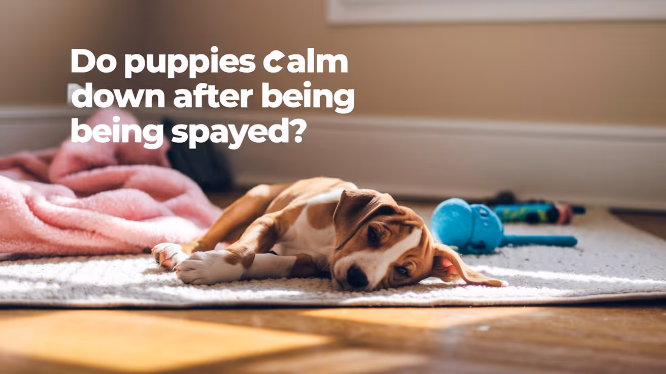
Do Puppies Calm Down After Being Spayed?
Learn how spaying affects puppy behavior and whether puppies calm down after the procedure.
Spaying a puppy and expecting her to calm down afterward is one of the most common expectations owners bring to the vet. Sometimes it is accurate. Often it is not quite right.
Puppies behave the way they do for several reasons: genetics, breed characteristics, developmental stage, exercise needs, and yes, hormones. Spaying addresses only the hormonal piece.
Here is what the evidence actually says about whether spaying calms puppies down, when it applies, and when it does not.
Quick answer: Spaying can reduce hormone-driven behaviors in female puppies, but most puppy energy is not hormone-driven. The wild, excitable behavior typical of young dogs is largely developmental and will naturally settle as the puppy matures, with or without surgery. Spaying eliminates heat cycle behaviors but does not reprogram a puppy's underlying energy level or temperament.
Key takeaways
- Most puppy energy is developmental, not hormonal: High activity in puppies reflects youth and brain development, not reproductive hormones.
- Heat cycle behaviors are eliminated by spaying: Restlessness, roaming, and urine marking during heat stop permanently after spaying.
- Natural maturation also produces calming: Puppies typically settle between 18 months and 3 years regardless of spay status.
- Timing of spaying affects long-term behavioral outcomes: Research suggests very early spaying may increase fearfulness in some dogs.
- Training and exercise calm puppies more reliably than surgery: These remain the primary tools for managing puppy behavior.
- Full post-spay hormonal adjustment takes two to three months: Not days.
Why puppies are energetic in the first place
Before connecting spaying to calming, it is worth understanding why puppies behave the way they do.
Puppy behavior is driven by several overlapping factors:
- Developmental stage: Puppies are neurologically immature. Their brains are still forming connections, their impulse control is limited, and they are driven to explore, play, and interact constantly.
- Breed genetics: A Border Collie puppy's drive, a Labrador's enthusiasm, a Jack Russell's intensity: these are genetic. They are not hormonal.
- Exercise and stimulation needs: Puppies that are not adequately exercised and mentally engaged redirect that energy into chaos.
- Learning and socialization: Puppies are in a constant state of learning. Without clear guidance, they default to whatever feels interesting.
- Hormonal influences: A relatively small proportion of puppy behavior is directly driven by reproductive hormones, and this increases as the puppy approaches sexual maturity.
Spaying addresses only item 5. Items 1 through 4 are untouched by surgery.
What spaying actually removes in a female puppy
Spaying removes the ovaries, which are the primary source of estrogen and progesterone. The effects are specific.
What spaying eliminates:
- Heat cycles, which in female dogs typically begin around 5 to 6 months of age in small breeds and later in large breeds
- The behavioral disruption of heat: restlessness, urine marking to attract males, roaming, vocalization
- False pregnancy cycles if the puppy would have been prone to these
- Cyclical mood changes linked to the hormonal fluctuations of estrus
What spaying does not eliminate:
- Breed-driven energy and drive
- Developmental excitability and poor impulse control that comes with youth
- Learned behaviors from the puppy's environment and training history
- Anxiety or fearfulness that has non-hormonal causes
The natural calming process: maturation
Here is a piece of the puzzle that often gets overlooked in the conversation about spaying and calming.
Puppies naturally become calmer as they mature.
Most dogs reach a noticeably more settled behavioral baseline between 18 months and 3 years of age. This happens regardless of spay status. The neurological development that improves impulse control, the reduction of developmental chaos, and the establishment of routines all contribute to this natural calming process.
Many owners who spay a puppy and then notice she is calmer six months later attribute the change to the surgery. In many cases, maturation deserves significant credit.
This is not to dismiss the real effects of spaying. It is to provide accurate context so that owners do not overestimate surgery's role or underestimate the contribution of time, training, and maturation.
What research says about puppies and spaying
The behavioral research on early spaying and puppies is worth understanding before making timing decisions.
Established findings:
- Spaying before the first heat cycle eliminates heat-related behaviors and reduces mammary cancer risk significantly.
- Behavioral problems were reduced or eliminated in the majority of spayed females in studies, but this applies primarily to hormone-driven behaviors.
More cautionary findings:
- Multiple studies have found that dogs spayed at a very young age, particularly before six months, show higher rates of fearfulness and anxiety than dogs spayed later.
- A study of Vizslas found that the younger the dog at the time of spaying, the earlier the mean age of onset of a behavioral disorder.
- Research using the C-BARQ assessment tool found increased fearfulness and touch sensitivity in spayed dogs compared to intact females.
These findings do not mean spaying causes behavioral problems in all puppies. They mean that very early spaying carries a different behavioral risk profile than spaying after maturity, and this is worth discussing with your vet when deciding on timing.
For the broader picture of full overview of behavior changes after spaying, including how the research applies to different ages and sexes, that guide provides the complete context.
Breed differences: why some puppies stay energetic after spaying
Breed temperament and genetic drive are not hormonal. Surgery does not change them.
A working Border Collie will need two hours of physical and mental exercise daily after spaying, just as she did before. An Australian Shepherd's herding instinct will remain intact. A Jack Russell Terrier's prey drive and intensity will not be reduced by the removal of her ovaries.
High-drive breeds are that way because of generations of selective breeding for those traits. Those traits sit in genetics, not in the reproductive system.
What this means practically:
If you have a high-energy breed puppy and are hoping spaying will reduce her energy to more manageable levels, realign those expectations before surgery. The energy level will remain. What changes is the absence of heat cycles and their associated disruption.
The timeline: when do puppies settle after spaying?
Two separate timelines operate here, and distinguishing them is important.
Post-spay hormonal adjustment:
- Days 1 to 14: Recovery from surgery. Any behavioral changes reflect pain and anesthesia.
- Weeks 2 to 6: Estrogen and progesterone levels declining.
- Months 2 to 3: Hormonal stabilization complete.
Developmental maturation:
- 6 to 12 months: Peak puppy chaos in most breeds.
- 12 to 18 months: Gradual improvement in impulse control and ability to settle.
- 18 months to 3 years: Most dogs reach a noticeably more settled behavioral baseline.
The calming that owners observe after spaying a puppy is often a combination of both timelines working together. The hormonal adjustment contributes. Maturation contributes. Exercise and training contribute. It is rarely attributable to surgery alone.
For the developmental nuances specific to younger dogs, the guide on calming effects in adult dogs covers the broader behavioral picture across different life stages.
What actually calms a puppy down
Whether or not your puppy is spayed, these are the most reliable interventions for managing puppy behavior:
1. Adequate physical exercise
A puppy that has genuinely burned physical energy is a calmer puppy at rest. Exercise needs vary by breed but most puppies are under-exercised relative to their needs.
2. Mental stimulation
Puzzle feeders, sniff walks, training sessions, and enrichment activities tire puppies in a way that physical exercise alone does not. Mental fatigue produces genuine calm.
3. Sleep
Puppies need significantly more sleep than adult dogs, often 16 to 18 hours per day. Over-tired puppies become dysregulated and difficult. Enforced rest periods reduce chaos.
4. Consistent training
Teaching a puppy to settle on a mat, to wait before doors, to sit before meals: these habits build impulse control that transfers across contexts. Training is the most durable path to a calm puppy.
5. Routine
Predictable daily schedules reduce ambient anxiety and help puppies know what to expect, which supports more settled behavior.
Spaying can contribute by removing the hormonal disruption of heat cycles. But it is most effective as one part of this picture, not as a standalone solution.
Frequently asked questions
Will my puppy calm down after being spayed?
She may become calmer in specific ways: heat cycle behaviors will be eliminated, and the cyclical hormonal disruption of estrus will stop. Whether she becomes generally calmer depends on what is driving her behavior. Developmental energy and breed-specific drive are not affected by spaying. For the best age to spay a puppy and how timing affects both health and behavioral outcomes, that guide covers the evidence-based timing recommendations in detail.
My puppy is three months old and incredibly wild. Should I spay her now to calm her down?
Spaying a puppy that young is unlikely to calm her down because the energy she is displaying is developmental, not hormonal. Female puppies typically do not experience their first heat until 5 to 6 months of age at the earliest. Spaying before that will not eliminate hormonal behavior because it has not yet started. What will help is exercise, training, mental stimulation, and sleep.
How long after spaying will my puppy start to calm down?
If the calming effect is going to occur, behavioral changes related to hormone reduction appear over two to six weeks and stabilize by two to three months. Any calming that happens faster than this is likely related to recovery rest and routine, not hormonal adjustment.
My puppy was spayed and she is just as hyper as before. Why?
Because spaying removes the hormonal layer of behavior, not the developmental and breed-driven layers. If her energy and excitability are not heat-driven, spaying was never going to address them. Training, exercise, and maturation are the tools for this type of puppy behavior.
What should I expect during my puppy's recovery from spaying?
The 10 to 14 day recovery period requires activity restriction, E-collar use, and daily incision monitoring. For a complete day-by-day guide on what to expect during puppy recovery, including how puppy recovery specifically differs from adult dog recovery, that guide covers every element of the post-surgical window.
Puppies are not energetic because of estrogen. They are energetic because they are puppies. Spaying addresses the hormonal layer of behavior, which for a young dog has barely had time to develop before surgery. The wild, chaotic, wonderful energy of a puppy is developmental. It settles with time, training, exercise, and maturation. Spaying contributes by preventing heat cycles and the disruption they bring. The rest of the work is yours.
Resources
The following sources were used as reference and background for this article:
- American Kennel Club. How Will Spaying Change My Dog? anasazivet.com
- Stanley Coren, Psychology Today. Are There Behavior Changes When Dogs Are Spayed or Neutered? psychologytoday.com
- Rebarkable. Do Puppies Calm Down After Being Spayed? rebarkable.com
- American Veterinary Medical Association (AVMA). Spaying and Neutering. avma.org
- PubMed. Changes in the behavior of dogs after castration. pubmed.ncbi.nlm.nih.gov

Dog Dislocated Shoulder Treatment Cost and Recovery
Learn about dog dislocated shoulder treatment costs, recovery time, and care tips to help your pet heal safely and comfortably.
A dog dislocated shoulder is a painful injury that can happen from trauma or accidents. It causes your dog to limp, cry, or avoid using the leg. Understanding the treatment cost and recovery process helps you prepare for your pet’s care.
This article explains how much dog dislocated shoulder treatment costs, what to expect during recovery, and how to support your dog’s healing. You will learn about diagnosis, treatment options, and aftercare tips to ensure the best outcome for your pet.
What causes a dog’s shoulder to dislocate?
Dogs can dislocate their shoulders due to sudden trauma or repeated stress. Knowing the causes helps you prevent future injuries and recognize symptoms early.
Shoulder dislocation happens when the upper arm bone slips out of its socket. This injury can be partial or complete, affecting your dog’s mobility and comfort.
- Trauma from accidents: Falls, car accidents, or rough play can force the shoulder joint out of place, causing sudden pain and lameness.
- Sports injuries: Active dogs involved in agility or running may strain their shoulder joint, increasing dislocation risk over time.
- Congenital joint weakness: Some dogs have naturally loose joints, making them more prone to dislocations even with minor stress.
- Degenerative joint disease: Arthritis or other joint problems weaken the shoulder, increasing the chance of dislocation during normal activities.
Understanding these causes helps you identify risk factors and seek prompt veterinary care if your dog shows signs of shoulder injury.
How is a dog dislocated shoulder diagnosed?
Diagnosing a dislocated shoulder in dogs requires a thorough physical exam and imaging tests. Early diagnosis ensures proper treatment and reduces complications.
Your vet will check for pain, swelling, and abnormal limb position. They may also test your dog’s range of motion and watch how it walks.
- Physical examination: The vet will palpate the shoulder to detect swelling, pain, or abnormal joint movement indicating dislocation.
- X-rays: Radiographs confirm the dislocation and help rule out fractures or other bone injuries around the shoulder.
- Ultrasound imaging: This may be used to assess soft tissue damage like ligament tears or muscle injuries near the shoulder joint.
- Joint fluid analysis: In some cases, fluid samples help detect infection or inflammation contributing to joint instability.
Accurate diagnosis guides the treatment plan and helps predict recovery time for your dog’s shoulder injury.
What treatment options are available for a dog dislocated shoulder?
Treatment depends on the severity of the dislocation and any associated injuries. Your vet will recommend the best option to restore joint stability and reduce pain.
Options range from conservative care to surgery. Early treatment improves outcomes and prevents chronic problems.
- Closed reduction: The vet manually repositions the shoulder joint under sedation or anesthesia without surgery, suitable for simple dislocations.
- Immobilization: After reduction, a sling or bandage may keep the joint stable while soft tissues heal, usually for 2-4 weeks.
- Surgical repair: Surgery may be needed if the dislocation is severe, recurrent, or involves ligament damage to stabilize the joint.
- Pain management: Medications like NSAIDs or opioids help control pain and inflammation during recovery.
Your vet will tailor the treatment to your dog’s specific injury and health status to ensure the best chance of full recovery.
How much does dog dislocated shoulder treatment cost?
The cost of treating a dog’s dislocated shoulder varies widely based on treatment type and location. Knowing typical expenses helps you plan financially for your pet’s care.
Costs include veterinary exams, imaging, medications, and possible surgery. Emergency visits may increase the price.
- Veterinary consultation: Initial exams typically cost between $50 and $150 depending on the clinic and region.
- Diagnostic imaging: X-rays usually range from $100 to $300; ultrasound may add $150 to $400 if needed.
- Closed reduction procedure: Non-surgical realignment can cost $200 to $600 including sedation and follow-up care.
- Surgical repair: Surgery costs vary from $1,000 to $3,000 depending on complexity, hospital fees, and aftercare.
Additional costs may include pain medications, physical therapy, and follow-up visits. Pet insurance or payment plans can help manage expenses.
What is the typical recovery time for a dog with a dislocated shoulder?
Recovery time depends on the injury severity and treatment method. Most dogs need several weeks to months to heal fully.
Proper rest and rehabilitation are essential to regain strength and prevent re-injury during recovery.
- Initial healing phase: Immobilization usually lasts 2-4 weeks to allow soft tissues to repair and reduce pain.
- Physical therapy: Gentle exercises and controlled activity start after immobilization to restore range of motion and muscle strength.
- Full recovery timeline: Most dogs recover within 6 to 12 weeks, but some may take longer depending on complications.
- Monitoring for complications: Watch for signs of persistent pain, swelling, or lameness that may require further treatment.
Following your vet’s recovery plan closely improves your dog’s chances of returning to normal activity without long-term problems.
How can you support your dog’s recovery at home?
Home care plays a vital role in your dog’s healing after a shoulder dislocation. You can help by providing a safe environment and following veterinary instructions carefully.
Proper care reduces stress on the injured joint and promotes comfort during recovery.
- Limit activity: Restrict running, jumping, and rough play to prevent re-injury while the shoulder heals.
- Use supportive devices: Slings or braces recommended by your vet help stabilize the joint and reduce pain.
- Administer medications: Give prescribed pain relievers and anti-inflammatories exactly as directed to control discomfort.
- Provide a comfortable resting area: A soft bed in a quiet space helps your dog rest and recover without unnecessary movement.
Regular follow-up visits allow your vet to track healing progress and adjust care as needed for the best outcome.
What are the risks if a dog’s shoulder dislocation is untreated?
Ignoring a dislocated shoulder can lead to chronic pain, joint instability, and permanent damage. Early treatment prevents these serious complications.
Untreated injuries may worsen over time, making future treatment more difficult and costly.
- Chronic lameness: Persistent limping and weakness reduce your dog’s quality of life and mobility.
- Joint arthritis: Untreated dislocations increase wear on cartilage, causing painful arthritis later.
- Muscle atrophy: Lack of use leads to muscle wasting around the shoulder, weakening the limb further.
- Recurring dislocations: Without proper repair, the shoulder may repeatedly dislocate, causing ongoing pain and damage.
Prompt veterinary care is essential to avoid these risks and help your dog regain normal function and comfort.
Conclusion
Dog dislocated shoulder treatment cost and recovery vary depending on injury severity and care needed. Early diagnosis and proper treatment improve healing and reduce long-term problems.
By understanding causes, treatment options, and home care, you can support your dog through recovery. Timely veterinary care and careful follow-up help your pet return to a happy, active life.
What signs indicate my dog has a dislocated shoulder?
Look for sudden limping, swelling around the shoulder, pain when moving the leg, and reluctance to bear weight on the affected limb.
Can a dislocated shoulder heal without surgery in dogs?
Yes, many simple dislocations heal with closed reduction and immobilization, but severe or recurrent cases often require surgery for stability.
How long should I restrict my dog’s activity after shoulder treatment?
Activity should be limited for at least 2 to 4 weeks during immobilization, followed by gradual reintroduction of movement under veterinary guidance.
Are there any home remedies to reduce my dog’s shoulder pain?
Only use vet-approved pain medications; cold compresses may help initially, but avoid unapproved treatments to prevent harm.
When should I contact my vet during my dog’s recovery?
Contact your vet if your dog shows increased pain, swelling, limping, or signs of infection like redness or discharge at the injury site.

Torn Meniscus Surgery Cost in Dogs Explained
Learn about torn meniscus surgery cost in dogs, including factors affecting price, procedure details, and recovery tips for your pet's health.
A torn meniscus in dogs is a common injury that affects the knee joint, causing pain and mobility issues. If your dog has this problem, you might wonder about the cost of surgery and what it involves. Understanding the expenses and treatment options can help you prepare for your pet's care.
This article explains the typical cost of torn meniscus surgery in dogs, the factors that influence pricing, and what you can expect during recovery. You will learn how to manage your dog's health and make informed decisions about treatment.
What is torn meniscus surgery in dogs?
Torn meniscus surgery in dogs is a procedure to repair or remove damaged cartilage in the knee joint. The meniscus acts as a cushion between bones, and injury can cause pain and lameness. Surgery aims to restore joint function and reduce discomfort.
The surgery is usually recommended when conservative treatments like rest and medication do not improve the dog's condition. It involves anesthesia and specialized techniques to address the tear.
- Purpose of surgery: To repair or remove the damaged meniscal cartilage to relieve pain and improve knee stability in dogs.
- Common causes: Meniscus tears often result from ligament injuries or trauma during activities like running or jumping.
- Surgical techniques: Options include meniscectomy (removal) or meniscal repair depending on the tear's location and severity.
- Post-surgery goals: Restore normal joint movement, reduce arthritis risk, and help the dog regain mobility.
Understanding the surgery helps you prepare for the treatment and care your dog will need.
How much does torn meniscus surgery cost in dogs?
The cost of torn meniscus surgery in dogs varies widely depending on location, clinic, and the dog's specific needs. On average, prices range from $1,500 to $4,000. This includes pre-surgical exams, anesthesia, surgery, and initial post-op care.
Knowing the cost breakdown can help you budget and discuss options with your veterinarian.
- Base surgery fee: Typically between $1,000 and $3,000, covering the surgical procedure and operating room use.
- Pre-surgical tests: Blood work and X-rays may cost $200 to $500 to assess the dog's health before surgery.
- Anesthesia and monitoring: Usually $300 to $700, essential for safe surgery and pain control.
- Post-operative care: Includes medications, bandages, and follow-up visits costing $200 to $500.
Costs may increase if complications arise or if advanced imaging like MRI is needed.
What factors affect the cost of meniscus surgery in dogs?
Several factors influence the total cost of torn meniscus surgery in dogs. These include the dog's size, the complexity of the injury, and the clinic's location. Understanding these helps you anticipate expenses and plan accordingly.
Discussing these factors with your vet can clarify the expected costs and available options.
- Dog's size and weight: Larger dogs may require more anesthesia and longer surgery time, increasing costs.
- Severity of tear: Complex or multiple tears need more surgical time and skill, raising the price.
- Veterinary clinic location: Urban or specialty clinics often charge more than rural general practices.
- Surgeon's experience: Board-certified surgeons may have higher fees but offer specialized care.
Knowing these factors helps you make informed choices about your dog's treatment plan.
What are the risks and benefits of torn meniscus surgery for dogs?
Surgery for a torn meniscus can improve your dog's quality of life but also carries some risks. Weighing these helps you decide if surgery is the best option for your pet.
The benefits often outweigh the risks when the injury causes significant pain or limits mobility.
- Benefit - Pain relief: Surgery can reduce joint pain and discomfort caused by the torn meniscus.
- Benefit - Improved mobility: Dogs often regain better movement and activity levels after recovery.
- Risk - Infection: Any surgery carries a small risk of infection requiring additional treatment.
- Risk - Anesthesia complications: Though rare, anesthesia can cause adverse reactions in some dogs.
Discussing risks and benefits with your vet ensures the best decision for your dog's health.
How should you prepare your dog for meniscus surgery?
Proper preparation before surgery helps reduce risks and supports a smooth procedure. Your veterinarian will give specific instructions tailored to your dog’s needs.
Following these steps can improve surgery outcomes and reduce stress for your pet.
- Pre-surgery fasting: Your dog should avoid food for 8-12 hours before surgery to prevent anesthesia complications.
- Health evaluation: Complete blood tests and physical exams ensure your dog is healthy enough for surgery.
- Medication review: Inform your vet about all medications or supplements your dog takes to avoid interactions.
- Arrange post-op care: Prepare a quiet, comfortable space and plan for restricted activity during recovery.
Good preparation helps your dog have a safer surgery and faster healing.
What is the recovery process after torn meniscus surgery in dogs?
Recovery after meniscus surgery requires careful management to ensure healing and prevent re-injury. The process usually takes 6 to 12 weeks, depending on the dog's age and health.
Following your vet’s instructions closely improves your dog’s chances of a full recovery.
- Restricted activity: Limit running, jumping, and stairs to protect the healing joint for several weeks.
- Physical therapy: Controlled exercises and rehabilitation help restore strength and flexibility.
- Pain management: Administer prescribed pain medications and monitor for discomfort signs.
- Follow-up visits: Regular check-ups allow your vet to assess healing and adjust care as needed.
Patience and consistency during recovery are key to your dog’s long-term joint health.
Conclusion
Torn meniscus surgery cost in dogs varies but generally ranges from $1,500 to $4,000 depending on many factors. Understanding the procedure, risks, and recovery helps you prepare financially and emotionally for your pet’s care.
With proper preparation and post-operative management, surgery can relieve your dog’s pain and improve mobility. Always consult your veterinarian to choose the best treatment plan for your dog’s torn meniscus injury.
What is the typical recovery time after torn meniscus surgery in dogs?
Recovery usually takes 6 to 12 weeks, during which activity must be restricted and physical therapy may be needed to regain strength and mobility.
Can torn meniscus surgery prevent arthritis in dogs?
While surgery can reduce joint damage and pain, it may not fully prevent arthritis but can slow its progression and improve quality of life.
Are there non-surgical options for treating a torn meniscus in dogs?
Yes, mild tears may be managed with rest, anti-inflammatory medications, and physical therapy, but surgery is often needed for severe cases.
How do I know if my dog needs meniscus surgery?
Your vet will diagnose based on clinical signs, physical exams, and imaging like X-rays or MRI to determine if surgery is necessary.
Is torn meniscus surgery painful for dogs?
Dogs receive anesthesia during surgery and pain medications afterward to minimize discomfort, making the procedure as pain-free as possible.
Do Dogs Calm Down After Being Spayed?
Learn how dogs behave after being spayed and when to expect them to calm down post-surgery.
It is one of the most common reasons owners choose to spay their female dog. The expectation of a calmer, more settled companion afterward.
Sometimes that is exactly what happens. Sometimes it does not. And in a small subset of cases, anxiety or excitability actually increases.
Understanding why gives you a more accurate picture of what to expect, which makes the outcome less surprising in either direction.
Quick answer: Many dogs do become calmer after spaying, but the effect is specific to hormone-driven behaviors, not overall temperament. Dogs that were restless, anxious, or excitable due to heat cycles typically show significant improvement. Dogs whose energy or anxiety has non-hormonal causes will not reliably calm down. Most behavioral changes, if they occur, appear over two to three months.
Key takeaways
- The calming effect is real but specific: It applies to behaviors driven by reproductive hormones, not all behaviors.
- Heat cycle behaviors disappear: Restlessness, roaming, frequent urination, and cyclical mood shifts all stop after spaying.
- Core energy level and personality do not change: A high-energy dog will still be high-energy after spaying.
- Breed and individual temperament matter: Some breeds retain high energy regardless of hormone status.
- The timeline is weeks to months, not days: Hormone levels decline gradually, so behavioral changes are gradual too.
- A small subset of dogs show increased fearfulness: This is documented in research and worth knowing about before surgery.
What "calming down" actually means after spaying
When people say they want their dog to calm down, they usually mean one of two things:
- They want heat cycle behaviors to stop: the roaming, the restlessness, the vocalization, the constant male attention.
- They want a generally more settled dog overall.
Spaying reliably delivers the first. The second is less predictable.
This distinction is the core of the honest answer to this question.
What spaying removes: the hormonal layer
Spaying removes the ovaries, which are the primary source of estrogen and progesterone. These hormones regulate the reproductive cycle and influence behaviors tied to it.
During heat cycles, which typically occur every six to eight months in adult dogs, estrogen surges create a set of predictable behaviors:
- Restlessness and inability to settle
- Increased vocalization
- Frequent urination in small amounts to signal reproductive status
- Strong drive to escape and find a mate
- Attracting intact male dogs
- Cyclical irritability or anxiety
All of these stop after spaying. The hormonal trigger is permanently removed.
Which dogs calm down most noticeably after spaying
Dogs whose difficult behaviors are primarily or significantly driven by the reproductive cycle show the most consistent improvement after spaying.
Dogs most likely to become calmer:
- Female dogs that showed significant behavior changes during heat cycles, including restlessness, anxiety, and clinginess
- Dogs prone to false pregnancies, with associated nesting, object guarding, and protective behavior
- Dogs that attempted to escape or roam during heat periods
- Dogs whose cyclical mood swings were disruptive to daily life
For these dogs, spaying removes a recurring source of behavioral disruption. The result is often a more predictable, stable dog.
Which dogs may not calm down after spaying
Dogs whose energy level, excitability, or anxiety comes from sources other than reproductive hormones will not reliably settle after spaying.
Dogs less likely to show a calming effect:
- High-energy working or sport breeds where drive is genetic, not hormonal
- Dogs with established anxiety, fearfulness, or reactivity from non-hormonal causes
- Dogs with learned problematic behaviors from their history and environment
- Dogs whose excitability comes from insufficient exercise or mental stimulation
A Border Collie that is highly energetic before spaying will still be highly energetic after. A Labrador that pulls on the leash before spaying will still pull after. These behaviors are not driven by estrogen.
The timeline: when does calming happen?
Behavioral changes from spaying do not happen within days of surgery. The immediate post-surgery period reflects recovery from anesthesia and pain, not hormonal adjustment.
Realistic timeline:
Owners who expect a noticeably calmer dog within days of surgery are working with an inaccurate timeline. The changes are gradual and unfold over weeks to months.
What research says about calming after spaying
The evidence is genuinely mixed, which is worth knowing rather than glossing over.
Supporting the calming effect:
A study of 591 dogs found behavioral problems reduced or eliminated in 59% of spayed females. Heat cycle-related behaviors showed the most consistent improvement. Many owners of females that were particularly difficult during heat cycles report significant positive changes after spaying.
Complicating the picture:
Two large-scale studies using the Canine Behavioral Assessment and Research Questionnaire (C-BARQ) found that sterilization was associated with a roughly 31% increase in fearfulness and a 33% increase in touch sensitivity in some dogs. These findings apply to both males and females.
What does this mean in practice? For a meaningful subset of dogs, spaying does not produce a calmer animal. It may even produce a more anxious one, particularly in dogs that had pre-existing fearfulness or were sterilized at a young age.
This does not mean spaying is wrong for these dogs. It means the behavioral outcome cannot be guaranteed, and expectations should be calibrated accordingly.
For the full picture of all the behavior changes to expect after spaying, including the research on unexpected outcomes, that guide covers the complete behavioral landscape across both sexes.
Does calming depend on the dog's age at spaying?
Yes, and this is one of the most important variables.
Dogs spayed before their first heat cycle:
The hormonal disruption of heat cycles is never established. Many owners report these dogs are consistently more settled than intact females across their lifetime, without ever having the difficult heat cycle period.
Dogs spayed after several heat cycles:
The calming effect from eliminating heat cycles is still real and significant. However, any behaviors that have become habitual through repetition during heat cycles may persist longer or require training to address.
Dogs spayed very young (before 6 months):
Research suggests a higher association with fearfulness in dogs sterilized very early, before hormonal maturity. For whether this applies specifically to puppies, the guide on calming effects specific to female dogs addresses this age-specific question in detail. For the puppy-specific picture, the guide on whether puppies calm down after being spayed covers the developmental nuances unique to younger dogs.
When the expected calming does not happen
If your dog does not seem noticeably calmer after spaying, consider these factors before concluding the surgery did not work.
Check the timeline: Have two to three months passed since surgery? If not, the hormonal adjustment may still be in progress.
Identify the behavior source: Is the behavior you wanted to address actually hormone-driven? If it is not, spaying was never going to address it.
Consider habituation: Some behaviors that started as hormone-driven become habitual over time. The hormone is removed but the habit persists. Consistent training addresses habituated behavior.
Consider an underlying anxiety: If your dog became more fearful or anxious post-surgery, this is a documented outcome in some dogs. A veterinary behaviorist or your regular vet can help evaluate what is happening and what options exist.
Understanding the truth behind the calming effect myth, including why the expectation of calming is sometimes oversold, helps put the variable outcomes in context.
What actually makes dogs calmer long-term
Whether or not spaying produces a calming effect, these factors reliably influence how settled and manageable a dog is:
- Adequate physical exercise: A dog that is genuinely tired is a calmer dog. Exercise needs are determined by breed and individual temperament.
- Mental stimulation: Breed-appropriate mental work, puzzle feeders, training sessions, and enrichment activities reduce restless, problematic behavior.
- Consistent training: Clear, reinforced expectations give dogs a behavioral framework. This is the single most reliable path to a calmer dog.
- Routine and predictability: Dogs settle more easily in stable, predictable environments.
Spaying contributes one element to this picture. It is not a standalone solution.
Frequently asked questions
How long after being spayed will my dog calm down?
If the calming effect is going to occur, most hormone-driven behavioral changes become apparent within two to six weeks as hormone levels decline. Full stabilization typically takes two to three months. Dogs that are still showing challenging behaviors three to four months after surgery are unlikely to improve further from the hormonal change alone.
Will spaying make my high-energy dog less active?
Probably not significantly. Energy level in dogs is primarily determined by breed, genetics, and conditioning. Spaying can cause a modest metabolic slowdown, but it does not reduce drive or enthusiasm. A high-energy breed will still need the same amount of exercise after spaying.
My dog got more anxious after being spayed. Why?
Research has documented increased fearfulness in a subset of dogs after sterilization, associated with a roughly 31% increase in fearfulness in some studies. This appears to occur more often in dogs sterilized at a young age and in dogs with pre-existing anxiety. If your dog shows increased anxiety after spaying, speak to your vet. A referral to a certified veterinary behaviorist may be helpful.
Will spaying help with aggression?
It depends on the type of aggression. Aggression linked to the heat cycle, competition with other female dogs, or false pregnancy guarding often decreases. Fear-based aggression, learned aggression, and resource guarding are not hormone-driven and are not reliably improved by spaying. For whether lower testosterone calms dogs down in the male counterpart to this question, that guide covers the testosterone-behavior link specifically.
Does breed affect whether spaying calms a dog down?
Yes. Breed temperament significantly influences baseline energy and anxiety levels. High-drive working breeds such as Border Collies, Australian Shepherds, and Belgian Malinois have energy and drive that is genetic, not hormonal. Removing reproductive hormones does not change these fundamental breed characteristics. Whether your specific dog falls into a calmer range after spaying depends on what was driving any difficult behavior in the first place.
The calming effect of spaying is real for dogs whose difficult behaviors are hormone-driven. For those dogs, eliminating the heat cycle and its associated hormonal disruption produces a meaningfully more stable animal. For dogs whose energy or anxiety comes from other sources, spaying is still beneficial for health reasons but should not be expected to produce behavioral transformation. The most accurate prediction is not based on "spaying calms dogs" but on "removing a specific hormonal influence changes the behaviors that influence drives."
Resources
The following sources were used as reference and background for this article:
- Stanley Coren, Psychology Today. Are There Behavior Changes When Dogs Are Spayed or Neutered? psychologytoday.com
- PubMed. Changes in the behavior of dogs after castration. pubmed.ncbi.nlm.nih.gov
- ManyPets. Do Dogs Change After Being Neutered or Spayed? manypets.com
- American Veterinary Medical Association (AVMA). Spaying and Neutering. avma.org
- Houndsy. How Spaying Affects Dog Behavior. houndsy.com

Why Does My Dog Have Scabs on Her Back?
Learn why your dog has scabs on her back, common causes, treatments, and when to see a vet for proper care.
Seeing scabs on your dog's back can be worrying. Scabs are signs of skin damage or irritation. They may result from allergies, infections, parasites, or injuries. Understanding why your dog has scabs is important to help her heal quickly and comfortably.
This article explains the common reasons dogs develop scabs on their backs. You will learn about causes, symptoms, treatments, and when to visit a veterinarian. Knowing these details helps you care for your dog better and prevent future skin problems.
What Causes Scabs on a Dog's Back?
Scabs form when the skin is injured or inflamed. Many conditions can cause scabs on a dog's back. Identifying the cause is key to effective treatment.
- Allergic reactions: Dogs can develop scabs due to allergies to food, pollen, or flea bites, causing itching and skin damage from scratching.
- Parasite infestations: Fleas, ticks, and mites bite the skin, leading to irritation, itching, and scab formation from constant scratching.
- Skin infections: Bacterial or fungal infections can cause sores and scabs as the skin tries to heal from the infection.
- Injuries or trauma: Scratches, bites, or wounds from rough play or accidents can scab over as they heal.
Knowing these causes helps you observe your dog’s behavior and environment to find the source of the problem.
How Can Allergies Lead to Scabs on My Dog's Back?
Allergies are a common cause of itchy skin and scabs in dogs. Allergic reactions cause inflammation and discomfort, leading dogs to scratch or bite their skin.
- Food allergies: Certain ingredients in dog food can trigger skin reactions, causing redness and scabs from scratching.
- Environmental allergies: Pollens, dust mites, or mold spores can irritate your dog's skin, especially on the back and neck areas.
- Flea allergy dermatitis: Flea saliva causes intense itching and scabbing even if only a few fleas are present.
- Contact allergies: Chemicals in cleaning products or plants can cause localized skin irritation and scabs.
Managing allergies often requires identifying triggers and using medications or diet changes to reduce symptoms.
What Role Do Parasites Play in Causing Scabs?
Parasites are a frequent cause of skin problems in dogs. Fleas, ticks, and mites irritate the skin and cause itching that leads to scabs.
- Fleas: Flea bites cause itching and allergic reactions, leading to scabs from excessive scratching and biting.
- Ticks: Ticks attach to the skin and can cause localized irritation and scabbing around the bite site.
- Mange mites: Mites burrow into the skin causing mange, which leads to hair loss, redness, and scabs.
- Prevention importance: Regular parasite control helps prevent infestations and the resulting skin damage.
Parasite control products and vet care are essential to stop scabs caused by parasites.
How Do Skin Infections Cause Scabs on Dogs?
Skin infections often develop after injury or from underlying conditions. They cause inflammation, pus, and scabs as the skin tries to heal.
- Bacterial infections: Bacteria enter broken skin causing hot spots, redness, and scabs that may ooze fluid.
- Fungal infections: Yeast or ringworm infections cause flaky, scabby patches often with hair loss.
- Secondary infections: Scratching from allergies or parasites can introduce bacteria, worsening scabs.
- Treatment necessity: Antibiotics or antifungal medications are often needed to clear infections and heal scabs.
Early vet diagnosis and treatment prevent infections from spreading or becoming chronic.
Can My Dog’s Behavior Cause Scabs on Her Back?
Behavioral factors like excessive licking, scratching, or biting can cause scabs. These behaviors often result from discomfort or stress.
- Itchiness response: Dogs scratch or bite itchy areas, causing skin damage and scabs.
- Stress or anxiety: Nervous dogs may lick or chew their backs compulsively, leading to scabs.
- Environmental irritants: Rough surfaces or allergens in bedding can cause irritation and scabbing.
- Observation importance: Watching your dog’s behavior helps identify if self-trauma causes scabs.
Addressing underlying causes and providing enrichment reduces harmful behaviors and skin damage.
When Should I Take My Dog to the Vet for Scabs?
Not all scabs require immediate veterinary care, but some signs mean you should see a vet promptly.
- Persistent scabs: Scabs that do not heal or keep returning need professional evaluation.
- Widespread skin lesions: If scabs cover large areas or spread quickly, vet care is needed.
- Signs of infection: Swelling, pus, bad odor, or pain around scabs indicate infection requiring treatment.
- Behavior changes: Excessive scratching, lethargy, or loss of appetite alongside scabs warrant a vet visit.
Early diagnosis and treatment improve outcomes and prevent complications.
How Can I Treat and Prevent Scabs on My Dog’s Back?
Treatment depends on the cause but generally includes soothing the skin and preventing further damage. Prevention focuses on good hygiene and parasite control.
- Topical treatments: Medicated shampoos and creams help reduce itching and promote healing of scabs.
- Parasite control: Regular flea and tick prevention stops infestations that cause scabs.
- Diet and supplements: Balanced nutrition and omega fatty acids support healthy skin and reduce allergies.
- Environmental care: Keep bedding clean and avoid irritants to prevent skin problems.
Following your vet’s advice and maintaining good skin care routines help your dog stay comfortable and scab-free.
Conclusion
Scabs on your dog's back can result from allergies, parasites, infections, or injuries. Identifying the cause is essential for proper treatment and healing. Watching your dog's behavior and environment helps find the source of scabs.
Timely veterinary care, good hygiene, and parasite control prevent scabs and keep your dog’s skin healthy. If scabs persist or worsen, consult your vet for diagnosis and treatment to ensure your dog’s comfort and well-being.
Why does my dog have scabs on her back?
Scabs on your dog’s back usually come from skin irritation caused by allergies, parasites, infections, or injuries leading to scratching and skin damage.
Can fleas cause scabs on my dog’s back?
Yes, flea bites cause itching and allergic reactions that make dogs scratch and develop scabs on their backs and other areas.
How do I know if my dog’s scabs are infected?
Signs of infection include redness, swelling, pus, bad smell, and pain around scabs. Infections need veterinary treatment to heal properly.
What home care can I do for my dog’s scabs?
Keep the area clean, use vet-recommended shampoos, prevent scratching, and maintain parasite control to support healing at home.
When should I see a vet about my dog’s scabs?
See a vet if scabs persist, spread, show infection signs, or if your dog is very itchy, lethargic, or losing appetite.
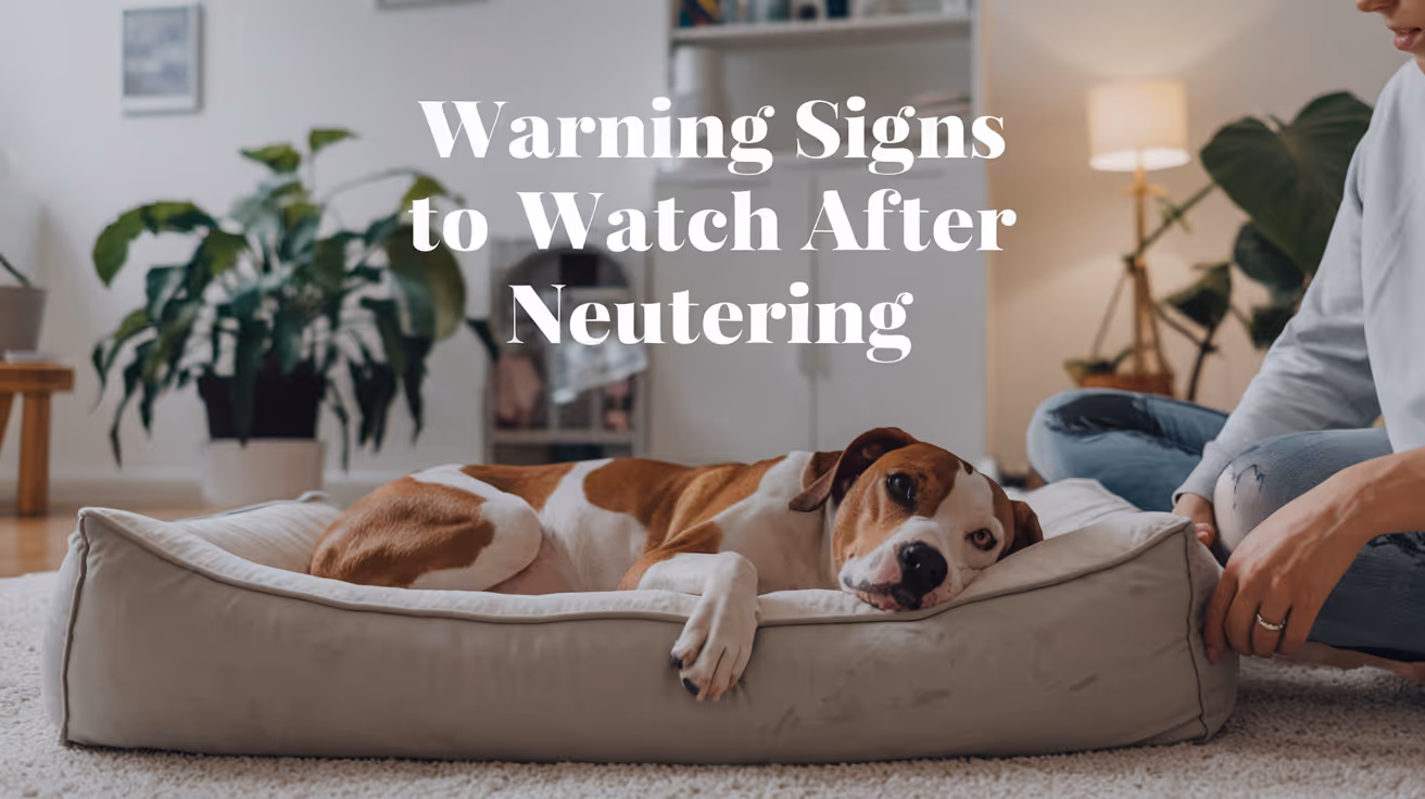
Warning Signs to Watch for After Neutering a Dog
Learn the key warning signs to watch for after neutering your dog to ensure a safe recovery and when to seek veterinary care.
Most dogs recover from neutering without a hitch. But some do not, and the difference between a minor complication and a serious one often comes down to how quickly you notice it.
The tricky part is that some things that look alarming are completely normal, and some things that look minor are not.
This guide gives you a clear, symptom-by-symptom breakdown of what is expected, what to monitor, and what requires an immediate call to your vet.
Quick answer: Mild grogginess, reduced appetite, and slight redness at the incision are all normal in the first 24 to 48 hours. Ongoing bleeding, yellow or green discharge, swelling that grows after day three, lethargy lasting more than 48 hours, or a wound that appears to be opening are all warning signs that need veterinary attention promptly.
Key takeaways
- Normal recovery includes mild grogginess, slight swelling, and reduced appetite for 24 to 48 hours.
- Warning signs that need a vet call include discharge, increasing swelling, continuous bleeding, and persistent lethargy.
- Emergency signs require immediate action: pale gums, collapse, or abdomen that appears bloated.
- Licking is the most preventable complication: the cone prevents most infection and wound problems.
- Daily incision checks catch problems early, when they are still easy to treat.
- When in doubt, call your vet. It is always better to check than to wait.
What normal recovery looks like
Understanding normal first makes it easier to spot abnormal.
In the first 24 to 48 hours after surgery, expect:
- Grogginess, drowsiness, or mild disorientation from anesthesia
- Reduced appetite or no interest in food
- Mild redness or slight puffiness at the incision site
- A small amount of clear or very faintly pink fluid at the wound in the first 24 to 72 hours
- Quieter behavior and preference for rest
- Possible shivering or restlessness as anesthesia wears off
All of these resolve on their own. By day two or three, your dog should be more alert, eating normally, and showing interest in their surroundings again.
Warning signs: incision and wound
The incision site is where most post-surgical problems show up first. Check it morning and evening throughout the recovery period.
Discharge
Swelling
Some swelling in the first two to three days is expected. The body's inflammatory response is part of normal healing.
Swelling that increases after day three, feels warm and firm to the touch, or is accompanied by redness and discharge is not normal. This pattern points to infection or hematoma and needs veterinary evaluation.
In large breed male dogs, scrotal swelling is a specific concern. Moderate scrotal swelling that resolves within a week is often normal. Swelling that grows, becomes hot, or is accompanied by pain or discharge is not.
Wound opening
The incision edges should be staying together, not pulling apart.
If you see any gap in the wound, exposed pink or red tissue beneath the skin, or sutures that appear to have pulled through, contact your vet immediately. This is called dehiscence and requires prompt attention.
Do not attempt to close an open wound yourself. Keep your dog calm, prevent licking, and get to your vet.
Warning signs: behavior and general health
Lethargy
Mild tiredness for the first 24 to 48 hours is expected. Anesthesia and surgery are taxing on the body.
Lethargy that persists beyond 48 hours, or a dog that is completely unresponsive, unable to stand, or does not improve over the first two days, is a warning sign. Call your vet.
Loss of appetite
Not wanting to eat on surgery day and the following morning is normal.
A dog that still refuses food at 48 hours after surgery, particularly if also vomiting, may be experiencing a complication. Call your vet if appetite has not returned by day two.
Vomiting
One or two episodes of vomiting the evening of surgery can be a side effect of anesthesia. This is within normal range.
Vomiting that continues beyond 24 hours after surgery, or that contains blood, requires a vet call. Persistent vomiting can indicate a reaction to pain medication, an obstruction, or another post-surgical issue.
Difficulty urinating or defecating
Some dogs take a day or two to have a bowel movement after surgery. This is usually due to fasting before the procedure and reduced activity afterward.
No bowel movement after three to five days is worth reporting. Difficulty or pain during urination, especially in male dogs, can indicate inflammation near the surgical site and should be checked promptly.
Warning signs: pain
Your dog will have been sent home with pain medication. Use it on schedule.
Signs that pain is not being adequately controlled include:
- Whining, yelping, or crying that persists beyond the first day
- Hunched posture or reluctance to sit or lie down
- Guarding the incision site by flinching or snapping when touched
- Panting excessively without exertion
- Trembling or shivering beyond the first few hours post-anesthesia
Contact your vet if pain seems severe after day three, or if prescribed medication does not appear to be working. Do not give human pain medications. Aspirin, ibuprofen, and acetaminophen are toxic to dogs and can cause serious harm or death.
Emergency warning signs: act immediately
These signs require emergency veterinary care, not a wait-and-see approach.
Call your vet or go to an emergency clinic immediately if you observe:
- Pale, white, or bluish gums
- Sudden collapse or inability to stand
- Abdomen that appears bloated, distended, or hard
- Heavy bleeding that does not stop
- Rapid or labored breathing
- Signs of extreme pain: screaming, freezing in place, inability to move
These can indicate internal bleeding, systemic infection, or other serious complications that are time-sensitive.
The most preventable warning sign: licking
Most of the infection and wound-breakdown cases that end up back at the vet have one thing in common: the cone came off.
Licking introduces bacteria directly into the wound, softens the tissue, and can pull sutures loose. A dog that licks its incision even once can undo days of clean healing.
The E-collar stays on for the full 10 to 14 days. If your dog refuses the hard plastic cone, soft inflatable collars or recovery suits are legitimate alternatives, provided they reliably prevent the dog from reaching the wound site.
For a full guide on what normal recovery looks like day by day and how to distinguish healing progress from early complications, tracking the incision against a clear timeline makes daily monitoring more actionable.
When to call vs. when to go straight to emergency care
Use this as a quick decision guide.
When in doubt, call. Most veterinary practices would rather hear from you about something minor than have you wait too long on something serious.
The role of aftercare in preventing complications
Most warning signs are preventable, not inevitable.
The dogs most likely to develop post-surgical complications are those given too much freedom too soon, or whose cones were removed early. Following a complete aftercare plan covering rest, incision monitoring, medications, and the cone is the most effective way to keep your dog out of trouble during recovery.
For a complete, step-by-step plan on full post-surgery care covering each day of the recovery window, what to feed, how to check the wound, and how to keep your dog calm, having that reference from day one prevents most of the issues covered in this article. Understanding surgical risks that can cause complications beyond the routine is also worth reading before surgery if you have a large breed or older dog.
Frequently asked questions
Is it normal for my dog to be lethargic after neutering?
Yes, for the first 24 to 48 hours. Anesthesia causes genuine fatigue, and your dog needs rest. Lethargy that does not improve by day two or three, or a dog that is completely unresponsive, warrants a call to your veterinarian.
What does an infected incision look like?
An infected incision typically shows yellow or green discharge, increasing redness and swelling after the first few days, warmth around the wound, and often a foul odor. Any of these signs alone justify a same-day call to your vet. For a detailed visual guide to signs of an infected incision, knowing what to look for at each stage of healing helps you act at the right time.
My dog is licking the incision despite wearing a cone. What should I do?
Check that the cone extends at least two inches past your dog's nose. If it is too short, your dog can still reach the incision. Consider switching to a soft inflatable collar or recovery suit if the hard cone is not fitting properly. Contact your vet if there are signs the wound has been compromised.
Should I be worried about scrotal swelling after neutering?
Some scrotal swelling in the first few days is normal. The scrotum fills with fluid as part of the healing response. Swelling that increases after day three, feels hot or very firm, or is accompanied by discharge or pain is abnormal and should be evaluated by your vet.
When should I go to an emergency vet after neutering?
Go immediately if your dog has pale or white gums, collapses, has a bloated or hard abdomen, or is bleeding heavily and continuously. These signs can indicate internal bleeding or systemic infection and are time-sensitive emergencies.
The difference between a smooth recovery and a complication is usually not bad luck. It is usually the cone coming off too early, too much activity on day four, or a wound check that was skipped. Pay attention for two weeks, and most dogs sail through without a single issue.
Resources
The following sources were used as reference and background for this article:
- Ace of Spays. Signs of Complications After Spay/Neuter Surgery: When to Contact Your Veterinarian. aceofspays.com
- Animal Humane Society. Spay/Neuter Post-Surgical Care and Recovery Instructions. animalhumanesociety.org
- Homeaglow. 10 Warning Signs After Spaying or Neutering Your Dog. homeaglow.com
- American Kennel Club. Post-Surgical Care for Dogs Following a Spay or Neuter. akc.org
- Union Hill Animal Hospital. Neutering Recovery Guide for Male Dogs. unionhillvet.com
- Rivergate Veterinary Clinic. Signs of Infection After Spaying or Neutering a Dog. rivergateveterinaryclinic.com
Will Spaying Calm a Female Dog?
Learn if spaying a female dog can calm her behavior and what changes to expect after surgery.
It is one of the most searched questions before booking a spay appointment. And the answer most owners get is a confident yes.
The complete answer is more qualified than that.
For female dogs whose difficult behavior is driven by heat cycles and reproductive hormones, spaying produces a real and often significant calming effect. For dogs whose energy, anxiety, or behavior problems come from other sources, the effect may be minimal or absent.
This guide gives you the specific, evidence-based answer so you know what to expect before and after surgery.
Quick answer: Spaying will calm a female dog if the behaviors you want to reduce are driven by reproductive hormones: heat cycle restlessness, roaming, urine marking, cyclical mood swings, and false pregnancy behaviors. It will not calm a dog whose energy, anxiety, or behavioral issues come from genetics, learned habits, or non-hormonal sources. The calming effect, when it occurs, develops gradually over two to three months.
Key takeaways
- The calming effect is hormone-specific: It applies to heat cycle behaviors, not all behaviors.
- Heat cycles cause real behavioral disruption: Restlessness, roaming, cyclical aggression, and false pregnancies are all eliminated by spaying.
- Core temperament does not change: A high-energy dog remains high-energy. A shy dog remains shy.
- The timeline is two to three months: Hormones decline gradually. Behavioral changes follow the same curve.
- Spaying is not a training substitute: Learned behaviors require training regardless of hormone status.
- A small subset of dogs may become more anxious: This documented outcome is worth discussing with your vet before surgery.
What drives difficult behavior in intact female dogs
Before answering whether spaying will calm your dog, it helps to understand what is making her difficult in the first place.
Intact female dogs experience cyclical hormonal changes driven by estrogen and progesterone. These cycles occur every six to eight months and create predictable but often exhausting behavioral patterns for owners.
During heat cycles, female dogs typically show:
- Restlessness, inability to settle, pacing
- Increased vocalization and whining
- Frequent urination in small amounts (urine marking to attract males)
- Strong, persistent drive to escape and find a mate
- Attracting intact male dogs from significant distances
- Cyclical irritability or clinginess
Between cycles, some dogs also experience pseudopregnancy, where they nest, guard objects as imaginary puppies, produce milk, and sometimes become aggressive when the "nest" is approached.
All of these behaviors are hormone-driven. Spaying eliminates the source of those hormones.
When spaying will calm a female dog
The answer is yes, clearly, for dogs whose difficult behaviors are primarily driven by the reproductive cycle.
Spaying reliably calms females who show:
- Significant behavioral changes during heat cycles
- Restlessness or inability to settle in the days and weeks of estrus
- Strong escape or roaming drive when in heat
- Cyclical mood swings, irritability, or clinginess linked to the hormonal cycle
- False pregnancy behaviors including nesting, object guarding, or milk production
- Heat-related aggression toward other dogs
For these dogs, removing the hormonal cycling removes the behavioral disruption. What remains is a more consistent, predictable dog whose mood and energy are not subject to the peaks and troughs of the reproductive cycle.
Many owners of females that were particularly challenging during heat cycles describe the post-spay result as dramatically positive.
When spaying may not calm a female dog
The answer is less certain for dogs whose behavior comes from sources other than reproductive hormones.
Spaying is less likely to produce a calming effect in dogs who show:
- High energy due to breed genetics (working breeds, sporting breeds, terriers)
- Anxiety or fearfulness unrelated to the heat cycle
- Learned behavioral problems from history or environment
- Reactivity or aggression toward people or dogs that is fear-based
- Restlessness caused by insufficient exercise or mental stimulation
Removing estrogen does not reduce a Border Collie's herding drive. It does not calm a dog whose anxiety comes from under-socialization. It does not fix pulling on the leash, jumping on visitors, or barking at the doorbell.
These behaviors require training and environmental management, not surgery.
The specific behaviors spaying eliminates vs. those it does not
What the research says
Most veterinary guidance presents the calming effect of spaying as reliable and universal. The research picture is somewhat more nuanced.
Studies of intact versus spayed females consistently show that heat-related behaviors are reliably reduced. This part of the conventional guidance is well-supported.
However, two large-scale studies using the Canine Behavioral Assessment and Research Questionnaire (C-BARQ) found that spayed females showed increased fearfulness compared to intact females in some analyses. The research found approximately a 31% increase in fearfulness in sterilized dogs of both sexes.
This does not mean spaying makes most dogs more anxious. But it means the outcome is not uniformly calming for every female, and in a subset of dogs, particularly those sterilized at a very young age or those with pre-existing anxiety, the behavioral outcome may be less positive than expected.
Understanding all the behavior changes to expect after spaying or neutering across both sexes gives this specific female question the broader research context it deserves. For the full picture of all behavior changes specific to female dogs after spaying, including the distinction between what spaying reliably changes and what requires training, that guide covers the female picture in full detail.
The timeline: when does calming happen?
Owners who expect a calmer dog within days of surgery are working with an inaccurate timeline.
Estrogen and progesterone do not disappear on surgery day. The ovaries are removed, but residual hormones still circulate in the bloodstream and take time to clear.
What to expect:
- Days 1 to 14: Recovery period. Any behavioral changes relate to pain and anesthesia, not hormonal adjustment.
- Weeks 2 to 6: Hormone levels declining. Early signs of reduced heat-driven behaviors may begin to appear.
- Weeks 6 to 12: Most hormonal behavioral changes are apparent.
- Months 2 to 3: Full hormonal stabilization. This is the point at which you can accurately assess your dog's post-spay behavioral baseline.
For the hormonal mechanism and why it takes this long, the guide on how long before hormone-driven behavior settles covers the clearance timeline in detail, including how this applies to the female hormonal system.
What else helps a female dog become calmer
Whether or not spaying produces a calming effect, these factors reliably influence how settled a female dog is:
Consistent daily exercise: Appropriate to her breed and age. A dog that has adequately burned physical energy is a calmer dog at rest.
Mental stimulation: Puzzle feeders, training sessions, scent work, and enrichment activities address the cognitive drive that underlies a lot of restless behavior.
Predictable routine: Dogs settle more easily in stable, consistent environments. Unpredictable schedules increase ambient anxiety.
Training: The most reliable long-term path to a calmer, more manageable dog. Clear expectations, consistently reinforced, shape behavior more durably than surgery.
Spaying contributes one element. It removes the hormonal disruption of the reproductive cycle. The rest of these factors remain under your control regardless of whether your dog is intact or spayed.
Frequently asked questions
Will my dog be noticeably calmer right after spaying?
Not immediately. The first week is recovery from surgery. Behavioral changes related to hormonal adjustment happen over weeks to months, not days. Two to three months after surgery is when you can accurately assess your dog's post-spay behavioral baseline.
My female dog is anxious and reactive. Will spaying help?
If her anxiety and reactivity are linked to the heat cycle, spaying may help. If they come from other sources, including under-socialization, fearful temperament, or learned responses, spaying is unlikely to make a meaningful difference. Consult your vet and consider a referral to a certified veterinary behaviorist if anxiety is a significant concern.
My female dog is aggressive. Will spaying calm her down?
It depends on the type of aggression. Aggression linked specifically to heat cycles, competition with other females, or false pregnancy guarding is often reduced. Fear-based aggression, territorial aggression, and resource guarding are not reliably improved by spaying and require behavioral intervention.
Will spaying calm a high-energy female dog?
Probably not significantly. High energy in female dogs is primarily determined by breed genetics and individual temperament, not reproductive hormones. A high-drive sporting breed will still need the same amount of exercise and engagement after spaying. For whether calming effects in adult dogs after spaying apply to your specific situation, that guide covers the nuances of the calming question across different dog types.
Are there female dogs that get worse after spaying?
Yes, in a subset. Some research documents increased fearfulness in spayed females compared to intact females, particularly in dogs spayed at a young age or those with pre-existing anxiety. If your dog becomes more anxious after spaying, speak to your vet. A referral to a certified veterinary behaviorist may be appropriate.
The honest answer to whether spaying will calm your female dog: probably yes, if what is making her difficult is her heat cycle. Probably not, if what is making her difficult is her breed, her upbringing, or her learned behaviors. Understanding which category your dog falls into before surgery sets accurate expectations for the result.
Resources
The following sources were used as reference and background for this article:
- Anasazi Veterinary. How Will Spaying Change My Dog? anasazivet.com
- RexiPets. Will Spaying Calm a Female Dog? rexipets.com
- Bliss Animal Hospital. Do Dogs Change After Spay or Neuter? blissanimalhospital.com
- Kainer Veterinary. Understanding the Health and Behavior Benefits of Spaying a Dog. kainervet.com
- Stanley Coren, Psychology Today. Are There Behavior Changes When Dogs Are Spayed or Neutered? psychologytoday.com
Do Dogs Calm Down After Being Spayed?
Learn how dogs behave after being spayed and when to expect them to calm down post-surgery.
It is one of the most common reasons owners choose to spay their female dog. The expectation of a calmer, more settled companion afterward.
Sometimes that is exactly what happens. Sometimes it does not. And in a small subset of cases, anxiety or excitability actually increases.
Understanding why gives you a more accurate picture of what to expect, which makes the outcome less surprising in either direction.
Quick answer: Many dogs do become calmer after spaying, but the effect is specific to hormone-driven behaviors, not overall temperament. Dogs that were restless, anxious, or excitable due to heat cycles typically show significant improvement. Dogs whose energy or anxiety has non-hormonal causes will not reliably calm down. Most behavioral changes, if they occur, appear over two to three months.
Key takeaways
- The calming effect is real but specific: It applies to behaviors driven by reproductive hormones, not all behaviors.
- Heat cycle behaviors disappear: Restlessness, roaming, frequent urination, and cyclical mood shifts all stop after spaying.
- Core energy level and personality do not change: A high-energy dog will still be high-energy after spaying.
- Breed and individual temperament matter: Some breeds retain high energy regardless of hormone status.
- The timeline is weeks to months, not days: Hormone levels decline gradually, so behavioral changes are gradual too.
- A small subset of dogs show increased fearfulness: This is documented in research and worth knowing about before surgery.
What "calming down" actually means after spaying
When people say they want their dog to calm down, they usually mean one of two things:
- They want heat cycle behaviors to stop: the roaming, the restlessness, the vocalization, the constant male attention.
- They want a generally more settled dog overall.
Spaying reliably delivers the first. The second is less predictable.
This distinction is the core of the honest answer to this question.
What spaying removes: the hormonal layer
Spaying removes the ovaries, which are the primary source of estrogen and progesterone. These hormones regulate the reproductive cycle and influence behaviors tied to it.
During heat cycles, which typically occur every six to eight months in adult dogs, estrogen surges create a set of predictable behaviors:
- Restlessness and inability to settle
- Increased vocalization
- Frequent urination in small amounts to signal reproductive status
- Strong drive to escape and find a mate
- Attracting intact male dogs
- Cyclical irritability or anxiety
All of these stop after spaying. The hormonal trigger is permanently removed.
Which dogs calm down most noticeably after spaying
Dogs whose difficult behaviors are primarily or significantly driven by the reproductive cycle show the most consistent improvement after spaying.
Dogs most likely to become calmer:
- Female dogs that showed significant behavior changes during heat cycles, including restlessness, anxiety, and clinginess
- Dogs prone to false pregnancies, with associated nesting, object guarding, and protective behavior
- Dogs that attempted to escape or roam during heat periods
- Dogs whose cyclical mood swings were disruptive to daily life
For these dogs, spaying removes a recurring source of behavioral disruption. The result is often a more predictable, stable dog.
Which dogs may not calm down after spaying
Dogs whose energy level, excitability, or anxiety comes from sources other than reproductive hormones will not reliably settle after spaying.
Dogs less likely to show a calming effect:
- High-energy working or sport breeds where drive is genetic, not hormonal
- Dogs with established anxiety, fearfulness, or reactivity from non-hormonal causes
- Dogs with learned problematic behaviors from their history and environment
- Dogs whose excitability comes from insufficient exercise or mental stimulation
A Border Collie that is highly energetic before spaying will still be highly energetic after. A Labrador that pulls on the leash before spaying will still pull after. These behaviors are not driven by estrogen.
The timeline: when does calming happen?
Behavioral changes from spaying do not happen within days of surgery. The immediate post-surgery period reflects recovery from anesthesia and pain, not hormonal adjustment.
Realistic timeline:
Owners who expect a noticeably calmer dog within days of surgery are working with an inaccurate timeline. The changes are gradual and unfold over weeks to months.
What research says about calming after spaying
The evidence is genuinely mixed, which is worth knowing rather than glossing over.
Supporting the calming effect:
A study of 591 dogs found behavioral problems reduced or eliminated in 59% of spayed females. Heat cycle-related behaviors showed the most consistent improvement. Many owners of females that were particularly difficult during heat cycles report significant positive changes after spaying.
Complicating the picture:
Two large-scale studies using the Canine Behavioral Assessment and Research Questionnaire (C-BARQ) found that sterilization was associated with a roughly 31% increase in fearfulness and a 33% increase in touch sensitivity in some dogs. These findings apply to both males and females.
What does this mean in practice? For a meaningful subset of dogs, spaying does not produce a calmer animal. It may even produce a more anxious one, particularly in dogs that had pre-existing fearfulness or were sterilized at a young age.
This does not mean spaying is wrong for these dogs. It means the behavioral outcome cannot be guaranteed, and expectations should be calibrated accordingly.
For the full picture of all the behavior changes to expect after spaying, including the research on unexpected outcomes, that guide covers the complete behavioral landscape across both sexes.
Does calming depend on the dog's age at spaying?
Yes, and this is one of the most important variables.
Dogs spayed before their first heat cycle:
The hormonal disruption of heat cycles is never established. Many owners report these dogs are consistently more settled than intact females across their lifetime, without ever having the difficult heat cycle period.
Dogs spayed after several heat cycles:
The calming effect from eliminating heat cycles is still real and significant. However, any behaviors that have become habitual through repetition during heat cycles may persist longer or require training to address.
Dogs spayed very young (before 6 months):
Research suggests a higher association with fearfulness in dogs sterilized very early, before hormonal maturity. For whether this applies specifically to puppies, the guide on calming effects specific to female dogs addresses this age-specific question in detail. For the puppy-specific picture, the guide on whether puppies calm down after being spayed covers the developmental nuances unique to younger dogs.
When the expected calming does not happen
If your dog does not seem noticeably calmer after spaying, consider these factors before concluding the surgery did not work.
Check the timeline: Have two to three months passed since surgery? If not, the hormonal adjustment may still be in progress.
Identify the behavior source: Is the behavior you wanted to address actually hormone-driven? If it is not, spaying was never going to address it.
Consider habituation: Some behaviors that started as hormone-driven become habitual over time. The hormone is removed but the habit persists. Consistent training addresses habituated behavior.
Consider an underlying anxiety: If your dog became more fearful or anxious post-surgery, this is a documented outcome in some dogs. A veterinary behaviorist or your regular vet can help evaluate what is happening and what options exist.
Understanding the truth behind the calming effect myth, including why the expectation of calming is sometimes oversold, helps put the variable outcomes in context.
What actually makes dogs calmer long-term
Whether or not spaying produces a calming effect, these factors reliably influence how settled and manageable a dog is:
- Adequate physical exercise: A dog that is genuinely tired is a calmer dog. Exercise needs are determined by breed and individual temperament.
- Mental stimulation: Breed-appropriate mental work, puzzle feeders, training sessions, and enrichment activities reduce restless, problematic behavior.
- Consistent training: Clear, reinforced expectations give dogs a behavioral framework. This is the single most reliable path to a calmer dog.
- Routine and predictability: Dogs settle more easily in stable, predictable environments.
Spaying contributes one element to this picture. It is not a standalone solution.
Frequently asked questions
How long after being spayed will my dog calm down?
If the calming effect is going to occur, most hormone-driven behavioral changes become apparent within two to six weeks as hormone levels decline. Full stabilization typically takes two to three months. Dogs that are still showing challenging behaviors three to four months after surgery are unlikely to improve further from the hormonal change alone.
Will spaying make my high-energy dog less active?
Probably not significantly. Energy level in dogs is primarily determined by breed, genetics, and conditioning. Spaying can cause a modest metabolic slowdown, but it does not reduce drive or enthusiasm. A high-energy breed will still need the same amount of exercise after spaying.
My dog got more anxious after being spayed. Why?
Research has documented increased fearfulness in a subset of dogs after sterilization, associated with a roughly 31% increase in fearfulness in some studies. This appears to occur more often in dogs sterilized at a young age and in dogs with pre-existing anxiety. If your dog shows increased anxiety after spaying, speak to your vet. A referral to a certified veterinary behaviorist may be helpful.
Will spaying help with aggression?
It depends on the type of aggression. Aggression linked to the heat cycle, competition with other female dogs, or false pregnancy guarding often decreases. Fear-based aggression, learned aggression, and resource guarding are not hormone-driven and are not reliably improved by spaying. For whether lower testosterone calms dogs down in the male counterpart to this question, that guide covers the testosterone-behavior link specifically.
Does breed affect whether spaying calms a dog down?
Yes. Breed temperament significantly influences baseline energy and anxiety levels. High-drive working breeds such as Border Collies, Australian Shepherds, and Belgian Malinois have energy and drive that is genetic, not hormonal. Removing reproductive hormones does not change these fundamental breed characteristics. Whether your specific dog falls into a calmer range after spaying depends on what was driving any difficult behavior in the first place.
The calming effect of spaying is real for dogs whose difficult behaviors are hormone-driven. For those dogs, eliminating the heat cycle and its associated hormonal disruption produces a meaningfully more stable animal. For dogs whose energy or anxiety comes from other sources, spaying is still beneficial for health reasons but should not be expected to produce behavioral transformation. The most accurate prediction is not based on "spaying calms dogs" but on "removing a specific hormonal influence changes the behaviors that influence drives."
Resources
The following sources were used as reference and background for this article:
- Stanley Coren, Psychology Today. Are There Behavior Changes When Dogs Are Spayed or Neutered? psychologytoday.com
- PubMed. Changes in the behavior of dogs after castration. pubmed.ncbi.nlm.nih.gov
- ManyPets. Do Dogs Change After Being Neutered or Spayed? manypets.com
- American Veterinary Medical Association (AVMA). Spaying and Neutering. avma.org
- Houndsy. How Spaying Affects Dog Behavior. houndsy.com
Torn Meniscus Surgery Cost in Dogs Explained
Learn about torn meniscus surgery cost in dogs, including factors affecting price, procedure details, and recovery tips for your pet's health.
A torn meniscus in dogs is a common injury that affects the knee joint, causing pain and mobility issues. If your dog has this problem, you might wonder about the cost of surgery and what it involves. Understanding the expenses and treatment options can help you prepare for your pet's care.
This article explains the typical cost of torn meniscus surgery in dogs, the factors that influence pricing, and what you can expect during recovery. You will learn how to manage your dog's health and make informed decisions about treatment.
What is torn meniscus surgery in dogs?
Torn meniscus surgery in dogs is a procedure to repair or remove damaged cartilage in the knee joint. The meniscus acts as a cushion between bones, and injury can cause pain and lameness. Surgery aims to restore joint function and reduce discomfort.
The surgery is usually recommended when conservative treatments like rest and medication do not improve the dog's condition. It involves anesthesia and specialized techniques to address the tear.
- Purpose of surgery: To repair or remove the damaged meniscal cartilage to relieve pain and improve knee stability in dogs.
- Common causes: Meniscus tears often result from ligament injuries or trauma during activities like running or jumping.
- Surgical techniques: Options include meniscectomy (removal) or meniscal repair depending on the tear's location and severity.
- Post-surgery goals: Restore normal joint movement, reduce arthritis risk, and help the dog regain mobility.
Understanding the surgery helps you prepare for the treatment and care your dog will need.
How much does torn meniscus surgery cost in dogs?
The cost of torn meniscus surgery in dogs varies widely depending on location, clinic, and the dog's specific needs. On average, prices range from $1,500 to $4,000. This includes pre-surgical exams, anesthesia, surgery, and initial post-op care.
Knowing the cost breakdown can help you budget and discuss options with your veterinarian.
- Base surgery fee: Typically between $1,000 and $3,000, covering the surgical procedure and operating room use.
- Pre-surgical tests: Blood work and X-rays may cost $200 to $500 to assess the dog's health before surgery.
- Anesthesia and monitoring: Usually $300 to $700, essential for safe surgery and pain control.
- Post-operative care: Includes medications, bandages, and follow-up visits costing $200 to $500.
Costs may increase if complications arise or if advanced imaging like MRI is needed.
What factors affect the cost of meniscus surgery in dogs?
Several factors influence the total cost of torn meniscus surgery in dogs. These include the dog's size, the complexity of the injury, and the clinic's location. Understanding these helps you anticipate expenses and plan accordingly.
Discussing these factors with your vet can clarify the expected costs and available options.
- Dog's size and weight: Larger dogs may require more anesthesia and longer surgery time, increasing costs.
- Severity of tear: Complex or multiple tears need more surgical time and skill, raising the price.
- Veterinary clinic location: Urban or specialty clinics often charge more than rural general practices.
- Surgeon's experience: Board-certified surgeons may have higher fees but offer specialized care.
Knowing these factors helps you make informed choices about your dog's treatment plan.
What are the risks and benefits of torn meniscus surgery for dogs?
Surgery for a torn meniscus can improve your dog's quality of life but also carries some risks. Weighing these helps you decide if surgery is the best option for your pet.
The benefits often outweigh the risks when the injury causes significant pain or limits mobility.
- Benefit - Pain relief: Surgery can reduce joint pain and discomfort caused by the torn meniscus.
- Benefit - Improved mobility: Dogs often regain better movement and activity levels after recovery.
- Risk - Infection: Any surgery carries a small risk of infection requiring additional treatment.
- Risk - Anesthesia complications: Though rare, anesthesia can cause adverse reactions in some dogs.
Discussing risks and benefits with your vet ensures the best decision for your dog's health.
How should you prepare your dog for meniscus surgery?
Proper preparation before surgery helps reduce risks and supports a smooth procedure. Your veterinarian will give specific instructions tailored to your dog’s needs.
Following these steps can improve surgery outcomes and reduce stress for your pet.
- Pre-surgery fasting: Your dog should avoid food for 8-12 hours before surgery to prevent anesthesia complications.
- Health evaluation: Complete blood tests and physical exams ensure your dog is healthy enough for surgery.
- Medication review: Inform your vet about all medications or supplements your dog takes to avoid interactions.
- Arrange post-op care: Prepare a quiet, comfortable space and plan for restricted activity during recovery.
Good preparation helps your dog have a safer surgery and faster healing.
What is the recovery process after torn meniscus surgery in dogs?
Recovery after meniscus surgery requires careful management to ensure healing and prevent re-injury. The process usually takes 6 to 12 weeks, depending on the dog's age and health.
Following your vet’s instructions closely improves your dog’s chances of a full recovery.
- Restricted activity: Limit running, jumping, and stairs to protect the healing joint for several weeks.
- Physical therapy: Controlled exercises and rehabilitation help restore strength and flexibility.
- Pain management: Administer prescribed pain medications and monitor for discomfort signs.
- Follow-up visits: Regular check-ups allow your vet to assess healing and adjust care as needed.
Patience and consistency during recovery are key to your dog’s long-term joint health.
Conclusion
Torn meniscus surgery cost in dogs varies but generally ranges from $1,500 to $4,000 depending on many factors. Understanding the procedure, risks, and recovery helps you prepare financially and emotionally for your pet’s care.
With proper preparation and post-operative management, surgery can relieve your dog’s pain and improve mobility. Always consult your veterinarian to choose the best treatment plan for your dog’s torn meniscus injury.
What is the typical recovery time after torn meniscus surgery in dogs?
Recovery usually takes 6 to 12 weeks, during which activity must be restricted and physical therapy may be needed to regain strength and mobility.
Can torn meniscus surgery prevent arthritis in dogs?
While surgery can reduce joint damage and pain, it may not fully prevent arthritis but can slow its progression and improve quality of life.
Are there non-surgical options for treating a torn meniscus in dogs?
Yes, mild tears may be managed with rest, anti-inflammatory medications, and physical therapy, but surgery is often needed for severe cases.
How do I know if my dog needs meniscus surgery?
Your vet will diagnose based on clinical signs, physical exams, and imaging like X-rays or MRI to determine if surgery is necessary.
Is torn meniscus surgery painful for dogs?
Dogs receive anesthesia during surgery and pain medications afterward to minimize discomfort, making the procedure as pain-free as possible.

Pinnal Vasculitis in Dogs: Signs, Causes & Management
Learn about pinnal vasculitis in dogs, including signs, causes, and effective management strategies to keep your pet healthy.
Pinnal vasculitis in dogs is a painful condition affecting the ears, causing inflammation and sores. It can be distressing for both pets and owners due to discomfort and visible symptoms. Understanding this condition helps you recognize it early and seek proper care.
This article explains the signs of pinnal vasculitis, its common causes, and how veterinarians manage the condition. You will learn how to identify symptoms, what triggers the disease, and the best treatment options to help your dog recover comfortably.
What is pinnal vasculitis in dogs?
Pinnal vasculitis is inflammation of the blood vessels in a dog's ear flap, or pinna. This inflammation causes redness, swelling, and sometimes ulcers or scabs on the ear surface. It can affect one or both ears and may cause your dog pain or itchiness.
The condition is not a disease itself but a symptom of underlying problems. It often signals immune system issues or infections that need veterinary attention.
- Inflammation of ear vessels: Pinnal vasculitis involves swelling and damage to small blood vessels in the ear flap, leading to visible skin changes and discomfort.
- Causes vary widely: It can result from infections, allergies, immune disorders, or trauma, making diagnosis important for proper treatment.
- Common in certain breeds: Breeds like German Shepherds and Dobermans may be more prone due to genetic or immune factors affecting their skin.
- Signs include sores and pain: Dogs often show redness, scabs, and may shake or scratch their ears due to irritation.
Recognizing pinnal vasculitis early helps prevent complications like chronic ear damage or secondary infections. Veterinary diagnosis usually involves skin tests and blood work.
What are the common signs of pinnal vasculitis in dogs?
Signs of pinnal vasculitis can vary but usually involve changes in the ear's appearance and your dog's behavior. Early symptoms may be subtle, so careful observation is important.
Most dogs show discomfort and visible skin changes on their ears, which can worsen without treatment.
- Redness and swelling: The ear flap looks inflamed and may feel warm or tender to touch, indicating active inflammation.
- Ulcers and scabs: Open sores or crusty areas develop on the ear surface due to damaged blood vessels and skin.
- Itching and scratching: Dogs often scratch or shake their head to relieve irritation, which can worsen the condition.
- Pain and sensitivity: Your dog may react when you touch the ears, showing signs of discomfort or distress.
Other signs may include hair loss around the ears and thickened skin if the condition becomes chronic. Early veterinary care can reduce these symptoms and improve comfort.
What causes pinnal vasculitis in dogs?
Pinnal vasculitis results from damage to blood vessels in the ear, often linked to other health issues. Identifying the cause is key to effective treatment and preventing recurrence.
Several factors can trigger this condition, ranging from infections to immune system problems.
- Immune-mediated diseases: Conditions like lupus or vasculitis cause the immune system to attack blood vessels, leading to inflammation in the ears.
- Bacterial or fungal infections: Infections can damage ear tissue and blood vessels, triggering vasculitis symptoms.
- Allergic reactions: Allergies to food, fleas, or environmental factors can cause inflammation and secondary vasculitis.
- Trauma or injury: Physical damage to the ear from scratching, bites, or accidents may lead to vessel inflammation.
Sometimes, no clear cause is found, which is called idiopathic pinnal vasculitis. Your vet will perform tests to rule out infections and immune diseases to tailor treatment.
How is pinnal vasculitis diagnosed in dogs?
Diagnosing pinnal vasculitis requires a thorough veterinary exam and specific tests. Early diagnosis helps start treatment before serious damage occurs.
Your vet will look at the ear carefully and may need samples to understand the underlying cause.
- Physical examination: The vet inspects the ears for redness, swelling, ulcers, and checks for pain or discomfort during handling.
- Skin biopsy: A small sample of affected skin may be taken to examine blood vessel inflammation under a microscope.
- Blood tests: These help detect immune system problems or infections that could cause vasculitis.
- Infection screening: Swabs from the ear may be cultured to identify bacteria or fungi causing secondary infections.
Combining these tests helps your vet determine the exact cause and plan the best treatment for your dog’s pinnal vasculitis.
What treatment options are available for pinnal vasculitis in dogs?
Treatment depends on the cause but usually involves reducing inflammation, controlling infections, and protecting the ears from further damage. Prompt care improves outcomes and comfort.
Your vet will tailor therapy based on test results and severity of symptoms.
- Anti-inflammatory medications: Steroids or other drugs reduce blood vessel inflammation and relieve pain and swelling in the ears.
- Antibiotics or antifungals: These treat any bacterial or fungal infections contributing to the vasculitis.
- Immune-suppressing drugs: For immune-mediated causes, medications help calm the immune system to prevent vessel damage.
- Protective ear care: Cleaning and protecting the ears from trauma or moisture helps healing and prevents worsening.
Long-term management may include regular vet visits and monitoring for flare-ups. Early treatment reduces the risk of permanent ear damage.
How can you manage pinnal vasculitis at home?
Alongside veterinary care, you can support your dog’s recovery at home with careful management. This helps reduce discomfort and speeds healing.
Consistent care and observation are essential to avoid complications.
- Gentle ear cleaning: Use vet-approved solutions to keep ears clean and dry, preventing infections that worsen vasculitis.
- Prevent scratching: Use an Elizabethan collar if needed to stop your dog from damaging inflamed ears further.
- Monitor symptoms: Watch for changes like increased redness or swelling and report them to your vet promptly.
- Follow medication schedules: Administer all prescribed drugs exactly as directed to ensure effective treatment.
Good nutrition and reducing exposure to allergens can also help manage underlying causes. Always consult your vet before trying new treatments or supplements.
What is the prognosis for dogs with pinnal vasculitis?
The outlook depends on the cause and how quickly treatment starts. Many dogs improve well with proper care, but some may have ongoing issues.
Chronic or severe cases need careful management to prevent permanent ear damage or discomfort.
- Early treatment improves outcomes: Dogs treated promptly usually heal well with minimal lasting damage to the ears.
- Chronic cases require monitoring: Long-term immune diseases may cause repeated flare-ups needing ongoing medication.
- Possible ear deformities: Severe vasculitis can cause scarring or thickening of the ear flap, affecting appearance and function.
- Quality of life considerations: Managing pain and preventing infections helps maintain your dog’s comfort and happiness.
Regular veterinary check-ups and following treatment plans are key to a good prognosis for dogs with pinnal vasculitis.
Conclusion
Pinnal vasculitis in dogs is a serious condition causing painful inflammation of the ear blood vessels. Recognizing the signs early and understanding the causes helps you seek timely veterinary care.
With proper diagnosis and treatment, most dogs recover well. Managing the condition at home and following your vet’s advice ensures your dog stays comfortable and healthy. Always consult your veterinarian if you notice ear problems to protect your pet’s well-being.
What should I do if I notice my dog scratching its ears a lot?
Excessive ear scratching can indicate irritation or pinnal vasculitis. Schedule a vet visit promptly to identify the cause and start appropriate treatment to prevent worsening.
Can pinnal vasculitis in dogs be cured completely?
Many cases improve fully with treatment, especially if caused by infections. Immune-related cases may need ongoing management but can still have good quality of life.
Is pinnal vasculitis contagious to other pets?
Pinnal vasculitis itself is not contagious, but underlying infections causing it might be. Keep affected dogs separated until a vet confirms the cause.
How long does treatment for pinnal vasculitis usually last?
Treatment length varies from a few weeks for infections to months for immune diseases. Follow your vet’s instructions and attend follow-ups for best results.
Can diet affect pinnal vasculitis in dogs?
Yes, food allergies can trigger or worsen pinnal vasculitis. Your vet may recommend dietary changes or hypoallergenic diets to help control symptoms.
Pug Nose Surgery Cost and Recovery Guide
Learn about pug nose surgery cost, recovery time, risks, and care tips to help your pet breathe better and heal safely.
Pugs are one of the most commonly affected breeds for a specific breathing problem: severely narrowed nostrils that restrict airflow from the very first breath.
The surgery to correct this, stenotic nares correction, is one of the most performed BOAS procedures in the US. For many pug owners, it is the first and most accessible surgical intervention for their dog's airway.
This guide covers what the procedure involves, what it costs, and what recovery looks like specifically for pugs.
Quick answer: Pug nose surgery (stenotic nares correction) typically costs between $500 and $2,000 for the procedure alone. When combined with soft palate correction in a full BOAS surgical package, total costs range from $2,000 to $4,000. Pugs are usually discharged the same day. Recovery involves two weeks of soft food, restricted activity, and E-collar use. Most pugs show noticeable breathing improvement within days.
Key takeaways
- Stenotic nares correction costs $500 to $2,000 as a standalone procedure: Full BOAS correction including soft palate work runs $2,000 to $4,000 for pugs.
- Laser surgery costs more but heals faster: Laser techniques ($1,000 to $2,000) reduce bleeding and swelling compared to scalpel methods ($500 to $1,200).
- Most pugs are discharged the same day: Stenotic nares correction is typically performed outpatient.
- The best outcomes happen in young dogs: Veterinarians recommend evaluation and surgery before age two where possible.
- Pet insurance can offset cost significantly: Enroll before any BOAS symptoms are documented.
- Most pugs breathe noticeably better within days: The improvement from nostril widening is often immediate and dramatic.
Why pugs specifically need this surgery
Pugs are among the most severely affected brachycephalic breeds for BOAS.
Their skull structure is extreme even by flat-faced breed standards: very wide, very short, with a deeply recessed muzzle. This produces some of the most significantly compressed nasal passages in the canine world.
Stenotic nares (severely narrowed nostrils) are nearly universal in pugs. Some individual pugs have nostrils so narrow that the nasal openings are barely visible as slits. Breathing through such nostrils requires significant muscular effort with every single breath.
For a full explanation of why pugs are particularly at risk alongside other brachycephalic breeds, including what the research shows about BOAS prevalence in pugs, that guide covers the breed-specific risk profile in detail.
What is pug nose surgery?
Pug nose surgery is the common name for stenotic nares correction, technically called rhinoplasty or alapexy depending on the specific technique used.
The surgery widens the nostrils by removing a small wedge of tissue from each nostril, permanently enlarging the nasal opening and allowing significantly more air to pass through.
The procedure does not change the overall appearance of the pug's face in any dramatic way. The nostrils are widened, but the characteristic flat-faced appearance remains. What changes is the amount of air that can move through.
Surgical techniques
Wedge resection: The most common technique. A small wedge of tissue is removed from each nostril using a scalpel. This reduces the inward collapse of the nostril wall and permanently widens the opening.
Alapexy: A technique that repositions and sutures the alar fold (the fleshy outer nostril wall) in a more open position rather than removing tissue. Used in some cases where tissue removal alone is insufficient.
Laser rhinoplasty: A laser is used instead of a scalpel to remove or reshape the nostril tissue. Laser techniques typically produce less bleeding, less post-operative swelling, and faster healing. They cost more, usually $1,000 to $2,000, compared to scalpel approaches at $500 to $1,200.
Understanding the condition behind the surgery
Stenotic nares is one component of a larger syndrome. Understanding the condition behind pug nose surgery in full helps pug owners understand why nostrils alone may not be the only issue addressed.
Many pugs need more than just nostril widening. The elongated soft palate is the other most commonly corrected defect in pugs, and veterinarians frequently recommend addressing both in the same anesthetic event.
What the surgery involves
Pug nose surgery is performed under general anesthesia.
Pre-surgical steps:
- Physical examination and veterinary history review
- Bloodwork to confirm fitness for anesthesia
- Imaging may be recommended to assess tracheal size and rule out concurrent issues
- Fasting the night before surgery
The procedure:
The surgery itself typically takes 30 to 60 minutes for nostril correction alone. When combined with soft palate resection, the total procedure time is one to two hours.
The veterinarian assesses the full airway at the time of anesthesia induction. This is why many surgeons examine the soft palate during the same event and address it if needed, rather than scheduling a separate anesthetic event.
After surgery:
Your pug will be monitored as anesthesia wears off. Most pugs are discharged the same day once the veterinarian is satisfied that breathing is stable.
Some surgeons recommend overnight monitoring for pugs, particularly those with suspected tracheal issues or concurrent health concerns. Discuss what your surgical team's protocol is before the procedure.
Cost breakdown for pug nose surgery
Costs vary by technique, geographic location, facility type, and whether additional procedures are performed.
Stenotic nares correction alone
Full BOAS correction for pugs (nares plus soft palate)
Additional costs to plan for
- Pre-surgical bloodwork: $80 to $200
- Pre-surgical radiographs or CT: $200 to $600 (recommended for pugs to assess tracheal size)
- Post-operative medications: $40 to $150
- Recheck appointments: $50 to $150 per visit
- Overnight hospitalization if needed: $200 to $500
For a broader picture of broader cost and risk comparison for brachycephalic breeds including how pug costs compare to bulldogs and what drives costs up or down, that guide provides the full financial context.
What drives the cost of pug nose surgery
Geographic location: Urban specialty hospitals in major cities cost more than rural or suburban general practices.
Surgeon qualifications: A board-certified veterinary surgeon charges more than a general practitioner. For straightforward cases in otherwise healthy young pugs, a general practitioner experienced in BOAS surgery is often appropriate. Complex cases benefit from specialist care.
Laser versus scalpel: Laser techniques cost approximately 60 to 100% more than scalpel, but the faster healing and reduced post-operative swelling are meaningful advantages.
Number of procedures: Nostril correction alone is significantly less expensive than a full BOAS correction. Combining procedures in one anesthetic event is more efficient, but it does increase the total bill.
Concurrent health issues: A pug with additional health concerns requires more intensive monitoring and may need longer hospitalization.
Pet insurance for pug nose surgery
Most comprehensive pet insurance policies that cover illnesses will cover stenotic nares surgery, provided the condition was not documented before enrollment.
Key points for pug owners:
- Enroll as early as possible, ideally before any BOAS signs are noted in veterinary records
- Many insurers classify stenotic nares as a hereditary or congenital condition, which may affect waiting periods or coverage limits
- Accident-only policies do not cover this surgery
- Review your policy's specific language about hereditary conditions before assuming coverage
Calling your insurer to confirm coverage before scheduling surgery is always advisable.
Recovery from pug nose surgery
Recovery from nostril correction alone is generally straightforward. Combined procedures require more careful aftercare.
First 24 hours
Your pug will come home groggy and may have a small amount of blood around the nostrils. This is normal for the first day.
Breathing may still sound noisy in the first day or two because of post-operative tissue swelling. This typically resolves as healing begins.
Keep your pug calm, quiet, and supervised. No excitement, no rough play, and no off-leash time.
Days 2 to 5
Appetite should return by day two. Feed soft food only: wet food or dry kibble soaked thoroughly until soft.
Small, frequent meals of four to five per day reduce GI strain and the risk of regurgitation while the throat heals.
Your veterinarian will likely schedule a recheck appointment at three to five days to assess early healing.
Days 5 to 14
Continue soaked kibble or soft food. No hard treats, chews, or bones.
Limit activity to short, calm leash walks on a Y-front harness. No running, jumping, or stairs where avoidable.
The 10 to 14 day recheck is when the surgeon assesses whether healing is progressing normally and when the E-collar can typically be removed.
Beyond two weeks
With veterinary clearance, a gradual return to normal diet and activity begins. Most pugs are significantly more comfortable by this point and may already be noticeably quieter in their breathing.
Full tissue healing takes up to six weeks internally.
What to expect from outcomes
Stenotic nares correction in pugs produces some of the most immediately visible improvements in BOAS surgery.
Because the nostrils are the first point of airway restriction, widening them produces a rapid and often dramatic improvement in nasal airflow. Many owners report being able to see and hear the difference within days of surgery.
Typical outcomes after pug nose surgery:
- Quieter breathing at rest, often significantly so
- Reduced snoring
- Better exercise tolerance
- Less overheating
- Improved sleep quality
If soft palate correction was also performed, the improvement is typically more comprehensive, as two major obstruction points have been addressed.
What does not change:
- The pug's characteristic flat-faced appearance
- The underlying brachycephalic conformation
- The need for permanent lifestyle management, including weight control, heat avoidance, and harness use
For guidance on when to consider surgery for your pug, including the signs that indicate surgery is the right next step and what a surgical evaluation involves, that guide covers the decision-making process in full. For lifestyle changes after airway surgery that apply permanently regardless of how well the surgery went, that guide covers the long-term management picture for all post-surgical BOAS dogs.
Frequently asked questions
At what age should a pug have nose surgery?
Veterinarians generally recommend evaluation between 6 and 12 months and surgery before age two for pugs with significant stenotic nares. Early surgery prevents secondary changes like everted laryngeal saccules and laryngeal collapse from developing. The procedure can be done at any age, but younger dogs tend to have faster recoveries and better long-term outcomes.
Will my pug still snort after nose surgery?
Stenotic nares correction significantly reduces but does not always eliminate all breathing noise. Pugs retain their flat-faced anatomy after surgery. The nostrils are wider, but other anatomical factors, particularly the soft palate, also contribute to breathing sounds. Dogs that have only nostril correction may still have some noise from the soft palate if that was not addressed simultaneously.
Can I combine pug nose surgery with spaying or neutering?
Yes, in many cases. Many veterinarians recommend combining the airway correction with a concurrent spay or neuter to minimize total anesthetic events. Whether this is appropriate depends on your pug's overall health, age, and the complexity of the BOAS correction needed. Discuss this option with your vet.
How do I know if my pug needs just nose surgery or a full BOAS correction?
A veterinary airway assessment, including examination under mild sedation to evaluate the soft palate, is the only way to know accurately. Many pugs have both stenotic nares and an elongated soft palate. Addressing only the nostrils when the soft palate is also significantly elongated produces partial improvement at best.
Is laser or scalpel surgery better for pugs?
Both are effective. Laser surgery costs more but produces less bleeding during the procedure and less post-operative swelling, which is advantageous in a breed that is already at elevated anesthetic risk. Scalpel techniques are well-established and produce excellent results in skilled hands. Discuss your surgeon's experience and preferred technique during the consultation.
Pug nose surgery is one of the most accessible and immediately rewarding interventions available to flat-faced breed owners. The procedure is relatively straightforward, recovery is manageable, and the breathing improvement is often rapid and unmistakable. For a pug with severely pinched nostrils, it is one of the most meaningful medical decisions you can make.
Resources
The following sources were used as reference and background for this article:
- Dogster. How Much Does Stenotic Nares Surgery Cost? dogster.com
- Hepper. How Much Does Stenotic Nares Surgery Cost? articles.hepper.com
- MetLife Pet Insurance. Stenotic Nares Surgery Cost for Dogs. metlifepetinsurance.com
- Insurify. Does Pet Insurance Cover BOAS Surgery? insurify.com
- InsuranceOpedia. How Much Does Stenotic Nares Surgery Cost? insuranceopedia.com
- PetMD. Brachycephalic Airway Syndrome in Dogs. petmd.com
- Vety. How Much Does BOAS Surgery Cost? vety.com

Furuncle in Dogs: Causes, Symptoms & Treatment
Learn about furuncles in dogs, including causes, symptoms, and effective treatments to keep your pet healthy and comfortable.
Furuncles in dogs are painful skin infections that can cause discomfort and health issues. These infections often arise from blocked hair follicles and can lead to swelling and pus formation. Understanding what causes furuncles and how to spot their symptoms is essential for timely treatment.
This article explains the main causes of furuncles in dogs, the signs to watch for, and the best treatment options. You will learn how to care for your dog and when to seek veterinary help to ensure your pet recovers quickly and safely.
What causes furuncles in dogs?
Furuncles develop when hair follicles become infected, usually due to bacteria entering through damaged skin. Several factors can increase the risk of furuncles forming in dogs. Knowing these causes helps prevent future infections and keeps your dog’s skin healthy.
- Bacterial infection: The most common cause is bacteria, especially Staphylococcus, entering hair follicles and causing inflammation and pus buildup.
- Skin trauma: Cuts, scratches, or insect bites can damage the skin and allow bacteria to invade, leading to furuncle formation.
- Underlying skin conditions: Allergies, mange, or dermatitis can weaken the skin’s defense and increase susceptibility to infections.
- Poor hygiene: Dirty or wet fur creates an environment where bacteria thrive, raising the chance of follicle infections.
Understanding these causes helps you take preventive steps such as keeping your dog clean and treating skin problems early to avoid furuncles.
What are the common symptoms of furuncles in dogs?
Recognizing furuncle symptoms early can prevent complications. These infections usually cause visible and physical signs that indicate your dog needs veterinary care. Symptoms vary but often include skin changes and discomfort.
- Swollen lumps: Raised, red, and painful bumps appear on the skin where hair follicles are infected.
- Pus discharge: The lumps may burst and release thick, yellow or white pus, indicating active infection.
- Hair loss: Fur around the infected area often falls out due to inflammation and damage.
- Itching and pain: Dogs may scratch or lick the area excessively, showing irritation and discomfort.
If you notice these symptoms, it is important to consult a veterinarian promptly to confirm the diagnosis and start treatment.
How is a furuncle diagnosed in dogs?
Veterinarians use several methods to diagnose furuncles accurately. Proper diagnosis ensures the right treatment plan and helps rule out other skin problems. Diagnosis typically involves physical exams and laboratory tests.
- Physical examination: The vet inspects the skin lumps, checking size, location, and signs of infection or pain.
- Skin cytology: A sample of pus or cells from the lesion is examined under a microscope to identify bacteria or inflammatory cells.
- Skin scraping: This test helps detect parasites or other skin conditions that may contribute to furuncle development.
- Bacterial culture: Samples may be sent to a lab to grow bacteria and determine the best antibiotic for treatment.
These diagnostic steps help confirm furuncles and guide effective treatment choices for your dog.
What treatment options are available for furuncles in dogs?
Treating furuncles requires addressing the infection and supporting skin healing. Treatment depends on the severity and underlying causes. Early care improves recovery and reduces the risk of spread or recurrence.
- Antibiotics: Oral or topical antibiotics are prescribed to kill bacteria and control infection effectively.
- Cleaning and draining: The vet may clean the area and drain pus to relieve pressure and promote healing.
- Anti-inflammatory drugs: Medications reduce pain and swelling, making your dog more comfortable.
- Addressing underlying issues: Treating allergies, parasites, or other skin problems helps prevent new furuncles from forming.
Following the vet’s instructions and completing the full course of treatment is essential to ensure your dog recovers fully.
How can you prevent furuncles in dogs?
Prevention focuses on maintaining healthy skin and reducing risk factors. Good care routines and early attention to skin problems help keep your dog free from painful infections like furuncles.
- Regular grooming: Brushing and bathing your dog keeps the coat clean and removes dirt and bacteria.
- Prompt wound care: Clean and treat any cuts or scratches quickly to prevent bacterial entry.
- Manage allergies: Work with your vet to control allergies that can cause skin irritation and infections.
- Maintain hygiene: Keep your dog’s bedding and living areas clean and dry to reduce bacterial growth.
Consistent preventive care reduces the chance of furuncles and supports your dog’s overall skin health.
When should you see a vet for your dog’s furuncle?
Timely veterinary care is important to avoid complications from furuncles. Some signs mean you should seek professional help immediately. Early intervention improves outcomes and prevents spread.
- Large or multiple lumps: If furuncles grow or appear in many places, veterinary evaluation is needed promptly.
- Persistent discharge: Ongoing pus or bleeding indicates the infection is active and requires treatment.
- Signs of pain or fever: If your dog shows discomfort, lethargy, or fever, urgent vet care is essential.
- No improvement: If symptoms do not improve after a few days of home care, a vet visit is necessary.
Early veterinary diagnosis and treatment help your dog heal faster and prevent serious skin infections.
Conclusion
Furuncles in dogs are painful infections caused by bacteria entering damaged hair follicles. Recognizing the causes and symptoms helps you act quickly to protect your dog’s skin health.
With proper diagnosis and treatment, most dogs recover well from furuncles. Preventive care and regular grooming reduce the risk of future infections. Always consult your veterinarian if you suspect your dog has a furuncle to ensure the best care and comfort.
What is the difference between a furuncle and a hot spot in dogs?
A furuncle is a deep bacterial infection of a hair follicle causing pus-filled lumps, while a hot spot is a surface skin irritation that is moist and inflamed but usually less deep.
Can furuncles in dogs heal without treatment?
Minor furuncles may improve with good hygiene, but most require veterinary treatment to prevent worsening or spreading of infection and to ensure proper healing.
Are certain dog breeds more prone to furuncles?
Breeds with thick or long coats, like Bulldogs and Retrievers, may be more prone due to skin folds or moisture retention, increasing infection risk.
How long does it take for a furuncle to heal in dogs?
With proper treatment, furuncles usually heal within 1 to 3 weeks, depending on severity and if underlying causes are managed.
Is it safe to use human antibiotic creams on dog furuncles?
Human antibiotic creams are not recommended without veterinary advice, as some ingredients can be harmful to dogs or ineffective for their infections.

