

TPLO Surgery Cost in San Francisco
Explore TPLO surgery cost in San Francisco, ranging from $4,500 to $8,000, influenced by surgeon skill, diagnostics, dog size, and rehab needs.

This article is for informational purposes only and is not a substitute for professional veterinary advice. Every case is unique, so always consult your veterinarian for guidance specific to your pet.
This content is intended for veterinary professionals for educational purposes. It does not replace clinical judgment or tailored advice. Always rely on your training, expertise, and the specific context of your patients.
TPLO surgery is a common procedure to repair a torn cranial cruciate ligament (CCL) in dogs. This surgery stabilizes the knee joint, helping dogs regain mobility and reduce pain. Understanding the cost of TPLO surgery is important for pet owners facing this decision.
Costs vary widely based on factors like the city, surgeon experience, diagnostic tests, dog size, implants used, and rehabilitation. In San Francisco, this article covers typical price ranges, what is included and excluded, key cost drivers, and tips for pet owners.
Typical TPLO Surgery Cost in San Francisco
Pricing for TPLO surgery in San Francisco varies significantly depending on the clinic, the surgeon's expertise, and the size of the dog. These factors influence the overall cost and quality of care.
- Low estimate in San Francisco — Some clinics offer TPLO surgery starting around $4,500. These lower prices may reflect less experienced surgeons or fewer included services. While more affordable, owners should consider potential compromises in post-op care or implant quality.
- Average cost range in San Francisco — Most pet owners pay between $5,500 and $7,000 for TPLO surgery. This range typically covers surgery by experienced veterinarians, standard implants, and basic post-operative care, representing the local market standard.
- High-end TPLO specialists in San Francisco — Premium clinics with board-certified surgeons and advanced surgical technology may charge $7,500 to $8,000 or more. These facilities often provide comprehensive care, including sophisticated implants and extensive rehabilitation services.
What the Cost Usually Covers
The TPLO surgery package generally includes several essential components that ensure the procedure's success and the dog's recovery.
- Surgery itself — The core of the cost covers the surgical procedure to stabilize the knee by cutting and rotating the tibia. This complex operation requires specialized skills and equipment, justifying its significant expense.
- Anaesthesia + monitoring — Safe anaesthesia and continuous monitoring during surgery are critical to prevent complications. This includes pre-anaesthetic evaluation and post-surgical recovery monitoring.
- Implants/plates — Stainless steel or titanium plates and screws are used to secure the bone. Titanium implants are more expensive but offer better biocompatibility and durability, affecting the overall cost.
- Post-op care and follow-up exams — Initial recovery care, including pain management and follow-up visits to assess healing, are usually included in the surgery package to ensure proper recovery.
What Might Not Be Included
Some costs are often overlooked by pet owners but can add significantly to the total expense of TPLO surgery.
- Pre-surgical diagnostics — X-rays, blood tests, and other diagnostics to assess the dog's condition before surgery may be billed separately. These tests are essential for surgical planning but can increase upfront costs.
- Post-surgical rehab therapy — Physical therapy or hydrotherapy to aid recovery is often an additional expense. These therapies improve mobility and reduce recovery time but are not always included in the initial quote. For more on recovery, see TPLO recovery tips.
- Medications beyond standard pain control — Extra medications for infections or complications may not be included in the base price, adding to the final bill.
- Additional cost if both legs need surgery — If a dog requires TPLO on both knees, costs can nearly double, as each surgery involves separate procedures and recovery periods.
Key Cost Drivers in San Francisco
Even within San Francisco, TPLO surgery costs vary due to several local factors that impact pricing and service quality.
- Dog size/weight — Larger dogs require bigger implants and longer surgery times, increasing costs. Smaller dogs generally incur lower expenses due to less complex procedures.
- General vet vs board-certified surgeon — Board-certified surgeons command higher fees due to specialized training and expertise, often resulting in better outcomes but at a premium price.
- City living costs + overhead in San Francisco — High rent, staff wages, and operational costs in San Francisco clinics contribute to higher surgery prices compared to other cities.
- Implant brand and surgical technology used — Clinics using advanced implants or cutting-edge surgical tools may charge more, reflecting investment in quality and innovation.
- Complication or infection risk — Cases with higher risk of complications may require additional care and monitoring, increasing the total cost of treatment.
Tips for Pet Owners in San Francisco
Careful financial planning and research can help pet owners manage TPLO surgery costs effectively in San Francisco.
- Ask for a detailed itemised estimate — Request a breakdown of all expected costs to avoid surprises. Knowing what each charge covers helps in budgeting and comparing clinics.
- Clarify if quote is for one leg or both — Confirm whether the estimate covers surgery on one knee or both, as this significantly affects the total cost and recovery plan.
- Explore pet insurance or financing options — Many insurers cover TPLO surgery partially or fully. Financing plans can also ease the financial burden. Learn more about pet insurance and TPLO coverage.
- Compare multiple clinics and ask about success rates — Research different providers, their experience, and outcomes to ensure quality care within your budget.
- Ask about rehab or physiotherapy packages — Some clinics offer bundled rehab services that can improve recovery and may be more cost-effective than paying separately. See physical therapy after TPLO for guidance.
Conclusion
TPLO surgery is a significant financial decision for dog owners in San Francisco. Costs vary widely based on many factors, making it important to plan ahead and understand what influences pricing to avoid unexpected expenses.
Comparing quotes from multiple clinics and confirming exactly what is included in the price can help owners make informed choices. Proper planning ensures the best outcome for both the dog’s health and the owner’s budget.
Frequently Asked Questions
How much does TPLO surgery cost in San Francisco?
TPLO surgery in San Francisco typically ranges from $4,500 to $8,000. The final cost depends on the clinic, surgeon expertise, dog size, and included services such as diagnostics and rehab.
Is TPLO worth the cost for dogs with CCL tears?
Yes, TPLO is often worth the investment as it provides better joint stability and faster recovery compared to other treatments. It helps dogs regain mobility and reduces arthritis risk long-term.
Can pet insurance cover TPLO surgery in San Francisco?
Many pet insurance plans cover TPLO surgery partially or fully, depending on the policy. Owners should check coverage details and pre-authorization requirements before surgery.
How do I know if a TPLO quote is reasonable?
A reasonable quote includes surgery, anaesthesia, implants, and post-op care. Comparing multiple estimates and asking for itemized costs helps ensure the price matches the quality and services offered.

Get a Free Poster
Enhance your workspace with a high-quality radiographs reference poster, designed for veterinary professionals. This free physical poster will be shipped directly to you—just fill out the form to request your copy.




Taking Great TPLO Radiographs
Click Below to Watch Live Video Demos


We'll send you a Free Wall Poster with all the steps
Step #1
Getting Ready
Ensuring a clean surgical field starts with proper skin preparation. This video demonstrates the best practices for:
- Shaving the patient – Achieving a close, even shave while minimizing skin irritation
- The Dirty Scrub – The initial skin prep step to remove surface debris and reduce bacterial load before the sterile scrub.
Following these techniques helps reduce infection risk and improve surgical outcomes. Watch the video to see how it’s done effectively!


Step #2
Reduce Your Risks
Many surgeons are shocked to find out that their patients are not protected from biofilms and resistant bacteria when they use saline and post-op antibiotics.
That’s Where Simini Comes In.
Why leave these risks and unmanaged? Just apply Simini Protect Lavage for one minute. Biofilms and resistant bacteria can be removed, and you can reduce two significant sources of infection.
Step #3
Take the Course
Preventing surgical infections is critical for patient safety and successful outcomes. This course covers:
- Aseptic techniques – Best practices to maintain a sterile field.
- Skin prep & draping – Proper methods to minimize contamination.
- Antibiotic stewardship – When and how to use perioperative antibiotics effectively.
Stay up to date with the latest evidence-based protocols. Click the link to start learning and earn CE credits!

Things to know
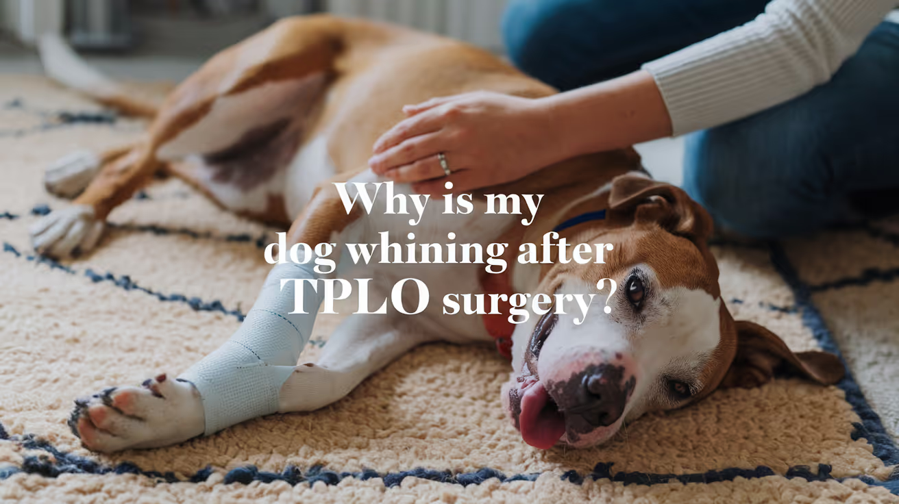
Dog Whining After TPLO Surgery: Causes and Care
Learn why your dog may whine after TPLO surgery, what it means, and how to help your pet recover comfortably and safely.
After your dog undergoes TPLO surgery, you might notice whining behavior that can worry you. Dog whining after TPLO surgery is common and can signal pain, discomfort, or anxiety during recovery. Understanding why your dog whines helps you provide better care and support during this critical healing phase.
This article explains the main reasons dogs whine after TPLO surgery, how to recognize when it is normal or concerning, and what steps you can take to ease your dog's discomfort. You will learn practical tips for managing pain, preventing complications, and promoting a smooth recovery.
Why is my dog whining after TPLO surgery?
Whining after TPLO surgery usually relates to pain or discomfort from the operation. Dogs cannot tell us how they feel, so whining is one way they express distress. It can also be a sign of anxiety or frustration due to restricted movement during recovery.
Recognizing the cause of whining helps you respond appropriately to your dog's needs and avoid unnecessary stress or complications.
- Postoperative pain: Pain from the surgical site is the most common reason for whining and usually peaks in the first few days after surgery.
- Restricted mobility: Limited ability to move or walk can cause frustration and whining as your dog adjusts to the recovery process.
- Anxiety or stress: Changes in routine, confinement, and discomfort can make your dog anxious, leading to vocalization like whining.
- Need for attention: Your dog may whine to seek comfort, reassurance, or physical contact from you during recovery.
Understanding these causes allows you to provide better comfort and care to your dog after TPLO surgery.
How can I tell if my dog’s whining is normal or a sign of a problem?
Not all whining after TPLO surgery is cause for alarm. Normal whining is usually mild and occurs during movement or when your dog is settling down. However, excessive or persistent whining may indicate complications or uncontrolled pain.
Knowing the difference helps you decide when to contact your veterinarian for advice or intervention.
- Normal whining: Occurs intermittently, especially when your dog tries to move or change position, and decreases over time.
- Signs of severe pain: Loud, continuous whining combined with limping, restlessness, or refusal to bear weight may signal uncontrolled pain.
- Signs of infection: Whining with swelling, redness, discharge, or foul odor at the surgical site requires immediate veterinary attention.
- Behavioral changes: Excessive whining with aggression, lethargy, or loss of appetite may indicate complications or distress.
Monitoring your dog closely and noting changes in whining patterns helps ensure timely care and prevents worsening conditions.
What pain management options are available after TPLO surgery?
Effective pain control is essential to reduce whining and promote healing after TPLO surgery. Your veterinarian will prescribe medications and suggest supportive measures to keep your dog comfortable.
Following the prescribed pain management plan closely improves your dog's recovery experience and reduces stress for both of you.
- Prescription painkillers: Non-steroidal anti-inflammatory drugs (NSAIDs) or opioids are commonly used to control postoperative pain safely.
- Cold therapy: Applying cold packs to the surgical area can reduce swelling and numb pain during the first 48 hours after surgery.
- Physical support: Using slings or harnesses helps your dog move without putting full weight on the operated leg, reducing pain during walking.
- Environmental comfort: Providing a quiet, soft resting area minimizes discomfort and encourages rest, which reduces whining.
Always follow your veterinarian's instructions on medication dosage and duration to avoid side effects or complications.
How should I care for my dog’s surgical site to reduce discomfort?
Proper wound care after TPLO surgery helps prevent infection and reduces pain that can cause whining. Keeping the surgical site clean and protected is vital during recovery.
Regular inspection and gentle care promote healing and comfort for your dog.
- Keep the incision dry: Avoid bathing or allowing water on the surgical site until your vet confirms it is safe to do so.
- Prevent licking or chewing: Use an Elizabethan collar or protective clothing to stop your dog from irritating the wound.
- Check for signs of infection: Look for redness, swelling, discharge, or foul smell and report concerns to your vet promptly.
- Follow dressing instructions: Change bandages as directed by your veterinarian to maintain cleanliness and support healing.
Consistent care of the surgical site reduces pain and helps your dog feel more comfortable during recovery.
What activity restrictions should I follow to help my dog heal?
Limiting your dog's activity after TPLO surgery is crucial to prevent injury and reduce pain that causes whining. Controlled movement supports proper healing and avoids complications.
Understanding and enforcing these restrictions ensures your dog recovers safely and comfortably.
- Strict confinement: Keep your dog in a small area or crate to limit running, jumping, or climbing for at least 6 to 8 weeks post-surgery.
- Leash walks only: Allow short, slow leash walks for bathroom breaks, avoiding off-leash activity or rough surfaces.
- Avoid stairs: Prevent stair climbing to reduce strain on the healing leg and minimize pain.
- Gradual reintroduction: Follow your veterinarian’s guidance to slowly increase activity as healing progresses, monitoring for any signs of discomfort.
Adhering to activity restrictions helps reduce whining caused by pain or injury during recovery.
When should I contact my veterinarian about my dog’s whining?
Knowing when to seek veterinary help is important if your dog’s whining signals a problem after TPLO surgery. Prompt attention can prevent complications and improve outcomes.
Being proactive about your dog’s symptoms ensures timely care and peace of mind.
- Persistent severe whining: If your dog whines continuously and shows signs of severe pain or distress, contact your vet immediately.
- Signs of infection: Whining accompanied by swelling, redness, discharge, or fever requires urgent veterinary evaluation.
- Changes in mobility: Sudden inability to bear weight or worsening lameness with whining should be assessed by a professional.
- Behavioral or appetite changes: Excessive whining with lethargy, aggression, or loss of appetite may indicate complications needing veterinary care.
Timely communication with your veterinarian helps ensure your dog’s recovery stays on track and reduces unnecessary suffering.
Conclusion
Dog whining after TPLO surgery is a common way your pet expresses pain, discomfort, or anxiety during recovery. Understanding the reasons behind the whining helps you provide better care and comfort to your dog.
By managing pain effectively, caring for the surgical site, restricting activity, and monitoring for complications, you can support your dog's healing and reduce distress. Always consult your veterinarian if you are concerned about your dog's whining or recovery progress.
FAQs
How long does whining usually last after TPLO surgery?
Whining typically decreases within the first two weeks as pain and discomfort lessen. Persistent or worsening whining beyond this period should be evaluated by a veterinarian.
Can I give my dog over-the-counter pain medication for TPLO recovery?
Do not give over-the-counter pain medications without veterinary approval, as some can be toxic to dogs or interfere with prescribed treatments.
Is it normal for my dog to whine at night after TPLO surgery?
Yes, dogs may whine at night due to discomfort or anxiety. Providing a comfortable, quiet space and following pain management helps reduce nighttime whining.
How can I comfort my dog when it whines after surgery?
Offer gentle petting, speak softly, and stay close to reassure your dog. Avoid excessive stimulation that may increase anxiety or pain.
When can my dog start physical therapy after TPLO surgery?
Physical therapy usually begins 1 to 2 weeks post-surgery, depending on healing progress. Follow your veterinarian’s recommendations for timing and exercises.
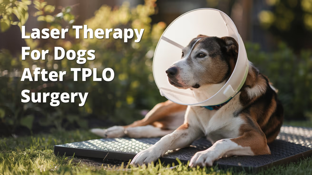
Laser Therapy for Dogs After TPLO Surgery
Learn how laser therapy helps dogs recover faster and with less pain after TPLO surgery for cruciate ligament repair.
Laser therapy also called photobiomodulation (PBMT) or low-level laser therapy (LLLT) is one of the most commonly offered adjunct treatments during TPLO recovery. Many specialist and rehabilitation centres include it routinely.
But the evidence for its benefits is more nuanced than the marketing suggests, and owners deserve an honest picture of what it does and does not reliably achieve.
Quick answer: Laser therapy after TPLO uses specific light wavelengths to reduce inflammation and support tissue healing. Evidence for early pain reduction is moderately supported; evidence for improved radiographic bone healing is weak. It is a safe adjunct but should not replace rehabilitation exercises, pain medication, or activity restriction.
Key takeaways
- Laser therapy reduces postoperative inflammation and may improve gait scores: a TPLO study found better hindlimb function at 8 weeks in treated dogs
- Evidence for improving radiographic bone healing is weak: three controlled studies found no statistically significant difference in healing time
- The 2024 AVMA randomized trial found no significant difference in CRP, weight bearing, pain scores, or SSI rates between PBMT and sham groups
- Sessions typically begin within the first few days of surgery and continue through the rehabilitation phase
- Laser therapy is safe with few contraindications: avoid eyes and active tumour sites; safe over the TPLO incision once closed
- It works best as part of a multimodal plan: exercise therapy and pain medication carry stronger evidence than laser alone
What laser therapy does
Laser therapy for dogs, also known as photobiomodulation, involves using specific wavelengths of light to penetrate tissues and promote cellular regeneration and healing. The laser light stimulates the production of ATP (adenosine triphosphate), enhancing cell repair and growth, reducing inflammation, and increasing blood circulation.
Photobiomodulation therapy has been shown to decrease inflammation, and increase analgesia, vascularization, and tissue healing after musculoskeletal injury or surgery.
The mechanism is photochemical: light energy at specific wavelengths (typically 630 to 980 nm) is absorbed by mitochondria.
This increases ATP production, modulates reactive oxygen species, and influences gene expression related to inflammation and healing.
The effects are local confined to the tissue depth the light reaches.
For post-TPLO use, the targets are: the surgical incision, the osteotomy site in the proximal tibia, and the surrounding periarticular soft tissues.
What the clinical evidence shows
Pain and function
Research following TPLO surgery showed that dogs receiving LLLT had better hindlimb function and gait scores at 8 weeks compared to controls. This is especially valuable in orthopedic recovery, where early weight-bearing can prevent muscle atrophy and joint stiffness.
In a controlled veterinary study, dogs with surgical incisions treated with laser therapy exhibited significantly less inflammatory cell infiltration and tissue necrosis within the first week post-op compared to untreated controls.
The 2024 randomized trial
54 client-owned dogs with CCL rupture undergoing unilateral TPLO surgery were enrolled. The study population was randomly assigned to either a treatment group receiving PBMT (24 dogs) or a control group (30 dogs). PBMT was performed immediately after induction, and at 6 hours, 24 hours, 48 hours, and 8 weeks postoperatively. Evaluation of CRP, pain scores, evidence of SSI, and percentage weight bearing were assessed at all time points.
The trial found the therapy showed promise but no statistically significant difference between groups on any primary outcome measure.
Bone healing
Three studies compared LLLT to a control and concluded that LLLT treatment did not make a significant difference in improving radiographic bone healing. The studies collectively provide weak evidence for this outcome.
This is an important distinction: laser therapy may support soft tissue healing, pain, and early function but it does not appear to accelerate the osteotomy healing visible on radiographs.
When to start and how often
Laser therapy uses focused light energy on the surgical site to support healing.
Most rehabilitation programmes begin laser therapy within the first 1 to 3 days after TPLO surgery, often at the surgical centre before discharge or at the first rehabilitation visit.
Typical post-TPLO laser protocol:
- Frequency: 3 to 5 sessions per week in the first 2 to 3 weeks
- Frequency: 1 to 2 sessions per week from weeks 3 to 8
- Session duration: 5 to 15 minutes depending on the laser system and dosing protocol
- Total sessions: typically 6 to 12 in the first 8-week recovery phase
The protocol varies by laser system, power output, and the individual patient's response.
Realistic expectations
Laser therapy is a useful adjunct in TPLO recovery. It is not a substitute for the treatments with stronger evidence: pain medication, activity restriction, and structured rehabilitation exercises.
Laser therapy could be particularly helpful for dogs with weight-bearing and gait issues while recovering from TPLO surgery after a cruciate injury.
Dogs with significant early swelling, wound sensitivity, or slow initial weight-bearing progress may benefit most. Dogs recovering well with standard multimodal analgesia and rehabilitation may show less measurable difference.
For the bone healing timeline that laser therapy supports during recovery, see TPLO bone healing time in dogs explained.
For the full recovery plan that laser therapy fits into, see 10 essential TPLO recovery tips for pet owners.
For the physical therapy that is the primary evidence-based adjunct, see when to start physical therapy after TPLO surgery.
For swelling management in the recovery period, see how long does swelling last after TPLO surgery.
Frequently asked questions
Is laser therapy safe over the TPLO incision?
Yes, once the incision is closed. Laser therapy is safe over sutured incisions and can be applied at the surgical site from the first post-operative day in most protocols.
Avoid direct application over open wounds or actively infected tissue.
How many laser sessions does a dog need after TPLO?
Typically 6 to 12 sessions across the first 8 weeks.
Start at 3 to 5 sessions per week for the first 2 to 3 weeks, tapering to 1 to 2 per week through the rehabilitation phase.
The exact protocol depends on the laser system and rehabilitation plan.
Does laser therapy replace pain medication after TPLO?
No. Laser therapy is an adjunct to pharmaceutical pain management, not a replacement. Post-TPLO pain management requires NSAIDs, and often gabapentin or other analgesics.
Laser therapy may reduce the pain burden and support earlier mobility but does not provide sufficient analgesia on its own.
Can I do laser therapy at home with a consumer device?
Consumer-grade red light therapy devices exist but operate at lower power densities than veterinary therapeutic lasers. The clinical evidence discussed in this article relates to veterinary-grade PBMT devices.
Home devices may offer some benefit but cannot replicate the dosing of professional equipment. Discuss with your rehabilitation veterinarian before purchasing.
Will laser therapy prevent my dog from needing more medication?
Possibly. If laser therapy reduces post-operative inflammation and pain, some dogs may need lower doses of pain medication or taper off sooner.
This should be guided by your veterinarian based on your dog's individual recovery trajectory.
Resources
- AVMA Journal. Photobiomodulation Therapy in Dogs Undergoing TPLO After Cranial Cruciate Ligament Rupture. avmajournals.avma.org
- Veterinary Evidence. Does LLLT Improve Radiographic Healing for Dogs with CCL Rupture Undergoing TPLO Surgery? veterinaryevidence.org
- AKC. Laser Therapy For Dogs: Uses, Side Effects, and Alternatives. akc.org
- Erchonia. Laser Therapy for Post-Surgical Recovery in Pets. erchonia.com
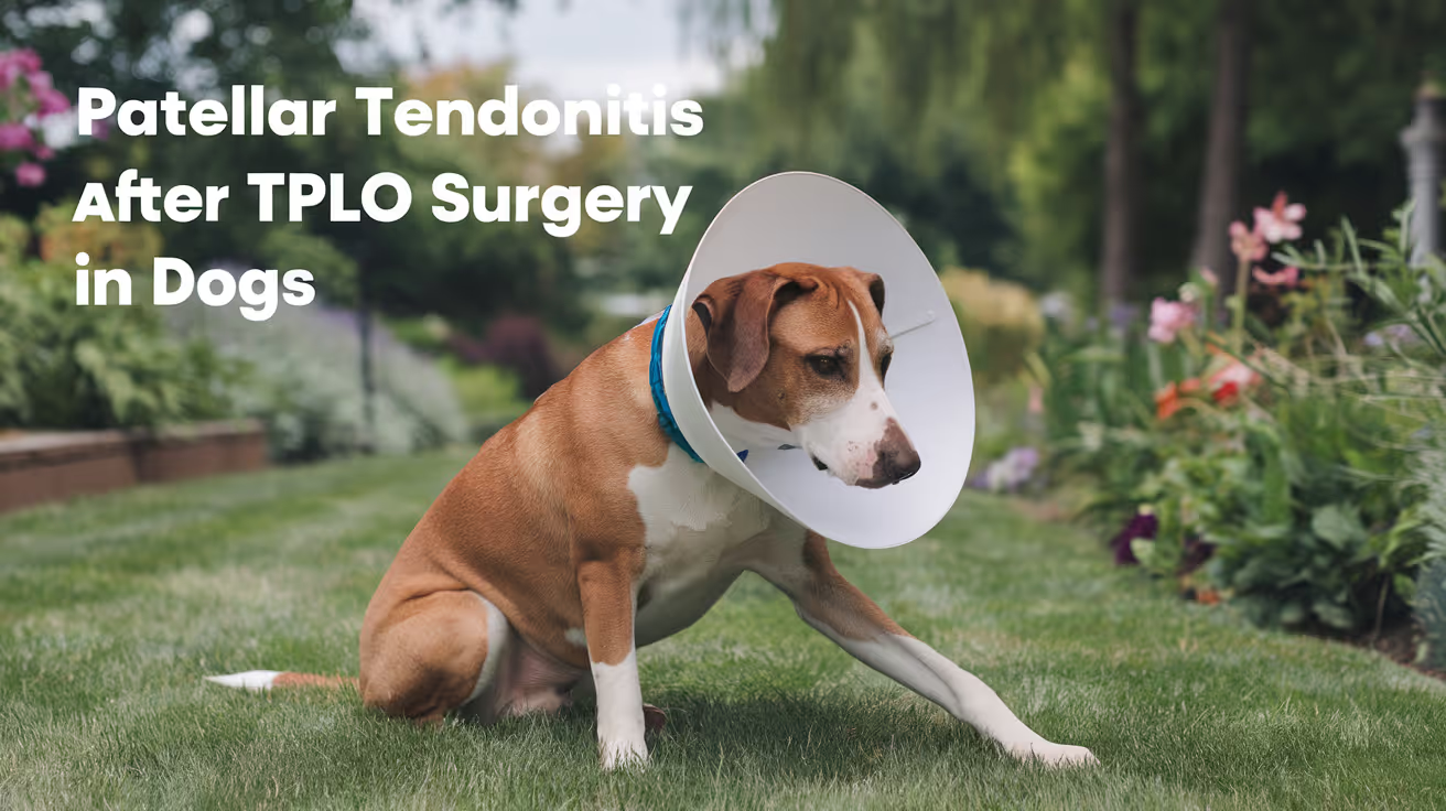
Patellar Tendonitis After TPLO Surgery in Dogs
Learn about patellar tendonitis after TPLO surgery in dogs, including causes, symptoms, treatment, and recovery tips for pet owners.
Patellar tendonitis more precisely called patellar desmopathy or patellar ligament desmitis in veterinary literature is a recognized complication of TPLO surgery. It is also one of the most under-discussed.
Research published in PMC shows radiographic evidence of patellar ligament thickening in 93% of dogs at 4 weeks after TPLO, though most cases are asymptomatic.
Understanding what this condition is, when it becomes a clinical problem, and how it is treated helps owners navigate one of the more commonly missed post-TPLO findings.
Quick answer: Patellar desmitis after TPLO is inflammation and thickening of the patellar ligament. Radiographic thickening occurs in 93% of TPLO dogs at 4 weeks but is usually asymptomatic. Clinically significant cases cause pain and increased lameness. Most are self-limiting; shock wave therapy accelerates resolution.
Key takeaways
- Radiographic patellar ligament thickening occurs in 93% of TPLO dogs at 4 weeks: most are asymptomatic and self-limiting
- Clinically significant patellar desmitis causes pain, swelling, and increased lameness beyond expected post-op discomfort
- TPLO changes stifle biomechanics and increases patellar tendon load: this explains why desmitis occurs more after TPLO
- Shock wave therapy (SWT) significantly reduced patellar ligament thickness at weeks 6 and 8 in a 2012 clinical trial
- Most cases are self-limiting: rest and NSAIDs resolve the majority of clinically significant cases within weeks to months
- Excessive post-operative activity is a modifiable risk factor: strict restriction in the first 8 weeks reduces desmitis risk
What is patellar desmitis after TPLO?
The patellar ligament runs from the kneecap (patella) to the tibial tuberosity — the bony bump just below the knee on the front of the tibia.
It is a critical structure for leg extension and stifle stability.
After TPLO surgery, the biomechanics of the stifle change fundamentally. The tibial plateau is rotated and held at a new angle.
This changes how forces are distributed around the knee, including the forces that pass through the patellar ligament.
The patellar ligament experiences altered tension and loading patterns after TPLO that it did not experience before surgery.
PMC research (2025 retrospective study) confirms desmopathy typically develops within 2.5 months after surgery, affecting the distal portion, and that TPLO causes more ligament thickening than other CCL repair methods.
A separate PMC study using elastosonography confirmed that patellar tendon tendinopathy can be easily detected clinically but that the exact damage evaluation is challenging without ultrasound or advanced imaging.
Why does TPLO cause patellar tendonitis?
Several mechanisms contribute:
Altered stifle biomechanics: TPLO changes the angle of the tibial plateau and the position of the tibial tuberosity relative to the femur. This alters patellar tendon geometry and tension during weight-bearing.
Surgical trauma: retraction and manipulation of soft tissues around the tibial tuberosity during the procedure causes local inflammatory changes that can involve the patellar tendon.
Intraoperative tension increase: rotating the proximal tibial segment may change the tension in the patellar tendon directly, especially during the initial healing phase.
Excessive postoperative exercise: Clinicians Brief's treatment review notes that excessive postoperative exercise is a commonly proposed contributing factor. Dogs allowed too much activity too early are more likely to develop clinically significant desmitis.
How common is patellar desmitis after TPLO?
Radiographic evidence of patellar ligament thickening at 4 weeks post-TPLO is reported in 93% of dogs in clinical studies.
The vast majority of these dogs have no clinical signs the radiographic finding does not translate into a symptomatic problem for most patients.
Clinically significant desmitis where the dog shows pain and lameness attributable to the patellar ligament specifically is less common but well-recognized by veterinary orthopedic surgeons.
Laguna Hills Animal Hospital notes that patellar tendinosis is relatively benign in most cases and improves with rest and medication.
Signs of clinically significant patellar desmitis
Unlike the asymptomatic radiographic finding, clinical patellar desmitis produces:
- Pain on direct palpation of the patellar ligament (between the kneecap and tibial tuberosity)
- Swelling or thickening directly over the ligament
- Lameness that is worse than expected for the dog's recovery stage
- Reluctance to fully extend the stifle during walking or exercise
- Stiffness after rest that improves briefly with movement
These signs typically appear in the first 6 to 10 weeks after TPLO.
Diagnosis
A veterinarian diagnoses patellar desmitis through:
Physical examination: palpation of the patellar ligament for thickening, pain response, and heat. A dog with significant desmitis will react to direct pressure on the ligament.
Radiographs: thickening of the soft tissue shadow over the patellar ligament on a lateral stifle projection. This is often identified at the routine 4 to 6 week post-TPLO radiographic recheck.
Ultrasound: provides direct visualization of the ligament, showing thickening, fiber disruption, and periligamentous edema. Ultrasound is more sensitive than radiographs and is the preferred modality for detailed assessment.
Treatment
Rest and activity restriction
The foundation of treatment for most cases. Reducing the mechanical load on the patellar ligament allows the inflammatory process to resolve.
Strict activity restriction should already be in place for post-TPLO recovery patellar desmitis is a reason to enforce it rigorously.
NSAIDs
Anti-inflammatory medication reduces pain and local inflammation. Meloxicam or carprofen at appropriate veterinary doses is the standard pharmaceutical approach for most dogs with symptomatic desmitis.
Shock wave therapy (SWT)
Clinicians Brief reviewed a trial of 30 large-breed TPLO dogs: SWT at weeks 4 and 6 significantly reduced patellar ligament thickness on radiographs at weeks 6 and 8 vs. controls.
SWT should be considered for dogs with clinically significant patellar desmitis.
Laser therapy
Low-level laser therapy (photobiomodulation) may reduce local inflammation and support tissue healing, though evidence specific to patellar desmitis after TPLO is limited compared to the SWT evidence base.
For the complete TPLO complications reference, see 15 common complications after TPLO surgery in dogs. For the incision infection guide, see TPLO incision infection symptoms and prevention.
For the general lameness guide that helps distinguish desmitis from other causes of post-TPLO lameness, see lameness after TPLO surgery in dogs.
For the TPLO failure rate context, see TPLO failure rate in dogs explained.
Frequently asked questions
Is patellar tendonitis after TPLO serious?
In most dogs, no. The radiographic finding is near-universal but typically asymptomatic and self-limiting.
Clinically significant desmitis causes pain and increased lameness but usually resolves with rest, NSAIDs, and sometimes shock wave therapy. It rarely causes permanent dysfunction.
How do I know if my dog has patellar tendonitis vs. normal post-TPLO soreness?
The key distinguishing feature is pain specifically on palpation of the patellar ligament the structure running from the kneecap to the bump just below the knee.
Normal post-TPLO soreness is diffuse and improves steadily. Patellar desmitis produces focal pain at this location, often with visible swelling over the ligament.
Can patellar desmitis be prevented?
Strict activity restriction in the first 8 weeks post-TPLO is the most effective preventive measure, since excessive early activity is a recognized contributing factor.
It cannot always be prevented altered biomechanics from the surgery itself play a role regardless of activity level but minimizing mechanical load during the healing phase reduces severity.
Does patellar tendonitis require a second surgery?
Almost never. The vast majority of cases resolve with conservative management. Shock wave therapy is effective in cases that need more active treatment.
Surgical intervention for patellar desmitis after TPLO is extremely rare.
Will this affect my dog's long-term outcome after TPLO?
For most dogs, no. Clinically significant patellar desmitis is typically a temporary complication that resolves during the recovery period.
Long-term stifle function in dogs that develop and recover from patellar desmitis is generally comparable to dogs that do not develop the condition.
Resources
- PMC. Retrospective Study on Patellar Desmopathy Following Surgical Resolution of CCL Rupture in 28 Dogs. ncbi.nlm.nih.gov
- PMC. Radiographic, Ultrasonographic and Shear Elastosonographic Changes in Patellar Ligament in Dogs Undergoing TPLO. ncbi.nlm.nih.gov
- Clinicians Brief. Treating Patellar Ligament Desmitis. cliniciansbrief.com
- Laguna Hills Animal Hospital. TPLO Surgery Complications. lhah.com
- ACVS. Cranial Cruciate Ligament Disease. acvs.org
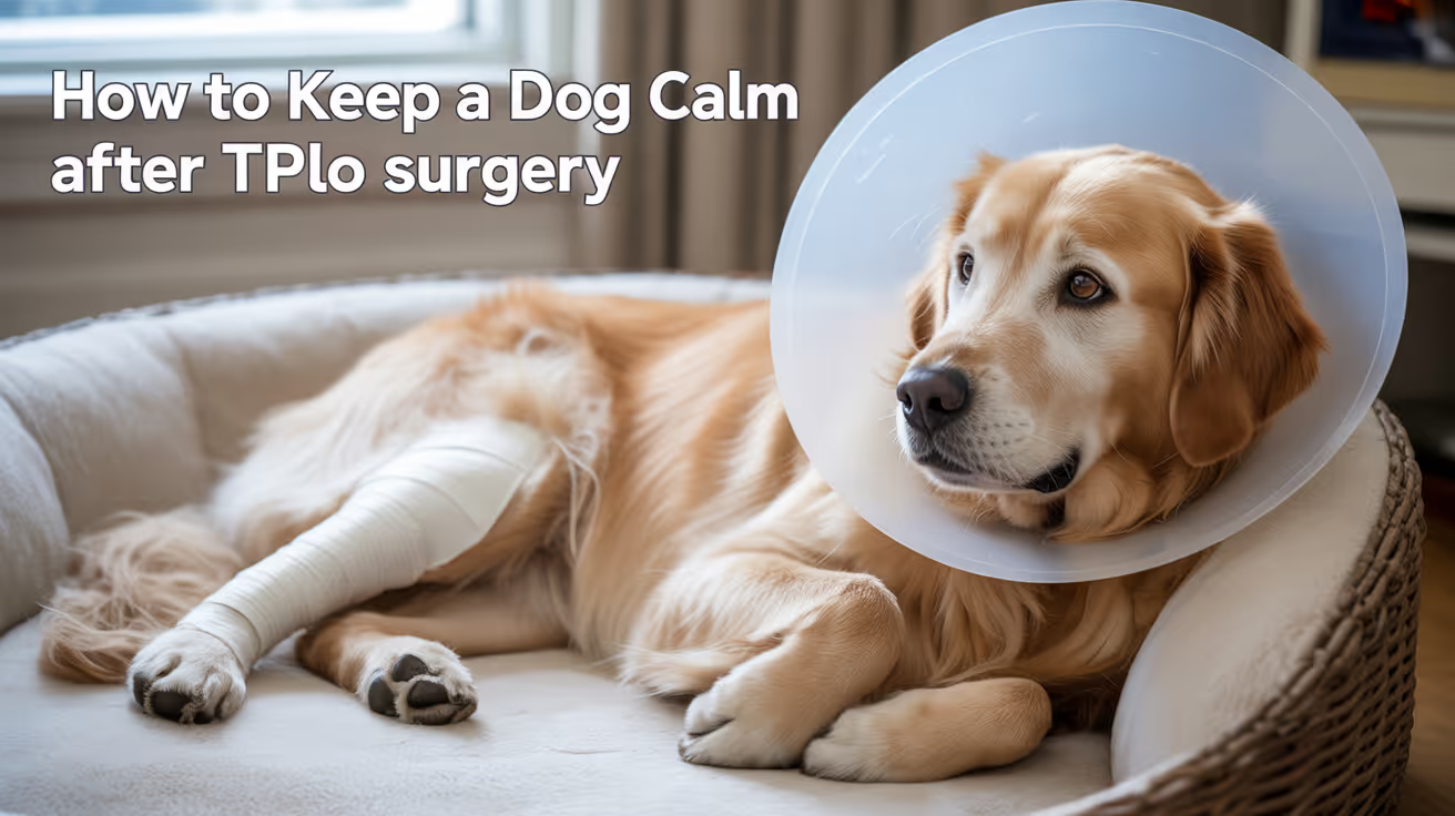
How to Keep a Dog Calm After TPLO Surgery
Learn how to keep your dog calm after TPLO surgery using rest, crate training, and soothing techniques for safe, stress-free recovery.
Keeping a dog calm during TPLO recovery is one of the hardest parts of the process -- and one of the most important.
A dog that feels physically capable of running at week 3 is not biologically ready to run at week 3. The osteotomy has not consolidated. The plate can fail.
Activity restriction is not optional.
Quick answer: Most effective strategies for keeping a dog calm after TPLO surgery: crate confinement during unsupervised periods, food puzzles and scent games, consistent pain management, a quiet low-stimulus environment, and trazodone or gabapentin for highly anxious dogs. Keep a leash on the dog indoors.
Key takeaways
- Crate confinement is the foundation of activity control: large enough to stand and turn but not pace; prevents the spontaneous running, jumping, and spinning that cause plate failure
- Mental stimulation substitutes for physical activity: food puzzles, lick mats, frozen Kongs, and scent games provide neural reward without physical impact
- Pain management compliance directly affects calmness: uncontrolled pain causes restlessness; giving NSAIDs and gabapentin on schedule keeps the dog comfortable enough to rest
- The environment must be actively managed: loud TV, visitors, and other pets cause arousal and movement; a quiet, predictable low-stimulus space reduces arousal
- Trazodone and gabapentin can be prescribed for highly anxious dogs: VetSurgInfo confirms trazodone is inexpensive, safe, and effective for anxiety and sedation during TPLO recovery
- Always use a leash indoors during early recovery: a dog that slips or startles can displace the plate; a house leash gives the owner control at all times
The crate: foundation of confinement
A crate is the safest confinement tool for TPLO recovery.
It limits the movements that risk plate failure -- spontaneous jumping, running to the door, sudden pivoting -- and gives the dog a defined, safe space.
Crate selection:
- Large enough for the dog to stand, turn, and lie down comfortably
- Not so large the dog can pace or run inside
- Soft bedding on non-slip material
- Location in the household main area, not isolated -- isolation increases anxiety
SustainableVet.org confirms: use a crate, pen, or small room with non-slip flooring. Crates prevent jumping, running, and twisting. Choose a crate large enough to stand and turn.
Exercise pen alternative: for dogs that do not tolerate crates, an exercise pen (X-pen) provides a larger confined space while still restricting free movement. Place the X-pen in the main living area so the dog is not isolated.
Small room alternative: a bathroom, laundry room, or small bedroom with a baby gate can work. Remove furniture the dog could jump on.
Mental stimulation: the most underused tool
A dog confined physically but not cognitively engaged becomes frustrated, which leads to escape attempts and activity that risks the repair.
Mental stimulation provides the same dopamine reward as physical activity with zero mechanical risk.
Effective mental stimulation options:
Food puzzles and slow feeders: scatter meals in a puzzle feeder or across a lick mat instead of in a bowl. A meal that takes 15 minutes to eat provides significant cognitive engagement.
Frozen Kongs: stuff with wet food, peanut butter (xylitol-free), or kibble soaked in broth; freeze for several hours. A frozen Kong can occupy a dog for 20 to 30 minutes.
Sniff games: hide small treats in a folded towel, a muffin tin covered with tennis balls, or a scatter across the crate bedding. Sniffing is cognitively tiring -- it is physiologically equivalent to sustained physical exercise in terms of neural engagement.
Chew toys: long-duration chews (bully sticks, raw bones appropriate for the dog's size, frozen chicken necks) keep the dog occupied without movement.
Animal Outpatient Surgery confirms: soft music, white noise, or pheromone diffusers create a serene atmosphere. Mental engagement through food puzzles and scent games reduces frustration from limited mobility.
Environmental management
The dog's immediate environment directly controls arousal level.
Reduce auditory stimuli: loud TV, doorbell sounds, and street noise cause the dog to alert and attempt to move. White noise machines or calm background music reduce stimulus peaks.
Manage household traffic: visitors who excite the dog, children who rough-house nearby, and other pets that want to interact all cause arousal. Limit access to the dog during early recovery.
Manage other pets: other dogs and cats approaching the recovering dog cause arousal and can trigger sudden jumping or running. Keep them separated during the early weeks, particularly unsupervised.
Non-slip flooring: rugs and yoga mats in the dog's movement area prevent slipping, which generates uncontrolled leg movements that can stress the plate.
Laveen Vet Center confirms: set up a quiet, confined area free from loud noises, active children, and other pets. White noise or soft music helps create a serene atmosphere.
Pain management and its role in calmness
An undertreated dog is a restless dog. Pain creates the exact behaviors -- panting, pacing, inability to settle, licking the leg -- that also indicate anxiety.
Distinguishing pain from anxiety is important because the response is different.
Pain management checklist:
- Give NSAIDs and gabapentin on the exact schedule prescribed, not as needed
- Never skip doses because the dog appears comfortable -- the medication is maintaining that comfort
- Give medications approximately 30 minutes before walks (TPLO home recovery protocols recommend this timing)
- Contact your vet if the dog cannot settle despite correct medication administration
VetSurgInfo confirms: gabapentin provides both analgesia and sedation; the sedating effect facilitates recovery and rest, particularly in the first 7 days.
Prescribed calming medications
For dogs with high anxiety, separation anxiety, or an inability to settle despite environmental and mental stimulation management, veterinary-prescribed medications are appropriate and safe.
Trazodone: a serotonin modulator used off-label in dogs for anxiety and sedation. VetSurgInfo confirms: dogs do very well on trazodone for anxiety and sedation during TPLO recovery; it is inexpensive and can facilitate recovery, rest, and calm during vet visits.
Gabapentin: the analgesic side effect of sleepiness helps with calmness in addition to pain control.
Alprazolam or other benzodiazepines: may be appropriate for situational anxiety (specific triggers like thunder).
SustainableVet.org confirms: vets can prescribe mild sedatives or calming medications for dogs who remain overly anxious; these drugs are safe when dosed correctly and can prevent harmful overactivity.
Never give human anxiety medications to dogs. Never use human supplements without vet approval -- some contain xylitol or other ingredients toxic to dogs.
Owner behavior and management
Owners unintentionally promote activity.
Do not excitedly greet the dog: excited greetings cause the dog to jump and spin. Calm, quiet greetings reduce arousal.
Leash the dog indoors: a house leash (a short, light leash attached to the collar) gives the owner control when the dog is out of the crate. If the dog startles or tries to move suddenly, the leash prevents uncontrolled movement.
Maintain a predictable routine: dogs are routine-oriented. Predictable meal times, bathroom walk times, and crate times reduce anticipatory anxiety. Variation creates excitement; routine creates calm.
Keep visits short: sitting with the dog is good. Prolonged exciting interaction is not. Brief, calm contact several times daily is better than one long exciting session.
For the recovery timeline, see what to expect after TPLO surgery in dogs. For infection prevention, see post-operative care mistakes that increase TPLO infection risk.
For swelling management, see fluid buildup after TPLO surgery in dogs. For exercise, see TPLO recovery exercises for dogs.
Frequently asked questions
My dog screams and cries in the crate for hours. What do I do?
Contact your vet. Sustained, intense crate distress may indicate: inadequate pain control (the dog is in pain, not just anxious), genuine severe separation anxiety, or insufficient mental stimulation.
Ask about trazodone or gabapentin for anxiety management. Do not give up on crate confinement -- the alternative is uncontrolled movement that risks the repair.
Is it okay to let my dog out of the crate to sit with me on the sofa?
Only if the dog is on a leash and cannot jump. Allow the dog to lie next to you on the floor or on a low dog bed beside you.
Do not allow the dog onto the sofa -- falling or jumping off generates the same plate-failure risk as running.
My dog seems much better at 3 weeks. Can I relax the restrictions?
No. Clinical improvement does not mean bone healing is complete. The osteotomy is not confirmed healed until radiographs at 8 to 12 weeks.
Plate failure typically happens when owners relax restrictions based on the dog appearing well. Maintain full restrictions until vet clearance.
What is the best medication for a high-energy dog that can't settle?
Trazodone is the most commonly prescribed agent for activity restriction in TPLO dogs. It is effective, safe, inexpensive, and can be used for the full restriction period.
Discuss with your vet at or before surgery -- it is easier to start early than to address a crisis at week 2.
Can I take my dog to visit friends or family during recovery?
Very limited visits in controlled, quiet environments where the dog is leashed and confined.
No visits to households with other active dogs, children who will excite the dog, or any environment where the dog cannot be kept calm. Unfamiliar environments increase anxiety and exploration behavior.
Resources
- SustainableVet. How to Keep a Dog Calm After TPLO Surgery. sustainablevet.org
- VetSurgInfo. TPLO Recovery Tips. vetsurginfo.com
- Animal Outpatient Surgery. How to Keep a Dog Calm After TPLO Surgery. animaloutpatientsurgery.com
- Laveen Vet Center. How to Keep a Dog Calm After TPLO Surgery. laveenvetcenter.com

13 Long-Term Effects of TPLO Surgery on Dogs
Discover the 13 long-term effects of TPLO surgery in dogs, including benefits, risks, and care tips for optimal recovery and joint health
TPLO produces predominantly positive long-term effects in most dogs, but it also produces a predictable set of changes that owners should understand and manage proactively.
Most of these effects are manageable with appropriate care. Knowing what to expect prevents owners from being surprised by normal post-TPLO findings and helps them recognize when a change requires veterinary attention.
Quick answer: The 13 key long-term effects of TPLO surgery: improved joint stability, progressive osteoarthritis, muscle atrophy, morning stiffness, cold weather sensitivity, scar tissue formation, patellar tendon thickening, permanent implant in situ, contralateral CCL rupture risk, late implant complications, behavioral changes from chronic pain, weight management challenges, and long-term monitoring requirements.
Key takeaways
- Improved joint stability is the intended and most common long-term effect: for 90 to 95% of dogs, TPLO eliminates CCL-related instability and restores near-normal function
- Progressive osteoarthritis develops in all CCL-affected stifles regardless of surgical technique: TPLO slows OA progression but does not stop it; long-term OA management is a universal requirement
- Muscle atrophy is common in the operated leg throughout recovery; the thigh may remain visibly smaller than the contralateral side for months if rehabilitation is inadequate
- Contralateral CCL rupture risk is high: SustainableVet.org confirms up to a 50% chance of tearing the opposite CCL within a few years; the same degenerative process affects both stifles
- The TPLO plate and screws are permanent in the vast majority of cases: removal is only performed when complications arise; routine plate removal is not standard practice
- Weight management is the most impactful long-term owner-controlled variable: excess weight accelerates OA progression; maintaining ideal body condition is the primary long-term prevention strategy
The 13 long-term effects
1. Improved joint stability and mobility
The primary intended effect. The osteotomy and plate fixation permanently change the tibial plateau geometry, eliminating the cranial tibial thrust that caused instability.
For 90 to 95% of dogs, this produces lasting functional improvement: return to normal gait, ability to run and play, and freedom from the pain of unstable joint loading.
SustainableVet.org confirms: over 90% of dogs regain full limb function within a year; with proper care, many dogs remain active for 8 to 10 years after surgery.
2. Progressive osteoarthritis
The most universal long-term challenge. CCL disease initiates an arthritis process in the stifle that continues regardless of surgical outcome.
TPLO reduces the rate of progression and the severity of clinical signs compared to other treatment options, but OA continues.
Clinical signs of progressing OA emerge gradually over months and years: stiffness after rest, reluctance to run, difficulty climbing, and behavioral changes associated with chronic low-grade pain.
Management: weight management, omega-3 fatty acids, glucosamine and chondroitin, NSAIDs for flares, hydrotherapy, physiotherapy.
3. Muscle atrophy and asymmetry
The operated leg loses muscle mass rapidly during the restriction period and slowly rebuilds during rehabilitation. Full recovery of muscle symmetry takes 4 to 6 months in most dogs.
In some dogs -- particularly those with inadequate rehabilitation or ongoing joint pain -- visible thigh circumference asymmetry persists long-term.
Vetplayas confirms: due to reduced use during recovery and altered movement patterns post-surgery, muscles around the operated leg may weaken and shrink.
4. Morning stiffness
Morning stiffness -- the dog rises from sleep stiffly and walks awkwardly for the first few minutes before loosening up -- is one of the most common long-term effects of post-TPLO OA.
It typically develops months to years after surgery and gradually worsens as the dog ages.
This stiffness should not be confused with acute lameness. It consistently improves within 5 to 10 minutes of movement.
If it does not improve, or if the dog refuses to bear weight on waking, contact your vet.
5. Cold weather sensitivity
Dogs with OA in the operated stifle commonly show increased stiffness and lameness during cold weather or when barometric pressure changes.
This reflects the sensitivity of arthritic joints to temperature and pressure changes -- the same phenomenon seen in human arthritis.
Management: NSAIDs or joint supplements maintained year-round; warming the dog's sleeping area; a brief gentle walk before expecting full function in cold conditions.
6. Scar tissue formation
The surgical dissection and bone healing produce scar tissue in and around the stifle. This fibrous tissue generally has a beneficial stabilizing effect, contributing to long-term joint stability.
In some dogs, however, periarticular fibrosis restricts range of motion and causes stiffness that persists long after bone healing is complete.
Regular low-impact exercise and physiotherapy help maintain range of motion and prevent excessive fibrous tissue restriction.
7. Patellar tendon thickening
Fibrous thickening of the patellar tendon is a recognized long-term change after TPLO. It is typically subclinical -- detected on palpation at follow-up examinations but not producing clinical lameness.
In a small number of dogs, it contributes to reduced range of motion.
8. Permanent implant in situ
The TPLO plate and screws are designed to remain in place for life. In the vast majority of dogs, the implant is never removed and causes no long-term problems.
SustainableVet.org confirms: these implants are meant to stay in place permanently. In rare cases, some dogs may develop infections around the implant or experience loosening of the hardware over time.
The plate is palpable under the skin on the medial tibial surface. This is normal and not a complication.
9. Contralateral CCL rupture
The degenerative process that caused one CCL to rupture typically affects both stifles.
SustainableVet.org confirms dogs have up to a 50% chance of tearing the CCL in the opposite knee within a few years.
RCVS confirms approximately 50% of dogs develop CCL disease in the other hind limb.
This is not a complication of TPLO -- it is the natural history of bilateral CCL disease.
Monitoring the contralateral stifle for early signs of instability, effusion, or lameness is appropriate in all TPLO dogs.
10. Late implant-associated complications
Implant-related complications -- loosening, late infection from hematogenous seeding, or implant irritation -- can occur years after the original surgery. These are uncommon but recognized long-term effects.
Signs: new lameness focused at the plate site, warmth or swelling over the plate, a new draining tract in previously healed skin. Any of these warrants prompt veterinary assessment.
11. Behavioral changes
Chronic joint pain from progressive OA, late meniscal injury, or implant irritation can produce subtle behavioral changes: reduced engagement in play, reluctance to walk, increased irritability, or reduced stair use.
These changes are often attributed to aging but may reflect treatable pain.
Annual or semi-annual pain assessments, including use of validated canine pain scales (Canine Brief Pain Inventory, Helsinki Chronic Pain Index), help detect behavioral pain signs before they become severe.
12. Weight management challenges
Activity restriction during recovery predisposes many dogs to weight gain. Excess weight gained during the 8 to 12 week restriction period is then carried on the healing joint for the following months.
Long-term obesity accelerates OA progression and is the most modifiable risk factor for long-term joint health.
Medcovet confirms: because activity is restricted, veterinary teams may recommend reducing food intake by about 30% during recovery to prevent weight gain and extra strain on the knee joint.
13. Long-term monitoring and supplement requirements
TPLO dogs benefit from long-term monitoring (every 6 to 12 months after full recovery) to assess:
- Arthritis progression radiographically and clinically
- Body condition score and weight
- Contralateral stifle stability
- Implant integrity (radiographs every 1 to 2 years in some protocols)
Joint supplements (omega-3 fatty acids at anti-inflammatory doses; glucosamine and chondroitin) and ongoing weight management are the primary preventive interventions for the long-term joint changes that TPLO does not prevent.
For the long-term outcomes guide, see long-term outcomes of TPLO surgery. For arthritis management, see arthritis after TPLO surgery in dogs.
For the signs that require attention, see signs of TPLO failure in dogs. For the failure rate overview, see TPLO failure rate in dogs.
Frequently asked questions
Will my dog develop arthritis even if the TPLO was successful?
Yes. All CCL-affected stifles develop progressive osteoarthritis regardless of surgical technique or outcome.
TPLO reduces the rate of progression and the severity of clinical signs compared to non-surgical management and lateral suture repair.
The goal of TPLO is not to prevent OA but to minimize it while restoring function.
How long will the TPLO plate last?
The plate is designed to be permanent. Published long-term follow-up data at 6.8 years shows 90.4% of dogs with good to excellent results -- with the plate in place.
Plate complications requiring removal occur in 3.5 to 7.5% of cases.
My dog had TPLO 2 years ago and is now limping. Is this related to the surgery?
Possibly. Two-year post-TPLO lameness most commonly results from progressive OA, late meniscal tear, late implant-associated infection (hematogenous seeding), or contralateral CCL disease.
A veterinary examination and radiographs of both stifles will identify the cause and guide treatment.
Does the opposite leg always need TPLO as well?
Not necessarily and not always at the same time. RCVS confirms approximately 50% of dogs develop CCL disease in the opposite hind limb.
When and whether the second stifle requires surgery depends on whether the second CCL has ruptured or become significantly unstable. Annual monitoring of the contralateral stifle is appropriate.
Can TPLO dogs still live active lives long-term?
Yes. Most do. SustainableVet.org confirms that with proper care, many dogs remain active for 8 to 10 years after surgery.
The keys are weight management, regular low-impact exercise, joint supplements, and addressing pain or complications promptly.
Resources
- SustainableVet. 13 Long-Term Effects of TPLO Surgery on Dogs. sustainablevet.org
- RCVS Canine Cruciate Registry. Tibial Plateau Levelling Osteotomy. rcvsknowledge.org
- TPLO Info. TPLO Literature and Studies. tploinfo.com
- SustainableVet. Long-Term Outcomes of TPLO Surgery. sustainablevet.org
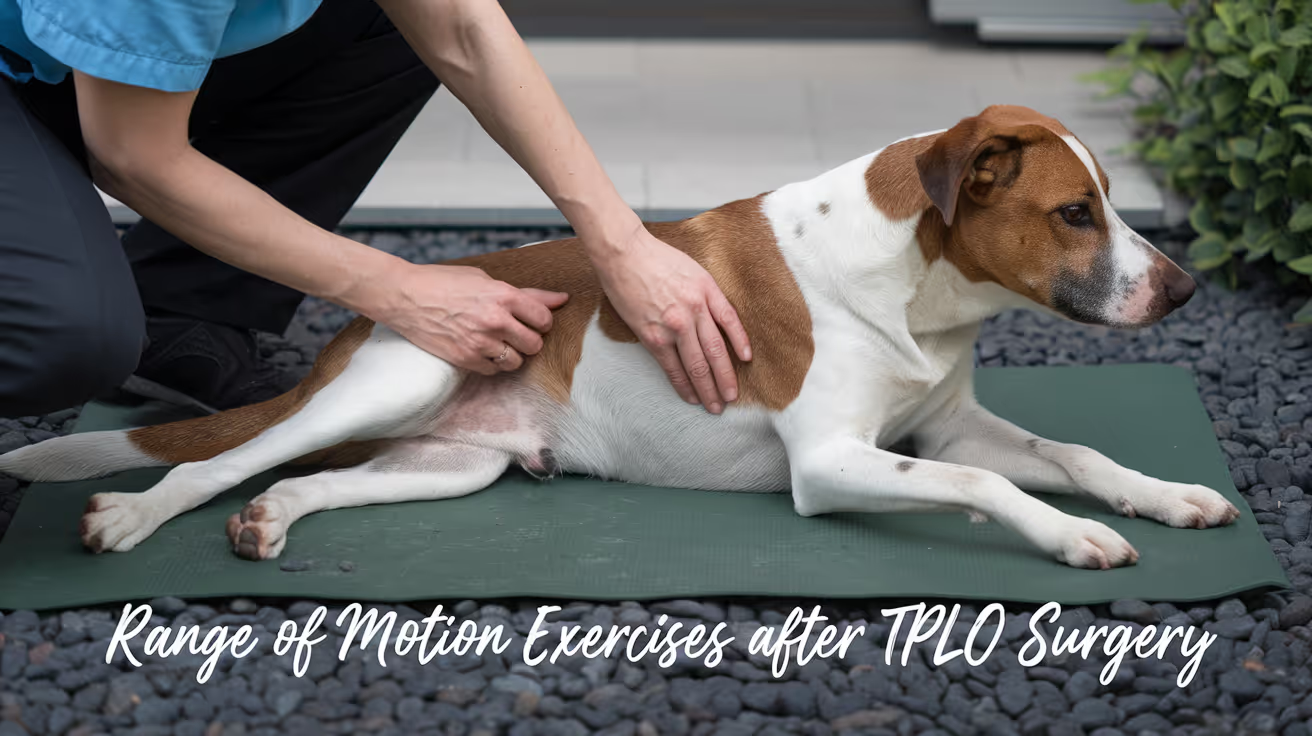
TPLO Range of Motion Exercises for Dogs
Learn effective TPLO range of motion exercises to help your dog recover safely and regain mobility after surgery.
Range of motion (ROM) exercises are one of the earliest and most consistently prescribed rehabilitation interventions after TPLO surgery.
They prevent joint stiffness, reduce scar tissue adhesion, and maintain the joint mobility that ultimately determines how well the dog walks and bears weight long-term.
A key research finding from a study of 412 TPLO patients: loss of stifle extension greater than 10 degrees was associated with significantly greater long-term lameness.
Quick answer: TPLO ROM exercises involve gently flexing and extending the stifle for 10 repetitions, 2 to 3 times daily, beginning 2 to 5 days post-surgery. Extension is critical: loss of more than 10 degrees is strongly linked to long-term lameness.
Key takeaways
- ROM exercises begin at 2 to 5 days post-TPLO: early initiation prevents scar tissue formation that progressively limits joint mobility
- Extension is more critical than flexion: loss of more than 10 degrees is strongly associated with increased long-term lameness
- Normal stifle ROM: extension 158 to 162 degrees, flexion 41 to 45 degrees; loss of more than 10 degrees warrants intervention
- Hydrotherapy significantly improves stifle ROM: 122 vs 105 degrees at week 12 in a randomized controlled trial
- Goniometry is the clinical tool: a goniometer measures joint angles and tracks ROM improvement at each rehabilitation assessment
- 10 repetitions, 2 to 3 times daily is the standard passive ROM protocol; stop if the dog vocalizes or tenses
What range of motion means after TPLO
Range of motion is the arc through which a joint can move from maximum flexion (the most bent position) to maximum extension (the most straight position).
In the stifle (knee), this is measured in degrees using a goniometer, a protractor-like device placed over the joint.
After TPLO surgery, the stifle is swollen, painful, and surrounded by muscles that have contracted to protect the injury.
Without regular movement, periarticular muscles tighten and scar tissue forms between tissue planes.
Clinical research confirms: dogs with normal stifle extension have greater weight-bearing on the operated limb at trot than those with restricted extension. ROM restoration is directly linked to functional outcome.
Normal stifle ROM values in dogs
Understanding what "normal" looks like helps set realistic goals for rehabilitation:
Extension: approximately 158 to 162 degrees (near full straightening of the joint)Flexion: approximately 41 to 45 degrees (the most bent position)
Loss of extension or flexion greater than 10 degrees from these reference values is clinically significant and associated with increased lameness (Veterian Key, 412-dog TPLO study).
Extension loss is worse than flexion loss: it is less tolerable, less amenable to rehabilitation, and more strongly correlated with osteoarthritis.
TPLO ROM exercise technique
Passive ROM (PROM) weeks 1 to 6
Passive ROM means you move the joint; the dog's muscles are relaxed.
Setup: dog lying on their side, operated leg facing up. Support the leg above and below the stifle with both hands.
Movement: gently flex the stifle (bring the lower leg toward the body), then extend it back toward neutral. Slow, smooth, continuous bicycle motion. Stay within the comfortable range do not push past resistance.
Repetitions: 10 per session, 2 to 3 sessions daily.
After: apply a cold pack for 5 to 10 minutes to reduce post-exercise inflammation.
This is identical to the PROM technique described in PROM exercises for dogs after TPLO surgery.
Active-assisted ROM weeks 4 to 8
As the dog begins bearing weight, active-assisted ROM incorporates the dog's own muscle activation:
Sit-to-stand: the dog moves from sit to stand repeatedly. Each sit flexes the stifle; each rise extends it. Begin with 5 to 10 repetitions, 2 to 3 times daily. This is one of the most effective active ROM exercises because the dog controls the movement within their own comfortable range.
Slow leash walks: controlled leash walking through a full gait cycle moves the stifle through its functional ROM repeatedly. Walking pace and surface affect how much ROM is required slow walks on level ground are most appropriate in early recovery.
Warm pack before exercise: from day 5 onward, apply a warm pack for 5 minutes before ROM sessions to relax the periarticular muscles and improve the range achieved.
Facilitated ROM weeks 6 to 12
As strength and confidence improve, exercises that challenge and expand ROM are introduced:
Cavaletti poles: stepping over poles at shoulder height (approximately 10 to 15 cm) requires active stifle flexion beyond normal walking ROM. Begin with 2 to 4 passes over 4 to 5 poles, increasing as strength improves.
Incline walking: walking up a gentle slope increases hindlimb extension demand; walking down increases flexion demand. Progress from 1 to 3 minutes per session at weeks 6 to 8 onward.
Passive stretching: gentle end-range holding of flexion and extension, 10 to 30 seconds per position, introduced at weeks 6 to 8 to address persistent stiffness. Do not force the joint; hold at comfortable resistance only.
The role of hydrotherapy in ROM recovery
A randomized controlled trial comparing hydrotherapy vs. land-based physiotherapy in post-TPLO dogs found that by week 12, hydrotherapy dogs achieved stifle ROM of 122 degrees compared to 105 degrees in land-only controls.
This is a clinically and statistically significant difference.
The underwater treadmill protocol involved twice-weekly sessions from week 3 onward, water at the level of the greater trochanter, 15 to 30 minute sessions.
Water buoyancy reduces joint load while the resistance and natural gait pattern in water challenge ROM more effectively than many land exercises at the same stage.
PMC (UWTM pilot study, 50 dogs): after 10 sessions, significant ROM improvement occurred in all joints including the stifle.
When to be concerned about ROM
Contact your veterinarian or rehabilitation therapist if:
- The dog resists or vocalizes during ROM exercises that were previously comfortable
- You notice a hard end-point to ROM (hard stop rather than soft resistance) suggesting joint capsule contracture
- ROM appears to be decreasing rather than improving at the 4- or 8-week recheck
- The dog is significantly lame and the stifle will not flex or extend near normal range
For the full PROM technique guide, see PROM exercises for dogs after TPLO surgery. For the complete rehab program that ROM fits into, see TPLO rehab exercises for dogs.
For the physical therapy guide, see when to start physical therapy after TPLO surgery. For the recovery exercises guide, see TPLO recovery exercises for dogs.
Frequently asked questions
How do I know if I'm achieving enough range of motion during home exercises?
Normal stifle flexion is approximately 41 to 45 degrees; normal extension is approximately 158 to 162 degrees.
If you are performing PROM correctly, you should feel gentle resistance at the end of each range not a hard stop, not complete looseness.
A rehabilitation practitioner can demonstrate the correct technique and measure your dog's current ROM with a goniometer at their first session.
My dog gets stiff after rest. Is this related to ROM?
Yes. Post-rest stiffness is one of the earliest signs of reduced ROM following TPLO. The joint has developed some adhesion and scar tissue that resolves partially with movement.
This is a signal to increase the frequency of ROM sessions and to ensure warm-up before walks.
If stiffness does not improve with ROM exercises over 1 to 2 weeks, contact your vet.
How long should ROM exercises continue after TPLO?
Passive ROM (PROM) is typically discontinued around weeks 6 to 8 when the dog achieves near-normal active ROM.
Active ROM exercises sit-to-stand, leash walks, cavaletti continue through weeks 8 to 16 and beyond until full function is restored. Ongoing ROM maintenance through normal activity continues for the dog's lifetime.
Can I use a goniometer at home to track progress?
Yes, but training is needed to use it accurately. Goniometers are inexpensive and available from physiotherapy suppliers. Your rehabilitation therapist can demonstrate the correct placement for the stifle joint.
Accurate home tracking provides useful objective data for your veterinarian alongside clinical assessments.
Does every TPLO dog need formal ROM therapy?
Home-based PROM performed 2 to 3 times daily produces good results for most dogs when performed correctly. Formal rehabilitation sessions add goniometric tracking, professional technique guidance, and modalities like hydrotherapy and laser.
Dogs that enter surgery with significant pre-surgical muscle atrophy or stiffness, or those not progressing at expected milestones, particularly benefit from formal rehabilitation.
Resources
- Veterian Key. Range-of-Motion and Stretching Exercises. veteriankey.com
- ScienceDirect. How Does CCL Rupture Treatment Affect Range of Motion in Dogs? sciencedirect.com
- PMC. Pilot Study on the Effects of UWTM on Canine Joint Range of Motion. ncbi.nlm.nih.gov
- TPLO Info. Rehab for Dogs After TPLO Surgery. tploinfo.com
- WM Referrals. Post-Operative Rehabilitation After TPLO. wm-referrals.com
What to Expect After TPLO Surgery in Dogs
Learn what to expect after TPLO surgery, including pain, swelling, healing time, and care tips for your dog’s smooth recovery.
TPLO recovery is a structured 8 to 12 week process. The surgery takes 1.5 to 2 hours; the recovery takes months.
Understanding what is normal at each stage allows owners to support recovery confidently, recognize complications early, and avoid mistakes that extend recovery or cause setbacks.
Quick answer: After TPLO surgery, expect 8 to 12 weeks of graduated activity restriction. Weeks 1 to 2: crate rest, short walks, e-collar on. Weeks 3 to 8: progressive walks and range-of-motion exercises. Weeks 8 to 12: confirm bone healing and resume normal activity. Full muscle recovery takes 6 months.
Key takeaways
- The 8 to 12 week bone healing period is non-negotiable: the plate holds the osteotomy until new bone consolidates; premature high-impact activity before radiographic healing can cause plate failure
- Weeks 1 and 2 are highest-risk for infection and wound complications: the incision is healing; activity and wound management must be strictly controlled
- Most dogs begin partial weight bearing within the first few days: early weight bearing maintains muscle mass and supports bone healing per TPLO Info
- The 2-week recheck, 6-week radiograph, and 12-week radiograph are essential milestones: each has a specific purpose; missing them risks missing early complications
- Full muscle recovery takes up to 6 months after bone healing is confirmed; bone union and functional recovery are not the same endpoint
- The e-collar must stay on for 10 to 14 days without exception: licking is the leading owner-controlled cause of TPLO surgical site infection
Immediately after surgery: days 1 to 3
Coming home
Dogs are typically discharged 24 to 48 hours after TPLO surgery, or sometimes the same day at facilities with same-day discharge protocols.
The dog will be sedated, disoriented from anesthesia, and may have bandaging on the surgical leg.
Expect:
- Groggy, disoriented behavior for 12 to 24 hours from anesthesia
- Some whining or restlessness (normal -- from anesthesia, not necessarily pain)
- Refusal to bear weight on the operated leg
- Mild to moderate swelling around the incision, peaking at days 2 to 3
Pain management
Give prescribed medications on the exact schedule provided. Typical medications include NSAIDs (meloxicam, carprofen, or similar) and gabapentin. Give NSAIDs with food to reduce gastric irritation.
TPLO Info confirms: dogs may experience slight discomfort from surgical inflammation in the days following surgery; this should be managed with appropriate pain medications given on schedule.
Confinement
Strict crate rest or confinement in a small, non-slippery room. No stairs, no jumping, no access to furniture. Leash walks outdoors for bathroom purposes only, 3 to 5 minutes, multiple times daily.
Weeks 1 to 2: incision healing phase
This is the highest-risk period for wound complications and infection.
Incision care:
- E-collar must be on at all times except supervised eating and drinking
- Keep the incision dry -- no baths, no wet grass walks
- Inspect the incision daily for spreading redness, cloudy or malodorous discharge, or wound dehiscence
- Normal: a small amount of clear serum discharge in the first 2 to 3 days; mild redness at the incision line fading from day 5 onward
- Abnormal: spreading redness, cloudy discharge, increasing warmth, or wound edges separating -- contact your vet immediately
Activity:
- Leash walks only, 5 minutes maximum, multiple times daily
- No running, jumping, stairs, or playing with other pets
- No unsupervised time without the e-collar
Weight bearing:
- Many dogs begin toe-touching within the first few days
- Some dogs refuse weight bearing for the full two weeks -- this is also normal
- Encouraging gentle, slow walking on a leash helps maintain muscle mass
2-week recheck:SustainableVet.org confirms: the 2-week visit confirms incision healing, removes sutures or staples if healing is complete, and reviews pain management and early mobility status.
Weeks 3 to 6: progressive walking phase
The incision is closed; the focus shifts to controlled progressive activity increase and gentle range of motion.
Activity:
- Increase leash walk duration gradually -- TPLO Info recommends increasing by up to 5 minutes per week from the week 2 baseline
- No off-leash activity, no running, no jumping, no stairs without a ramp
- No playing with other pets
Range-of-motion exercises:
- Passive range-of-motion (PROM) exercises can begin in most dogs at weeks 2 to 4 if the vet approves
- Gentle flexion and extension of the stifle for 5 to 10 repetitions, 2 to 3 times daily
- Supports joint flexibility and prevents stiffness during the bone healing period
Weight bearing:
- By weeks 3 to 4, most dogs are consistently bearing weight on the operated leg
- Occasional toe-touching or three-legged walking during activity is still normal
What to watch for:
- Lameness that worsens after a period of improvement (trajectory reversal)
- New swelling at the plate site, separate from the healed incision
- Any discharge from the healed wound area
Weeks 6 to 8: early consolidation phase
6-week radiograph (critical milestone):SustainableVet.org confirms: the 6-week visit includes follow-up radiographs to evaluate bone alignment and early plate stability; if healing looks good, limited physical therapy or short walks may begin.
If the radiograph shows good progression of bone healing, activity is increased. If healing is delayed, restrictions continue with a re-radiograph at 8 weeks.
Activity (if healing is confirmed):
- Leash walks increasing to 15 to 20 minutes twice daily
- Rehabilitation exercises as directed
- Hydrotherapy (if available) is an excellent low-impact option for muscle building
Weeks 8 to 12: bone consolidation and return to activity
Follow-up radiograph at 8 to 10 weeks:Medcovet confirms: a follow-up radiograph is commonly performed at weeks 8 to 10 to assess bone healing; if healing is good, the vet may approve a gradual increase in activity.
12-week visit (major milestone):SustainableVet.org confirms: the 12-week visit includes final radiographs to confirm bone fusion and plate integrity; the vet may lift most activity restrictions at this stage.
Activity (if healing confirmed at 12 weeks):
- Progressive return to normal activity
- Running, jumping, and play can gradually resume over weeks 12 to 16
- Supervised play and controlled activity continue until full muscle recovery
Animal Outpatient Surgery confirms: full muscle recovery takes up to 6 months to restore full strength and coordination in the operated leg.
Medications and supplements
NSAIDs: given for 2 to 4 weeks post-surgery or as directed. Never exceed the prescribed dose; never use human NSAIDs (ibuprofen, aspirin) -- they are toxic to dogs.
Gabapentin: helps with pain and anxiety in the early recovery period; typically tapered off after 2 to 4 weeks.
Joint supplements: glucosamine and omega-3 fatty acids are commonly recommended for long-term joint health. Starting them at surgery is reasonable; discuss with your vet.
Signs requiring immediate veterinary contact
At any stage of recovery, contact your vet immediately if you see:
- Spreading redness or warmth at the incision or plate site
- Cloudy, yellow, green, or malodorous discharge from the wound
- Wound edges separating
- Severe worsening of lameness
- Fever, lethargy, or loss of appetite beyond the first 2 to 3 days
- Any draining tract opening near the surgical site
For the week-by-week rehabilitation exercises guide, see TPLO recovery exercises for dogs. For the infection prevention guide, see how can TPLO infections be prevented post-operatively?.
For the PROM exercises guide, see PROM exercises for dogs after TPLO surgery. For the earliest infection signs, see earliest signs of TPLO infection.
Frequently asked questions
When will my dog be back to normal after TPLO?
Bone healing is typically confirmed by radiograph at 8 to 12 weeks. Most activity restrictions are lifted at the 12-week mark.
Full muscle recovery and functional normalization takes up to 6 months from surgery.
TPLO has a high success rate (more than 90%) and most dogs return to their pre-injury activity level after full recovery.
My dog is not using the leg at all after surgery. Is that normal?
Some dogs refuse to bear weight for the full first 2 weeks. This is within normal range.
If the dog was bearing some weight and has now stopped completely after day 5, contact your vet to rule out a complication.
PROM exercises and gentle encouraged walking help prompt early weight bearing.
Can my dog go up stairs during recovery?
Not during weeks 1 to 6. Stairs generate impact and torsion on the healing osteotomy. Carry small dogs over stairs or use a ramp.
Larger dogs may need a sling or harness to assist navigating a small number of essential steps.
When can my dog swim or do hydrotherapy?
Hydrotherapy (underwater treadmill or controlled pool swimming) typically begins after incision healing is confirmed at 2 weeks. Check with your vet.
Swimming in open water (lakes, rivers) is not appropriate until the vet explicitly clears the dog, typically after the 12-week radiograph.
When should I be concerned about my dog's pain during recovery?
Increasing pain after initial improvement -- the dog more lame or more distressed today than yesterday -- is the key warning sign. Normal recovery pain decreases progressively.
Worsening pain warrants veterinary assessment to rule out infection, implant complication, or late meniscal injury.
Resources
- TPLO Info. What to Expect After Your Dog's TPLO Surgery. tploinfo.com
- Animal Outpatient Surgery. What to Expect After Your Dog's TPLO Surgery. animaloutpatientsurgery.com
- Medcovet. Essential Guide to Dog Recovery TPLO Surgery. medcovet.com
- VCA Animal Hospitals. Tibial Plateau Leveling Osteotomy (TPLO) in Dogs. vcahospitals.com

13 Signs Your Dog May Need TPLO Surgery
Is your dog limping or slowing down? Discover 13 early signs that may indicate your dog needs TPLO surgery for a torn cruciate ligament
CCL (cranial cruciate ligament) rupture is one of the most common orthopedic injuries in dogs.
Canine CCL rupture often develops gradually through progressive degeneration. Early signs are subtle and easy to dismiss as aging or a minor sprain.
Recognizing the signs early improves outcomes because partial tears can progress to complete ruptures if not addressed.
Quick answer: Common signs a dog may need TPLO surgery: persistent rear limb limping, stiffness after rest, toe-touching gait, sitting with one rear leg extended (positive sit test), stifle swelling and warmth, and sudden non-weight bearing. A drawer test and radiographs confirm CCL rupture.
Key takeaways
- Hind limb lameness is the most common presenting sign: any dog with a persistent rear leg limp warrants veterinary evaluation; CCL rupture is the most common orthopedic cause
- The classic sitting posture change is highly specific: a dog now sitting with one rear leg extended to the side rather than tucked under is compensating for stifle pain (positive sit test)
- Intermittent lameness that comes and goes may indicate a partial tear: partial tears progress to complete ruptures in most dogs without treatment
- Stiffness after rest or sleep is a common early sign: the dog seems fine after moving around but is stiff when first rising
- Non-weight bearing or acute severe lameness after activity indicates a complete CCL rupture; the dog holds the leg entirely off the ground
- Both stifles are at risk: 30 to 40% of dogs develop CCL rupture in both knees over time; monitor the opposite side in any dog with known CCL disease
The 13 signs
1. Hind limb limping after exercise
The dog walks normally at rest but limps noticeably after running, playing, or climbing stairs. The limping may resolve after 10 to 30 minutes of rest.
This pattern suggests the joint can handle low-level loading but is symptomatic under increased mechanical stress.
SustainableVet.org confirms: occasional limping may come and go, especially after exercise or long walks; some days may seem better than others, but this inconsistency is a sign of joint problems.
2. Toe-touching gait (barely bearing weight)
The dog walks with most weight on three legs, barely resting the toes of the affected rear limb on the ground.
The toe touches down for stability and ground contact but carries almost no weight.
SustainableVet.org confirms: the dog may lightly place just the toes of the affected leg on the ground without putting full weight on it; this is a common sign of knee pain.
Coldwater Online confirms: sitting with the leg kicked out to the side is a classic signal.
3. Sitting with one rear leg extended to the side (positive sit test)
A dog with stifle pain cannot comfortably flex the knee to a normal sitting position. Instead, the affected leg extends out to the side while the dog sits.
This is reproducible and highly suggestive of stifle pathology.
AESC confirms: dogs with CCL tears often sit with one leg stuck out to the side rather than tucked normally.
4. Stiffness after rest or sleep
The dog gets up from sleeping or lying and is noticeably stiff and lame for the first few minutes. After moving around, the stiffness resolves.
This is the classic pattern of joint inflammation: fluid redistribution during rest causes stiffness that is mobilized by movement.
Coldwater Online confirms: stiffness after rest, especially in the morning or after a nap, is a classic early signal.
5. Reluctance to go up or down stairs
Climbing stairs requires repetitive stifle flexion and load bearing. A dog with a partial or complete CCL tear shows hesitation or refusal to use stairs, often pausing at the base or top.
6. Reluctance to jump into the car or onto furniture
Loading into the car from the ground requires a pushing-off motion from the rear legs with sudden stifle extension. Dogs with CCL pain become reluctant to perform this movement.
7. Muscle atrophy of the rear leg
The quadriceps and hamstring muscles of the affected leg visibly reduce in size compared to the opposite leg. This occurs because the dog guards the painful limb and reduces loading over time.
Noticeable asymmetry in thigh circumference is a sign of chronic or progressive CCL disease.
8. Swelling at the stifle joint
The stifle (knee) joint becomes palpably enlarged.
The medial compartment (inside of the knee) often shows a visible soft tissue swelling called a medial buttress -- fibrous tissue that develops in response to chronic joint instability.
The joint may feel warm compared to the opposite stifle.
MetLife Pet Insurance confirms: the stifle area may appear swollen or feel warm to the touch.
9. Audible popping or clicking from the knee
Meniscal damage accompanies CCL rupture in approximately 40 to 60% of chronic cases.
When the unstable tibia slides across the femoral condyles, it may crush or tear the meniscus, producing an audible clicking or popping sound.
AESC confirms: popping noises as the knee joint moves may be heard and are a sign of possible meniscal involvement.
10. Sudden complete non-weight bearing after activity
The dog was running, playing, or jumping, then suddenly cried out and completely stopped using the rear leg. This acute presentation indicates a sudden complete CCL rupture.
Unlike gradual partial tears, complete acute ruptures are painful and dramatic at onset.
Sandringham Vet confirms: most dogs with a CCL tear will avoid putting weight on the affected leg; difficulty standing up or sitting from a resting position is common after complete rupture.
11. Chronic intermittent lameness with improvement then worsening
The dog limps, seems to improve, then worsens again. This cyclical pattern often reflects a partial tear progressing with activity then settling with rest.
Without treatment, partial tears progress to complete ruptures in most dogs.
12. Both rear legs showing signs simultaneously
Some dogs present with bilateral CCL disease -- both stifles partially or completely ruptured.
These dogs show a characteristic bunny-hopping gait (using both rear legs together rather than alternating) or severe difficulty rising.
Midvalley Animal Clinic notes 30 to 40% of patients will develop CCL rupture in both knees over time.
13. Hindquarters weakness or instability during activity
The dog's rear end seems weak or gives way during turning, running, or on uneven terrain. This reflects dynamic instability of the stifle under load rather than pain at rest.
How the diagnosis is confirmed
Signs alone are not sufficient for definitive diagnosis. Veterinary confirmation requires:
Physical examination tests
Tibial thrust test: the vet applies cranial-directed force to the hock while stabilizing the femur. In a CCL-deficient stifle, the tibia slides forward under this force.
Drawer test: with the dog sedated or relaxed, the vet grasps the femur and tibia and attempts to slide them in opposite directions. Forward movement of the tibia relative to the femur (cranial drawer) confirms CCL rupture.
Chewy confirms: your veterinarian will check for tibial thrust -- abnormal sliding of femur and tibia. A positive drawer test confirms CCL rupture is very likely.
Radiographs
X-rays assess joint fluid accumulation (which causes fat pad displacement on the lateral view -- the "fat pad sign"), early arthritis changes, and tibial plateau angle (TPA) measurement for surgical planning.
Radiographs do not directly image the CCL but confirm the secondary changes of CCL disease.
Arthroscopy or arthrotomy
At the time of surgery, the surgeon directly inspects the CCL and menisci. This provides the definitive diagnosis and allows concurrent meniscal treatment.
For the TPLO surgery overview, see what causes TPLO surgery to be needed in dogs. For alternatives, see alternatives to TPLO surgery for dogs.
For TPLO vs lateral suture, see lateral suture vs TPLO for dogs. For surgery preparation, see preparing for your dog's TPLO surgery.
Frequently asked questions
Can a dog with a CCL tear recover without surgery?
Partial tears in very small dogs (under 10 to 15 kg) sometimes stabilize with strict rest and conservative management.
For most dogs, particularly those over 15 to 20 kg, conservative management does not restore normal joint stability and the lameness persists or worsens.
Complete ruptures in any dog over about 15 kg typically require surgery for full functional recovery.
How quickly does a CCL tear progress if untreated?
A partial tear typically progresses to a complete rupture within weeks to months if activity is not restricted. Arthritis develops progressively from the moment the joint becomes unstable.
Early treatment reduces arthritis progression and improves long-term outcomes.
My dog limped once and then seemed fine. Should I go to the vet?
Yes, if the limp was in a rear leg and involved guarding or reduced weight bearing. A single episode of rear limb lameness lasting more than a few minutes warrants evaluation.
Early CCL tears can produce episodic lameness that resolves with rest -- evaluation while the dog appears normal can still reveal joint instability or effusion on examination.
Does the dog need sedation for the drawer test?
Sedation often improves accuracy. A dog in pain or a large, tense dog may resist examination sufficiently to produce a false-negative drawer test.
Sedation allows full relaxation and more reliable assessment of drawer and tibial thrust.
Which breeds are most at risk for CCL rupture?
Dispomed confirms high-risk breeds include Rottweilers, Newfoundlands, Staffordshire Terriers, Mastiffs, Akitas, Saint Bernards, Labrador Retrievers, and Chesapeake Bay Retrievers. Neutered dogs are at higher risk than intact dogs of the same breed.
Obesity and poor physical condition also increase risk.
Resources
- SustainableVet. 13 Signs Your Dog May Need TPLO Surgery. sustainablevet.org
- AESC Parker. TPLO Surgery in Dogs: Frequently Asked Questions. aescparker.com
- MetLife Pet Insurance. TPLO Surgery for Dogs. metlifepetinsurance.com
- ACVS. Cranial Cruciate Ligament Disease. acvs.org
Synthes TPLO Plate Overview and Use
Learn about Synthes TPLO plates, their design, surgical use, and benefits in canine cruciate ligament repair.
Cruciate ligament injuries are common in dogs, often requiring surgical repair to restore joint stability. The Synthes TPLO plate is a specialized orthopedic implant designed to support tibial plateau leveling osteotomy (TPLO) surgeries, which correct knee joint mechanics in affected dogs.
This article explains what the Synthes TPLO plate is, how it works, and why it is widely used in veterinary orthopedic surgery. You will learn about its design features, surgical application, and postoperative care to understand its role in successful cruciate ligament repair.
What is a Synthes TPLO Plate?
The Synthes TPLO plate is a metal implant used in veterinary surgery to stabilize the tibia after a TPLO procedure. It is specifically designed to fit the anatomy of a dog's tibia and support the bone during healing.
This plate helps maintain the new angle of the tibial plateau after osteotomy, allowing the knee to function without abnormal stress on the cruciate ligament.
- Purpose-built design: The plate is contoured to match the shape of the canine tibia, ensuring a precise fit and optimal stability during healing.
- Material strength: Made from stainless steel or titanium, the plate provides durable support to withstand the forces on the knee joint during recovery.
- Locking screw technology: It uses locking screws that secure the plate firmly to the bone, reducing the risk of loosening or shifting after surgery.
- Varied sizes: Available in multiple sizes to accommodate different dog breeds and sizes, allowing customized surgical repair.
These features make the Synthes TPLO plate a reliable choice for orthopedic surgeons treating cruciate ligament injuries in dogs.
How Does the Synthes TPLO Plate Work in Surgery?
During a TPLO surgery, the tibial plateau is cut and rotated to change its slope, reducing the strain on the cruciate ligament. The Synthes TPLO plate is then applied to stabilize the bone segments.
The plate holds the bone in the new position while it heals, allowing the dog to regain normal knee function without instability.
- Osteotomy stabilization: The plate bridges the cut bone, maintaining alignment and preventing movement during healing.
- Load distribution: It evenly distributes mechanical forces across the bone to reduce stress concentration and promote bone growth.
- Secure fixation: Locking screws anchor the plate firmly, preventing micromovements that could delay healing or cause implant failure.
- Facilitates early mobility: By stabilizing the joint, the plate allows controlled weight-bearing soon after surgery, aiding recovery.
Proper placement and fixation of the Synthes TPLO plate are critical for surgical success and long-term joint health.
What Are the Benefits of Using Synthes TPLO Plates?
The Synthes TPLO plate offers several advantages over other fixation methods in TPLO surgery. These benefits contribute to improved outcomes and faster recovery for dogs.
Understanding these benefits helps pet owners appreciate why this implant is often recommended by veterinary surgeons.
- Enhanced stability: The plate provides rigid fixation, reducing the risk of postoperative complications like implant loosening or bone displacement.
- Improved healing: Stable fixation promotes faster bone healing and reduces inflammation around the surgical site.
- Reduced pain: By stabilizing the joint, the plate minimizes abnormal joint movement that causes pain during recovery.
- Long-term joint function: Proper alignment and healing help restore normal knee mechanics, reducing the chance of arthritis development.
These benefits make the Synthes TPLO plate a preferred choice for treating cranial cruciate ligament rupture in dogs.
What Are the Surgical Steps for Using a Synthes TPLO Plate?
Successful use of the Synthes TPLO plate requires careful surgical technique. The procedure involves precise bone cutting, plate selection, and screw placement.
Following these steps helps ensure the implant functions correctly and the dog recovers well.
- Preoperative planning: Surgeons measure the tibial plateau and select the appropriate plate size to match the dog's anatomy.
- Performing osteotomy: The tibial plateau is cut using specialized saws to allow rotation and slope adjustment.
- Plate application: The Synthes TPLO plate is positioned on the bone and temporarily fixed with screws to confirm alignment.
- Final fixation: Locking screws are inserted to secure the plate firmly, ensuring stable fixation of the bone segments.
Each step requires precision and experience to minimize complications and optimize healing.
What Are the Postoperative Care Guidelines After Synthes TPLO Plate Surgery?
After surgery, proper care is essential to support healing and prevent implant-related problems. Owners must follow veterinary instructions closely.
Postoperative care focuses on activity restriction, pain management, and monitoring for complications.
- Restricted activity: Limit the dog's movement to prevent stress on the surgical site and allow bone healing.
- Pain control: Administer prescribed analgesics to keep the dog comfortable during recovery.
- Wound monitoring: Check the incision regularly for signs of infection such as redness, swelling, or discharge.
- Follow-up visits: Schedule regular veterinary appointments for X-rays to assess bone healing and implant position.
Adhering to these guidelines improves the chances of a successful outcome and full return to function.
What Are the Potential Complications of Synthes TPLO Plate Use?
While the Synthes TPLO plate is effective, some complications can occur. Awareness of these risks helps owners and surgeons manage them promptly.
Most complications relate to surgical technique, implant issues, or postoperative care.
- Infection risk: Surgical site infections can occur, requiring antibiotics or implant removal in severe cases.
- Implant failure: Plate or screw loosening or breakage may happen if the dog is too active or bone healing is poor.
- Delayed healing: Factors like poor blood supply or infection can slow bone repair, prolonging recovery time.
- Joint stiffness: Inadequate rehabilitation may cause reduced knee mobility and muscle wasting.
Close monitoring and timely intervention reduce the impact of these complications on the dog's recovery.
Conclusion
The Synthes TPLO plate is a key tool in veterinary orthopedic surgery for repairing cruciate ligament injuries in dogs. Its design and locking screw technology provide stable fixation that supports effective bone healing and joint function.
Understanding the plate's use, benefits, and care requirements helps pet owners support their dog's recovery and improve surgical outcomes. With proper surgical technique and postoperative management, the Synthes TPLO plate offers a reliable solution for restoring knee stability and mobility in affected dogs.
FAQs
How long does it take for a dog to heal after TPLO surgery with a Synthes plate?
Healing typically takes 8 to 12 weeks, depending on the dog's size and health. Regular veterinary check-ups monitor bone healing and implant stability during this period.
Can the Synthes TPLO plate be removed after healing?
Plate removal is usually not necessary unless complications arise. Most dogs keep the implant permanently without issues once healing is complete.
Is the Synthes TPLO plate suitable for all dog breeds?
The plate comes in various sizes to fit most breeds, but very small or very large dogs may require customized implants or alternative treatments.
What signs indicate complications after TPLO surgery?
Watch for swelling, redness, discharge, increased pain, or lameness worsening. These signs warrant prompt veterinary evaluation to address possible infection or implant problems.
How soon can a dog bear weight on the leg after Synthes TPLO plate surgery?
Most dogs begin partial weight-bearing within days after surgery, progressing to full weight-bearing over weeks as healing advances under veterinary guidance.
How to Keep a Dog Calm After TPLO Surgery
Learn how to keep your dog calm after TPLO surgery using rest, crate training, and soothing techniques for safe, stress-free recovery.
Keeping a dog calm during TPLO recovery is one of the hardest parts of the process -- and one of the most important.
A dog that feels physically capable of running at week 3 is not biologically ready to run at week 3. The osteotomy has not consolidated. The plate can fail.
Activity restriction is not optional.
Quick answer: Most effective strategies for keeping a dog calm after TPLO surgery: crate confinement during unsupervised periods, food puzzles and scent games, consistent pain management, a quiet low-stimulus environment, and trazodone or gabapentin for highly anxious dogs. Keep a leash on the dog indoors.
Key takeaways
- Crate confinement is the foundation of activity control: large enough to stand and turn but not pace; prevents the spontaneous running, jumping, and spinning that cause plate failure
- Mental stimulation substitutes for physical activity: food puzzles, lick mats, frozen Kongs, and scent games provide neural reward without physical impact
- Pain management compliance directly affects calmness: uncontrolled pain causes restlessness; giving NSAIDs and gabapentin on schedule keeps the dog comfortable enough to rest
- The environment must be actively managed: loud TV, visitors, and other pets cause arousal and movement; a quiet, predictable low-stimulus space reduces arousal
- Trazodone and gabapentin can be prescribed for highly anxious dogs: VetSurgInfo confirms trazodone is inexpensive, safe, and effective for anxiety and sedation during TPLO recovery
- Always use a leash indoors during early recovery: a dog that slips or startles can displace the plate; a house leash gives the owner control at all times
The crate: foundation of confinement
A crate is the safest confinement tool for TPLO recovery.
It limits the movements that risk plate failure -- spontaneous jumping, running to the door, sudden pivoting -- and gives the dog a defined, safe space.
Crate selection:
- Large enough for the dog to stand, turn, and lie down comfortably
- Not so large the dog can pace or run inside
- Soft bedding on non-slip material
- Location in the household main area, not isolated -- isolation increases anxiety
SustainableVet.org confirms: use a crate, pen, or small room with non-slip flooring. Crates prevent jumping, running, and twisting. Choose a crate large enough to stand and turn.
Exercise pen alternative: for dogs that do not tolerate crates, an exercise pen (X-pen) provides a larger confined space while still restricting free movement. Place the X-pen in the main living area so the dog is not isolated.
Small room alternative: a bathroom, laundry room, or small bedroom with a baby gate can work. Remove furniture the dog could jump on.
Mental stimulation: the most underused tool
A dog confined physically but not cognitively engaged becomes frustrated, which leads to escape attempts and activity that risks the repair.
Mental stimulation provides the same dopamine reward as physical activity with zero mechanical risk.
Effective mental stimulation options:
Food puzzles and slow feeders: scatter meals in a puzzle feeder or across a lick mat instead of in a bowl. A meal that takes 15 minutes to eat provides significant cognitive engagement.
Frozen Kongs: stuff with wet food, peanut butter (xylitol-free), or kibble soaked in broth; freeze for several hours. A frozen Kong can occupy a dog for 20 to 30 minutes.
Sniff games: hide small treats in a folded towel, a muffin tin covered with tennis balls, or a scatter across the crate bedding. Sniffing is cognitively tiring -- it is physiologically equivalent to sustained physical exercise in terms of neural engagement.
Chew toys: long-duration chews (bully sticks, raw bones appropriate for the dog's size, frozen chicken necks) keep the dog occupied without movement.
Animal Outpatient Surgery confirms: soft music, white noise, or pheromone diffusers create a serene atmosphere. Mental engagement through food puzzles and scent games reduces frustration from limited mobility.
Environmental management
The dog's immediate environment directly controls arousal level.
Reduce auditory stimuli: loud TV, doorbell sounds, and street noise cause the dog to alert and attempt to move. White noise machines or calm background music reduce stimulus peaks.
Manage household traffic: visitors who excite the dog, children who rough-house nearby, and other pets that want to interact all cause arousal. Limit access to the dog during early recovery.
Manage other pets: other dogs and cats approaching the recovering dog cause arousal and can trigger sudden jumping or running. Keep them separated during the early weeks, particularly unsupervised.
Non-slip flooring: rugs and yoga mats in the dog's movement area prevent slipping, which generates uncontrolled leg movements that can stress the plate.
Laveen Vet Center confirms: set up a quiet, confined area free from loud noises, active children, and other pets. White noise or soft music helps create a serene atmosphere.
Pain management and its role in calmness
An undertreated dog is a restless dog. Pain creates the exact behaviors -- panting, pacing, inability to settle, licking the leg -- that also indicate anxiety.
Distinguishing pain from anxiety is important because the response is different.
Pain management checklist:
- Give NSAIDs and gabapentin on the exact schedule prescribed, not as needed
- Never skip doses because the dog appears comfortable -- the medication is maintaining that comfort
- Give medications approximately 30 minutes before walks (TPLO home recovery protocols recommend this timing)
- Contact your vet if the dog cannot settle despite correct medication administration
VetSurgInfo confirms: gabapentin provides both analgesia and sedation; the sedating effect facilitates recovery and rest, particularly in the first 7 days.
Prescribed calming medications
For dogs with high anxiety, separation anxiety, or an inability to settle despite environmental and mental stimulation management, veterinary-prescribed medications are appropriate and safe.
Trazodone: a serotonin modulator used off-label in dogs for anxiety and sedation. VetSurgInfo confirms: dogs do very well on trazodone for anxiety and sedation during TPLO recovery; it is inexpensive and can facilitate recovery, rest, and calm during vet visits.
Gabapentin: the analgesic side effect of sleepiness helps with calmness in addition to pain control.
Alprazolam or other benzodiazepines: may be appropriate for situational anxiety (specific triggers like thunder).
SustainableVet.org confirms: vets can prescribe mild sedatives or calming medications for dogs who remain overly anxious; these drugs are safe when dosed correctly and can prevent harmful overactivity.
Never give human anxiety medications to dogs. Never use human supplements without vet approval -- some contain xylitol or other ingredients toxic to dogs.
Owner behavior and management
Owners unintentionally promote activity.
Do not excitedly greet the dog: excited greetings cause the dog to jump and spin. Calm, quiet greetings reduce arousal.
Leash the dog indoors: a house leash (a short, light leash attached to the collar) gives the owner control when the dog is out of the crate. If the dog startles or tries to move suddenly, the leash prevents uncontrolled movement.
Maintain a predictable routine: dogs are routine-oriented. Predictable meal times, bathroom walk times, and crate times reduce anticipatory anxiety. Variation creates excitement; routine creates calm.
Keep visits short: sitting with the dog is good. Prolonged exciting interaction is not. Brief, calm contact several times daily is better than one long exciting session.
For the recovery timeline, see what to expect after TPLO surgery in dogs. For infection prevention, see post-operative care mistakes that increase TPLO infection risk.
For swelling management, see fluid buildup after TPLO surgery in dogs. For exercise, see TPLO recovery exercises for dogs.
Frequently asked questions
My dog screams and cries in the crate for hours. What do I do?
Contact your vet. Sustained, intense crate distress may indicate: inadequate pain control (the dog is in pain, not just anxious), genuine severe separation anxiety, or insufficient mental stimulation.
Ask about trazodone or gabapentin for anxiety management. Do not give up on crate confinement -- the alternative is uncontrolled movement that risks the repair.
Is it okay to let my dog out of the crate to sit with me on the sofa?
Only if the dog is on a leash and cannot jump. Allow the dog to lie next to you on the floor or on a low dog bed beside you.
Do not allow the dog onto the sofa -- falling or jumping off generates the same plate-failure risk as running.
My dog seems much better at 3 weeks. Can I relax the restrictions?
No. Clinical improvement does not mean bone healing is complete. The osteotomy is not confirmed healed until radiographs at 8 to 12 weeks.
Plate failure typically happens when owners relax restrictions based on the dog appearing well. Maintain full restrictions until vet clearance.
What is the best medication for a high-energy dog that can't settle?
Trazodone is the most commonly prescribed agent for activity restriction in TPLO dogs. It is effective, safe, inexpensive, and can be used for the full restriction period.
Discuss with your vet at or before surgery -- it is easier to start early than to address a crisis at week 2.
Can I take my dog to visit friends or family during recovery?
Very limited visits in controlled, quiet environments where the dog is leashed and confined.
No visits to households with other active dogs, children who will excite the dog, or any environment where the dog cannot be kept calm. Unfamiliar environments increase anxiety and exploration behavior.
Resources
- SustainableVet. How to Keep a Dog Calm After TPLO Surgery. sustainablevet.org
- VetSurgInfo. TPLO Recovery Tips. vetsurginfo.com
- Animal Outpatient Surgery. How to Keep a Dog Calm After TPLO Surgery. animaloutpatientsurgery.com
- Laveen Vet Center. How to Keep a Dog Calm After TPLO Surgery. laveenvetcenter.com

Lameness After TPLO Surgery in Dogs
Learn about causes, diagnosis, and treatment of lameness after TPLO surgery in dogs to help your pet recover fully.
Lameness after TPLO surgery in dogs is a common concern for pet owners. TPLO, or Tibial Plateau Leveling Osteotomy, is a surgical procedure to stabilize the knee after a cranial cruciate ligament rupture. Sometimes, dogs show limping or difficulty walking after this surgery, which can worry owners.
This article explains why lameness happens after TPLO surgery, how veterinarians diagnose it, and what treatments help dogs recover. You will learn what signs to watch for and how to support your dog’s healing process effectively.
What causes lameness after TPLO surgery in dogs?
Lameness after TPLO surgery can arise from several factors. Understanding these causes helps you recognize if your dog’s limping is normal or needs veterinary attention. Some causes are related to surgery recovery, while others may indicate complications.
- Postoperative pain: Pain from surgery and inflammation can cause limping during the first weeks, which usually improves with pain management and rest.
- Soft tissue swelling: Swelling around the knee joint after surgery can limit movement and cause discomfort, leading to temporary lameness.
- Implant irritation: The metal plate and screws used in TPLO can sometimes irritate surrounding tissues, causing discomfort and limping.
- Infection risk: Surgical site infections can cause pain, swelling, and lameness, requiring prompt veterinary care.
Other causes include meniscal injury, implant failure, or improper bone healing. Monitoring your dog closely after surgery helps detect these issues early.
How do veterinarians diagnose lameness after TPLO surgery?
Diagnosing the cause of lameness after TPLO involves a thorough physical exam and diagnostic tests. Your veterinarian will assess your dog’s gait, pain level, and surgical site condition.
- Physical examination: Checking the operated leg for swelling, heat, pain, and range of motion helps identify inflammation or injury.
- Gait analysis: Observing how your dog walks or stands can reveal the severity and pattern of lameness.
- Radiographs (X-rays): X-rays evaluate bone healing, implant position, and detect complications like fractures or loosening.
- Joint fluid analysis: In some cases, sampling joint fluid helps detect infection or inflammation inside the knee.
These diagnostic steps guide the veterinarian in choosing the best treatment plan for your dog’s recovery.
What treatments help dogs with lameness after TPLO surgery?
Treatment depends on the cause and severity of lameness. Most dogs improve with conservative care, but some need additional interventions.
- Pain management: Using prescribed pain medications and anti-inflammatories reduces discomfort and supports healing.
- Restricted activity: Limiting running, jumping, and rough play during recovery prevents stress on the surgical site.
- Physical therapy: Controlled exercises and rehabilitation improve muscle strength and joint mobility, aiding recovery.
- Surgical revision: In cases of implant failure, infection, or meniscal injury, additional surgery may be necessary to correct problems.
Following your veterinarian’s instructions closely ensures the best outcome for your dog’s lameness after TPLO surgery.
When is lameness after TPLO surgery normal versus a concern?
Some lameness is expected after TPLO surgery, especially in the first few weeks. However, persistent or worsening limping needs evaluation.
- Normal recovery lameness: Mild limping that improves gradually over 6 to 12 weeks with rest and medication is typical.
- Warning signs: Increasing pain, swelling, heat, or inability to bear weight on the leg indicates complications.
- Delayed healing: If lameness does not improve after 3 months, further diagnostics are needed to check bone healing.
- Infection symptoms: Fever, discharge from the surgical site, or foul odor require urgent veterinary attention.
Early communication with your veterinarian helps address concerns and prevents long-term problems.
How can you support your dog’s recovery after TPLO surgery?
Supporting your dog’s recovery involves careful home care and following veterinary advice. Your role is vital to reduce lameness and promote healing.
- Follow medication schedule: Administer all prescribed painkillers and antibiotics exactly as directed to control pain and prevent infection.
- Limit exercise: Keep your dog confined to a small area and avoid stairs or slippery floors to protect the surgical site.
- Use supportive devices: Consider harnesses or slings to help your dog walk safely during early recovery stages.
- Attend follow-ups: Regular veterinary visits allow monitoring of healing progress and timely intervention if needed.
Patience and consistency in care improve your dog’s comfort and speed recovery after TPLO surgery.
What are the long-term outcomes for dogs with lameness after TPLO surgery?
Most dogs regain good function after TPLO surgery despite initial lameness. Long-term success depends on proper healing and rehabilitation.
- Improved mobility: Successful TPLO surgery stabilizes the knee, allowing dogs to return to normal activity levels over time.
- Arthritis risk: Some dogs develop arthritis in the operated joint, which may cause mild chronic lameness later.
- Rehabilitation benefits: Physical therapy improves muscle strength and joint health, reducing long-term lameness risks.
- Owner vigilance: Monitoring for new limping or pain helps catch problems early and maintain quality of life.
With proper care, dogs can enjoy active lives after recovering from lameness post-TPLO surgery.
Conclusion
Lameness after TPLO surgery in dogs is common but often temporary. It usually results from pain, swelling, or normal healing processes. Understanding the causes and treatments helps you support your dog’s recovery effectively.
If lameness persists or worsens, prompt veterinary evaluation is essential to identify complications like infection or implant issues. Following your veterinarian’s advice on medication, activity restriction, and rehabilitation maximizes your dog’s chances for a full recovery and return to normal activity.
FAQs
How long does lameness last after TPLO surgery?
Lameness usually improves gradually over 6 to 12 weeks after surgery with proper care. Some dogs may take longer depending on individual healing rates.
When should I contact my vet about lameness after TPLO?
Contact your vet if lameness worsens, your dog cannot bear weight, or you notice swelling, heat, discharge, or fever around the surgical site.
Can physical therapy help with lameness after TPLO?
Yes, physical therapy strengthens muscles and improves joint mobility, which helps reduce lameness and supports faster recovery.
Is infection common after TPLO surgery?
Infection is a possible but uncommon complication. Signs include redness, swelling, discharge, and fever, requiring prompt veterinary treatment.
Will my dog fully recover normal use of the leg?
Most dogs regain good leg function after TPLO surgery, especially with proper care and rehabilitation, though some may have mild long-term arthritis.

TPLO Plate Rejection Symptoms in Dogs
Learn to recognize TPLO plate rejection symptoms in dogs, including signs, causes, and treatment options for better recovery.
TPLO plate rejection is one of the less common but more serious complications that can follow tibial plateau leveling osteotomy surgery.
The term "rejection" is sometimes used loosely distinguishing between true immune-mediated rejection, infection, and implant loosening matters because the treatment differs significantly for each.
Quick answer: True TPLO plate rejection is rare — implants are highly biocompatible. It produces persistent swelling, heat, pain over the plate, and sometimes a draining tract. Infection produces identical symptoms. Contact your vet if lameness worsens or discharge appears weeks to months after surgery.
Key takeaways
- True immune-mediated plate rejection is rare: TPLO plates are made from highly biocompatible titanium or stainless steel
- Symptoms overlap significantly with infection: pain, swelling, heat, and discharge occur in both; bacterial culture distinguishes them
- Infection is the most common implant complication: TPLO infection rates range from 0.8 to 14.3%; true rejection is a small subset
- Signs typically appear weeks to months after surgery: worsening lameness after initial improvement is the classic red flag
- A draining tract (fistula) over the plate is a hallmark sign of implant-associated infection or rejection
- True rejection requires plate removal: once the osteotomy heals (at least 12 weeks), the plate is removed; bone heals without it
What is TPLO plate rejection?
The TPLO plate and screws are made from medical-grade titanium or stainless steel.
These materials are designed to be biologically inert the body should recognize them as neutral foreign material and wall them off without reaction.
Laguna Hills Animal Hospital explains that in some cases, the TPLO plate is rejected by the dog's body because it is perceived as a foreign object.
True immune-mediated rejection occurs when the immune system mounts an inflammatory response against the implant material itself. This is distinct from:
- Periprosthetic infection: bacteria colonize the implant surface and cause chronic infection around the plate; this is far more common than true rejection
- Mechanical failure: the plate bends, breaks, or screws loosen due to excessive force; this is a structural problem, not an immune reaction
- Normal post-surgical inflammation: expected in the first 2 to 3 weeks; should be improving, not worsening
In clinical practice, the distinction between true rejection and low-grade infection is often made by bacterial culture of the fluid or tissue at the site.
True aseptic rejection produces inflammation without bacterial growth; periprosthetic infection produces the same symptoms with positive cultures.
Symptoms of TPLO plate rejection
Dog Discoveries' veterinary reference describes plate rejection as: the knee becomes inflamed and sore around the plate, the dog feels constant pain and irritation, and the dog gets worse rather than better.
Signs of trouble are generally seen weeks to months after surgery rather than in the immediate post-operative period.
Key symptoms to monitor:
Worsening or persistent lameness: a dog that was improving and then regresses, or one whose lameness fails to improve beyond the expected baseline, is showing a key red flag. This is distinct from the expected early post-TPLO lameness that should be progressively improving week by week.
Localized swelling over the plate: visible or palpable swelling directly over the plate site (proximal medial tibia) that does not resolve with rest and is not reducing over time.
Warmth and heat at the surgical site: warmth beyond the first 2 to 3 post-operative weeks suggests ongoing inflammation.
Draining tract (fistula): a small hole in the skin near the surgical site that produces persistent clear, bloody, or purulent discharge. This is a hallmark of implant-associated complications either infection or reaction. Vet Playas confirms persistent drainage or discharge, especially with a foul odor, may indicate infection or plate rejection.
Pain on palpation: the dog reacts to direct pressure over the plate site.
Failure to heal: the dog is not progressing as expected at radiographic rechecks; osteotomy bridging is delayed or absent.
How plate rejection is diagnosed
Clinical examination: systematic palpation of the plate site, assessment of lameness grade, and comparison to expected recovery trajectory.
Radiographs: assess osteotomy healing, implant position, screw integrity, and periimplant bone quality. Periprosthetic bone loss suggests infection or chronic reaction.
Culture and sensitivity: fluid or tissue from the surgical site is cultured to determine whether bacteria are present and which antibiotics are effective. Culture-negative inflammation supports true rejection; culture-positive results confirm infection.
Blood work: elevated inflammatory markers (C-reactive protein, white blood cell count) confirm systemic involvement.
Treatment
For infection (most common implant complication)
Culture-directed antibiotic therapy is initiated. If the osteotomy is not yet healed, the plate must remain in place removing the plate before the osteotomy heals causes the bone segments to lose support.
Targeted antibiotics and local wound management may control infection until healing is sufficient.
If the infection does not resolve with antibiotics, surgical debridement or implant removal (if the bone has healed) may be required.
For the full infection management guide, see TPLO plate infection signs and treatment.
For true aseptic rejection
Plate removal is the definitive treatment. Laguna Hills Animal Hospital confirms that the bone plate must remain in place for at least 12 weeks to allow the bone to heal.
Once healing is confirmed radiographically typically at 10 to 12 weeks the plate can be safely removed.
The bone does not require the plate long-term; it is a scaffold to hold the osteotomy while it heals.
When to contact your veterinarian
Contact your vet if:
- Lameness worsens or plateaus instead of progressively improving
- You notice new swelling, heat, or discharge at the surgical site weeks after surgery
- A draining tract appears at or near the incision
- Your dog shows signs of pain at the plate site on palpation
- Radiographs at rechecks show unexpected findings around the plate or screws
For the implant failure guide, see TPLO implant failure signs and causes. For the plate removal recovery guide, see TPLO plate removal recovery guide.
For the full complications reference, see 15 common complications after TPLO surgery in dogs. For the plate infection article with antibiotic treatment details, see TPLO plate infection signs and treatment.
For when the plate needs to come out due to infection, see when does a TPLO implant need to be removed due to infection.
Frequently asked questions
How common is TPLO plate rejection in dogs?
True immune-mediated rejection is uncommon. TPLO plates are manufactured from highly biocompatible titanium or stainless steel.
The more common implant complication is infection (0.8 to 14.3% of TPLO cases depending on the study), which produces similar symptoms to rejection.
Metal allergy or true rejection accounts for a small subset of these cases.
How do I know if it's plate rejection or infection?
The symptoms overlap significantly. Bacterial culture of fluid from the site distinguishes them: culture-positive means infection; culture-negative inflammation in the right clinical context suggests true rejection.
Both require veterinary evaluation do not attempt to manage either at home.
Can the plate stay in if my dog is rejecting it?
No. If true rejection is diagnosed and the plate is causing ongoing inflammation, it must be removed once the osteotomy has healed sufficiently.
Most dogs do not require the plate after 10 to 12 weeks once the bone has consolidated. Plate removal is a less complex procedure than the original TPLO.
Will my dog need another surgery if the plate is rejected?
Plate removal surgery is typically required if true rejection is confirmed and the osteotomy has healed. This is less invasive than the original TPLO.
In most cases the dog recovers well after plate removal, and long-term outcomes are good.
Can rejection be prevented?
Pre-surgical screening for metal sensitivities can help select the best implant material. Strict aseptic technique during surgery minimizes infection that can mimic rejection.
Activity restriction during recovery reduces mechanical stress on the implant that increases complication risk.
Resources
- Dog Discoveries. Signs of TPLO Plate Rejection in Dogs. dogdiscoveries.com
- Vetplayas. Unveiling TPLO Plate Rejection Symptoms. vetplayas.com
- Laguna Hills Animal Hospital. TPLO Surgery Complications. lhah.com
- ACVS. Cranial Cruciate Ligament Disease. acvs.org

