

Redness After TPLO Surgery in Dogs: Causes & Care
Learn why redness occurs after TPLO surgery in dogs, how to recognize normal healing versus complications, and when to seek veterinary care.

This article is for informational purposes only and is not a substitute for professional veterinary advice. Every case is unique, so always consult your veterinarian for guidance specific to your pet.
This content is intended for veterinary professionals for educational purposes. It does not replace clinical judgment or tailored advice. Always rely on your training, expertise, and the specific context of your patients.
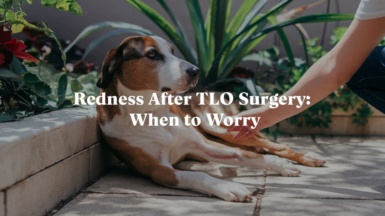
Some redness around a TPLO incision is a completely normal part of healing.
The skin and underlying tissues respond to any surgical trauma with an inflammatory response redness is one of the visible signs of that response.
The question is always: is this normal healing redness, or is this infection?
Quick answer: Mild redness at the TPLO incision is normal for 2 to 3 days, fading over 1 to 2 weeks. Call your vet if redness spreads, is warm, produces discharge, worsens after day 3, or your dog develops fever.
Key takeaways
- Mild redness at the incision is normal for 2 to 3 days post-surgery: it should be fading by day 3 to 5
- Normal redness is localized to the incision line: redness spreading outward from the wound edge is not normal
- Infection redness is spreading, warm, and accompanied by discharge: these features together require veterinary evaluation
- Suture reaction redness is localized and itchy: the pattern is different from infection and typically resolves after suture removal
- Licking is the most preventable wound complication: saliva introduces bacteria; the E-collar must stay on until suture removal
- Redness after week 2 warrants veterinary evaluation: new redness after day 14 suggests a complication
Normal redness after TPLO surgery
The inflammatory phase of wound healing is the body's first response to surgical trauma. Blood flow increases to the wound, bringing immune cells and clotting factors.
This increased blood flow makes the skin appear pink or faintly red. This is expected and necessary for healing.
SustainableVet.org confirms: normal redness after TPLO surgery is usually mild and limited to the incision area. It should improve daily as the wound heals.
Characteristics of normal healing redness:
- Confined to the immediate edges of the incision not spreading outward
- Mild pale pink to light red, not deep red or purple
- Decreasing over time improving from day 3 onward
- Not warm to the touch (normal healing generates mild local warmth, but not the significant heat of infection)
- No discharge from the wound other than possibly a small amount of serum in the first 24 to 48 hours
- The dog is not bothered by gentle examination of the area
The redness typically peaks at 48 to 72 hours post-surgery and should be noticeably fading by the end of the first week.
Abnormal redness: signs of infection
Infection redness is different from normal healing redness in several characteristic ways:
Spreading: the redness extends outward from the incision margin into the surrounding skin. This cellulitis pattern spreading red discoloration in surrounding tissue is a sign that bacteria are spreading through the tissue planes.
Warmth: the infected area feels significantly warmer than surrounding skin or the opposite leg. Infection drives significant local heat through the inflammatory cytokine response.
Discharge: infected wounds produce discharge that may be clear-to-yellow early, progressing to yellow-green, cloudy, or frankly purulent. Foul odor is a hallmark of anaerobic bacterial involvement.
Worsening, not improving: normal redness fades. Infected redness intensifies. If redness is worse at day 5 than at day 2, infection is strongly suspected.
Systemic signs: fever (rectal temperature above 102.5 F), lethargy, loss of appetite, and reluctance to bear weight alongside redness suggest the infection is causing systemic effects.
SustainableVet.org confirms: infection risk includes bacteria entering the incision, causing redness that worsens along with discharge or odor.
The TPLO incision infection guide confirms: infection risk occurs in approximately 3 to 10% of TPLO cases. Signs of infection include redness, swelling, warmth, discharge, and delayed healing.
Other causes of post-TPLO redness
Suture or staple reaction
Some dogs develop a local inflammatory reaction to the suture material more commonly with older suture types or in dogs with sensitive skin.
This produces redness specifically along the suture line, often with mild swelling and sometimes itching (prompting the dog to lick or chew at the site).
It is localized, does not produce the spreading pattern of infection, and typically resolves after suture removal.
Contact dermatitis
Redness from a reaction to bandage material, antiseptic solution applied to the wound, or topical medications can mimic early infection. The pattern follows the contact area rather than the wound edge.
Seroma formation
A seroma that presses against the overlying skin may cause mild skin discoloration and the appearance of redness over the fluid-filled swelling. The area is soft, fluctuant, and typically not warm.
For the full seroma guide, see seroma in dogs after TPLO surgery.
What to do if you notice redness
First 3 days: normal range
Monitor daily. Take a photograph to track progression. Ensure the E-collar is on at all times. Check that no discharge or spreading is occurring.
Days 3 to 10: should be improving
If redness is not clearly fading by day 5 to 7, contact your veterinarian.
Photograph the wound to share with the vet by phone or telehealth before deciding whether an in-person visit is needed.
Do not apply any topical products to the wound without veterinary guidance antiseptics can delay healing and worsen skin reaction.
After day 10: any new or persistent redness needs evaluation
By day 10 to 14, most surgical incisions are well into the proliferative healing phase.
Persistent redness at this stage, or new-onset redness that was not present before, warrants a veterinary appointment rather than home monitoring.
Incision care during recovery
- Keep the incision dry: no bathing, no swimming, no wet grass
- E-collar on at all times until sutures are removed (typically day 10 to 14)
- Check the wound twice daily: look at it, do not touch it unless instructed
- No ointments or antiseptics unless specifically prescribed
- Contact your vet before applying anything to the wound
For the incision infection guide with full symptom detail, see TPLO incision infection symptoms and prevention. For the full complications reference, see 15 common complications after TPLO surgery in dogs.
For the seroma guide (fluid that can cause discoloration), see seroma in dogs after TPLO surgery. For the full TPLO recovery guide, see what to expect after TPLO surgery in dogs.
Frequently asked questions
How do I know if my dog's incision redness is normal?
Normal redness is mild, pink, confined directly to the wound edges, and fading by day 3 to 5.
Abnormal redness is spreading outward from the incision, deepening in color, warm to the touch, or accompanied by discharge or odor. If you are unsure, photograph the wound and contact your veterinarian.
My dog's incision looks red and my dog keeps licking it. What should I do?
Replace or refit the E-collar immediately if the dog has been reaching the wound. Even a few minutes of licking introduces bacteria.
Examine the wound for any discharge, spreading redness, or wound breakdown. If you see any of these, contact your vet the same day.
If the wound looks intact, continue monitoring closely with the E-collar secured.
Can redness appear weeks after surgery?
Yes. Late-onset redness appearing 2 to 6 weeks post-surgery often signals a developing infection rather than normal healing.
Common causes include low-grade suture infection, developing seroma, or a bacterial infection that has been slowly progressing. Any new redness appearing after the initial 2-week healing phase warrants veterinary evaluation.
Is red skin around the TPLO staples normal?
A small amount of mild pinkness immediately at the staple insertion points is normal.
Spreading redness between staple sites, significant swelling along the staple line, or discharge from staple holes is not normal and requires veterinary evaluation before the scheduled staple removal visit.
What happens if redness from infection is left untreated?
Superficial infections can progress to deeper tissue infections, then to implant-associated infections and osteomyelitis.
Laguna Hills Animal Hospital confirms that even superficial soft tissue infections have been shown to progress to implant infections and bone infections when not treated promptly and appropriately.
Resources
- SustainableVet. TPLO Incision Infection Symptoms and Prevention. sustainablevet.org
- Dial A Vet. Redness and Swelling Around TPLO Incision. dialavet.com
- Laguna Hills Animal Hospital. TPLO Surgery Complications. lhah.com
- VCA Animal Hospitals. Tibial Plateau Leveling Osteotomy (TPLO) in Dogs. vcahospitals.com

Get a Free Poster
Enhance your workspace with a high-quality radiographs reference poster, designed for veterinary professionals. This free physical poster will be shipped directly to you—just fill out the form to request your copy.






Taking Great TPLO Radiographs
Click Below to Watch Live Video Demos


We'll send you a Free Wall Poster with all the steps
Step #1
Getting Ready
Ensuring a clean surgical field starts with proper skin preparation. This video demonstrates the best practices for:
- Shaving the patient – Achieving a close, even shave while minimizing skin irritation
- The Dirty Scrub – The initial skin prep step to remove surface debris and reduce bacterial load before the sterile scrub.
Following these techniques helps reduce infection risk and improve surgical outcomes. Watch the video to see how it’s done effectively!


Step #2
Reduce Your Risks
Many surgeons are shocked to find out that their patients are not protected from biofilms and resistant bacteria when they use saline and post-op antibiotics.
That’s Where Simini Comes In.
Why leave these risks and unmanaged? Just apply Simini Protect Lavage for one minute. Biofilms and resistant bacteria can be removed, and you can reduce two significant sources of infection.
Step #3
Take the Course
Preventing surgical infections is critical for patient safety and successful outcomes. This course covers:
- Aseptic techniques – Best practices to maintain a sterile field.
- Skin prep & draping – Proper methods to minimize contamination.
- Antibiotic stewardship – When and how to use perioperative antibiotics effectively.
Stay up to date with the latest evidence-based protocols. Click the link to start learning and earn CE credits!

Things to know
Seroma in Dogs After TPLO Surgery: Causes & Care
Learn about seroma in dogs after TPLO surgery, including causes, symptoms, treatment, and prevention tips for pet owners.
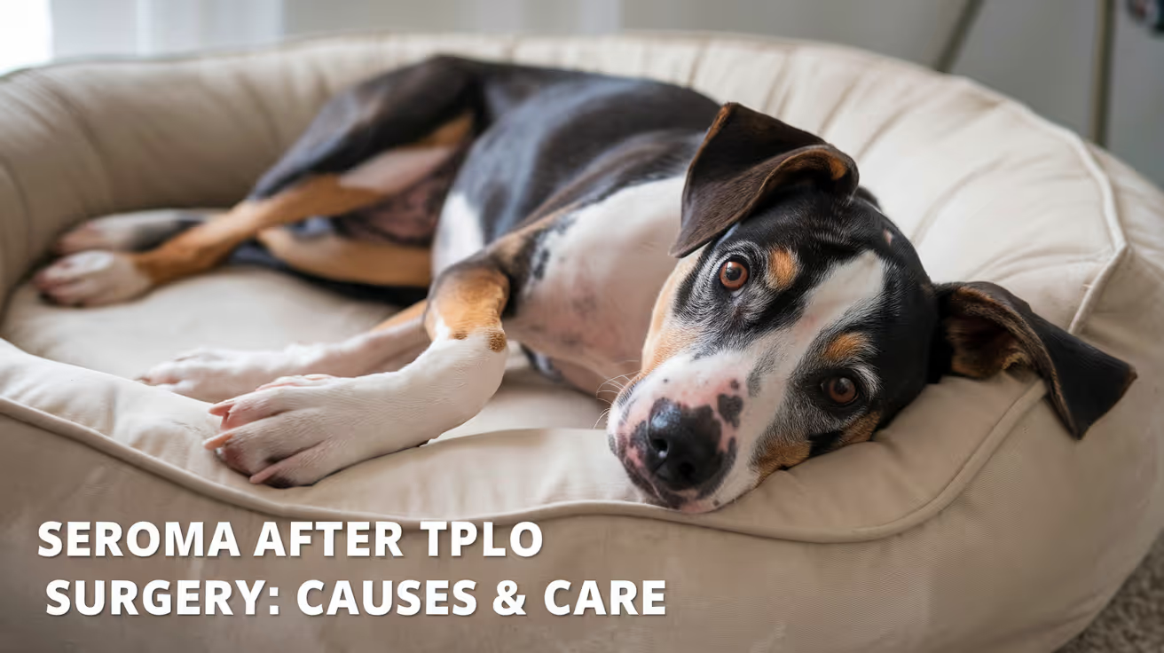
A seroma is one of the most common soft tissue complications after TPLO surgery in dogs.
Finding a soft lump under the skin near the surgical site is alarming, but in most cases it is a benign finding that resolves with strict rest and time.
Understanding the difference between a seroma and a more serious complication helps you respond appropriately.
Quick answer: A seroma is clear serum fluid under the TPLO incision, not an infection. Small seromas resolve in 1 to 3 weeks with rest; larger ones need sterile aspiration. Call your vet if the swelling is hot, fast-growing, or producing discharge.
Key takeaways
- A seroma is clear serum fluid under the skin — not an infection; a benign accumulation from disrupted blood and lymphatic vessels
- Excessive activity is the leading preventable cause: unnecessary movement produces more fluid and enlarges the seroma
- Small seromas resolve with strict rest in 1 to 3 weeks: the lymphatic system reabsorbs the fluid without intervention
- Large seromas may need needle aspiration: a sterile outpatient procedure by your vet; never drain at home
- Warm packs twice daily help reabsorption: 10 minutes of warmth twice daily is recommended for established seromas
- A seroma that becomes warm, red, painful, or produces discharge needs immediate vet attention it may have become infected
What is a seroma?
Vetinfo explains that a seroma is a swelling resulting from the accumulation of fluid under the skin.
It is made of serous fluid (blood plasma without most of the cells), so it is usually watery with a slight pink tinge rather than dark blood or cloudy infected fluid.
After TPLO surgery, the osteotomy site and surrounding soft tissues leave behind a space between tissue layers called dead space where fluid from disrupted capillaries and lymphatics can collect.
The fluid pools there before the body's healing processes close the space.
Seromas feel soft, fluctuant (squishy), and move under finger pressure. They are usually not painful when small and are not warm to the touch.
These features distinguish them from infections, which are typically warm, firm, painful, and may produce purulent discharge.
What causes seromas after TPLO?
Excessive activity
This is the most important and most preventable cause. Vetinfo confirms that seromas occur when a dog is active immediately after surgery instead of remaining inactive during recovery.
Every unnecessary step disrupts the tissue planes that need to approximate and seal, producing more fluid.
The surgeon Dr.
Matt confirms on JustAnswer that the best way to reduce seromas is to restrict activity as much as possible at least 8 weeks of restricted activity after TPLO is recommended.
Dead space at the surgical site
TPLO surgery involves significant soft tissue dissection around the proximal tibia. Closing this space with sutures reduces but does not eliminate dead space.
Any gap between tissue layers fills with fluid before normal healing closes it.
Surgical drainage
In some cases, surgical drains are placed during the procedure to allow fluid to escape rather than collect.
Not all surgeons routinely place drains some reserve them for dogs considered at higher risk of seroma formation.
Patient factors
Larger dogs produce more dead space and more fluid. Dogs with more subcutaneous fat are at higher risk.
Dogs that are particularly active during recovery despite attempts at confinement are most likely to develop significant seromas.
Recognizing a seroma
Classic signs:
- Soft, squishy swelling directly under or near the incision site
- The swelling moves under pressure it feels like a water balloon
- Normal skin color over the swelling (not red or purple)
- Not painful when gentle pressure is applied
- Not warm to the touch
- No discharge from the skin over the swelling
When to suspect infection instead:
- The swelling is warm, red, or firm rather than soft
- A draining tract or discharge appears from the skin
- The dog shows increased pain, fever, lethargy, or loss of appetite
- The swelling is growing rapidly
Treatment
Small seromas: observation and rest
Cuteness notes that most often the body absorbs the fluid and the swelling diminishes. For small seromas (typically under 3 cm), the approach is:
- Enforce strict activity restriction no running, jumping, or free roaming
- Cold compresses in the first few days (reduces new fluid production)
- Warm compresses from days 5 onward 10 minutes, twice daily to promote fluid reabsorption
- Monitor size and character daily
Large or persistent seromas: needle aspiration
Large seromas that are uncomfortable or not responding to rest may be drained by your veterinarian using a sterile needle and syringe. This is a simple outpatient procedure.
Cuteness confirms that veterinarians may drain or aspirate the seroma to make the dog more comfortable.
Aspiration provides relief but does not address the underlying dead space. The seroma may refill sometimes multiple aspirations are required. After aspiration, continued strict rest is essential.
Compression bandaging
A gentle compression bandage applied by your vet reduces dead space and discourages fluid refilling after aspiration.
Improper bandaging can cause pressure sores or circulatory compromise, so bandages must be applied and monitored by a veterinarian.
Surgical drain placement
If a seroma repeatedly refills after aspiration, a closed suction drain may be placed to allow ongoing drainage until the dead space closes.
This is uncommon but is an option for persistent or large seromas.
For the full fluid buildup guide covering all types of post-TPLO fluid accumulation, see fluid buildup after TPLO surgery in dogs.
For incision complications including infection, see TPLO incision infection symptoms and prevention. For the full complications reference, see 15 common complications after TPLO surgery in dogs.
For the incision redness guide to distinguish seroma from infection, see redness after TPLO surgery in dogs: causes and care.
Frequently asked questions
Can a seroma go away on its own?
Yes in the majority of cases, small seromas resolve without intervention as the body reabsorbs the fluid over 1 to 3 weeks.
Strict activity restriction is essential; without it, the seroma will continue to refill as fast as it drains.
Do not assume a seroma will resolve if the dog is not being properly confined.
Should I drain the seroma myself at home?
Never. Home drainage risks introducing bacteria into a sterile fluid collection, converting a benign seroma into an infected abscess. Only a veterinarian should aspirate a seroma under sterile conditions.
If you believe the seroma needs draining, contact your vet.
My dog's seroma looks like it ruptured through the skin. What do I do?
Spontaneous seroma rupture does occasionally occur. The fluid drains through the skin, leaving a small wound.
Contact your vet immediately for guidance the wound will need to be kept clean and monitored for infection. A veterinarian Dr.
Matt reports this occurrence on JustAnswer, confirming prompt vet contact is appropriate.
How long does a seroma last after TPLO?
Small seromas typically resolve in 1 to 3 weeks with strict activity restriction. Larger ones may take 4 to 6 weeks and may require aspiration.
If a seroma persists beyond 4 weeks without improvement, a veterinary recheck is warranted.
Is a seroma the same as an infection?
No. A seroma is sterile fluid no bacteria. An infection involves bacterial colonization and produces different characteristics: warmth, redness, purulent discharge, systemic illness signs.
Bacterial culture distinguishes them definitively if clinical assessment is uncertain.
Resources
- Vetinfo. Seroma After-Surgery Care for Dogs. vetinfo.com
- Cuteness. What Are the Treatments for Seroma Formation in Dogs? cuteness.com
- VCA Animal Hospitals. Seroma in Dogs. vcahospitals.com
- TPLO Info. Recovery FAQs. tploinfo.com
TPLO Failure Rate in Dogs Explained
Learn about TPLO failure rates in dogs, causes, prevention, and recovery tips for better surgical outcomes.
Tibial Plateau Leveling Osteotomy (TPLO) is a common surgery to fix cranial cruciate ligament injuries in dogs. Many pet owners worry about the TPLO failure rate in dogs and what it means for their pet’s recovery. Understanding the risks and outcomes can help you make informed decisions for your dog’s health.
This article explains what TPLO failure means, how often it happens, and what factors affect it. You will learn how to recognize complications, prevent failure, and support your dog after surgery for the best results.
What is the TPLO failure rate in dogs?
The TPLO failure rate in dogs varies but is generally low when performed by experienced surgeons. Failure means the surgery did not fully restore knee stability or complications occurred that affect recovery.
Studies show failure rates range from 2% to 10%, depending on factors like surgical technique and dog size. Most dogs recover well and regain normal function after TPLO.
- Low overall failure: Most dogs have successful outcomes with TPLO, with failure rates usually under 10% in clinical studies.
- Variation by surgeon: Surgeons with more experience tend to have lower failure rates due to better technique and planning.
- Dog factors matter: Larger dogs or those with severe ligament damage may have a higher risk of failure after TPLO.
- Definition of failure: Failure includes persistent lameness, implant problems, or the need for revision surgery after TPLO.
Understanding these rates helps you set realistic expectations and discuss risks with your vet before surgery.
What causes TPLO failure in dogs?
Several factors can lead to TPLO failure. Knowing these causes helps prevent problems and improve recovery chances.
Failures often result from surgical errors, infection, or poor healing. Other causes include implant issues and the dog’s activity level after surgery.
- Surgical technique errors: Incorrect bone cuts or implant placement can cause instability or implant failure after TPLO.
- Infection risk: Postoperative infections can delay healing and lead to surgery failure if not treated promptly.
- Poor bone healing: Factors like age, nutrition, or underlying disease can slow bone healing and cause failure.
- Excessive activity: Dogs that are too active too soon may damage the surgical site and cause failure.
Preventing these causes requires careful surgical planning, good postoperative care, and close monitoring.
How can TPLO failure be prevented in dogs?
Prevention of TPLO failure starts with choosing a skilled surgeon and following postoperative instructions closely. Proper care reduces complications and supports healing.
Owners play a key role in managing their dog’s activity and health during recovery to avoid failure.
- Experienced surgeon choice: Select a board-certified surgeon with extensive TPLO experience to reduce technical errors.
- Strict activity control: Limit your dog’s movement and exercise as advised to protect the surgical site during healing.
- Infection prevention: Keep the incision clean and watch for signs of infection to catch problems early.
- Follow-up visits: Regular vet check-ups help monitor healing and detect complications before failure occurs.
Following these steps improves your dog’s chance of a successful TPLO surgery and recovery.
What are the signs of TPLO failure in dogs?
Recognizing failure signs early lets you seek veterinary care promptly. Signs often involve ongoing lameness or swelling around the knee.
Not all postoperative discomfort means failure, but persistent or worsening symptoms should be evaluated by a vet.
- Persistent lameness: Continued limping or inability to bear weight on the leg weeks after surgery may indicate failure.
- Swelling or pain: Increased swelling, heat, or pain around the knee can signal infection or implant problems.
- Unusual noises: Clicking or popping sounds from the knee might mean implant loosening or joint instability.
- Reduced range of motion: Difficulty bending or extending the knee joint may suggest complications affecting recovery.
If you notice any of these signs, contact your veterinarian immediately for assessment and treatment.
How is TPLO failure treated in dogs?
Treatment depends on the cause of failure. Some cases require revision surgery, while others may respond to medical management.
Your vet will perform exams and imaging to determine the best approach to restore function and relieve pain.
- Revision surgery: Some dogs need a second surgery to fix implant issues or correct bone alignment after failure.
- Antibiotic therapy: Infections causing failure require antibiotics and sometimes implant removal to heal properly.
- Pain management: Medications and physical therapy help control pain and improve mobility during recovery.
- Supportive care: Weight management and controlled exercise support healing and reduce stress on the knee joint.
Early treatment improves outcomes and helps your dog regain normal activity levels.
What is the recovery outlook after TPLO failure in dogs?
Recovery after TPLO failure can be more challenging but is often successful with proper care. Many dogs regain good function after treatment.
Recovery time may be longer, and some dogs need ongoing management to maintain comfort and mobility.
- Longer healing time: Dogs with failure often require extended recovery periods compared to uncomplicated TPLO cases.
- Physical therapy benefits: Rehabilitation exercises improve strength and joint function after failure treatment.
- Possible chronic issues: Some dogs may develop arthritis or mild lameness despite treatment.
- Owner commitment: Consistent care and monitoring are essential for a positive recovery after failure.
With patience and veterinary support, many dogs live active, happy lives following TPLO failure treatment.
What factors affect TPLO failure rates in different dog breeds?
Breed and size influence TPLO outcomes. Larger and more active breeds may face higher failure risks due to greater joint stress.
Understanding breed-specific risks helps tailor surgical and postoperative care for better success.
- Large breed challenges: Heavy dogs put more pressure on the knee, increasing the chance of implant failure or delayed healing.
- Active breed risks: Energetic dogs may be harder to restrict post-surgery, raising failure risk from premature activity.
- Bone quality differences: Some breeds have denser or more fragile bones affecting healing after TPLO.
- Genetic predispositions: Certain breeds may be more prone to ligament injuries and complications after surgery.
Discuss your dog’s breed and lifestyle with your vet to plan the best approach for TPLO surgery and recovery.
Conclusion
The TPLO failure rate in dogs is generally low but depends on many factors like surgeon skill, dog size, and postoperative care. Understanding these helps you prepare for surgery and recovery.
By choosing an experienced surgeon, following care instructions, and watching for signs of failure, you can help your dog heal well and return to a happy, active life after TPLO surgery.
What is the typical TPLO failure rate in dogs?
TPLO failure rates usually range between 2% and 10%, with most dogs recovering successfully after surgery.
Can infection cause TPLO failure in dogs?
Yes, infections at the surgical site can lead to delayed healing and failure if not treated quickly with antibiotics and care.
How soon can dogs return to activity after TPLO?
Dogs typically need 8 to 12 weeks of restricted activity to allow proper healing and reduce the risk of failure.
Is revision surgery common after TPLO failure?
Revision surgery is sometimes necessary to correct implant or alignment issues when TPLO fails to restore knee stability.
Do larger dogs have higher TPLO failure rates?
Larger dogs often have increased failure risk due to greater joint stress and challenges in controlling postoperative activity.

Common Myths About TPLO Surgery Explained
Discover the truth about TPLO surgery for dogs as we debunk common myths and provide evidence-based insights for informed pet care decisions
TPLO is the most commonly performed surgery for CCL rupture in dogs and the most commonly misunderstood.
Misconceptions about TPLO cause unnecessary hesitation, lead some owners to choose inferior alternatives, and create unrealistic expectations about recovery.
This guide addresses the 10 most common myths directly -- with evidence-based clarification for each.
Quick answer: The most persistent TPLO myths: that dogs can manage without surgery, that TPLO is only for large breeds, that recovery takes a few weeks, that the plate must be removed after healing, and that TPLO prevents arthritis. All are false. TPLO has a 90 to 95% success rate.
Key takeaways
- Myth: "My dog can heal without surgery": dog knee anatomy differs fundamentally from human; without surgery, severe OA, meniscal tearing, and chronic lameness are expected in most dogs over 15 kg
- Myth: "TPLO is only for large dogs": SustainableVet.org confirms TPLO is just as effective for small and toy breeds; the biomechanical correction is appropriate regardless of size
- Myth: "Recovery only takes a few weeks": bone healing takes 8 to 12 weeks; full muscle recovery takes 4 to 6 months; 8 to 12 weeks is the minimum restriction period
- Myth: "The plate needs to be removed after healing": the TPLO plate is designed to be permanent; removal is only performed when complications arise
- Myth: "TPLO prevents arthritis": all CCL-affected stifles develop progressive OA regardless of surgical technique; TPLO reduces OA progression but does not prevent it
- Myth: "Dogs are in severe pain after TPLO": SustainableVet.org confirms this is a misconception; modern multi-modal pain management controls post-surgical pain effectively
Myth 1: "My dog can heal without surgery"
The truth: dogs and humans have fundamentally different knee anatomy. In humans, the flat tibial plateau and strong surrounding musculature allow some CCL tears to be managed conservatively with physical therapy and bracing. In dogs, the tibial plateau slopes steeply downward, generating a cranial shear force that the absent CCL cannot resist. Without surgical correction of this force, the joint remains mechanically unstable with every weight-bearing step.
TPLO Info confirms: dogs and humans respond very differently to CCL tears. Without surgery, severe OA can develop, meniscal tearing can occur, and other side effects may be present.
Conservative management (strict rest, weight management, joint supplements, physical therapy) can provide temporary improvement, particularly in small dogs and partial tears.
For most dogs over 15 kg with complete CCL rupture, conservative management does not restore normal joint stability, and progressive lameness, OA, and joint deterioration are the expected outcome.
Who can be managed conservatively?
- Very small dogs (under 10 to 15 kg) with partial tears and low activity levels
- Dogs with significant comorbidities where anesthetic risk outweighs surgical benefit
- Owners who decline surgery after being fully informed of expected outcomes
For all other dogs -- medium and large breeds, young dogs, active dogs, complete ruptures -- surgery produces substantially better outcomes than conservative management.
Myth 2: "TPLO is only for large dogs"
The truth: TPLO was originally developed for large and giant breeds because they were the most severely affected by CCL disease and the most poorly served by lateral suture stabilization. But TPLO corrects the underlying biomechanical problem (excessive tibial plateau angle) in any dog, regardless of size.
SustainableVet.org confirms: TPLO was initially developed for large dogs due to biomechanical challenges, but has been found just as effective for small and toy breeds.
For small dogs, both TPLO and lateral suture stabilization can achieve good outcomes.
The decision for a small dog should be based on the dog's TPA, activity level, and individual assessment -- not a blanket assumption that TPLO is unnecessary.
Myth 3: "TPLO recovery only takes a few weeks"
The truth: bone healing takes 8 to 12 weeks. This is the period of activity restriction. But bone healing and functional recovery are not the same thing. Full muscle rebuilding and joint function restoration takes 4 to 6 months from surgery.
Canada Vet Express confirms: recovery is gradual; most dogs begin walking within a few days and return to normal activity within 3 to 4 months with proper care.
The 8 to 12 week bone healing timeline means:
- Activity restrictions are in place for 8 to 12 weeks minimum
- Radiographic confirmation of healing is required before restrictions are lifted
- Progressive return to full activity follows the radiographic clearance
After restrictions are lifted, the operated leg continues to rebuild strength and coordination for months. For sport dogs and working dogs, the return-to-sport protocol extends to 7 to 9 months from surgery.
Myth 4: "The TPLO plate needs to be removed after the bone heals"
The truth: the plate is designed to remain in place permanently. It does not need to be removed when the osteotomy heals. Routine plate removal is not standard practice in veterinary orthopedics.
SustainableVet.org confirms: these implants are meant to stay in place permanently, but in rare cases they can cause problems; removal is only performed when complications arise.
When plate removal is performed:
- Deep implant-associated infection requiring source removal
- Implant failure (breakage, loosening causing instability)
- Persistent implant irritation producing clinical lameness specifically attributable to the plate
In the vast majority of dogs, the plate is never removed. It remains in place for life with no adverse effects.
Myth 5: "TPLO prevents arthritis"
The truth: TPLO reduces and slows arthritis progression. It does not prevent it. All CCL-affected stifles develop progressive OA regardless of surgical technique or outcome.
SustainableVet.org confirms: mild arthritis is expected even in successful cases.
Published long-term data (TPLO Info, citing long-term retrospective studies) confirms moderate but significant OA progression in all TPLO cohorts with follow-up extending to 6.8 years.
TPLO produces substantially less OA progression than TTA or lateral suture stabilization, but OA progression is universal.
The appropriate expectation: TPLO minimizes OA development and clinical impact. Long-term management (weight management, exercise modification, joint supplements, and periodic NSAID use for flares) is required in all TPLO dogs.
Myth 6: "Dogs are in severe pain after TPLO"
The truth: modern veterinary anesthesia and pain management protocols produce well-controlled post-operative pain in TPLO dogs. Dogs typically begin bearing some weight within 24 to 48 hours. Pain that is not controlled is a clinical problem requiring medication adjustment -- not an expected feature of TPLO recovery.
SustainableVet.org confirms: it is a misconception that dogs experience severe pain after TPLO surgery; modern multi-modal pain management controls post-surgical pain effectively.
Post-operative pain management typically includes: NSAIDs (carprofen, meloxicam), gabapentin, opioids during hospitalization, local anesthetic nerve blocks at surgery.
Dogs in adequate pain control rest comfortably, bear some weight within days, and show appetite by day 3.
Uncontrolled post-operative pain (dog cannot settle, crying, refuses all weight bearing beyond day 3) indicates a need for pain medication adjustment -- contact your vet rather than accepting it as inevitable.
Myth 7: "TPLO is too risky for most dogs"
The truth: TPLO has a 90 to 95% success rate. The complication rate across all studies is 10 to 34%, but the vast majority of complications are minor (superficial infection, seroma, mild swelling). Serious complications requiring revision surgery occur in fewer than 10% of cases.
Dispomed confirms: overall complication rates are 14 to 34%, but a second surgery is required in less than 10% of cases.
No surgery is without risk.
But for a dog with a torn CCL, the risk of TPLO is substantially lower than the certainty of progressive joint deterioration, pain, and reduced quality of life without surgical treatment.
Myth 8: "TPLO is only for show dogs or working dogs"
The truth: TPLO is appropriate for any dog with CCL rupture that will benefit from restored joint stability -- including middle-aged household pets with no competitive goals.
SustainableVet.org confirms: some pet owners mistakenly believe that TPLO surgery is only for show dogs or high-performance athletes; this is not true; TPLO benefits any dog with CCL disease.
The goal of TPLO is freedom from joint pain and restored ability to perform normal daily activities -- walking, playing, using stairs, sitting and rising comfortably.
These are benefits for every dog, not just sport dogs.
Myth 9: "All dogs with CCL tears need TPLO immediately"
The truth: TPLO is not always an emergency. The timing of surgery should be guided by the veterinary assessment. Partial tears in small dogs may be monitored with conservative management. Dogs with significant concurrent illness may benefit from medical optimization before surgery. The appropriate goal is surgical timing that optimizes anesthetic risk and surgical conditions, not surgical urgency at any cost.
SustainableVet.org confirms: while TPLO surgery is the best treatment for CCL tears, it isn't necessary for every case; for mild or partial tears in less active dogs, alternatives can be effective.
However, in dogs with complete rupture, particularly larger breeds, delay generally produces worse outcomes as OA progresses and the meniscus is exposed to ongoing mechanical insult from the unstable joint.
Myth 10: "If TPLO fails, the dog will never recover"
The truth: most TPLO complications are treatable. Superficial infections resolve with antibiotics. Late meniscal tears are treated with partial meniscectomy. Implant failures can be revised. Osteomyelitis, the most challenging complication, requires prolonged treatment but is often successfully managed.
SustainableVet.org confirms: revision TPLO surgery can be performed if the initial procedure fails; this may involve removing or replacing implants, repositioning the tibial cut, or addressing infections.
The critical factor in outcome from a complication is early detection and prompt treatment. Most complications that are identified and treated early resolve without long-term sequelae.
For the full TPLO overview, see what is TPLO surgery in dogs?. For the success rate data, see TPLO failure rate in dogs. For recovery, see what to expect after TPLO surgery.
For alternatives, see alternatives to TPLO surgery for dogs.
Frequently asked questions
Is TPLO actually the best treatment for CCL rupture?
For most dogs over 15 to 20 kg, TPLO is the most evidence-supported surgical option, producing the highest rates of long-term function and owner satisfaction in the published literature.
For small dogs, lateral suture stabilization achieves comparable results at lower cost. The choice depends on the individual dog's size, activity level, TPA, and health status.
Can I wait and see how my dog does without surgery?
You can choose conservative management.
The expected outcome in most dogs over 15 kg with complete CCL rupture who do not have surgery is progressive lameness, rapidly advancing OA, ongoing meniscal damage, and reduced quality of life.
Early surgical intervention consistently produces better long-term outcomes than delayed or non-surgical management.
Is the recovery really as strict as described?
Yes. The restriction protocol exists because the plate holds the osteotomy until new bone consolidates -- a process that takes 8 to 12 weeks regardless of how well the dog appears.
A single running or jumping episode before healing is confirmed can displace the plate and require revision surgery.
Will my dog be the same dog after TPLO?
Most owners report their dog is effectively the same dog -- or better than before the CCL rupture -- within 6 to 12 months of surgery.
The 90 to 95% success rate reflects a high proportion of dogs returning to their previous activity level.
Some dogs develop mild age-related stiffness years later as OA progresses, but this is manageable with appropriate care.
Does TPLO cure the CCL problem permanently?
TPLO permanently corrects the biomechanical cause of CCL-related instability in the operated stifle. It does not repair or replace the CCL -- it makes the CCL unnecessary.
It does not prevent CCL disease in the contralateral stifle.
Resources
- SustainableVet. Common Myths About TPLO Surgery Explained. sustainablevet.org
- TPLO Info. 5 Misconceptions About TPLO Surgery. tploinfo.com
- Dispomed. TPLO Surgery in Dogs: Success Rates and Alternatives. dispomed.com
- ACVS. Cranial Cruciate Ligament Disease. acvs.org
How Long Does TPLO Surgery Take?
Learn how long TPLO surgery takes, what to expect during the procedure, and recovery timelines for your dog's knee surgery.
"Failure rate" is used loosely when owners discuss TPLO, which can cause significant confusion. A complication is not the same as a failure.
Understanding what the published data actually shows and distinguishing minor complications from true surgical failures gives owners a much clearer picture of what to expect.
Quick answer: The overall TPLO complication rate is 10 to 34% across studies; most are minor. Major complications requiring reoperation occur in 2 to 7%. Infection is the most common major complication. Long-term good outcomes are reported in 90 to 93% of dogs.
Key takeaways
- Overall complication rate is 10 to 34% across published studies; most complications are minor and resolve without additional surgery
- Major complication rate requiring reoperation is 2 to 7%: a 1,000-dog study found 14.8% overall with 6.6% major complications
- SSI is the most common major complication: 2.9% to 25.9% across studies; giant breeds over 50 kg are at highest risk
- Small dogs under 15 kg have significantly lower complication rates: 4.44% in one 90-dog study, with no revisions
- Good to excellent long-term outcomes in 90 to 93% of dogs: the complication rate does not equal the failure rate
- Surgeon experience is the most modifiable risk factor: complication rates are consistently lower at high-volume specialty centers with board-certified surgeons
Defining "failure" vs. "complication"
This distinction is critical for interpreting the literature and for setting owner expectations accurately.
Complication: any deviation from the expected post-operative course. This includes minor findings like superficial wound dehiscence, mild swelling, or radiographic evidence of delayed osteotomy healing with no clinical signs. Many publications include these minor events in their complication rates.
Major complication: an event requiring additional surgical intervention or causing extended lameness. Major complications represent the subset of complications that meaningfully affect recovery and outcome.
Failure: the most restrictive definition surgery that did not achieve its goal of stifle stabilization, resulting in persistent instability and requiring revision with a different technique. True failure in this sense is uncommon after TPLO.
What published complication data shows
Overall complication rates
Vetamac confirms: complication rates following TPLO surgery range from 14% to 34%, with less than 10% of cases requiring a second surgery.
PMC (intraoperative bacterial culture study) confirms: the complication rate of TPLO varies between 10% and 34%, with 2 to 4% requiring surgical revision.
Today's Veterinary Practice reports a 14.8% complication rate in 1,000 TPLO patients (6.6% major — requiring repeat surgery or extended lameness), including 2.8% late meniscal injury and 6.6% infections.
Infection rates
Infection is the most common major complication and the one with the greatest variation by dog size and surgical setting.
PMC confirms: SSI rate after TPLO is reported to be 2.9% to 25.9% across studies, which is higher than other clean orthopedic surgeries (2.0 to 6.7%).
Surgical site infection rates of 21.3% to 25.9% have been reported specifically for giant-breed dogs weighing over 50 kg.
The implant removal rate due to infection is 3 to 7.4% per published literature (PMC PRP study; ResearchGate implant removal study).
Complication rates by dog size
Dog size is a consistent predictor of complication risk.
PLOS One retrospective (90 dogs under 15 kg): overall complication rate 4.44%, with no complications requiring surgical revision.
The study confirms: the complication rate in dogs under 15 kg is less than that historically reported in heavier dogs.
PubMed retrospective (small and medium dogs, 2.0 and 2.7 mm plates): overall complication rate 36%.
The most common complications were radiographic evidence of delayed healing (9%), patellar tendon thickening (8%), and tibial tuberosity fracture (6%). Only 7% of complications were major.
PLOS One confirms: the TPLO complication rates range from 10% to 34% and vary in severity from swelling and bruising to fractures and osteomyelitis.
Implant-related complications reportedly occur in under 10% of all TPLO procedures.
Long-term success rates
Complication rates must be interpreted alongside long-term success rates.
The retrospective cohort study comparing TPLO, Tightrope, and TTA found: function greater than 1 year after surgery was 93.1% for TPLO, 92.7% for Tightrope, and 89.2% for TTA.
SustainableVet.org confirms: good to excellent long-term outcomes are achieved in 90.4% of patients up to 6.8 years after TPLO surgery; 80 to 90% of owners are satisfied with their dog's long-term recovery.
A dog can have a complication even a significant one like an infection and still achieve a good long-term outcome after the complication is resolved.
The complication rate and the long-term success rate address different aspects of the outcome.
Factors that affect complication and failure rates
Surgeon experience and volume
This is the most consistently cited modifiable risk factor. High-volume specialty centers and board-certified surgeons consistently report lower complication rates than general practitioners performing occasional TPLO procedures.
Surgical efficiency, technique precision, and post-operative protocol standardization all contribute.
Dog size and body weight
Larger dogs generate higher joint forces, have larger dead space at the surgical site, and place more mechanical demand on the implant during healing.
These factors contribute to the higher complication rates seen in large and giant breeds.
Excessive tibial plateau angle (TPA)
Dogs with TPA above 35 degrees (excessive TPA, or eTPA) have higher complication rates.
Today's Veterinary Practice notes: owner-perceived outcome was superior for eTPA group dogs when TPLO resulted in TPA of 14 degrees or less compared to those with TPA greater than 14 degrees.
Postoperative complications were more common in dogs with eTPA.
Post-operative activity restriction compliance
Dogs that are over-active during recovery have higher rates of implant complications, seroma formation, and wound complications.
Strict adherence to the prescribed restriction protocol is the owner's primary contribution to reducing complication risk.
For the complications overview, see 15 common complications after TPLO surgery in dogs. For the infection signs guide, see TPLO plate infection signs and treatment.
For the long-term outcomes guide, see long-term outcomes of TPLO surgery. For the plate removal guide, see TPLO plate removal recovery guide.
Frequently asked questions
What is the TPLO failure rate?
Depends on how "failure" is defined. If failure means any complication: 10 to 34% across studies. If failure means major complications requiring surgery or causing extended lameness: 2 to 7%.
If failure means persistent instability requiring revision with a different technique: uncommon and not consistently reported across large series.
Is TPLO safe for large breed dogs?
Yes, but with a higher complication risk than small dogs. Vetamac confirms: for large breeds, TPLO is still the gold standard, providing better long-term stability than alternatives.
The higher complication rate in large dogs reflects the greater mechanical demands and surgical challenges, not a fundamental unsafety of the procedure.
What percentage of TPLO dogs need a second surgery?
Across published studies, 2 to 10% require a second surgical intervention. Most second surgeries address infection (requiring plate removal), late meniscal tears (requiring partial meniscectomy), or implant complications.
Does my dog's vet experience affect failure rate?
Yes, substantially. Surgeons performing fewer than a set number of TPLO procedures per year have higher complication rates than high-volume specialists.
This is one reason referral to a board-certified orthopedic surgeon (ACVS diplomate) is recommended for TPLO.
If my dog has a complication, does that mean the TPLO failed?
Not necessarily. Many dogs who develop complications including infections requiring plate removal still achieve good to excellent long-term function after the complication is resolved.
The complication rate and the long-term outcome are different measures.
Resources
- Today's Veterinary Practice. Canine Cranial Cruciate Disease: Evidence-Based Look at Current Treatment Modalities. todaysveterinarypractice.com
- PLOS One. Risk Factors for Short-Term Postoperative Complications in Dogs Weighing Less Than 15 kg. journals.plos.org
- PubMed. Short-Term Complications Associated With TPLO in Dogs Using 2.0 and 2.7 mm Plates. pubmed.ncbi.nlm.nih.gov
- PMC. Clinical Relevance of Positive Intraoperative Bacterial Culture in TPLO in Dogs. ncbi.nlm.nih.gov
- Vetamac. TPLO Surgery for Dogs: Success Rates and Alternative Options. vetamac.com
Why Is My Dog's Leg Clicking After TPLO Surgery?
Wondering why your dog’s leg is clicking after TPLO surgery? Learn common causes, when to worry, and what to expect during recovery
A clicking or popping sound from the stifle after TPLO surgery is a common observation that owners report, especially during the first weeks of recovery.
It is not automatically a sign of a problem.
The clinical significance depends on what accompanies the click: is the dog bearing weight and recovering normally, or is clicking associated with lameness, pain, or a trajectory reversal?
Quick answer: Post-TPLO clicking most often reflects normal joint adjustment, soft tissue movement, or cavitation. The most clinically important cause is a meniscal tear, producing a meniscal click with pain and returning lameness. Clicking without lameness or pain in early recovery is usually benign.
Key takeaways
- Most post-TPLO clicking is benign: the click typically comes from joint adjustment to new mechanics; the dog usually feels fine even when a sound is present
- The meniscal click is the clinically significant type: dogs with meniscal tears exhibit a meniscal click accompanied by lameness and pain
- Clicking with lameness or pain warrants prompt veterinary evaluation: clicking with swelling, limping, or avoiding the leg could signal instability, implant issues, or a torn meniscus
- Clicking that is isolated, early in recovery, and not associated with lameness is usually normal and improves as healing progresses
- Late-onset clicking in a previously normal recovery is more concerning than early clicking; it suggests a new event such as meniscal tear or implant loosening
- Implant loosening is a rare but serious cause of clicking: abnormal joint mechanics from loosened screws or plate produce clicking accompanied by pain and radiographic changes
What causes clicking after TPLO surgery
Normal joint adjustment
After TPLO, the tibial plateau is repositioned to a new angle. The soft tissues, tendons, and joint capsule require weeks to adapt to this new geometry.
During this adaptation period, movement can produce audible sounds as structures slide over each other or snap slightly over new contours.
TPLO Info confirms: after CCL repair, the joints adjust to new motion. The click comes from the knees. The dog usually feels fine even when a sound is present.
Soft tissue movement over the implant
The TPLO plate sits against the medial tibial surface. Tendons and fascial layers that previously moved over the normal tibial contour now move over a plate with a different profile.
This can produce audible snapping or clicking sounds during flexion and extension, particularly in the first weeks before soft tissue accommodation occurs.
Gas bubble release (cavitation)
Joint capsule distension from post-surgical effusion can trap gas in the joint space. Movement releases this gas in small pops -- the same mechanism as knuckle cracking in humans.
This is painless and harmless.
Meniscal click (most significant cause)
The medial meniscus is vulnerable to injury in CCL disease. At the time of TPLO, the surgeon assesses the meniscus and removes damaged tissue.
However, meniscal injury can occur after TPLO -- either from previously undetected damage that progresses, or from new injury in a joint that has not yet fully stabilized.
Laguna Woods Vets confirms: dogs with meniscal tears frequently exhibit a meniscal click. If damage is noted during TPLO, the meniscus is removed. If not removed, it may tear later.
SustainableVet.org confirms: symptoms of meniscal damage include clicking when the dog moves, lameness, reluctance to use the affected leg, and discomfort when bending the knee.
The meniscal click is distinguished from benign clicking by:
- Accompanying lameness (often a sudden return of limping after initial improvement)
- Pain when the stifle is manipulated on examination
- Worsening trajectory rather than improvement
Implant-related clicking (uncommon)
Plate or screw loosening generates abnormal tibial motion during weight bearing, which can produce clicking.
This is uncommon in an uncomplicated TPLO but more likely if the dog has been overactive during recovery.
Signs include pain localized to the plate site and radiographic evidence of implant migration or peri-implant bone changes.
SustainableVet.org confirms: implant failure is rare but serious; plates or screws can loosen or break if the dog is too active early, leading to discomfort and joint noises.
Normal vs. concerning clicking: a practical guide
Likely normal (benign):
- Clicking present from the first days after surgery
- No lameness associated with the click
- Dog is bearing weight and improving week by week
- Click is quiet and intermittent
- Dog does not react (wince, pull back, vocalize) when the stifle is manipulated
Warrants veterinary evaluation:
- Clicking that appears new after a period of recovery without clicking
- Clicking accompanied by lameness or reluctance to bear weight
- Dog flinches or vocalizes when the stifle is flexed
- Clicking that is getting louder or more frequent over time
- Lameness that had been improving and then reverses
SustainableVet.org confirms: contact your vet if the clicking sound is persistent or getting worse over time; a mild clicking early in recovery is normal but it should improve as healing progresses.
Meniscal tear after TPLO: what happens next
If a meniscal tear is suspected or confirmed, management depends on severity.
Mild cases: pain management with NSAIDs, joint supplements, and controlled exercise restriction. The meniscus may stabilize with conservative management if the tear is small and the joint is otherwise stable.
Cases with ongoing pain or lameness: partial meniscectomy (surgical removal of the torn portion of the meniscus) is required. This is a second surgical procedure requiring general anesthesia and a repeat recovery period.
SustainableVet.org confirms: if a meniscal tear causes ongoing pain and instability, surgery is often needed; this may involve a partial meniscectomy.
Larger dogs and clicking
Larger dogs are more likely to produce audible joint sounds during recovery. Greater body mass means greater joint loading forces, which amplify soft tissue and implant movement sounds.
SustainableVet.org confirms: larger dogs are more likely to make joint sounds during recovery; their extra weight puts more pressure on the healing leg, which can make clicking more noticeable.
This does not mean clicking in large dogs is more likely to be pathological -- only that it is more audible when it occurs.
When to contact your vet about clicking
Contact your vet promptly (same day or within 24 hours) if:
- Clicking is accompanied by lameness or non-weight bearing
- A dog that was improving begins to worsen (trajectory reversal)
- The click is producing a visible pain response
- The click appears late in recovery (after weeks of silent healing)
Contact at your next scheduled appointment (can wait):
- Intermittent clicking early in recovery without lameness
- Click that is quieter week by week
- Dog is bearing full weight and improving in function
For the full TPLO complications guide, see common complications after TPLO surgery. For the toe-tapping guide, see dog toe tapping after TPLO surgery.
For the lameness guide, see lameness after TPLO surgery in dogs. For the leg shaking guide, see dog leg shaking after TPLO surgery.
Frequently asked questions
My dog's knee has been clicking for 3 weeks but she is not limping at all. Should I be worried?
Three weeks of clicking with no lameness and a normal, improving recovery trajectory is most likely benign joint adjustment. Monitor for lameness, pain on stifle palpation, or worsening of the click.
Mention it at your next scheduled vet appointment -- this is not an emergency call.
My dog's leg clicked once loudly while getting up, and then she held the leg up. What do I do?
Contact your vet the same day. A sudden loud click followed by acute lameness may indicate a meniscal injury event. This presentation warrants veterinary examination and likely radiographs.
Can clicking after TPLO get better on its own?
Yes -- benign mechanical clicking from joint adjustment and soft tissue accommodation typically resolves or reduces significantly over the first 4 to 8 weeks.
A meniscal click does not resolve without treatment of the meniscal tear.
Does clicking mean the plate is failing?
Not typically. Implant failure clicking is accompanied by pain and worsening lameness. Benign clicking in a comfortable, normally-recovering dog does not indicate implant failure.
Radiographs at the scheduled 6-week and 12-week appointments assess implant integrity.
My dog clicks on every step. Is that different from occasional clicking?
Consistent clicking on every step, particularly if it has a regular, reproducible pattern during weight bearing, is more likely to have a structural cause (meniscal click) than intermittent clicking.
Mention this to your vet and assess whether lameness is present.
Resources
- TPLO Info. Why Is My Dog's Leg Clicking After TPLO Surgery? tploinfo.com
- SustainableVet. Why Is My Dog's Leg Clicking After TPLO Surgery? sustainablevet.org
- Laguna Woods Vets (LHAH). TPLO Surgery Complications. lhah.com
- ACVS. Cranial Cruciate Ligament Disease. acvs.org
Dog Toe Tapping After TPLO Surgery Explained
Learn why your dog may tap toes after TPLO surgery, what it means, and how to support recovery with expert veterinary advice.
After your dog undergoes TPLO surgery, you might notice unusual behaviors like toe tapping. This can be worrying if you do not understand why it happens or what it means for your dog's recovery. Toe tapping after TPLO surgery is a common sign that your dog is adjusting to changes in limb use and nerve sensation.
This article explains what causes toe tapping after TPLO surgery, how to recognize normal versus concerning signs, and what steps you can take to help your dog heal properly. You will learn about the healing process, pain management, and when to contact your veterinarian for further care.
Why does my dog tap toes after TPLO surgery?
Toe tapping after TPLO surgery often results from nerve irritation or changes in limb sensation. The surgery alters the knee joint mechanics, which can temporarily affect how your dog feels and moves the leg. This tapping is usually a sign your dog is testing the limb and regaining control.
Understanding the cause helps you differentiate normal recovery behavior from signs of complications.
- Nerve sensitivity: Surgery can irritate nerves around the knee, causing your dog to tap toes as a response to altered sensations or mild discomfort.
- Muscle weakness: After TPLO, muscles supporting the leg may be weak, leading to unsteady foot placement and toe tapping during weight shifting.
- Proprioception changes: The dog's sense of limb position may be temporarily impaired, causing tapping as the brain relearns limb control.
- Habitual behavior: Some dogs develop toe tapping as a habit during the recovery phase when adjusting to new limb mechanics.
Most toe tapping is a normal part of the healing process and improves as nerve function and muscle strength return.
How long does toe tapping last after TPLO surgery?
The duration of toe tapping varies depending on the individual dog and the extent of surgery. Typically, toe tapping decreases as healing progresses over weeks to months. Monitoring your dog's behavior helps track recovery.
Knowing the expected timeline can ease your concerns and guide you when to seek veterinary advice.
- Early recovery phase: Toe tapping is most common in the first 2 to 4 weeks post-surgery when nerves and muscles are healing.
- Mid recovery phase: Between 4 to 8 weeks, tapping should reduce as strength and sensation improve with physical therapy.
- Late recovery phase: After 8 weeks, toe tapping usually disappears, indicating good nerve and muscle recovery.
- Persistent tapping: If toe tapping continues beyond 3 months, it may signal complications requiring veterinary evaluation.
Regular follow-ups with your vet ensure your dog's recovery stays on track and toe tapping resolves appropriately.
Is toe tapping after TPLO surgery painful for my dog?
Toe tapping itself is not usually painful but may indicate mild discomfort or nerve irritation. Your dog might tap toes to relieve sensations or test limb stability. Proper pain management during recovery minimizes discomfort and promotes healing.
Recognizing pain signs helps you provide better care and communicate effectively with your veterinarian.
- Mild discomfort: Nerve healing can cause tingling or mild pain, leading to toe tapping as a coping mechanism.
- Pain signs: Excessive licking, whining, or limping along with tapping may indicate pain needing treatment.
- Pain control: Use prescribed pain medications and follow your vet’s instructions to keep your dog comfortable.
- Physical therapy: Gentle exercises improve circulation and reduce nerve irritation, lessening discomfort and tapping.
Always report increased pain or unusual behaviors to your veterinarian for timely intervention.
What should I do if my dog taps toes after TPLO surgery?
If your dog taps toes after TPLO surgery, observe the behavior and support recovery with proper care. Most toe tapping is normal, but you should ensure your dog rests adequately and follows rehabilitation protocols.
Taking the right steps helps your dog regain normal limb function safely and comfortably.
- Monitor behavior: Keep a daily log of toe tapping frequency and any other signs like swelling or limping to share with your vet.
- Follow rehab plan: Adhere to prescribed physical therapy and controlled exercise to strengthen muscles and improve coordination.
- Manage pain: Administer pain medications as directed and watch for signs of discomfort requiring vet attention.
- Protect limb: Prevent jumping or running that could stress the healing knee and worsen symptoms.
Consult your veterinarian if toe tapping worsens or is accompanied by other concerning signs to ensure proper healing.
When should I worry about toe tapping after TPLO surgery?
While toe tapping is often normal, some signs indicate complications requiring veterinary care. Recognizing these signs early prevents worsening problems and supports your dog’s recovery.
Knowing when to seek help ensures your dog receives timely treatment for any issues.
- Increased tapping intensity: Sudden or worsening toe tapping may signal nerve damage or pain needing evaluation.
- Swelling or redness: Signs of infection or inflammation around the surgical site require immediate veterinary attention.
- Persistent limping: If your dog avoids putting weight on the leg for more than a few days, it may indicate complications.
- Behavior changes: Excessive licking, biting at the leg, or signs of distress suggest pain or nerve problems needing assessment.
Contact your veterinarian promptly if you notice these signs to prevent long-term damage.
How can physical therapy help with toe tapping after TPLO surgery?
Physical therapy plays a key role in reducing toe tapping by improving muscle strength, coordination, and nerve function. Tailored exercises help your dog regain normal limb use and reduce abnormal movements.
Incorporating therapy into recovery accelerates healing and enhances your dog’s quality of life.
- Strength building: Targeted exercises increase muscle tone around the knee, stabilizing the joint and reducing tapping.
- Proprioception training: Activities that improve limb position sense help your dog control foot placement better.
- Pain reduction: Gentle massage and stretching relieve nerve irritation contributing to tapping behavior.
- Improved mobility: Controlled movement prevents stiffness and encourages normal gait patterns during recovery.
Work with a veterinary rehabilitation specialist to design a safe and effective therapy plan for your dog.
Conclusion
Toe tapping after TPLO surgery is a common behavior linked to nerve healing, muscle weakness, and changes in limb sensation. It usually decreases over weeks as your dog recovers strength and coordination. Understanding this helps you support your dog’s healing process with patience and proper care.
Always monitor your dog’s behavior closely and follow your veterinarian’s advice on pain management and physical therapy. Promptly report any worsening signs to ensure your dog heals safely and returns to normal activity.
FAQs
Is toe tapping normal after TPLO surgery?
Yes, toe tapping is a normal sign of nerve and muscle recovery after TPLO surgery. It usually improves within a few weeks as your dog regains limb control.
Can toe tapping mean my dog is in pain?
Toe tapping itself is not usually painful but may indicate mild discomfort or nerve irritation. Watch for other pain signs and consult your vet if concerned.
How long should I expect toe tapping to last?
Toe tapping typically lasts 2 to 8 weeks post-surgery and should decrease as your dog heals. Persistent tapping beyond 3 months needs veterinary evaluation.
What can I do to help reduce toe tapping?
Follow your vet’s rehab plan, manage pain with medications, and provide gentle physical therapy to improve strength and nerve function.
When should I contact my veterinarian about toe tapping?
Contact your vet if toe tapping worsens, is accompanied by swelling, persistent limping, or signs of pain to rule out complications.
Staph Infection After TPLO Surgery: Signs & Risks
Staph infections after TPLO surgery can cause serious issues. Learn how to spot early symptoms, treat infections, and prevent complications
Staphylococcus species are the most common cause of surgical site infections (SSI) after TPLO surgery in dogs.
The primary culprit is Staphylococcus pseudintermedius -- a bacterium that normally lives on the skin, nares, and perianal area of healthy dogs.
Understanding why this organism is so prevalent, how it produces infection, and what makes methicillin-resistant strains particularly challenging is essential for owners of TPLO dogs and the veterinary professionals treating them.
Quick answer: S. pseudintermedius is the most common cause of post-TPLO infection. MRSP accounted for 28% of infections in a 659-TPLO study. Signs include redness, swelling, warmth, and discharge -- or only lameness regression and plate-site swelling for deep implant infections. Culture and sensitivity testing is required; empirical treatment risks selecting for further resistance.
Key takeaways
- S. pseudintermedius is the most frequently identified pathogen in TPLO infections: a normal skin commensal in dogs; the PMC 769-TPLO study identified it in 37 of 65 SSI cases
- MRSP accounted for 28% of infections in a 659-TPLO study: a high proportion of resistant organisms in a single surgical population; MRSP does not respond to many first-line antibiotics
- Biofilm formation on the TPLO plate makes staph infections difficult to treat: once established on the metal plate, bacteria are protected from antibiotics; deep implant infections often require plate removal
- Signs of staph infection range from visible wound changes to none: superficial infections produce redness, warmth, and discharge; implant infections may show only lameness regression and plate-site swelling
- Diagnosis requires bacterial culture and sensitivity testing: MRSP cannot be diagnosed visually; empirical antibiotic treatment without culture risks selecting for further resistance
- Pre-operative MRSP carriage significantly increases SSI risk: preoperative MRSP colonization is an independent risk factor; dogs with known carriage require enhanced perioperative protocol
Why S. pseudintermedius is so prevalent in TPLO SSI
It is a normal resident of dog skin
S. pseudintermedius colonizes the skin, nares, and perianal area of most dogs as part of their normal flora. It is present on the surgical preparation area even after antiseptic skin preparation.
The preparation reduces the bacterial load but does not sterilize the skin.
SustainableVet.org confirms: S. pseudintermedius is naturally found on a dog's skin; in healthy dogs it rarely causes issues, but after TPLO surgery it can infect the surgical incision and implants.
The TPLO plate provides a preferred colonization surface
Metal implants dramatically lower the minimum infective dose -- the number of bacteria required to establish infection. On a plate surface, S. pseudintermedius can form biofilm with far fewer organisms than required to infect soft tissue alone.
SustainableVet.org confirms: TPLO surgeries are particularly prone to infection because of the metal implants, which provide a surface for bacteria to stick to and form biofilms.
Licking introduces oral cavity organisms to the incision
The oral cavity of dogs is also colonized with S. pseudintermedius. When a dog licks the incision, it introduces additional bacteria directly to the wound.
This is the most common mechanism of avoidable post-TPLO SSI.
ResearchGate MRSA TPLO case report confirms: the dog licked the surgical site, causing dehiscence, and MRSA was detected from the purulent discharge. This underscores the critical importance of e-collar compliance.
MRSP: the resistant strain that complicates treatment
MRSP (methicillin-resistant Staphylococcus pseudintermedius) is resistant to beta-lactam antibiotics via the mecA gene -- the same mechanism as MRSA in humans. Cephalexin, amoxicillin-clavulanate, and most penicillins are ineffective.
Why MRSP is a particular challenge in TPLO
Most perioperative antibiotic protocols use cefazolin (a cephalosporin). MRSP is resistant to cephalosporins by definition.
If a dog unknowingly colonized with MRSP receives cefazolin prophylaxis, the MRSP is not suppressed. If it reaches the plate, it establishes infection against which standard first-line antibiotics are ineffective.
SustainableVet.org confirms: MRSP has developed resistance to many commonly used antibiotics, making infections harder to treat and increasing the risk of complications.
MRSP incidence in TPLO infections
ResearchGate TPLO MRSA/MRSP study (659 TPLO procedures, SSI rate 11%): MRSP accounted for 20 of 71 infections (28%). This is a high proportion of resistant organisms in a post-surgical population.
Antibiotic options for MRSP
First-line options for susceptible S. pseudintermedius (cephalexin, amoxicillin-clavulanate) do not work for MRSP. Culture and sensitivity testing is required to identify effective agents. Common options based on sensitivity results include:
- Chloramphenicol: effective against many MRSP; requires bone marrow monitoring
- Clindamycin: effective if sensitivity confirmed; not all MRSP strains
- Doxycycline: bacteriostatic; used for some superficial MRSP infections
- Rifampin (rifampicin): excellent biofilm penetration; always combined with another agent to prevent resistance; used for implant-associated MRSP
- Vancomycin: last resort; the MRSA TPLO case report describes a rifampin-vancomycin combination resolving MRSA infection that persisted despite implant removal and vancomycin alone
SustainableVet.org confirms: one of the biggest challenges in treating staph infections after TPLO is the presence of multidrug-resistant bacteria, particularly MRSP; this strain has developed resistance to many commonly used antibiotics.
Signs of post-TPLO staph infection
Superficial incisional infection (SSI-S)
- Redness spreading beyond the incision line (after day 5)
- Warmth at the incision site increasing rather than decreasing
- Swelling that is not resolving or is increasing
- Discharge: cloudy, yellow, green, or malodorous from the incision
- Wound edges that are not healing or are separating
Deep implant-associated infection (SSI-D)
- New or worsening lameness after a period of improvement
- Swelling over the plate site (not necessarily at the incision)
- Warmth specifically at the plate site on palpation
- A draining tract opening near but not at the incision (typically weeks to months post-surgery)
- No discharge from the original incision despite significant clinical signs
SustainableVet.org confirms: signs of infection include redness, swelling, warmth around the incision, pus-like discharge, an unpleasant odor, and increased pain or tenderness.
Diagnosis
Wound swab culture: taken from discharge at the incision or from the draining tract. Provides the organism identification and sensitivity profile.
Deep tissue culture: more accurate than wound swabs because it avoids surface contamination. Obtained at surgery (arthrotomy or plate removal).
Plate surface culture: the most sensitive diagnostic for implant-associated infection. Obtained when the plate is removed.
Imaging: radiographs assess peri-implant bone changes (radiolucent halos, periosteal reaction); advanced imaging (CT) may be used for complex deep infections.
The PMC 769-TPLO study confirmed: bacterial culture was positive in 72% of samples submitted when SSI was first suspected.
Treatment
Superficial infection: culture-directed oral antibiotics for 2 to 4 weeks; wound cleaning; e-collar compliance to prevent licking; local wound care.
Deep implant-associated infection: surgical debridement (lavage and debridement of the plate site); culture; culture-directed antibiotics for 4 to 8 weeks; plate retention if the osteotomy is not yet healed (with the understanding that complete resolution may require later plate removal); plate removal once the osteotomy has healed.
MRSP-specific: culture results guide antibiotic selection. Rifampin combinations are used for biofilm-associated implant infections. Veterinary infectious disease consultation is appropriate for complex MRSP cases.
For the infection prevention guide, see how can TPLO infections be prevented post-operatively?. For MRSP specifically, see can MRSP or MRSA occur after TPLO surgery?.
For the SSI rate data, see how common are infections after TPLO surgery?. For the antibiotic guide, see antibiotics commonly used for TPLO infections.
Frequently asked questions
My dog had a culture and it came back as S. pseudintermedius. Is that MRSP?
Not necessarily. S. pseudintermedius can be methicillin-susceptible (MSSP) or methicillin-resistant (MRSP). The sensitivity panel on the culture report will indicate whether the organism is susceptible to cephalexin and other beta-lactam antibiotics.
If susceptible, it is MSSP and responds to standard first-line drugs. If resistant to cephalosporins, it is MRSP and requires second-line agents.
Can I prevent staph infection after TPLO?
Licking prevention (e-collar compliance for 10 to 14 days) is the single most impactful owner-controlled prevention measure. Licking is the most common avoidable cause of post-TPLO SSI.
Wound dryness, clean bedding, limited environmental contamination, and daily wound inspection are additional preventive measures.
My dog finished the antibiotic course but the discharge came back. What does that mean?
Recurrence after antibiotic completion most commonly indicates biofilm-associated implant infection that was suppressed but not eradicated by antibiotics. Plate removal is likely needed for definitive resolution.
Contact your vet immediately -- do not wait to see if it resolves again.
Can my dog spread MRSP to other dogs or people?
MRSP is primarily a veterinary pathogen. It is less commonly transmitted to humans than MRSA, but zoonotic transmission has been documented.
Standard hygiene measures (hand washing after wound care, gloves for wound dressing changes, avoiding face contact with the infected wound) are appropriate precautions during treatment.
How long does it take to clear a deep staph infection after TPLO?
Deep implant-associated infections typically require 4 to 8 weeks of culture-directed antibiotics plus plate removal once the osteotomy is healed.
MRSP infections that involve biofilm and require rifampin combinations may take 3 to 6 months of total treatment. Osteomyelitis extends this further.
Long courses are necessary because biofilm-associated bacteria are protected from antibiotics and resistant populations can regrow when treatment stops.
Resources
- PMC. Surgical Site Infection After 769 TPLOs. pmc.ncbi.nlm.nih.gov
- ResearchGate. Treatment of MRSA Infection Following TPLO in a Dog. researchgate.net
- SustainableVet. Staph Infection After TPLO Surgery. sustainablevet.org
- SustainableVet. Is Staph the Most Common Infection After TPLO Surgery? sustainablevet.org
Fluid Buildup After TPLO Surgery in Dogs
Learn about fluid buildup after TPLO surgery in dogs, its causes, symptoms, treatment, and prevention to ensure your pet's smooth recovery.
Some degree of fluid accumulation after TPLO surgery is normal and expected.
The surgery involves cutting and rotating the tibia a significant tissue injury that triggers an inflammatory response, part of which is fluid production.
The question is not whether fluid will appear, but whether the fluid you are seeing is a normal healing response or a sign of something that needs attention.
Quick answer: Fluid buildup after TPLO is usually a seroma — clear serum under the skin that is not an infection. Small seromas resolve in 1 to 3 weeks with rest. Call your vet if swelling is hot, rapidly growing, producing discharge, or accompanied by fever.
Key takeaways
- Seroma is the most common fluid buildup after TPLO: clear serum under the skin, not an infection, resolving with rest in 1 to 3 weeks
- Small seromas are managed with rest: the body reabsorbs the fluid naturally; no intervention is needed for most small seromas
- Large or persistent seromas may require needle aspiration: this must be done under sterile veterinary conditions; never drain at home
- Ankle swelling at 3 to 5 days post-surgery is normal: gravity pulls post-surgical fluid downward to the ankle; TPLO Info confirms this is routine
- Warning signs requiring vet contact: rapidly increasing swelling, warmth, redness, pus, foul odor, fever, or lethargy
- Activity restriction is the most important prevention: excessive movement is the leading cause of seroma formation after TPLO
What is fluid buildup after TPLO surgery?
Fluid buildup after TPLO surgery typically takes one of two forms: seroma or post-surgical edema.
A seroma is a collection of serum the clear, protein-rich fluid component of blood that leaks from disrupted blood vessels and lymphatic channels during surgery.
It pools in the dead space beneath the skin at the surgical site. Seromas feel soft, fluctuant (squishy), and are usually not warm or painful when small. They are not infections.
Post-surgical edema is broader tissue swelling caused by the inflammatory response to surgery.
This is expected in the first few days after any major orthopedic procedure and typically peaks at 48 to 72 hours before gradually resolving.
Both are distinct from hematoma (a collection of blood) and from infectious fluid (which appears purulent, has an odor, and accompanies systemic illness signs).
Why fluid builds up after TPLO
TPLO surgery creates significant soft tissue disruption.
The curved osteotomy cut, muscle retraction, and plate placement create dead space under the skin.
Fluid accumulates in this space before the tissues close together during healing.
Key contributing factors:
Excessive activity after surgery: the most preventable cause. Every time the operated leg moves beyond gentle weight-bearing, tissue planes are disrupted and more serum leaks into the dead space. TPLO Info confirms that the most common post-surgical cause of seroma is excessive movement of the knee.
Surgical technique: a longer or more complex procedure increases tissue disruption and the resulting dead space. Most surgeons use subcutaneous sutures and tissue closure techniques to minimize this, but some seroma formation remains common even with optimal technique.
Dog size and body composition: larger dogs and those with more subcutaneous fat tend to have more dead space at the osteotomy site and are more prone to seroma formation.
Licking or chewing at the incision: disrupts the healing wound edges and can introduce bacteria into a seroma, converting a benign fluid collection into an infected abscess. The E-collar must remain on at all times.
Normal vs. abnormal fluid buildup: how to tell the difference
Treatment options
Observation and strict rest: for small seromas (under 2 to 3 cm), the standard approach is to enforce activity restriction and allow the body to reabsorb the fluid. Small seromas typically resolve within 1 to 3 weeks. Cold compresses in the first few days reduce new inflammation.
Needle aspiration: large or uncomfortable seromas may be drained by your veterinarian using a sterile needle and syringe. This is a simple outpatient procedure but must be performed aseptically to avoid introducing infection. Never drain a seroma at home.
Compression bandaging: gentle compressive bandaging reduces the dead space available for fluid accumulation and can help prevent reformation after aspiration. This is typically applied by the veterinarian.
Surgical drain placement: if fluid continues to accumulate despite aspiration, a closed suction drain can be placed temporarily. This is rarely needed but is an option for persistent, large seromas.
Antibiotics: only if bacterial infection is confirmed or strongly suspected. Most seromas do not require antibiotics. Prescribing antibiotics for a non-infected seroma is not appropriate and contributes to antimicrobial resistance.
When to contact your veterinarian
Contact your vet promptly if you notice any of the following:
- Swelling that is rapidly increasing in size, especially in the first week
- The swollen area becomes warm, red, or painful to touch
- Any discharge, pus, or unusual odor from the incision or the swollen area
- Your dog develops fever, lethargy, loss of appetite, or stops bearing weight
- The seroma persists beyond 3 to 4 weeks without improvement
For the related article on incision site complications, see seroma in dogs after TPLO surgery. For the full complications overview, see 15 common complications after TPLO surgery in dogs.
For incision infection specifics, see TPLO incision infection symptoms and prevention.
Frequently asked questions
Is it normal to have a lump under the TPLO incision?
Yes, in most cases. A soft, non-painful lump that develops within the first 1 to 2 weeks under the TPLO incision is typically a seroma.
As long as it is not hot, rapidly growing, or producing discharge, it is usually a benign post-operative finding. Monitor it daily and report any changes to your vet.
How long does post-TPLO swelling last?
Most post-surgical edema resolves within 3 to 7 days as the acute inflammatory phase subsides.
Seromas take longer typically 1 to 3 weeks for small ones, and potentially 4 to 6 weeks for larger ones if they reform after aspiration.
Can I massage the fluid away?
Gentle massage around (not directly over) the incision can encourage local circulation, but vigorous massage of a seroma risks rupturing it and introducing bacteria.
TPLO Info notes that ankle swelling that develops 3 to 5 days post-surgery can be massaged and iced. For the surgical site itself, follow your vet's specific guidance.
What happens if a seroma is left untreated?
Most small seromas reabsorb on their own without problems.
A larger seroma that is not treated may persist for weeks or months, become uncomfortable due to pressure, or rarely become infected if bacteria enter through the overlying skin.
Monitoring and reporting changes to your vet is the key strategy.
Can my dog's activity level cause more fluid to build up?
Yes. Excessive activity is the most preventable cause of seroma formation and recurrence after aspiration. Every unnecessary movement of the operated leg disrupts healing tissue planes and drives more fluid production.
Strict confinement and leash-only activity are essential.
Resources
- TPLO Info. Recovery FAQs. tploinfo.com
- Vetplayas. Understanding Swelling After TPLO Surgery. vetplayas.com
- VCA Animal Hospitals. Seroma in Dogs. vcahospitals.com
- ACVS. Cranial Cruciate Ligament Disease. acvs.org
What is TPLO Surgery in Dogs?
What is TPLO in dogs? A surgery to fix CCL injuries, restore mobility, and prevent arthritis. Learn the procedure, benefits, risks, and recovery timeline.
TPLO (tibial plateau leveling osteotomy) is the most widely performed surgery for cranial cruciate ligament (CCL) rupture in dogs.
It is the procedure most commonly recommended by board-certified veterinary surgeons for medium and large dogs, and it has the most extensive published evidence base of any CCL repair technique.
Understanding what TPLO is, why it works, and what recovery involves helps owners make informed decisions and prepare for the process.
Quick answer: TPLO (tibial plateau leveling osteotomy) corrects CCL rupture by changing tibial plateau geometry to eliminate the shear force making the CCL unnecessary. A curved cut is made through the tibia, rotated to 5 degrees, and secured with a locking plate while the bone heals. Success rate is 90 to 95%.
Key takeaways
- TPLO stands for tibial plateau leveling osteotomy: it levels the tibial plateau through a bone cut to stabilize the stifle without relying on the damaged CCL
- TPLO corrects CCL rupture by eliminating the shear force the CCL resists: it changes the tibial plateau geometry so the joint is stable without a CCL
- The procedure achieves a post-operative tibial plateau angle of approximately 5 degrees: VCA confirms the tibial plateau is rotated until appropriately level (ideally 5 degrees), eliminating cranial tibial thrust
- TPLO has a 90 to 95% success rate: RCVS confirms 90 to 95% of dogs regain complete or near-complete function; 93% of owners report satisfaction at 12 months
- Recovery requires 8 to 12 weeks of activity restriction for bone healing; progressive return to full activity follows; muscle recovery takes up to 6 months
- TPLO is the most commonly recommended procedure for CCL rupture by ACVS surgeons in dogs over approximately 15 to 20 kg
The CCL and why it ruptures
The cranial cruciate ligament (CCL) is the equivalent of the human ACL in dogs.
It connects the femur (thigh bone) to the tibia (shin bone) inside the stifle (knee) joint and serves three primary functions:
- Preventing the tibia from sliding forward relative to the femur during weight bearing
- Limiting internal rotation of the tibia
- Preventing hyperextension of the stifle
Unlike most human ACL tears (which are acute traumatic events), CCL rupture in dogs is typically degenerative. The fibers weaken progressively over months to years before the ligament fails completely.
By the time complete rupture occurs, most of the fibers have already been compromised.
This is why the contralateral (opposite) CCL is also at risk -- the same degenerative process typically affects both stifles.
When the CCL ruptures, the tibial plateau's natural slope creates an unconstrained cranial shear force during weight bearing.
The femur slides down the tibial plateau, the tibia shifts forward, and the dog is in pain and unable to use the leg normally.
CCL rupture is one of the most common orthopedic injuries in dogs. High-risk breeds include Labrador Retrievers, Rottweilers, Staffordshire Terriers, Mastiffs, Newfoundlands, and German Shepherds.
Neutered dogs have a higher risk than intact dogs of the same breed.
How TPLO works: the biomechanical principle
The normal canine tibial plateau slopes downward from front to back at approximately 23 to 29 degrees (the tibial plateau angle, or TPA).
When the dog bears weight, this slope creates a cranial shear force -- the tibia tries to slide forward relative to the femur. In a normal stifle, the CCL resists this force.
When the CCL is absent, the force is unopposed and the joint is unstable.
TPLO does not attempt to restore the CCL. Instead, it eliminates the shear force entirely.
By rotating the tibial plateau to approximately 5 degrees, the joint surface becomes level enough that the patellar tendon -- rather than the CCL -- provides the stabilizing force during weight bearing.
The joint is mechanically stable without any ligament. The need for the CCL is eliminated by changing the geometry of the tibial plateau.
Dispomed confirms: TPLO addresses CCL rupture by altering the biomechanics of the stifle. The tibial plateau normally slopes downward; TPLO creates a flat or level plateau, decreasing sliding motion at this joint.
The TPLO procedure: step by step
Pre-operative planning
Before surgery, the dog is sedated and lateral stifle radiographs are taken.
The surgeon measures the TPA from these images and calculates the amount of rotation required to achieve the target post-operative TPA of approximately 5 degrees.
The surgeon also selects the appropriate saw blade radius and implant size based on these measurements and the dog's body weight.
Anesthesia and preparation
The dog is placed under general anesthesia. The surgical site is clipped and prepared with antiseptic solution. IV antibiotics (typically cefazolin) are given before the incision.
Joint inspection and meniscal assessment
The surgeon makes an incision over the medial aspect of the proximal tibia and performs a small arthrotomy (opening into the joint) to inspect the menisci.
Damaged meniscal tissue is removed at this stage.
Chewy confirms: during surgery, the joint is examined and the surgeon might remove damaged tissue before proceeding with the osteotomy.
The osteotomy
Using a specialized oscillating saw blade, the surgeon makes a curved (arcuate) cut through the proximal tibia at the level of the tibial plateau.
The radius of this cut is determined by the pre-operative planning.
Rotation
The cut bone segment (containing the tibial plateau) is rotated backward (caudally) by the calculated amount to achieve the target TPA.
VCA Animal Hospitals confirms: the top section of the tibia is rotated backward until deemed appropriately level -- typically 2 to 14 degrees, with 5 degrees as the ideal angle.
Plate fixation
A TPLO plate (a contoured locking plate) is secured to the tibia across the osteotomy using locking screws. The plate holds the rotated tibial plateau in position while the bone heals.
Closure
The joint capsule, fascia, subcutaneous tissue, and skin are closed in layers. The incision is bandaged.
Success rate and outcomes
RCVS Canine Cruciate Registry confirms: TPLO has a 90 to 95% success rate, with 90 to 95% of dogs regaining complete or near-complete function; 93% of owners reporting satisfaction at 12 months.
A 2013 study found that TPLO patients achieved 93% restoration of limb function at 1 year.
TPLO produces better long-term outcomes than lateral suture stabilization in most studies of large dogs, and similar long-term outcomes to TTA in comparable patient populations.
RCVS confirms: all dogs with CCL rupture are expected to develop at least some osteoarthritis, but this is reduced or delayed in dogs that have had surgical stabilization.
Recovery overview
- Weeks 0 to 2: crate rest, 5-minute bathroom leash walks only, e-collar on at all times
- Weeks 2 to 6: graduated leash walk increase, passive range-of-motion exercises
- Week 6: radiographic assessment of bone healing; activity increase if healing is progressing
- Weeks 8 to 12: final radiographic confirmation of bone healing; activity restrictions lifted if healing is confirmed
- Months 3 to 6: progressive return to full activity; muscle recovery continues
For the full recovery guide, see what to expect after TPLO surgery in dogs. For the surgery comparisons, see TPLO vs CBLO vs TTA: a guide to cruciate surgery options.
For preparation, see preparing for your dog's TPLO surgery. For the signs that TPLO may be needed, see 13 signs your dog may need TPLO surgery.
Frequently asked questions
Is TPLO the only surgery for CCL rupture in dogs?
No. The main alternatives are lateral suture stabilization (extracapsular repair), TTA (tibial tuberosity advancement), and CBLO (CORA-based leveling osteotomy). Lateral suture achieves good outcomes in dogs under approximately 35 pounds.
TTA produces similar long-term outcomes to TPLO in appropriately selected dogs. CBLO is used mainly for immature dogs and revision surgery cases.
Can a dog live without surgery for CCL rupture?
Small dogs (under 10 to 15 kg) sometimes stabilize adequately with conservative management (strict rest, weight management, joint support).
For most dogs over 15 kg, conservative management does not restore normal joint stability and the dog remains in pain with progressive arthritis.
Surgery is strongly recommended for most medium and large dogs.
How long does TPLO surgery take?
The procedure typically takes 1.5 to 2 hours. Bilateral simultaneous TPLO takes 3 to 4 hours.
Does the TPLO plate stay in permanently?
Yes, in most cases. The plate is not routinely removed after the osteotomy heals. It is removed if it causes complications (infection, implant irritation, failure).
What is the cost of TPLO surgery?
TPLO typically costs $3,500 to $6,000 per stifle depending on the facility, surgeon, geographic location, and the dog's size.
This typically includes the surgical procedure, anesthesia, hospitalization, implants, and the immediate post-operative period.
Resources
- RCVS Canine Cruciate Registry. Tibial Plateau Levelling Osteotomy (TPLO). rcvsknowledge.org
- VCA Animal Hospitals. TPLO. vcahospitals.com
- Dispomed. TPLO Surgery in Dogs: Success Rates and Alternatives. dispomed.com
- ACVS. Cranial Cruciate Ligament Disease. acvs.org

TPLO Implant Failure Signs and Causes
Learn the common signs and causes of TPLO implant failure in dogs to ensure timely treatment and recovery.
Tibial Plateau Leveling Osteotomy (TPLO) is a common surgery to fix cruciate ligament injuries in dogs. However, sometimes the implants used in TPLO can fail. Recognizing the signs of TPLO implant failure early is important to prevent further damage and pain for your dog.
This article explains the main signs and causes of TPLO implant failure. You will learn what symptoms to watch for and why implants might fail. Understanding these points helps you act quickly and get the best care for your pet.
What are the common signs of TPLO implant failure?
Implant failure after TPLO surgery can show in several ways. Early detection of these signs helps your vet decide if further treatment is needed. Watch your dog closely after surgery for any unusual symptoms.
- Persistent lameness: If your dog keeps limping or refuses to put weight on the leg weeks after surgery, it may indicate implant loosening or breakage.
- Swelling and pain: Noticeable swelling around the surgery site along with pain can suggest implant irritation or infection causing failure.
- Visible deformity: A change in leg shape or abnormal movement can mean the implant is broken or the bone is not healing properly.
- Warmth and redness: Increased warmth or redness at the surgical area often points to infection, which can weaken the implant’s hold.
These signs require prompt veterinary evaluation. Ignoring them can lead to worsening damage and longer recovery times.
Why do TPLO implants fail after surgery?
Several factors can cause TPLO implant failure. Understanding these helps in preventing problems and improving surgical outcomes. Causes include mechanical, biological, and technical issues.
- Improper implant placement: Incorrect positioning during surgery can cause uneven stress and early implant loosening or breakage.
- Infection at surgery site: Bacterial infection weakens bone healing and can cause implant instability or failure.
- Poor bone quality: Dogs with weak or diseased bones may not hold the implant well, leading to failure.
- Excessive activity too soon: Allowing the dog to run or jump before full healing puts extra strain on the implant and risks failure.
Knowing these causes helps vets and owners take steps to reduce implant failure risk.
How can implant failure be diagnosed after TPLO?
Diagnosing implant failure involves a combination of physical exams and imaging tests. Your vet will look for clinical signs and confirm implant status with tools.
- Physical examination: The vet checks for pain, swelling, and abnormal leg movement indicating implant problems.
- X-rays: Radiographs show implant position, bone healing, and any fractures or loosening of the implant.
- CT scans: In complex cases, CT provides detailed images of bone and implant condition.
- Blood tests: These help detect infection or inflammation that may contribute to implant failure.
Early and accurate diagnosis is key to planning effective treatment.
What treatments are available for TPLO implant failure?
Treatment depends on the cause and severity of implant failure. Options range from conservative care to surgery. Your vet will recommend the best plan.
- Rest and restricted activity: Limiting movement helps reduce stress on the implant and supports healing if failure is minor.
- Antibiotics for infection: If infection is present, antibiotics are essential to control bacteria and protect the implant.
- Revision surgery: In cases of implant breakage or severe loosening, surgery to replace or remove the implant may be needed.
- Physical therapy: Controlled rehab exercises improve joint function and muscle strength after treatment.
Timely treatment improves recovery chances and reduces complications.
How can TPLO implant failure be prevented?
Preventing implant failure starts before surgery and continues through recovery. Owners and vets must work together to ensure success.
- Choose experienced surgeons: Skilled surgeons reduce risks by placing implants correctly and managing surgery well.
- Follow post-op care instructions: Strictly limiting activity and following medication schedules help implants heal securely.
- Monitor for infection signs: Early detection and treatment of infection prevent implant weakening.
- Regular follow-up visits: Routine check-ups with X-rays confirm proper healing and implant stability.
Good communication and care improve outcomes and lower failure rates.
What risks do dogs face if TPLO implant failure is untreated?
Ignoring implant failure can cause serious problems for your dog. The implant supports bone healing and joint stability, so failure affects mobility and health.
- Chronic pain: Failed implants cause ongoing discomfort, reducing your dog’s quality of life.
- Joint instability: Without a stable implant, the knee joint may become unstable, leading to arthritis or further injury.
- Bone fractures: Weak implants increase fracture risk around the surgery site.
- Infection spread: Untreated infection can spread to other tissues or the bloodstream, causing severe illness.
Prompt veterinary care is essential to avoid these risks and help your dog recover fully.
Conclusion
TPLO implant failure is a serious complication that can affect your dog’s recovery after cruciate ligament surgery. Recognizing the signs like persistent lameness, swelling, and deformity helps you seek veterinary care quickly.
Understanding the causes such as infection, poor implant placement, and early activity guides prevention and treatment. With proper care, most dogs recover well and regain normal function. Stay alert and follow your vet’s advice to protect your pet’s health.
FAQs
How soon after TPLO surgery can implant failure occur?
Implant failure can happen anytime but is most common within the first 6 to 12 weeks after surgery during the bone healing phase.
Can implant failure cause permanent lameness in dogs?
If untreated, implant failure can lead to chronic pain and joint instability, which may cause long-term lameness or arthritis in dogs.
Is implant removal always necessary if failure occurs?
Not always; minor loosening or infection may be treated conservatively, but severe failure often requires surgical implant removal or replacement.
What signs indicate infection around a TPLO implant?
Signs include redness, swelling, warmth, discharge from the incision, fever, and increased pain at the surgery site.
How can I help my dog recover safely after TPLO surgery?
Follow your vet’s activity restrictions, give prescribed medications, attend follow-up visits, and watch for any unusual symptoms to ensure safe recovery.

Ankle Swelling After TPLO Surgery in Dogs
Learn about causes, symptoms, and treatment of ankle swelling after TPLO surgery in dogs to ensure proper recovery and care.
Many owners are surprised to find their dog's ankle swollen after TPLO surgery especially when the surgery was performed on the knee, which is well above the ankle.
This gravity-dependent swelling is one of the most common post-TPLO findings, and in most cases it is a completely normal part of recovery.
Knowing what causes it, when it is expected, and when it becomes a reason to call your vet puts you in a much better position during recovery.
Quick answer: Ankle swelling 3 to 5 days post-TPLO is normal — gravity pools post-surgical fluid at the ankle. Resolves in 1 to 2 weeks with rest and cold packing. Call your vet if swelling is hot, fast-growing, or discharging.
Key takeaways
- Ankle swelling at 3 to 5 days post-TPLO is normal: fluid from the knee surgical site tracks down the leg; TPLO Info confirms this is routine
- Gravity pulls fluid to the ankle: dogs do not elevate the leg during recovery, so post-surgical fluid migrates downward
- Normal ankle swelling is mild, soft, and non-painful: not hot, red, rapidly growing, or producing any discharge
- Cold packing and gentle massage help resolve ankle swelling: they reduce inflammation and encourage fluid movement
- Excessive activity worsens ankle swelling: running, jumping, or sustained weight-bearing beyond short leash walks increases fluid accumulation
- Infection is the main abnormal cause: infected swelling is warm, painful, and accompanied by fever or lethargy
Why does ankle swelling happen after TPLO?
TPLO surgery is performed at the proximal tibia just below the knee. However, the inflammatory response and fluid production that result from surgery are not confined to the exact surgical site.
Gravity-dependent fluid migration: post-surgical serum and inflammatory fluid produced at the knee level migrate downward along tissue planes. Because dogs do not elevate their legs during recovery unlike a human keeping a leg raised on a pillow this fluid pools in the lower leg and ankle region.
TPLO Info confirms ankle swelling at 3 to 5 days post-TPLO is completely normal and recommends massage and icing if this occurs.
Lymphatic disruption: the surgery disrupts local lymphatic vessels that normally drain fluid from the lower leg. Until these pathways regenerate or compensate, fluid accumulation in the lower leg is common.
Reduced movement: when a dog is resting with the leg dependent (hanging down), fluid accumulates. Brief controlled leash walks stimulate the lymphatic pump and muscle contractions that help move fluid back up the leg.
Normal ankle swelling: what to expect
Normal post-TPLO ankle swelling typically:
- Appears at day 3 to 5 post-surgery
- Is soft and pitting (you can press on it and it indents temporarily)
- Is not warm to the touch
- Does not produce any discharge
- Does not cause significant additional pain beyond the expected post-operative discomfort
- Gradually reduces over 1 to 2 weeks with rest and activity restriction
VCA Animal Hospitals notes that mild to moderate swelling is expected after TPLO surgery as the body responds to tissue trauma and the healing process begins.
Abnormal ankle swelling: warning signs
Contact your veterinarian if you observe:
- Swelling that is rapidly increasing especially in the first 1 to 2 weeks
- Warmth or heat in the swollen area beyond mild
- Redness or skin discoloration around the ankle or lower leg
- Discharge or pus from the surgical incision or surrounding skin
- Your dog refusing to bear any weight on the leg
- Fever, lethargy, or loss of appetite alongside the swelling
- Swelling that is growing rather than shrinking after the first week
These signs suggest infection, hematoma, or a complication beyond normal gravity-dependent fluid pooling.
How to manage ankle swelling at home
Cold packing: apply a cold pack (frozen peas wrapped in a thin towel) to the ankle for 10 to 15 minutes, 2 to 3 times daily. Cold reduces local inflammation and provides mild pain relief. Always wrap the cold pack never apply ice directly to skin.
Gentle massage: TPLO Info recommends gentle massage if ankle swelling develops. Using a light hand, massage from the ankle upward toward the knee to encourage fluid to move back toward the body. Do not massage the surgical incision site itself.
Activity restriction: the most important intervention. Every unnecessary step the dog takes increases fluid production and worsens swelling. Strict confinement with leash-only bathroom walks is essential for the first 6 to 8 weeks.
Short controlled leash walks: paradoxically, very short leash walks (5 to 10 minutes) are better than complete immobility. Gentle controlled movement stimulates the lymphatic pump and helps clear fluid more effectively than prolonged rest. Do not allow the dog to run, jump, or play.
Timeline: when should ankle swelling resolve?
Most normal ankle swelling resolves within 1 to 2 weeks of surgery when activity restriction is maintained.
Vetplayas notes that swelling is a common response to surgical intervention and typically improves as healing progresses.
Persistent ankle swelling beyond 2 to 3 weeks, or swelling that worsens rather than improves, warrants a veterinary recheck.
This may indicate a seroma has formed in the lower leg, or that the dog is more active than recommended.
For the full fluid buildup and seroma guide, see fluid buildup after TPLO surgery in dogs.
For the full swelling timeline and duration guide, see how long does swelling last after TPLO surgery. For the complete complications overview, see 15 common complications after TPLO surgery in dogs.
For the incision infection guide, see TPLO incision infection symptoms and prevention.
Frequently asked questions
Is ankle swelling after TPLO always from the surgery?
In the first 1 to 3 weeks post-surgery, yes ankle swelling almost always relates directly to the procedure.
Rarely, a dog might develop an unrelated issue (insect sting, foreign body, contact dermatitis) but the timing overwhelmingly points to post-surgical fluid migration in most cases.
Should I wrap my dog's ankle if it's swollen after TPLO?
Only if your vet specifically recommends it. Improper bandaging can cut off circulation or cause pressure sores.
If you are concerned about the ankle swelling, contact your vet before applying any bandage or wrap at home.
My dog's ankle is swollen but the knee looks fine. Is that normal?
Yes. The surgical site at the knee may look relatively normal while the ankle is more visibly swollen because gravity pulls fluid downward.
This pattern is completely consistent with normal post-TPLO fluid migration and is not a sign that something is wrong at the ankle itself.
Can I use a compression sock on my dog's ankle?
Not without veterinary guidance. Human compression garments are not designed for canine anatomy and can cause pressure injury, especially around the hock joint.
If compression is indicated, your vet will apply an appropriate veterinary bandage.
When should I take my dog back to the vet for ankle swelling?
If the swelling appears within the first 1 to 2 weeks and is mild, soft, and not causing distress monitor at home with cold packing and massage.
If it is growing, warm, accompanied by redness or discharge, or if your dog is in significant pain, contact your vet promptly rather than waiting for a scheduled recheck.
Resources
- TPLO Info. Recovery FAQs. tploinfo.com
- VCA Animal Hospitals. Tibial Plateau Leveling Osteotomy (TPLO) in Dogs. vcahospitals.com
- Vetplayas. Understanding Swelling After TPLO Surgery. vetplayas.com
- Midwest Veterinary Specialists. Post-Operative Care Following TPLO. midwestveterinaryspecialists.com
When Can Dogs Resume Agility Training Post-TPLO?
Learn when and how to safely resume dog agility training after TPLO surgery. Get expert-backed recovery tips, rehab steps, and risk precautions
Returning an agility dog to sport after TPLO surgery requires more than standard recovery. The standard 8 to 12 week bone healing protocol restores a dog to normal household function.
Returning to agility -- a high-impact sport involving jumping, sharp turns, weave poles, and contact obstacles -- requires a further 4 to 6 months of structured conditioning on top of that foundation.
Quick answer: Most agility dogs return to training 7 to 9 months post-TPLO. The Balanced Dog confirms 80% returned before 9 months (average 7.5 months). Bone healing at 8 to 12 weeks is the starting point. Return to sport requires muscle symmetry, pain-free movement, and a graduated protocol with vet clearance.
Key takeaways
- The return-to-agility timeline averages 7 to 9 months: The Balanced Dog confirms 80% returned before 9 months, average 7.5 months; 3 to 6 months reflects light training, not full competition
- Bone healing at 8 to 12 weeks is the starting point, not the endpoint: high-impact agility may require 4 to 6 more months; healed bone does not mean rebuilt muscle
- Agility-specific demands require agility-specific conditioning: jumping, tight turns, weave poles, and contacts generate forces far greater than walking; the dog must be conditioned for these before returning
- All dogs with agility return goals need a return-to-sport protocol: it adds sport-specific movement, progressive jumping, and fitness benchmarks beyond the standard recovery protocol
- Veterinary clearance at each phase is non-negotiable: vet or surgeon clearance is required before resuming training; no benchmark should be self-assessed
- Not all dogs will reach pre-surgery performance: most regain significant function but some need training changes to reduce joint stress
Why agility is a higher bar than normal recovery
Normal TPLO recovery targets return to household mobility: walking, light running, stairs, and play. Agility sport involves:
- Jumping: impact forces on landing of 2 to 3 times body weight
- Tight turns at speed: high lateral shear forces on the stifle
- Weave poles: rapid lateral flexion and extension
- Contact obstacles (A-frame, dog walk, teeter): climbing and controlled descent under dynamic loading
Each of these demands requires not just healed bone but fully rebuilt muscle, coordinated neuromuscular control, and joint proprioception. These take months to restore after TPLO.
SustainableVet.org confirms: dogs in sports or agility should reintroduce jumps and running only after veterinary clearance; swimming or uphill walking maintains muscle tone during the conditioning phase.
The return-to-sport timeline
Phase 1: bone healing (weeks 0 to 12)
Standard TPLO recovery. Strict confinement, graduated leash walking, and passive range-of-motion exercises. No running, jumping, or agility activity of any kind.
Radiographic bone healing is confirmed at the 8 to 12 week recheck. This is the gate that opens Phase 2.
Phase 2: rebuilding foundation fitness (months 3 to 5)
After bone healing is confirmed, the focus shifts to rebuilding the muscle mass and neuromuscular coordination that atrophied during the restriction period.
Activities during Phase 2:
- Progressive leash walking with duration increasing weekly
- Hydrotherapy (underwater treadmill) for muscle building without joint impact
- Hill walking (controlled inclines) to load the hip extensors and stifle stabilizers
- Balance and proprioception work (wobble boards, cavaletti poles)
- Core strengthening exercises
TPLO Info confirms: overall return to agility takes around 3 to 6 months; this period of rebuilding is what fills the gap between bone healing and sport re-entry.
Phase 3: sport-specific conditioning (months 5 to 7)
Once the dog has rebuilt baseline fitness, sport-specific conditioning begins. This is distinct from the standard recovery protocol.
Activities:
- Controlled jumping starting very low (4 inches for a dog that jumps 24 inches in competition) and increasing progressively over 6 to 8 weeks
- Slow figure-8 work and curved running to restore stifle lateral mechanics
- Individual weave pole work at slow speed before full weave sequence
- Contact obstacle approach and exit work at slow, controlled speed
- Video gait assessment to identify any asymmetry or compensation patterns
Petcarepartners.co confirms: most vets suggest a waiting period of 4 to 6 months for running and fetch; for agility, add a further structured sport-specific phase on top of this.
Phase 4: return to full competition (months 7 to 9+)
Full competition resumption follows veterinary clearance of the sport-specific conditioning phase. The criteria for clearance include:
- Pain-free movement through full range of stifle motion
- Muscle symmetry (thigh circumference equal bilaterally)
- Confident single-leg weight bearing during agility-specific movements
- No lameness following a full practice session
- Surgeon clearance based on physical and radiographic assessment
The Balanced Dog confirms: it typically takes 7 to 9 months to return to sport; 80% of dogs returned to agility before 9 months with a good rehabilitation and return-to-sport program.
Factors that affect the return timeline
Dog's age: younger dogs (under 4 years) typically rebuild muscle faster than older dogs. Senior dogs may take 10 to 12 months or longer.
Dog's pre-surgical fitness: well-conditioned sport dogs with strong baseline muscle mass rebuild faster than deconditioned dogs. Body condition score at the time of surgery matters.
Adherence to the restriction protocol: dogs that were overactive during weeks 1 to 6 may have developed micro-complications or delayed healing that shifts the timeline.
Concurrent meniscal tear: dogs that had a meniscal tear treated at the time of TPLO may need additional time, particularly if a late meniscal tear occurs during the return-to-sport phase.
Bilateral CCL disease: dogs with CCL rupture on the second side during or after recovery for the first require the full protocol on the second side before bilateral agility work resumes.
SustainableVet.org confirms: average recovery time to agility is about seven months; some dogs start controlled activity at six months, while others take up to two years.
Using a certified canine rehabilitation therapist
For agility dogs, professional rehabilitation is strongly recommended rather than a home-only protocol. Certified canine rehabilitation practitioners (CCRPs) provide:
- Biomechanical gait analysis to detect compensatory movement patterns before return to sport
- Underwater treadmill protocols calibrated to the specific demands of agility
- Sport-specific functional testing to determine readiness
- Individualized return-to-sport programs
The Balanced Dog confirms: surgery is just the start; a good-quality rehabilitation and return-to-sport program is instrumental in giving the dog the best chance at returning to competitive agility.
For the recovery timeline, see what to expect after TPLO surgery in dogs. For physical therapy timing, see when to start physical therapy after TPLO surgery.
For recovery exercises, see TPLO recovery exercises for dogs. For the failure rate overview, see TPLO failure rate in dogs.
Frequently asked questions
My agility dog had TPLO 6 months ago and is moving well. Can she start jumping?
Six months is within the average return-to-sport window but requires veterinary assessment before starting jumping.
The criteria -- muscle symmetry, pain-free full range of motion, confident single-leg weight bearing -- must be met and confirmed by your vet or rehabilitation therapist before any jumping begins.
Do not self-assess readiness.
Will my dog ever compete at the same level as before TPLO?
Many dogs do return to pre-injury competitive levels, especially those with strong pre-surgical conditioning and an excellent rehabilitation program.
SustainableVet.org confirms most dogs regain significant function and can participate in agility, though not all will reach pre-injury performance levels. The Balanced Dog study confirms 80% returned before 9 months.
Should I lower my dog's jump height after TPLO?
Discuss with your vet. Some dogs perform best at a jump height one level below their pre-surgery height to reduce cumulative joint loading. Others return to their full height without adjustment.
This is an individual decision based on the dog's age, size, arthritis level, and post-recovery movement quality.
Can I use a FitPaws or conditioning equipment at home?
Yes -- proprioception and balance equipment (wobble boards, FitPaws equipment, balance pads) is appropriate during Phase 2 and Phase 3 under the direction of your rehabilitation therapist.
These tools help rebuild the neuromuscular coordination needed for agility-specific movements. Always use on non-slip flooring.
What happens if my dog tears the other CCL during agility return?
Bilateral CCL disease is common (30 to 40% of dogs develop it). If the second stifle becomes symptomatic during the return-to-agility phase, the rehabilitation program pauses and the second stifle is assessed.
The return-to-agility timeline resets based on the second surgery date. Most dogs with bilateral TPLO can still return to agility.
Resources
- The Balanced Dog. Returning to Dog Agility After TPLO Surgery. the-balanced-dog.com
- TPLO Info. Can You Resume Dog Agility Training After TPLO Surgery? tploinfo.com
- SustainableVet. When Can Dogs Resume Agility Training Post-TPLO? sustainablevet.org
- ACVS. Cranial Cruciate Ligament Disease. acvs.org

