

TPLO Surgery Cost in Cincinnati
Explore TPLO surgery cost in Cincinnati, ranging from $3,000 to $6,000. Factors like surgeon skill, dog size, and rehab affect pricing.

This article is for informational purposes only and is not a substitute for professional veterinary advice. Every case is unique, so always consult your veterinarian for guidance specific to your pet.
This content is intended for veterinary professionals for educational purposes. It does not replace clinical judgment or tailored advice. Always rely on your training, expertise, and the specific context of your patients.

TPLO surgery is a common procedure to repair a torn cranial cruciate ligament (CCL) in dogs. This surgery stabilizes the knee joint, helping dogs regain mobility and reduce pain. Understanding the cost of TPLO surgery is important for pet owners facing this diagnosis.
Costs vary widely based on location, surgeon experience, diagnostic tests, dog size, implants used, and post-operative rehabilitation. In Cincinnati, this article covers price ranges, what is included and excluded, key cost drivers, and tips for owners planning for TPLO surgery.
Typical TPLO Surgery Cost in Cincinnati
Pricing for TPLO surgery in Cincinnati varies depending on the veterinary clinic, the surgeon's expertise, and the size of your dog. Each factor influences the final cost significantly.
- Low estimate in Cincinnati — Some clinics offer TPLO surgery starting around $3,000. These lower prices may reflect less experienced surgeons or fewer included services. While affordable, owners should confirm what is covered to avoid unexpected expenses.
- Average cost range in Cincinnati — Most pet owners pay between $4,000 and $5,000 for TPLO surgery. This range typically includes surgery, anesthesia, implants, and basic post-op care. It reflects standard care by experienced surgeons in well-equipped clinics.
- High-end TPLO specialists in Cincinnati — Premium clinics with board-certified surgeons and advanced surgical technology may charge $5,500 to $6,000 or more. These facilities often provide comprehensive care, including detailed diagnostics and rehabilitation services.
What the Cost Usually Covers
TPLO surgery packages generally include several key components essential for a successful outcome. Understanding these helps owners know what they are paying for.
- Surgery itself — The procedure involves cutting and rotating the tibia to stabilize the knee. This complex surgery requires skill and precision, contributing significantly to the overall cost.
- Anaesthesia + monitoring — Safe anesthesia and continuous monitoring during surgery are critical to minimize risks. This specialized care adds to the expense but ensures your dog's safety.
- Implants/plates — Stainless steel or titanium plates and screws are used to secure the bone. Titanium implants cost more but may offer better durability and lower risk of allergic reactions.
- Post-op care and follow-up exams — Follow-up visits to monitor healing and manage pain are usually included. These appointments ensure proper recovery and early detection of complications.
What Might Not Be Included
Some costs are often overlooked by pet owners when budgeting for TPLO surgery. Being aware of these helps prevent surprises.
- Pre-surgical diagnostics — X-rays, blood tests, and other diagnostics before surgery may be billed separately. These tests assess your dog's overall health and surgical suitability.
- Post-surgical rehab therapy — Physical therapy or hydrotherapy after surgery is usually an additional expense. Rehab supports faster recovery but is not always included in the initial quote. For more details, see the TPLO cost guide.
- Medications beyond standard pain control — Additional medications for infection or inflammation may increase costs. These are often prescribed after surgery but not included in the base price.
- Additional cost if both legs need surgery — If your dog requires TPLO on both knees, expect roughly double the cost. Some clinics may offer package deals, but this is not guaranteed.
Key Cost Drivers in Cincinnati
Even within Cincinnati, TPLO surgery prices can vary due to several important factors. Understanding these helps owners make informed decisions.
- Dog size/weight — Larger dogs require bigger implants and longer surgery times, increasing costs. Smaller dogs may have lower fees due to simpler procedures.
- General vet vs board-certified surgeon — Board-certified surgeons have advanced training and often charge more for their expertise. General practitioners may offer lower prices but with varying outcomes.
- City living costs + overhead in Cincinnati — Clinic rent, staff salaries, and equipment costs in Cincinnati influence pricing. Higher overhead means higher fees for pet owners.
- Implant brand and surgical technology used — Premium implant brands and modern surgical tools raise costs but can improve results and reduce complications.
- Complication or infection risk — Cases with higher risk or complications may require additional care and expenses, increasing the total cost.
Tips for Pet Owners in Cincinnati
Careful financial planning is essential when considering TPLO surgery for your dog. These tips can help you prepare and avoid surprises.
- Ask for a detailed itemised estimate — Request a breakdown of all expected costs before surgery. This helps you understand what is included and identify potential extra charges.
- Clarify if quote is for one leg or both — Confirm whether the price covers surgery on one knee or both. This avoids confusion if your dog needs bilateral surgery.
- Explore pet insurance or financing options — Some pet insurance plans cover TPLO surgery. Financing plans may also be available to spread out payments. Learn more about pet insurance and TPLO coverage.
- Compare multiple clinics and ask about success rates — Getting quotes from several veterinarians helps you find the best value. Ask about surgeon experience and typical outcomes to ensure quality care.
- Ask about rehab or physiotherapy packages — Inquire if clinics offer bundled rehab services. Early physical therapy can improve recovery but may add to costs. For guidance, see TPLO recovery tips.
Conclusion
TPLO surgery is a significant financial commitment for Cincinnati pet owners. Costs vary widely depending on clinic, surgeon, and dog-specific factors. Planning ahead and understanding pricing helps prevent unexpected expenses.
Comparing detailed quotes and confirming what is included ensures you choose the best care for your dog. Being informed allows you to make confident decisions about your pet's health and recovery.
Frequently Asked Questions
How much does TPLO surgery cost in Cincinnati?
TPLO surgery in Cincinnati typically ranges from $3,000 to $6,000. The final price depends on the clinic, surgeon expertise, dog size, and included services such as diagnostics and rehab.
Is TPLO worth the cost for dogs with CCL tears?
TPLO is often worth the investment as it stabilizes the knee and improves long-term mobility. It reduces arthritis risk and pain compared to conservative treatments.
Can pet insurance cover TPLO surgery in Cincinnati?
Many pet insurance plans cover TPLO surgery, but coverage varies. Check your policy details or consult your insurer to understand benefits and limitations.
How do I know if a TPLO quote is reasonable?
A reasonable quote includes surgery, anesthesia, implants, and follow-up care. Compare multiple estimates and ask for itemized costs to ensure transparency and fair pricing.

Get a Free Poster
Enhance your workspace with a high-quality radiographs reference poster, designed for veterinary professionals. This free physical poster will be shipped directly to you—just fill out the form to request your copy.




Taking Great TPLO Radiographs
Click Below to Watch Live Video Demos


We'll send you a Free Wall Poster with all the steps
Step #1
Getting Ready
Ensuring a clean surgical field starts with proper skin preparation. This video demonstrates the best practices for:
- Shaving the patient – Achieving a close, even shave while minimizing skin irritation
- The Dirty Scrub – The initial skin prep step to remove surface debris and reduce bacterial load before the sterile scrub.
Following these techniques helps reduce infection risk and improve surgical outcomes. Watch the video to see how it’s done effectively!


Step #2
Reduce Your Risks
Many surgeons are shocked to find out that their patients are not protected from biofilms and resistant bacteria when they use saline and post-op antibiotics.
That’s Where Simini Comes In.
Why leave these risks and unmanaged? Just apply Simini Protect Lavage for one minute. Biofilms and resistant bacteria can be removed, and you can reduce two significant sources of infection.
Step #3
Take the Course
Preventing surgical infections is critical for patient safety and successful outcomes. This course covers:
- Aseptic techniques – Best practices to maintain a sterile field.
- Skin prep & draping – Proper methods to minimize contamination.
- Antibiotic stewardship – When and how to use perioperative antibiotics effectively.
Stay up to date with the latest evidence-based protocols. Click the link to start learning and earn CE credits!

Things to know
Arthrex TPLO Weight Chart Explained
Understand the Arthrex TPLO weight chart and how it guides post-op care for dogs after TPLO surgery.
After TPLO surgery, one of the most important questions owners and rehabilitation teams ask is: how much weight should my dog be putting on the operated leg right now?
The Arthrex TPLO weight chart provides a structured, week-by-week answer to that question, giving both surgeons and owners a shared reference point for tracking recovery progress.
Quick answer: The Arthrex TPLO weight chart tracks the percentage of body weight a dog bears on the operated leg at each recovery stage — from 0 to 10% in the first weeks to 100% by 8 to 12 weeks post-surgery.
Key takeaways
- The weight chart tracks weight-bearing percentage at each stage, from 0 to 10% in weeks 1 to 2 to 100% by weeks 8 to 12
- It is a rehabilitation monitoring tool, not a surgical planning tool: the rotation chart governs intraoperative correction; this tracks recovery
- Progression must be gradual: advancing too fast risks implant failure; too slow leads to atrophy and stiffness
- The chart integrates with clinical examination: 8-week radiographs, gait assessment, and muscle mass evaluation are considered alongside chart milestones
- Individual dogs vary: the chart provides population-level benchmarks; your surgeon's instructions take precedence
- The chart is used alongside the plate size and rotation charts: together they form the Arthrex TPLO system framework
What the weight chart is and what it is not
The Arthrex TPLO weight chart is a post-operative rehabilitation tool.
It shows recommended weight-bearing percentages at specific time points after surgery a reference guide that helps veterinarians, rehabilitation therapists, and owners track whether a dog's recovery is progressing appropriately.
It is not the same as the rotation chart (which guides intraoperative tibial rotation) or the plate size chart (which guides implant selection before surgery).
These three charts together form the Arthrex TPLO planning and monitoring system.
For the rotation chart guide, see Arthrex TPLO rotation chart explained. For the plate overview, see Arthrex TPLO plate overview and use.
Understanding the weight-bearing percentages
The chart uses percentage of body weight as its unit of measurement.
This is a more useful measure than time alone because it relates directly to the mechanical load placed on the healing osteotomy and implant.
0 to 10% weight-bearing (weeks 1 to 2): toe-touching and minimal contact only. The dog is not actively loading the leg during this phase. Bathroom trips on leash are permitted, but no sustained weight-bearing.
10 to 30% (weeks 2 to 4): the dog begins placing meaningful weight on the leg during slow leash walks. This corresponds to the short 5 to 10 minute leash walks prescribed at this stage. The healing osteotomy is starting to consolidate but is not yet ready for full load.
30 to 60% (weeks 4 to 8): progressive loading during longer walks, sit-to-stand exercises, and early rehabilitation activities. Most dogs are visibly improving in gait symmetry during this phase.
60 to 100% (weeks 8 to 12): full weight-bearing is approached and typically achieved after the radiographic recheck at week 8 confirms adequate osteotomy healing. Activity levels increase under veterinary guidance.
How to use the chart in practice
As a clinical benchmark
Surgeons use the weight chart to assess whether a dog is progressing as expected at each recheck visit.
A dog that is well below the expected weight-bearing percentage for their stage may have inadequate pain control, implant complications, or infection.
A dog exceeding the expected percentage may be at risk of mechanical overload if the bone has not yet consolidated enough to tolerate the load.
As an owner reference
Owners are not expected to measure exact weight-bearing percentages at home this requires force plate analysis or pressure-sensitive walkway equipment.
The chart provides a conceptual framework: early recovery shows only toe-touching; by weeks 4 to 6, the dog places meaningful weight during walks; by weeks 10 to 12, gait symmetry improves steadily.
What affects weight-bearing progress
Bone healing rate: younger dogs and smaller breeds typically consolidate the osteotomy faster than older or giant breed dogs.
Pain control: inadequate analgesia is the most common cause of delayed weight-bearing. A dog in pain will protect the operated leg regardless of how well the surgery went. Contact your surgeon if your dog is significantly below expected weight-bearing benchmarks.
Muscle atrophy: dogs that enter surgery with significant muscle atrophy from prolonged pre-surgical lameness progress more slowly and benefit most from structured rehabilitation.
Body weight: heavier dogs place more absolute load on the healing osteotomy at any given percentage. This is one reason weight management is emphasized throughout the TPLO recovery period.
Rehabilitation: dogs receiving structured rehabilitation (PROM, sit-to-stand, hydrotherapy) consistently achieve better weight-bearing progression than those managed with rest alone.
When the chart says one thing and your dog does another
The weight chart represents population-level expectations. Individual dogs routinely deviate from the expected timeline for reasons that are often identifiable and manageable.
If your dog is significantly under-progressing: contact your surgeon to rule out infection, implant problems, or inadequate pain management before assuming slow healing.
If your dog appears to be weight-bearing well ahead of schedule: this does not mean the osteotomy is healed faster than expected.
Radiographic confirmation at week 8 is still required regardless of how the dog looks clinically. Apparent early function does not mean the bone has consolidated enough to withstand full unrestricted activity.
For the full weight-bearing recovery timeline guide, see how soon can a dog walk after TPLO surgery.
For the rotation chart that is used alongside the weight chart in the Arthrex system, see Arthrex TPLO rotation chart explained.
For the plate size chart used in conjunction with both, see TPLO plate size chart explained.
Frequently asked questions
Is the Arthrex TPLO weight chart the same as a standard recovery timeline?
Not exactly. A recovery timeline tells you what activities to do and avoid at each stage.
The weight chart specifically tracks how much weight the dog is placing on the operated leg at each stage, expressed as a percentage of body weight.
The two are related but provide different information.
Can I measure my dog's weight-bearing at home?
Not precisely. Force plate and pressure-sensitive walkway systems that measure actual weight distribution require specialist veterinary equipment.
At home, you can observe whether the dog is toe-touching, placing the foot flat, and walking with increasing symmetry which approximates the chart's progression categories without exact percentages.
What if my dog refuses to use the leg at all two weeks after TPLO?
Non-weight-bearing at two weeks is a warning sign. TPLO Austin confirms that if your pet is not bearing weight within two weeks of surgery, contact your veterinarian.
The most common causes are inadequate pain control, infection, or implant positioning concerns all of which are addressable if caught early.
Do all TPLO surgeons use the Arthrex weight chart?
No. The Arthrex system is one of the two major TPLO implant systems in the US (alongside Synthes). Surgeons using Synthes implants use Synthes-specific planning tools.
Some surgeons use individualized rehabilitation protocols rather than system-specific charts. The underlying recovery milestones are similar regardless of which chart or system is used.
What happens at the 8-week recheck in terms of weight-bearing?
At the 8-week recheck, radiographs assess osteotomy healing. If healing is adequate, the surgeon approves increased activity typically 20 to 30 minute leash walks and the beginning of return-to-normal activity.
The weight chart at this point expects the dog to be approaching or achieving 100% weight-bearing on the operated leg.
Resources
- Arthrex Veterinary. TPLO Surgical Technique. arthrex.com
- VCA Animal Hospitals. Tibial Plateau Leveling Osteotomy (TPLO) in Dogs. vcahospitals.com
- TPLO Austin. Step 6: TPLO Surgery Recovery. tploaustin.com
- ACVS. Cranial Cruciate Ligament Disease. acvs.org
CORA-Based Leveling Osteotomy vs TPLO in Dogs
Compare CORA-Based Leveling Osteotomy and TPLO for canine cruciate ligament surgery, including techniques, benefits, and recovery.
When dogs suffer from cranial cruciate ligament (CCL) rupture, surgical options like CORA-Based Leveling Osteotomy (CBLO) and Tibial Plateau Leveling Osteotomy (TPLO) are common. Choosing the right surgery can be confusing for pet owners. This article explains the differences between CBLO and TPLO clearly.
Both surgeries aim to stabilize the knee and reduce pain, but they use different techniques. Understanding these can help you decide the best treatment for your dog. You will learn how each surgery works, their benefits, risks, and recovery expectations.
What is CORA-Based Leveling Osteotomy (CBLO)?
CBLO is a newer surgical method designed to correct the knee joint angle by cutting and rotating the tibia bone. It uses the Center of Rotation of Angulation (CORA) to plan the correction precisely. This technique aims to restore normal joint mechanics and reduce arthritis progression.
The surgery involves measuring the tibial plateau angle and calculating the exact bone cut needed. This precision helps improve joint stability and function after surgery.
- Precise correction planning: CBLO uses detailed imaging to calculate the exact bone cut angle, improving surgical accuracy and joint alignment.
- Restores joint mechanics: By correcting the tibial angle precisely, CBLO helps the knee function more naturally, reducing abnormal stresses.
- Reduces arthritis risk: Better joint alignment may slow arthritis development compared to other techniques.
- Suitable for various dogs: CBLO can be adapted for different breeds and sizes, making it versatile for many patients.
CBLO is gaining popularity because it focuses on restoring the knee’s natural biomechanics. This can lead to better long-term outcomes for dogs with CCL injuries.
What is Tibial Plateau Leveling Osteotomy (TPLO)?
TPLO is a well-established surgery that changes the slope of the tibial plateau to stabilize the knee without relying on ligament repair. It involves cutting the tibia and rotating the bone to a flatter angle. This reduces the forward thrust that causes instability in CCL injuries.
TPLO has been used for decades and is considered a gold standard for many surgeons treating CCL rupture. It is effective in restoring function and reducing pain.
- Changes tibial slope: TPLO flattens the tibial plateau angle to prevent the femur from sliding forward during movement.
- Improves joint stability: By altering bone geometry, TPLO stabilizes the knee without needing ligament reconstruction.
- Proven track record: TPLO has decades of clinical use with good success rates in many dog breeds.
- Requires specialized equipment: The surgery needs specific saws and plates designed for the procedure.
TPLO remains a trusted option for many veterinarians due to its consistent results and ability to restore mobility in affected dogs.
How do CBLO and TPLO differ in surgical technique?
Both CBLO and TPLO involve cutting and rotating the tibia, but their planning and goals differ. CBLO uses the CORA method for precise angle correction, while TPLO focuses on leveling the tibial plateau to a standard angle.
These differences affect how the bone is cut, rotated, and fixed with plates and screws during surgery.
- Planning approach: CBLO uses CORA calculations for individualized correction; TPLO uses a fixed target angle for leveling.
- Bone cut shape: CBLO often uses an angled cut based on CORA; TPLO uses a curved cut around the tibial plateau.
- Rotation amount: CBLO rotation varies by dog’s anatomy; TPLO rotation aims for a 5-degree tibial plateau slope.
- Implant fixation: Both use plates and screws, but implant design may differ based on osteotomy shape.
Understanding these technical differences helps surgeons choose the best method for each patient’s anatomy and needs.
What are the benefits of CBLO compared to TPLO?
CBLO offers several advantages due to its precise correction method. It aims to restore normal joint mechanics more closely than TPLO, which may improve long-term joint health.
These benefits can influence recovery speed, pain levels, and arthritis progression after surgery.
- Individualized correction: CBLO tailors the bone cut to each dog’s unique anatomy, potentially improving joint function.
- Potentially less arthritis: Better joint alignment may reduce abnormal wear and slow arthritis development.
- Preserves bone stock: CBLO may remove less bone compared to TPLO, preserving more natural structure.
- Improved biomechanics: Restoring the natural joint angle can lead to better limb use and comfort.
While CBLO is newer, early studies suggest it may offer improved outcomes for some dogs compared to TPLO.
What are the risks and complications of CBLO and TPLO?
Both surgeries carry risks common to orthopedic procedures, including infection, implant failure, and delayed healing. Some risks vary based on technique and patient factors.
Knowing these risks helps owners prepare and monitor their dogs after surgery.
- Infection risk: Both surgeries involve bone cuts and implants, which can become infected if not properly managed.
- Implant complications: Plates or screws may loosen or break, requiring revision surgery in some cases.
- Delayed bone healing: Healing time can vary, and some dogs may experience slower recovery.
- Postoperative pain: Both procedures cause pain that needs management with medication and care.
Choosing a skilled surgeon and following postoperative instructions reduce these risks significantly.
How do recovery and rehabilitation compare between CBLO and TPLO?
Recovery after CBLO and TPLO involves rest, controlled activity, and physical therapy. Both surgeries require careful management to ensure proper bone healing and joint function.
Rehabilitation protocols may differ slightly based on the surgery type and surgeon preference.
- Initial rest period: Both require strict rest for 6 to 8 weeks to allow bone healing without stress.
- Physical therapy: Controlled exercises help regain strength and range of motion gradually.
- Pain management: Medications and cold therapy reduce pain and swelling during recovery.
- Long-term outcome: Most dogs regain good limb function within 3 to 6 months post-surgery.
Following the veterinarian’s rehabilitation plan is crucial for a successful recovery regardless of the surgery chosen.
Which dogs are best suited for CBLO or TPLO?
Dog size, breed, age, and anatomy influence which surgery is most appropriate. Both CBLO and TPLO can treat CCL rupture effectively but may be better suited to different patients.
Veterinarians evaluate each dog’s knee anatomy and lifestyle before recommending surgery.
- Large breeds: TPLO has a long history of success in large and active breeds like Labradors and German Shepherds.
- Variable anatomy: CBLO’s precise correction suits dogs with unusual tibial angles or complex deformities.
- Older dogs: Both surgeries can be performed safely, but recovery may be slower in senior dogs.
- Activity level: Active dogs may benefit from the biomechanical advantages of CBLO or TPLO depending on anatomy.
Discussing your dog’s specific case with a veterinary surgeon helps determine the best surgical option.
Conclusion
Choosing between CORA-Based Leveling Osteotomy and TPLO depends on your dog’s anatomy, breed, and lifestyle. Both surgeries aim to stabilize the knee and reduce pain from cruciate ligament rupture.
CBLO offers precise correction tailored to the dog’s unique bone structure, potentially improving joint mechanics and reducing arthritis risk. TPLO is a proven, reliable surgery with decades of success in many breeds. Consulting your veterinarian will help you decide the best option for your dog’s health and recovery.
FAQs
What is the main difference between CBLO and TPLO?
CBLO uses precise angle correction based on the CORA method, while TPLO levels the tibial plateau to a fixed angle to stabilize the knee.
Is one surgery better for small dogs?
Both surgeries can be used in small dogs, but CBLO’s individualized correction may be more suitable for unusual bone shapes.
How long does recovery take after these surgeries?
Recovery typically takes 3 to 6 months, with strict rest and physical therapy essential for proper healing.
Are there risks of arthritis after CBLO or TPLO?
Both surgeries reduce arthritis risk by stabilizing the knee, but CBLO may better preserve joint mechanics, potentially slowing arthritis progression.
Can dogs return to normal activity after surgery?
Most dogs regain good limb function and return to normal activity levels with proper rehabilitation after either CBLO or TPLO.
Dog Incontinence After TPLO Surgery: Causes & Care
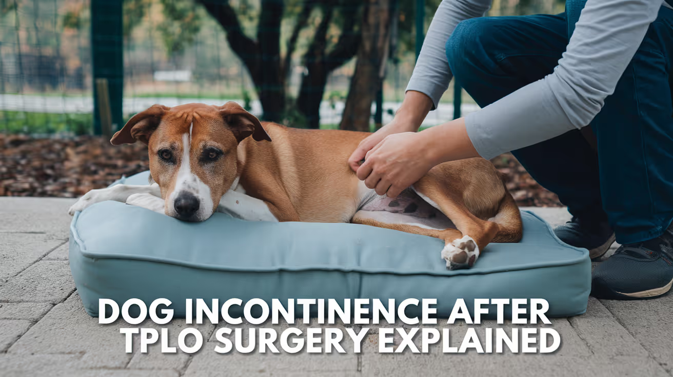
Learn about dog incontinence after TPLO surgery, its causes, treatment options, and recovery tips for your pet’s comfort and health.
Urinary incontinence after TPLO surgery is not a common complication, but it does occur and is understandably alarming for owners.
In most cases it is a temporary consequence of anesthesia, pain medication, stress, or reduced mobility not a sign of a serious surgical problem.
Knowing the causes, the timeline, and when to call your vet helps you respond appropriately.
Quick answer: Incontinence after TPLO is usually temporary from anesthesia, pain medications, reduced mobility, or stress. Most cases resolve within days to weeks. Call your vet if it persists beyond 2 to 4 weeks or comes with bloody urine or fever.
Key takeaways
- Immediate post-TPLO incontinence is most often from anesthesia or nerve irritation: both are temporary and resolve within days to weeks
- Opioid pain medications cause urinary retention or reduced bladder awareness in some dogs this resolves as medications are tapered
- Postoperative swelling may temporarily irritate nerves involved in bladder control; this resolves with swelling
- Reduced mobility is a practical contributor: a confined, painful dog may not signal in time or reach the door quickly
- Stress after major surgery can cause accidents in previously house-trained dogs; this is behavioral, not neurological
- Genuine nerve damage from TPLO is rare: incontinence persisting beyond 6 weeks warrants neurological evaluation
Why dogs may become incontinent after TPLO surgery
Anesthesia effects
General anesthesia temporarily suppresses normal nerve function throughout the body, including the neural pathways that control bladder filling awareness and sphincter tone.
As anesthetic drugs are metabolized over the 12 to 24 hours after surgery, some dogs have reduced bladder control.
This is the most common cause of immediate post-surgical incontinence. It resolves as anesthesia fully clears, typically within 24 to 48 hours.
Nausea, disorientation, and general sedation from anesthesia also mean the dog may not signal clearly when they need to go outside.
Scheduled outdoor bathroom trips every 2 to 4 hours in the first 24 to 48 hours post-surgery help manage this.
Opioid and sedative medications
Opioid pain medications commonly prescribed after TPLO affect bladder control in two ways: they reduce awareness of bladder fullness, and in some dogs cause urinary retention.
Both effects resolve as medications are tapered in the days following surgery.
Retention and incontinence are opposite problems retention is not urinating at all, while incontinence is involuntary urination but both can occur at different times as different medications have their effects.
As pain medications are tapered in the days after surgery, these effects resolve for most dogs.
Postoperative swelling and nerve irritation
The proximal tibia is surrounded by neurovascular structures. Postoperative swelling around the surgical site may temporarily compress or irritate nerves that travel to the bladder and urethra.
This is more likely in the first week after surgery when swelling is at its peak.
This type of incontinence improves as swelling resolves and typically does not indicate permanent nerve damage.
Reduced mobility
A dog recovering from TPLO is confined, in pain, and may not reach the door quickly enough when the bladder signals urgency.
This is a practical mobility problem, not a neurological one.
Frequent scheduled outdoor trips every 2 to 4 hours in the first week reduce accidents significantly. Waterproof mattress covers and washable bedding help manage the recovery period hygienically.
Stress and behavioral response
Major surgery is a significant stressor. Pain, an unfamiliar environment, an E-collar, restricted movement, and disrupted routine can cause previously house-trained dogs to have accidents unrelated to neurological dysfunction.
This type of incontinence typically resolves as the dog settles into a recovery routine at home, usually within 1 to 2 weeks.
Pre-existing urinary conditions
Dogs with pre-existing urinary sphincter mechanism incompetence (common in middle-aged spayed females), bladder infections, or weak bladder control may show worsened incontinence after surgery.
Anesthesia, medications, and reduced mobility all unmask or worsen underlying conditions.
If your dog was occasionally leaking urine before surgery, report this to your vet during the pre-surgical assessment so appropriate management can be planned for the recovery period.
Types of post-TPLO incontinence by timing
Immediate (within 24 to 48 hours): almost always anesthesia effects or opioid medication. Expected and self-limiting.
Early post-operative (days 3 to 14): medication effects, swelling-related nerve irritation, mobility limitation, or stress. Usually self-limiting with appropriate management.
Delayed onset (weeks 2 to 6): may indicate a developing urinary tract infection (UTI), which can occur after catheterization during surgery or from reduced immune response. A urinalysis is warranted if incontinence appears or worsens after initial improvement.
Chronic (beyond 6 weeks): uncommon. If incontinence persists beyond 6 weeks without improvement, veterinary evaluation for nerve damage or underlying urinary tract disease is appropriate.
Home management during recovery
- Scheduled outdoor bathroom trips every 2 to 4 hours for the first 1 to 2 weeks
- Waterproof mattress protectors and washable bedding in the recovery area
- Non-slip surfaces to reduce the effort required to stand and move toward the door
- Monitor urine color and odor cloudy, bloody, or foul-smelling urine suggests UTI
- Keep the genital area clean to prevent skin irritation from urine contact
- Use a belly band (for males) or absorbent dog diapers if incontinence is significant
When to contact your veterinarian
Call your vet if:
- The dog cannot urinate at all for more than 12 to 24 hours after surgery
- Urine is bloody, cloudy, or has a strong unusual odor (UTI signs)
- Incontinence is accompanied by straining, crying during urination, or frequent squatting without producing urine
- Incontinence worsens after initial improvement
- The dog has persistent incontinence beyond 2 to 4 weeks without any improvement
- Incontinence is combined with significant worsening lameness or systemic signs
For the not-peeing guide that covers urinary retention after TPLO, see dog not peeing after TPLO surgery: causes and care.
For the full TPLO complications reference, see 15 common complications after TPLO surgery in dogs.
For TPLO plate infection signs that may be contributing to systemic effects, see TPLO plate infection signs and treatment.
For post-op whining that may relate to the same pain driving accidents, see dog whining after TPLO surgery: causes and care.
Frequently asked questions
Is incontinence normal after TPLO surgery?
Temporary incontinence particularly in the first 24 to 48 hours is a recognized post-surgical finding related to anesthesia and opioid pain medications.
It is not an expected routine outcome, but it is not rare. If it resolves within a few days, it was almost certainly anesthesia-related.
Could my dog have nerve damage from TPLO surgery?
True nerve damage causing persistent incontinence from TPLO is rare. TPLO is performed at the proximal tibia, which is some distance from the pelvic nerves controlling bladder function.
Temporary nerve irritation from swelling is much more common than actual nerve injury and resolves as swelling subsides.
My dog was continent before TPLO and is now leaking at night. Why?
Nighttime leaking specifically is typical of urinary sphincter mechanism incompetence (USMI) a condition where the urethral sphincter relaxes during sleep or rest.
If this is new after surgery, anesthesia and opioid medications may have temporarily worsened underlying borderline sphincter function.
If it continues beyond 2 to 3 weeks, a vet visit for urinalysis and discussion of phenylpropanolamine (PPA) or other treatment is appropriate.
How do I know if my dog has a UTI after TPLO?
Signs of UTI include: cloudy or bloody urine, strong or unusual odor, frequent small amounts of urination, straining to urinate, and licking at the genital area more than normal.
Some dogs with UTI also show lethargy and decreased appetite. Urinalysis and culture confirm the diagnosis.
Should I limit my dog's water intake to reduce accidents?
No. Do not restrict water. Hydration is important for recovery and kidney function. Restricting water can concentrate the urine and increase UTI risk.
Instead, manage accidents through scheduled outdoor trips and waterproof bedding.
Resources
- VCA Animal Hospitals. Tibial Plateau Leveling Osteotomy (TPLO) in Dogs. vcahospitals.com
- ACVS. Cranial Cruciate Ligament Disease. acvs.org
- PetMD. Urinary Incontinence in Dogs. petmd.com
- Midwest Veterinary Specialists. Post-Operative Care Following TPLO. midwestveterinaryspecialists.com
Bruising After TPLO Surgery in Dogs
Learn about bruising after TPLO surgery in dogs, its causes, care tips, and when to seek veterinary help.
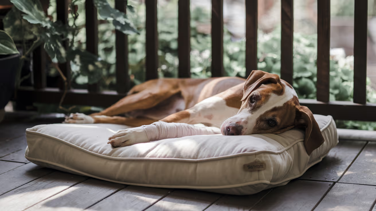
Bruising after TPLO surgery in dogs is a common concern for many pet owners. TPLO, or Tibial Plateau Leveling Osteotomy, is a surgical procedure to stabilize a dog's knee after a cruciate ligament injury. After surgery, owners often notice bruising around the incision site or leg, which can be alarming.
This article explains why bruising happens after TPLO surgery, how to care for your dog during recovery, and when to contact your veterinarian. Understanding bruising helps you support your dog's healing process effectively.
Why Does Bruising Occur After TPLO Surgery in Dogs?
Bruising happens when blood vessels under the skin break and leak blood into surrounding tissues. After TPLO surgery, bruising is usually due to the trauma of surgery and tissue manipulation. The surgical area may show discoloration as part of normal healing.
It is important to recognize that some bruising is expected, but excessive or worsening bruising may indicate complications. Knowing the causes helps you monitor your dog’s recovery closely.
- Surgical trauma: The cutting and repositioning of bone and soft tissue during TPLO can cause small blood vessels to rupture, leading to bruising around the surgical site.
- Inflammation response: The body’s natural healing process causes inflammation, which can increase blood flow and cause visible bruising and swelling near the incision.
- Blood thinning medications: If your dog is on medications like aspirin or NSAIDs, these can increase bruising risk by affecting clotting mechanisms.
- Movement after surgery: Excessive activity or pressure on the leg soon after surgery can worsen bruising by disrupting healing tissues and blood vessels.
Understanding these causes helps you differentiate normal bruising from signs of complications that need veterinary attention.
What Does Normal Bruising Look Like After TPLO Surgery?
Normal bruising after TPLO surgery usually appears as a purple, blue, or reddish discoloration around the incision or leg. It often develops within 24 to 48 hours post-surgery and gradually fades over 1 to 2 weeks.
Knowing the typical appearance and timeline of bruising helps you track your dog’s healing and identify abnormal changes early.
- Color changes: Bruises typically start dark purple or blue, then change to green, yellow, and finally fade as healing progresses over days to weeks.
- Size and spread: Normal bruising is usually localized near the incision and does not spread extensively to other parts of the leg or body.
- Swelling presence: Mild swelling often accompanies bruising but should not cause severe discomfort or limb deformity.
- Pain level: Some tenderness is expected, but your dog should not show signs of severe pain or limping beyond normal post-surgical recovery.
Monitoring these signs helps ensure bruising is part of normal healing and not a sign of infection or bleeding complications.
How Should You Care for Bruising After TPLO Surgery?
Proper care reduces bruising severity and supports healing after TPLO surgery. Following your veterinarian’s instructions and providing gentle care helps your dog recover comfortably and safely.
Simple steps at home can minimize bruising and swelling while preventing complications.
- Rest and restricted activity: Limit your dog’s movement to prevent pressure on the surgical site and avoid worsening bruising or swelling.
- Cold compress application: Applying a cold pack wrapped in cloth for 10–15 minutes several times daily during the first 48 hours can reduce swelling and bruising.
- Proper bandage care: Keep any surgical bandages clean and dry, and follow your vet’s instructions on changing or removing them to avoid irritation.
- Medication adherence: Administer all prescribed painkillers and anti-inflammatory drugs exactly as directed to control pain and reduce inflammation.
Consistent care helps your dog heal faster and reduces the risk of bruising-related complications.
When Should You Be Concerned About Bruising After TPLO Surgery?
While bruising is normal, certain signs indicate that your dog may need urgent veterinary evaluation. Recognizing these warning signs early can prevent serious complications.
If you notice any of the following, contact your veterinarian promptly for advice or examination.
- Rapidly expanding bruises: Bruising that spreads quickly or becomes very large may signal active bleeding or hematoma formation requiring medical attention.
- Excessive swelling and heat: Swelling that worsens with warmth and redness around the incision could indicate infection or inflammation needing treatment.
- Severe pain or lameness: If your dog shows intense pain, refuses to bear weight, or has a limp worsening over time, this may suggest complications.
- Discharge or foul odor: Any pus, bleeding, or bad smell from the surgical site is a sign of infection and requires immediate veterinary care.
Early detection of problems helps ensure timely treatment and better recovery outcomes.
Can Bruising Affect Your Dog’s Recovery After TPLO Surgery?
Bruising itself usually does not affect the overall success of TPLO surgery if it is mild and managed properly. However, severe bruising or related complications can delay healing and cause discomfort.
Understanding the impact of bruising helps you provide better care and communicate effectively with your veterinarian during recovery.
- Minor bruising benefits: Mild bruising is a normal sign of healing and usually resolves without affecting joint stability or function.
- Severe bruising risks: Large bruises or hematomas can increase pain, limit mobility, and sometimes require drainage or additional treatment.
- Infection risk: Bruised tissue is more vulnerable to infection, so keeping the area clean and monitoring closely is essential.
- Physical therapy considerations: Bruising may limit early physical therapy, so adjusting rehabilitation plans based on your dog’s condition is important.
Working closely with your vet ensures bruising does not interfere with your dog’s full recovery and return to normal activity.
How Can You Prevent Bruising After TPLO Surgery in Dogs?
While some bruising is unavoidable due to surgery, certain steps can reduce its severity and support smooth healing. Prevention focuses on careful handling and following veterinary guidance.
Taking precautions before and after surgery helps protect your dog’s tissues and blood vessels from excessive damage.
- Pre-surgery health check: Ensuring your dog is healthy and not on blood-thinning medications before surgery reduces bruising risk.
- Gentle post-op handling: Avoid rough play, jumping, or excessive pressure on the leg during recovery to prevent new bruises.
- Follow medication instructions: Use prescribed drugs properly and avoid over-the-counter medications without vet approval to prevent clotting issues.
- Regular veterinary follow-up: Attend all scheduled check-ups so your vet can monitor healing and address bruising or other concerns early.
Prevention and careful management promote a safer, faster recovery with minimal bruising complications.
Conclusion
Bruising after TPLO surgery in dogs is a common and usually normal part of the healing process. It results from surgical trauma and inflammation, appearing as discoloration near the incision site. Mild bruising typically resolves within two weeks without affecting recovery.
Proper care, including rest, cold compresses, and medication adherence, helps reduce bruising and supports healing. However, owners should watch for signs of excessive bruising, swelling, pain, or infection and contact their veterinarian promptly if these occur. Understanding bruising after TPLO surgery allows you to care for your dog confidently and ensure a smooth recovery.
FAQs
Is bruising after TPLO surgery normal for all dogs?
Yes, mild bruising is common after TPLO surgery due to tissue trauma and inflammation. However, the amount varies by individual and surgical factors.
How long does bruising last after TPLO surgery?
Bruising usually appears within 1–2 days after surgery and fades over 1 to 2 weeks as the tissues heal naturally.
Can I use cold packs on my dog’s bruising after surgery?
Yes, applying cold compresses wrapped in cloth for 10–15 minutes several times daily during the first 48 hours helps reduce swelling and bruising.
When should I call the vet about bruising after TPLO?
Contact your vet if bruising spreads rapidly, swelling worsens, pain is severe, or if there is discharge or foul odor from the incision.
Does bruising affect my dog’s ability to walk after TPLO surgery?
Mild bruising usually does not affect walking, but severe bruising or pain may cause limping and require veterinary evaluation.
How Long Does Swelling Last After TPLO Surgery
Find out how long swelling lasts after TPLO surgery, what’s normal, and when to call your vet for possible infection or complications.
Post-surgical swelling after TPLO is normal, expected, and self-limiting when managed correctly. Most incision-area swelling peaks within the first 2 to 5 days and resolves by 2 weeks.
Swelling that does not follow this trajectory -- that is not declining, or that appears new after initial improvement -- is the key indicator that requires veterinary attention.
Quick answer: Normal TPLO swelling peaks at days 2 to 5 and resolves within 1 to 2 weeks with cold therapy and rest. Ankle swelling from gravity-dependent fluid migration appears at days 3 to 5. Swelling increasing after day 5, or with warmth, redness, or discharge, warrants veterinary evaluation.
Key takeaways
- Normal TPLO swelling peaks at days 2 to 5: SustainableVet.org confirms mild incision swelling usually goes away within two weeks
- Ankle swelling is a normal gravity-dependent phenomenon: TPLO Info confirms ankle swelling typically appears at days 3 to 5; it is fluid migrating downward from the surgical site
- Cold therapy in the first 48 to 72 hours is the primary management tool: 10 to 15 minutes, 3 to 4 times daily, reduces swelling and pain
- Swelling worsening after day 5 or accompanied by warmth, redness, or discharge indicates infection, seroma, or hematoma; contact your vet within 24 hours
- Mild swelling can persist for weeks at a low level in larger dogs and those with poor activity restriction compliance; trajectory matters more than absolute level
- Nighttime swelling is common and normal: dogs move less at rest, allowing fluid to settle; this should improve by morning or after repositioning
Normal swelling: timeline and characteristics
Days 1 to 3: acute post-surgical swelling
Swelling begins within hours of surgery and results from:
- Surgical tissue trauma (incision through skin, subcutaneous tissue, and fascia)
- Bone cutting at the osteotomy site
- Inflammatory response to the metal plate and screws
- Fluid accumulating in the disrupted tissue spaces
This early swelling is diffuse, soft, and warm to the touch. It is the expected cellular response to surgery.
Days 2 to 5: peak swelling
Swelling typically peaks between days 2 and 5. The dog's leg may look noticeably larger at this stage, and some extension of swelling down the lower leg toward the ankle is normal.
Vetplayas confirms: swelling typically peaks within the first few days after surgery but should gradually subside over time.
Days 3 to 5: ankle swelling appears
A distinct swelling often appears at the ankle (tarsus) between days 3 and 5, sometimes alarming owners who notice it away from the surgical site. This is entirely normal.
TPLO Info confirms: it is completely normal for swelling to develop around the ankle approximately 3 to 5 days following surgery on the TPLO leg.
This swelling can be massaged and iced if it occurs and will resolve over a few days.
TPLO Austin confirms this finding identically. The mechanism is gravity-dependent migration of tissue fluid from the surgical site through the fascial planes of the lower leg.
Days 5 to 14: progressive resolution
From day 5 onward, swelling should consistently decrease with each daily inspection. Cold therapy, rest, and anti-inflammatory medication all contribute to this resolution.
SustainableVet.org confirms: mild swelling around the incision site usually goes away within two weeks. The trajectory must be consistently improving.
Beyond 2 weeks: residual low-level swelling
Some dogs retain mild soft swelling around the lower leg beyond 2 weeks, particularly larger dogs with greater tissue volume and dogs who have been less compliant with activity restriction.
This is usually minor and does not indicate a problem if:
- The swelling is soft and not warm
- There is no discharge or redness
- The dog is bearing weight and improving in lameness
Cold therapy: the primary management tool
Cold therapy is the evidence-based first-line intervention for post-surgical swelling.
Protocol:
- Wrap an ice pack or bag of frozen peas in a thin cloth or towel
- Apply to the incision and plate site for 10 to 15 minutes
- Repeat 3 to 4 times daily
- Begin on day 1 post-surgery
- Continue for the first 48 to 72 hours (the acute inflammatory phase)
SustainableVet.org confirms: cold therapy, like applying an ice pack wrapped in a towel for 10 to 15 minutes several times a day during the first 72 hours, helps reduce inflammation.
After the initial period, warm compresses can be used to promote circulation.
Never apply ice directly to the skin or incision -- frostbite and wound damage can result. Remove the pack immediately if the skin appears pale or very cold.
For the ankle specifically, TPLO Info confirms: ankle swelling can be massaged and iced. Gentle proximal-to-distal massage (toward the toes) applied after icing helps lymphatic drainage.
Concerning swelling: signs requiring veterinary contact
Swelling that is not declining after day 5
Normal swelling is consistently declining from day 5 onward. Swelling that is the same or larger than on day 3, measured at day 7, is not normal.
New swelling after apparent resolution
If swelling had reduced and then reappears -- particularly at the plate site rather than the incision -- this suggests a developing seroma, hematoma, or early infection.
SustainableVet.org confirms: you should be concerned if swelling increases after the first week or is accompanied by heat, redness, oozing, or a bad odor.
Warmth, firmness, redness, or discharge alongside swelling
These features distinguish infection from normal surgical edema. Normal edema is diffuse, soft, not dramatically warm, and has no discharge. Infection produces focal warmth, firmness, redness spreading beyond the incision, and discharge.
Swelling at the plate site (not the incision) weeks after surgery
This can indicate an implant-associated infection with seroma or early osteomyelitis. Requires radiographic assessment and deep sampling.
Seroma: a specific type of post-surgical swelling
A seroma is a localized collection of clear serous fluid under the skin, forming when tissue spaces fill with tissue fluid that has not been reabsorbed.
Seromas after TPLO are common and are distinct from infection.
Signs of seroma:
- A soft, fluctuant (fluid-filled) swelling at or near the incision
- Typically appears 1 to 3 weeks post-surgery
- Fluid is clear or pale yellow when aspirated
- No warmth, redness, or discharge from the incision itself
Most seromas resolve without treatment. Your vet will assess whether aspiration or monitoring is appropriate.
For the fluid buildup guide, see fluid buildup after TPLO surgery in dogs. For the ankle swelling guide, see ankle swelling after TPLO surgery in dogs.
For the infection signs guide, see TPLO plate infection signs and treatment. For the bruising guide, see bruising after TPLO surgery in dogs.
Frequently asked questions
Is it normal for the whole leg to swell after TPLO?
Yes, in the early days. Diffuse swelling of the lower leg, extending from the surgical site toward the ankle, is normal in the first 1 to 2 weeks.
Gravity and the inflammatory response both contribute. This resolves as the inflammatory process settles and the dog begins controlled weight bearing.
My dog's leg looks huge at day 3. Is that too much?
Day 3 is within the normal peak swelling period.
Unless the swelling is dramatically asymmetric compared to the other leg, is very hot, or is producing discharge, observe and continue cold therapy as directed.
If you are concerned, contact your vet to describe the swelling -- they can advise whether in-person assessment is needed.
Should I massage the swollen leg?
Gentle lymphatic massage (soft strokes toward the body, from the ankle upward) after icing can help reduce distal swelling.
Do not massage directly over the incision or plate site, and do not massage aggressively. Ask your vet or rehabilitation therapist to demonstrate the correct technique.
My dog's swelling is going up and down -- bigger in the evening, smaller in the morning. Is that normal?
Yes. SustainableVet.org confirms that mild nighttime swelling occurs because dogs move less while resting, allowing fluid to settle. Morning improvement after repositioning and movement is normal.
The concern is when the morning maximum is larger than the previous day's morning maximum -- a consistently worsening trend.
When does warm therapy replace cold therapy?
Cold therapy is most beneficial in the first 48 to 72 hours (the acute inflammatory phase). After 72 hours, warm compresses promote circulation and may help reabsorb residual fluid.
The transition is typically around days 3 to 5. Never apply warmth to an incision that looks infected or discharging -- this can worsen infection.
Resources
- TPLO Info. TPLO Recovery FAQ. tploinfo.com
- SustainableVet. Post-Surgery Recovery After TPLO in Dogs. sustainablevet.org
- TPLO Austin. TPLO Incision Care After Surgery. tploaustin.com
- VCA Animal Hospitals. Tibial Plateau Leveling Osteotomy (TPLO) in Dogs. vcahospitals.com
How to Confine Your Dog Post-TPLO Surgery
Keep your dog safe after TPLO surgery with smart confinement tips. Learn crate setup, barriers, and daily care for smooth healing
Confinement after TPLO surgery is not optional. It is the primary structural safeguard preventing the plate from failing before the osteotomy heals.
A dog that is allowed to run, jump, or spin in the first 8 weeks risks plate displacement -- a complication that requires revision surgery and extends recovery significantly.
Effective confinement is a practical skill that owners can prepare for before surgery.
Quick answer: Confine in a crate large enough to stand, turn, and lie but not to pace. An exercise pen works for crate-resistant dogs. A baby-gated small room is a third option. Non-slip flooring is essential. Outdoor access requires a leash. Strict confinement continues for 6 to 8 weeks.
Key takeaways
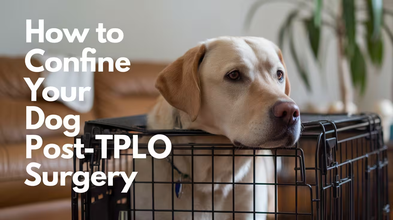
- The crate is the gold standard confinement tool: limits jumping, spinning, and running that risk plate displacement; large enough to stand and turn but not to pace
- Non-slip flooring is required in all confinement spaces: slipping generates uncontrolled leg movements that stress the plate
- Leash on the dog at all times outside the crate: every unsupervised second without a leash risks slipping or running; a house leash gives immediate control
- Exercise pens (X-pens) are a practical alternative for crate-resistant dogs: more space than a crate while still preventing free access to the rest of the house
- The confinement protocol changes weekly: strict crate-only in weeks 1 to 2, then gradual expansion as bone healing is confirmed on radiographs
- Clean bedding weekly: dirty bedding harbors S. pseudintermedius; the incision is in close contact with bedding throughout recovery
Why confinement matters
The TPLO plate holds the rotated tibial plateau in position while new bone grows across the osteotomy. The plate is strong but not indestructible.
Bone is not fully consolidated across the osteotomy for 8 to 12 weeks.
During this healing period, a dog that runs, jumps onto a sofa, slips on hardwood flooring, or spins to chase another pet can apply forces to the plate that exceed its tolerance.
Plate bending, screw loosening, or osteotomy displacement are the results. Each requires revision surgery.
SustainableVet.org confirms: dogs typically need 6 to 8 weeks of restricted activity before returning to light exercise; strict confinement ensures the bone plate stabilizes and heals properly.
Option 1: the crate
Why crates work best
A crate completely controls the dog's environment. The dog cannot run to the door, jump on the sofa, or spin in excitement. Every movement is small, controlled, and low-impact.
SustainableVet.org confirms: a crate is one of the safest ways to confine a dog after TPLO surgery; crates help prevent jumping, running, or sudden twisting that can damage the healing leg.
Crate selection
Size: the crate must be large enough for the dog to stand fully, turn around, and lie in any direction comfortably. It should NOT be large enough for the dog to trot or run inside it -- an oversized crate allows the dog to generate enough speed within the crate to create impact when stopping.
Flooring: avoid wire-bottomed crates. The dog's leg can slip through wire flooring. Use solid-floored crates or add a mat over wire flooring.
Bedding: comfortable, non-slip bedding. Memory foam dog mats are excellent for TPLO dogs -- they cushion pressure points during long rest periods.
Location: place the crate in the main family living area, not in an isolated room. Isolation increases separation anxiety and whining. A dog that can see and hear the family is calmer and more able to rest.
TPLO Info confirms: dogs do not like dirty bedding, so wash crate items at least once a week.
Transitioning a crate-naive dog
If the dog has never been crated, introduce the crate before surgery:
- Place meals in the crate with the door open for several days before surgery
- Gradually extend time inside with the door closed, building from 10 minutes to 1 hour
- Never force the dog in or use the crate for punishment
A dog that enters the crate willingly before surgery recovers with significantly less confinement-related distress.
Option 2: the exercise pen (X-pen)
An X-pen is a freestanding modular fence creating a confined area larger than a crate. It can be configured in different shapes and sizes.
TPLO Info confirms: X-pens give enough room to move while keeping the dog in one place. Larger dogs may attempt to jump over the fence and require monitoring.
Advantages: more space than a crate, reducing frustration in dogs that do not tolerate crates; can be set up in any room.
Disadvantages: requires monitoring for jumping attempts; the dog can build more momentum in a larger space; not suitable for very large or athletic dogs who can clear the fence.
Height: use a 48-inch or taller X-pen for medium and large dogs; even a non-athletic dog may clear a 36-inch pen if motivated.
Flooring inside the X-pen: place yoga mats or a rubber-backed rug inside to cover hard flooring.
When to use an X-pen: recommended after the initial 2 weeks of strictest confinement, when the dog has established initial healing and the wound is closed. Not ideal for weeks 1 to 2.
Option 3: a baby-gated small room
A bathroom, laundry room, or small bedroom can serve as a recovery room when gated with a baby gate.
Dog Knee Injury confirms: prepare your home for post-surgery by setting up a recovery space with a comfortable dog bed, non-slip flooring, and a baby gate to create a barrier.
Requirements:
- Non-slip flooring throughout (add yoga mats or rugs)
- No furniture the dog can jump onto or off
- Accessible water and food bowls at floor level
- The gate must prevent the dog exiting unsupervised
Advantages: familiar environment, easy human access, more comfortable than a crate for larger dogs.
Disadvantages: more space means more opportunity for movement; the dog can circle and pace in a room, which a crate prevents; requires strict monitoring.
Outdoor access: leash only, no exceptions
Every outdoor bathroom trip requires a leash. No exceptions.
SustainableVet.org confirms: always use a leash when outside, even for bathroom breaks; open-door access to the yard allows a dog to run, chase, or slip before the owner can intervene.
Leash length: short -- no retractable leashes. The owner must be able to prevent sudden running immediately. A 4 to 6 foot leash is appropriate.
Bathroom trip duration: 5 minutes maximum in weeks 1 to 2. Gradual increase per the vet's protocol.
Wet weather: rain and wet grass introduce bacteria to the incision area and also create slipping hazard. Time trips to avoid heavy rain where possible; carry a dry towel.
Week-by-week confinement protocol
Weeks 0 to 2 (strictest):
- Crate at all times except supervised bathroom leash walks
- 5-minute walks, 3 to 5 times daily
- E-collar on at all times
- No access to any area without owner supervision
Weeks 2 to 6 (moderate):
- Crate or X-pen when unsupervised
- Supervised time in the confinement room with owner present
- Leash walks increasing gradually per vet's direction
- E-collar until incision fully confirmed healed at 2-week recheck
Weeks 6 to 12 (graduated relaxation):
- Confinement continues but may expand to a room-sized area under supervision
- Activity increases based on radiographic findings at 6-week recheck
- Off-leash access to the house (not yard) under close supervision for some dogs, vet-directed
After 12 weeks (post-clearance):
- Full activity restriction lifted after radiographic bone healing confirmation
- Progressive return to normal environment and activity
- Still avoid high-impact play until full muscle recovery at 4 to 6 months
For the full recovery timeline, see what to expect after TPLO surgery in dogs. For keeping a dog calm during confinement, see how to keep a dog calm after TPLO surgery.
For physical therapy timing, see when to start physical therapy after TPLO surgery. For infection prevention, see post-operative care mistakes that increase TPLO infection risk.
Frequently asked questions
My dog has never been in a crate. Can I start at surgery?
Yes, but start before surgery if possible -- even a few days of crate introduction before surgery significantly reduces post-operative confinement distress.
If you cannot prepare beforehand, introduce the crate gently in the first day or two at home, using meals and treats to create positive associations.
How do I know if the crate is the right size?
The dog should be able to stand with their head up, turn in a full circle, and lie in multiple positions without being cramped.
They should NOT be able to trot or run within the crate space. If in doubt, err toward smaller.
My dog cries in the crate for hours. Is the confinement causing psychological harm?
Extended, severe crate distress may indicate inadequate pain management (the dog is in pain, not just frustrated) or genuine severe separation anxiety. Contact your vet.
Trazodone and gabapentin can significantly reduce confinement distress. Do not abandon confinement -- the physical risk of uncontrolled activity is greater than the psychological discomfort of confinement.
Can my dog sleep in my bed during recovery?
No. Jumping off the bed -- even a single time -- can displace the plate.
The dog must sleep in the crate or on a floor-level dog bed in the recovery area throughout the restriction period.
Can I let my dog outside in a fenced yard for bathroom breaks without a leash?
No. Dogs routinely run, spin, and perform sudden movements in yards even when told to go to the bathroom.
A single run to chase a squirrel or greet someone at the fence is enough to cause plate failure before the osteotomy heals. Leash only, every time.
Resources
- SustainableVet. How to Confine Your Dog Post-TPLO Surgery. sustainablevet.org
- TPLO Info. How to Confine Your Dog Post-TPLO Surgery. tploinfo.com
- Dog Knee Injury. How to Confine Your Dog After TPLO Surgery. dogkneeinjury.com
- VCA Animal Hospitals. Tibial Plateau Leveling Osteotomy (TPLO) in Dogs. vcahospitals.com
Fluid Buildup After TPLO Surgery in Dogs
Learn about fluid buildup after TPLO surgery in dogs, its causes, symptoms, treatment, and prevention to ensure your pet's smooth recovery.
Some degree of fluid accumulation after TPLO surgery is normal and expected.
The surgery involves cutting and rotating the tibia a significant tissue injury that triggers an inflammatory response, part of which is fluid production.
The question is not whether fluid will appear, but whether the fluid you are seeing is a normal healing response or a sign of something that needs attention.
Quick answer: Fluid buildup after TPLO is usually a seroma — clear serum under the skin that is not an infection. Small seromas resolve in 1 to 3 weeks with rest. Call your vet if swelling is hot, rapidly growing, producing discharge, or accompanied by fever.
Key takeaways
- Seroma is the most common fluid buildup after TPLO: clear serum under the skin, not an infection, resolving with rest in 1 to 3 weeks
- Small seromas are managed with rest: the body reabsorbs the fluid naturally; no intervention is needed for most small seromas
- Large or persistent seromas may require needle aspiration: this must be done under sterile veterinary conditions; never drain at home
- Ankle swelling at 3 to 5 days post-surgery is normal: gravity pulls post-surgical fluid downward to the ankle; TPLO Info confirms this is routine
- Warning signs requiring vet contact: rapidly increasing swelling, warmth, redness, pus, foul odor, fever, or lethargy
- Activity restriction is the most important prevention: excessive movement is the leading cause of seroma formation after TPLO
What is fluid buildup after TPLO surgery?
Fluid buildup after TPLO surgery typically takes one of two forms: seroma or post-surgical edema.
A seroma is a collection of serum the clear, protein-rich fluid component of blood that leaks from disrupted blood vessels and lymphatic channels during surgery.
It pools in the dead space beneath the skin at the surgical site. Seromas feel soft, fluctuant (squishy), and are usually not warm or painful when small. They are not infections.
Post-surgical edema is broader tissue swelling caused by the inflammatory response to surgery.
This is expected in the first few days after any major orthopedic procedure and typically peaks at 48 to 72 hours before gradually resolving.
Both are distinct from hematoma (a collection of blood) and from infectious fluid (which appears purulent, has an odor, and accompanies systemic illness signs).
Why fluid builds up after TPLO
TPLO surgery creates significant soft tissue disruption.
The curved osteotomy cut, muscle retraction, and plate placement create dead space under the skin.
Fluid accumulates in this space before the tissues close together during healing.
Key contributing factors:
Excessive activity after surgery: the most preventable cause. Every time the operated leg moves beyond gentle weight-bearing, tissue planes are disrupted and more serum leaks into the dead space. TPLO Info confirms that the most common post-surgical cause of seroma is excessive movement of the knee.
Surgical technique: a longer or more complex procedure increases tissue disruption and the resulting dead space. Most surgeons use subcutaneous sutures and tissue closure techniques to minimize this, but some seroma formation remains common even with optimal technique.
Dog size and body composition: larger dogs and those with more subcutaneous fat tend to have more dead space at the osteotomy site and are more prone to seroma formation.
Licking or chewing at the incision: disrupts the healing wound edges and can introduce bacteria into a seroma, converting a benign fluid collection into an infected abscess. The E-collar must remain on at all times.
Normal vs. abnormal fluid buildup: how to tell the difference
Treatment options
Observation and strict rest: for small seromas (under 2 to 3 cm), the standard approach is to enforce activity restriction and allow the body to reabsorb the fluid. Small seromas typically resolve within 1 to 3 weeks. Cold compresses in the first few days reduce new inflammation.
Needle aspiration: large or uncomfortable seromas may be drained by your veterinarian using a sterile needle and syringe. This is a simple outpatient procedure but must be performed aseptically to avoid introducing infection. Never drain a seroma at home.
Compression bandaging: gentle compressive bandaging reduces the dead space available for fluid accumulation and can help prevent reformation after aspiration. This is typically applied by the veterinarian.
Surgical drain placement: if fluid continues to accumulate despite aspiration, a closed suction drain can be placed temporarily. This is rarely needed but is an option for persistent, large seromas.
Antibiotics: only if bacterial infection is confirmed or strongly suspected. Most seromas do not require antibiotics. Prescribing antibiotics for a non-infected seroma is not appropriate and contributes to antimicrobial resistance.
When to contact your veterinarian
Contact your vet promptly if you notice any of the following:
- Swelling that is rapidly increasing in size, especially in the first week
- The swollen area becomes warm, red, or painful to touch
- Any discharge, pus, or unusual odor from the incision or the swollen area
- Your dog develops fever, lethargy, loss of appetite, or stops bearing weight
- The seroma persists beyond 3 to 4 weeks without improvement
For the related article on incision site complications, see seroma in dogs after TPLO surgery. For the full complications overview, see 15 common complications after TPLO surgery in dogs.
For incision infection specifics, see TPLO incision infection symptoms and prevention.
Frequently asked questions
Is it normal to have a lump under the TPLO incision?
Yes, in most cases. A soft, non-painful lump that develops within the first 1 to 2 weeks under the TPLO incision is typically a seroma.
As long as it is not hot, rapidly growing, or producing discharge, it is usually a benign post-operative finding. Monitor it daily and report any changes to your vet.
How long does post-TPLO swelling last?
Most post-surgical edema resolves within 3 to 7 days as the acute inflammatory phase subsides.
Seromas take longer typically 1 to 3 weeks for small ones, and potentially 4 to 6 weeks for larger ones if they reform after aspiration.
Can I massage the fluid away?
Gentle massage around (not directly over) the incision can encourage local circulation, but vigorous massage of a seroma risks rupturing it and introducing bacteria.
TPLO Info notes that ankle swelling that develops 3 to 5 days post-surgery can be massaged and iced. For the surgical site itself, follow your vet's specific guidance.
What happens if a seroma is left untreated?
Most small seromas reabsorb on their own without problems.
A larger seroma that is not treated may persist for weeks or months, become uncomfortable due to pressure, or rarely become infected if bacteria enter through the overlying skin.
Monitoring and reporting changes to your vet is the key strategy.
Can my dog's activity level cause more fluid to build up?
Yes. Excessive activity is the most preventable cause of seroma formation and recurrence after aspiration. Every unnecessary movement of the operated leg disrupts healing tissue planes and drives more fluid production.
Strict confinement and leash-only activity are essential.
Resources
- TPLO Info. Recovery FAQs. tploinfo.com
- Vetplayas. Understanding Swelling After TPLO Surgery. vetplayas.com
- VCA Animal Hospitals. Seroma in Dogs. vcahospitals.com
- ACVS. Cranial Cruciate Ligament Disease. acvs.org

TPLO Plate Rejection Symptoms in Dogs
Learn to recognize TPLO plate rejection symptoms in dogs, including signs, causes, and treatment options for better recovery.
TPLO plate rejection is one of the less common but more serious complications that can follow tibial plateau leveling osteotomy surgery.
The term "rejection" is sometimes used loosely distinguishing between true immune-mediated rejection, infection, and implant loosening matters because the treatment differs significantly for each.
Quick answer: True TPLO plate rejection is rare — implants are highly biocompatible. It produces persistent swelling, heat, pain over the plate, and sometimes a draining tract. Infection produces identical symptoms. Contact your vet if lameness worsens or discharge appears weeks to months after surgery.
Key takeaways
- True immune-mediated plate rejection is rare: TPLO plates are made from highly biocompatible titanium or stainless steel
- Symptoms overlap significantly with infection: pain, swelling, heat, and discharge occur in both; bacterial culture distinguishes them
- Infection is the most common implant complication: TPLO infection rates range from 0.8 to 14.3%; true rejection is a small subset
- Signs typically appear weeks to months after surgery: worsening lameness after initial improvement is the classic red flag
- A draining tract (fistula) over the plate is a hallmark sign of implant-associated infection or rejection
- True rejection requires plate removal: once the osteotomy heals (at least 12 weeks), the plate is removed; bone heals without it
What is TPLO plate rejection?
The TPLO plate and screws are made from medical-grade titanium or stainless steel.
These materials are designed to be biologically inert the body should recognize them as neutral foreign material and wall them off without reaction.
Laguna Hills Animal Hospital explains that in some cases, the TPLO plate is rejected by the dog's body because it is perceived as a foreign object.
True immune-mediated rejection occurs when the immune system mounts an inflammatory response against the implant material itself. This is distinct from:
- Periprosthetic infection: bacteria colonize the implant surface and cause chronic infection around the plate; this is far more common than true rejection
- Mechanical failure: the plate bends, breaks, or screws loosen due to excessive force; this is a structural problem, not an immune reaction
- Normal post-surgical inflammation: expected in the first 2 to 3 weeks; should be improving, not worsening
In clinical practice, the distinction between true rejection and low-grade infection is often made by bacterial culture of the fluid or tissue at the site.
True aseptic rejection produces inflammation without bacterial growth; periprosthetic infection produces the same symptoms with positive cultures.
Symptoms of TPLO plate rejection
Dog Discoveries' veterinary reference describes plate rejection as: the knee becomes inflamed and sore around the plate, the dog feels constant pain and irritation, and the dog gets worse rather than better.
Signs of trouble are generally seen weeks to months after surgery rather than in the immediate post-operative period.
Key symptoms to monitor:
Worsening or persistent lameness: a dog that was improving and then regresses, or one whose lameness fails to improve beyond the expected baseline, is showing a key red flag. This is distinct from the expected early post-TPLO lameness that should be progressively improving week by week.
Localized swelling over the plate: visible or palpable swelling directly over the plate site (proximal medial tibia) that does not resolve with rest and is not reducing over time.
Warmth and heat at the surgical site: warmth beyond the first 2 to 3 post-operative weeks suggests ongoing inflammation.
Draining tract (fistula): a small hole in the skin near the surgical site that produces persistent clear, bloody, or purulent discharge. This is a hallmark of implant-associated complications either infection or reaction. Vet Playas confirms persistent drainage or discharge, especially with a foul odor, may indicate infection or plate rejection.
Pain on palpation: the dog reacts to direct pressure over the plate site.
Failure to heal: the dog is not progressing as expected at radiographic rechecks; osteotomy bridging is delayed or absent.
How plate rejection is diagnosed
Clinical examination: systematic palpation of the plate site, assessment of lameness grade, and comparison to expected recovery trajectory.
Radiographs: assess osteotomy healing, implant position, screw integrity, and periimplant bone quality. Periprosthetic bone loss suggests infection or chronic reaction.
Culture and sensitivity: fluid or tissue from the surgical site is cultured to determine whether bacteria are present and which antibiotics are effective. Culture-negative inflammation supports true rejection; culture-positive results confirm infection.
Blood work: elevated inflammatory markers (C-reactive protein, white blood cell count) confirm systemic involvement.
Treatment
For infection (most common implant complication)
Culture-directed antibiotic therapy is initiated. If the osteotomy is not yet healed, the plate must remain in place removing the plate before the osteotomy heals causes the bone segments to lose support.
Targeted antibiotics and local wound management may control infection until healing is sufficient.
If the infection does not resolve with antibiotics, surgical debridement or implant removal (if the bone has healed) may be required.
For the full infection management guide, see TPLO plate infection signs and treatment.
For true aseptic rejection
Plate removal is the definitive treatment. Laguna Hills Animal Hospital confirms that the bone plate must remain in place for at least 12 weeks to allow the bone to heal.
Once healing is confirmed radiographically typically at 10 to 12 weeks the plate can be safely removed.
The bone does not require the plate long-term; it is a scaffold to hold the osteotomy while it heals.
When to contact your veterinarian
Contact your vet if:
- Lameness worsens or plateaus instead of progressively improving
- You notice new swelling, heat, or discharge at the surgical site weeks after surgery
- A draining tract appears at or near the incision
- Your dog shows signs of pain at the plate site on palpation
- Radiographs at rechecks show unexpected findings around the plate or screws
For the implant failure guide, see TPLO implant failure signs and causes. For the plate removal recovery guide, see TPLO plate removal recovery guide.
For the full complications reference, see 15 common complications after TPLO surgery in dogs. For the plate infection article with antibiotic treatment details, see TPLO plate infection signs and treatment.
For when the plate needs to come out due to infection, see when does a TPLO implant need to be removed due to infection.
Frequently asked questions
How common is TPLO plate rejection in dogs?
True immune-mediated rejection is uncommon. TPLO plates are manufactured from highly biocompatible titanium or stainless steel.
The more common implant complication is infection (0.8 to 14.3% of TPLO cases depending on the study), which produces similar symptoms to rejection.
Metal allergy or true rejection accounts for a small subset of these cases.
How do I know if it's plate rejection or infection?
The symptoms overlap significantly. Bacterial culture of fluid from the site distinguishes them: culture-positive means infection; culture-negative inflammation in the right clinical context suggests true rejection.
Both require veterinary evaluation do not attempt to manage either at home.
Can the plate stay in if my dog is rejecting it?
No. If true rejection is diagnosed and the plate is causing ongoing inflammation, it must be removed once the osteotomy has healed sufficiently.
Most dogs do not require the plate after 10 to 12 weeks once the bone has consolidated. Plate removal is a less complex procedure than the original TPLO.
Will my dog need another surgery if the plate is rejected?
Plate removal surgery is typically required if true rejection is confirmed and the osteotomy has healed. This is less invasive than the original TPLO.
In most cases the dog recovers well after plate removal, and long-term outcomes are good.
Can rejection be prevented?
Pre-surgical screening for metal sensitivities can help select the best implant material. Strict aseptic technique during surgery minimizes infection that can mimic rejection.
Activity restriction during recovery reduces mechanical stress on the implant that increases complication risk.
Resources
- Dog Discoveries. Signs of TPLO Plate Rejection in Dogs. dogdiscoveries.com
- Vetplayas. Unveiling TPLO Plate Rejection Symptoms. vetplayas.com
- Laguna Hills Animal Hospital. TPLO Surgery Complications. lhah.com
- ACVS. Cranial Cruciate Ligament Disease. acvs.org
How Long Does Swelling Last After TPLO Surgery
Find out how long swelling lasts after TPLO surgery, what’s normal, and when to call your vet for possible infection or complications.
Post-surgical swelling after TPLO is normal, expected, and self-limiting when managed correctly. Most incision-area swelling peaks within the first 2 to 5 days and resolves by 2 weeks.
Swelling that does not follow this trajectory -- that is not declining, or that appears new after initial improvement -- is the key indicator that requires veterinary attention.
Quick answer: Normal TPLO swelling peaks at days 2 to 5 and resolves within 1 to 2 weeks with cold therapy and rest. Ankle swelling from gravity-dependent fluid migration appears at days 3 to 5. Swelling increasing after day 5, or with warmth, redness, or discharge, warrants veterinary evaluation.
Key takeaways
- Normal TPLO swelling peaks at days 2 to 5: SustainableVet.org confirms mild incision swelling usually goes away within two weeks
- Ankle swelling is a normal gravity-dependent phenomenon: TPLO Info confirms ankle swelling typically appears at days 3 to 5; it is fluid migrating downward from the surgical site
- Cold therapy in the first 48 to 72 hours is the primary management tool: 10 to 15 minutes, 3 to 4 times daily, reduces swelling and pain
- Swelling worsening after day 5 or accompanied by warmth, redness, or discharge indicates infection, seroma, or hematoma; contact your vet within 24 hours
- Mild swelling can persist for weeks at a low level in larger dogs and those with poor activity restriction compliance; trajectory matters more than absolute level
- Nighttime swelling is common and normal: dogs move less at rest, allowing fluid to settle; this should improve by morning or after repositioning
Normal swelling: timeline and characteristics
Days 1 to 3: acute post-surgical swelling
Swelling begins within hours of surgery and results from:
- Surgical tissue trauma (incision through skin, subcutaneous tissue, and fascia)
- Bone cutting at the osteotomy site
- Inflammatory response to the metal plate and screws
- Fluid accumulating in the disrupted tissue spaces
This early swelling is diffuse, soft, and warm to the touch. It is the expected cellular response to surgery.
Days 2 to 5: peak swelling
Swelling typically peaks between days 2 and 5. The dog's leg may look noticeably larger at this stage, and some extension of swelling down the lower leg toward the ankle is normal.
Vetplayas confirms: swelling typically peaks within the first few days after surgery but should gradually subside over time.
Days 3 to 5: ankle swelling appears
A distinct swelling often appears at the ankle (tarsus) between days 3 and 5, sometimes alarming owners who notice it away from the surgical site. This is entirely normal.
TPLO Info confirms: it is completely normal for swelling to develop around the ankle approximately 3 to 5 days following surgery on the TPLO leg.
This swelling can be massaged and iced if it occurs and will resolve over a few days.
TPLO Austin confirms this finding identically. The mechanism is gravity-dependent migration of tissue fluid from the surgical site through the fascial planes of the lower leg.
Days 5 to 14: progressive resolution
From day 5 onward, swelling should consistently decrease with each daily inspection. Cold therapy, rest, and anti-inflammatory medication all contribute to this resolution.
SustainableVet.org confirms: mild swelling around the incision site usually goes away within two weeks. The trajectory must be consistently improving.
Beyond 2 weeks: residual low-level swelling
Some dogs retain mild soft swelling around the lower leg beyond 2 weeks, particularly larger dogs with greater tissue volume and dogs who have been less compliant with activity restriction.
This is usually minor and does not indicate a problem if:
- The swelling is soft and not warm
- There is no discharge or redness
- The dog is bearing weight and improving in lameness
Cold therapy: the primary management tool
Cold therapy is the evidence-based first-line intervention for post-surgical swelling.
Protocol:
- Wrap an ice pack or bag of frozen peas in a thin cloth or towel
- Apply to the incision and plate site for 10 to 15 minutes
- Repeat 3 to 4 times daily
- Begin on day 1 post-surgery
- Continue for the first 48 to 72 hours (the acute inflammatory phase)
SustainableVet.org confirms: cold therapy, like applying an ice pack wrapped in a towel for 10 to 15 minutes several times a day during the first 72 hours, helps reduce inflammation.
After the initial period, warm compresses can be used to promote circulation.
Never apply ice directly to the skin or incision -- frostbite and wound damage can result. Remove the pack immediately if the skin appears pale or very cold.
For the ankle specifically, TPLO Info confirms: ankle swelling can be massaged and iced. Gentle proximal-to-distal massage (toward the toes) applied after icing helps lymphatic drainage.
Concerning swelling: signs requiring veterinary contact
Swelling that is not declining after day 5
Normal swelling is consistently declining from day 5 onward. Swelling that is the same or larger than on day 3, measured at day 7, is not normal.
New swelling after apparent resolution
If swelling had reduced and then reappears -- particularly at the plate site rather than the incision -- this suggests a developing seroma, hematoma, or early infection.
SustainableVet.org confirms: you should be concerned if swelling increases after the first week or is accompanied by heat, redness, oozing, or a bad odor.
Warmth, firmness, redness, or discharge alongside swelling
These features distinguish infection from normal surgical edema. Normal edema is diffuse, soft, not dramatically warm, and has no discharge. Infection produces focal warmth, firmness, redness spreading beyond the incision, and discharge.
Swelling at the plate site (not the incision) weeks after surgery
This can indicate an implant-associated infection with seroma or early osteomyelitis. Requires radiographic assessment and deep sampling.
Seroma: a specific type of post-surgical swelling
A seroma is a localized collection of clear serous fluid under the skin, forming when tissue spaces fill with tissue fluid that has not been reabsorbed.
Seromas after TPLO are common and are distinct from infection.
Signs of seroma:
- A soft, fluctuant (fluid-filled) swelling at or near the incision
- Typically appears 1 to 3 weeks post-surgery
- Fluid is clear or pale yellow when aspirated
- No warmth, redness, or discharge from the incision itself
Most seromas resolve without treatment. Your vet will assess whether aspiration or monitoring is appropriate.
For the fluid buildup guide, see fluid buildup after TPLO surgery in dogs. For the ankle swelling guide, see ankle swelling after TPLO surgery in dogs.
For the infection signs guide, see TPLO plate infection signs and treatment. For the bruising guide, see bruising after TPLO surgery in dogs.
Frequently asked questions
Is it normal for the whole leg to swell after TPLO?
Yes, in the early days. Diffuse swelling of the lower leg, extending from the surgical site toward the ankle, is normal in the first 1 to 2 weeks.
Gravity and the inflammatory response both contribute. This resolves as the inflammatory process settles and the dog begins controlled weight bearing.
My dog's leg looks huge at day 3. Is that too much?
Day 3 is within the normal peak swelling period.
Unless the swelling is dramatically asymmetric compared to the other leg, is very hot, or is producing discharge, observe and continue cold therapy as directed.
If you are concerned, contact your vet to describe the swelling -- they can advise whether in-person assessment is needed.
Should I massage the swollen leg?
Gentle lymphatic massage (soft strokes toward the body, from the ankle upward) after icing can help reduce distal swelling.
Do not massage directly over the incision or plate site, and do not massage aggressively. Ask your vet or rehabilitation therapist to demonstrate the correct technique.
My dog's swelling is going up and down -- bigger in the evening, smaller in the morning. Is that normal?
Yes. SustainableVet.org confirms that mild nighttime swelling occurs because dogs move less while resting, allowing fluid to settle. Morning improvement after repositioning and movement is normal.
The concern is when the morning maximum is larger than the previous day's morning maximum -- a consistently worsening trend.
When does warm therapy replace cold therapy?
Cold therapy is most beneficial in the first 48 to 72 hours (the acute inflammatory phase). After 72 hours, warm compresses promote circulation and may help reabsorb residual fluid.
The transition is typically around days 3 to 5. Never apply warmth to an incision that looks infected or discharging -- this can worsen infection.
Resources
- TPLO Info. TPLO Recovery FAQ. tploinfo.com
- SustainableVet. Post-Surgery Recovery After TPLO in Dogs. sustainablevet.org
- TPLO Austin. TPLO Incision Care After Surgery. tploaustin.com
- VCA Animal Hospitals. Tibial Plateau Leveling Osteotomy (TPLO) in Dogs. vcahospitals.com
Dog Not Peeing After TPLO Surgery: Causes & Care
Learn why your dog may not pee after TPLO surgery and how to manage this common post-op issue safely and effectively.
It is common to notice changes in a dog's urination pattern in the first 24 to 48 hours after TPLO surgery.
Most of these changes are temporary and related to the effects of anesthesia, pain medications, and reduced mobility.
However, not urinating at all for more than 12 to 24 hours is a clinical concern that requires prompt veterinary contact.
Quick answer: Reduced urination in the first 12 to 24 hours after TPLO is usually caused by anesthesia effects, opioid medications, pain, or stress. Contact your vet immediately if your dog has not urinated within 12 hours post-surgery, or if you observe straining, vocalization, or abdominal distension.
Key takeaways
- Reduced urination in the first 12 to 24 hours is normal: anesthesia suppresses bladder awareness and opioids reduce the sensation of fullness
- Contact your vet if no urination occurs within 12 hours post-surgery: this is the critical threshold, per published veterinary guidance do not wait
- Straining, vocalizing, or abdominal swelling requires same-day veterinary evaluation: these signs suggest urinary retention requiring catheterization
- Opioid pain medications are the most common pharmacological cause: they reduce bladder filling sensation and sphincter tone in some dogs
- Surgical catheterization during the procedure may temporarily affect urination for 12 to 24 hours after catheter removal
- Increased water intake without urination is particularly concerning: it suggests the dog is producing urine but cannot eliminate it a potential retention emergency
Why dogs may not urinate normally after TPLO
Anesthesia effects
General anesthesia suppresses the entire nervous system, including the neural pathways that signal bladder fullness and initiate voiding.
As anesthetic agents clear over the first 12 to 24 hours, normal bladder signaling gradually returns.
Most dogs produce less urine during anesthesia because of reduced blood pressure and fluid redistribution.
When fluid therapy resumes and hydration normalizes after surgery, urine production increases but the dog must also have recovered enough neurological function to void normally.
Opioid pain medications
Opioid analgesics (hydromorphone, morphine, buprenorphine) prescribed after TPLO affect urination in two main ways.
They reduce the perception of bladder fullness, so the dog does not signal readiness to urinate as promptly as usual.
In some dogs they also increase urethral sphincter tone, making voiding more difficult.
JustAnswer veterinary consultation confirms: opioids can cause urinary retention by relaxing or altering bladder muscles, making it hard for the dog to pee.
This effect is temporary and typically resolves as opioid dosing decreases over the first 48 to 72 hours.
Pain and reluctance to posture
Urination requires the dog to posture squatting or lifting a leg both of which require hindlimb use and core stability that are painful in the first days after TPLO.
A dog that finds posturing too painful may suppress the urge to urinate or produce only small amounts.
Providing a non-slip surface, a low slung outdoor space (not steps or slopes), and supporting the dog with a belly sling during outdoor trips reduces posturing pain and encourages normal urination.
Surgical catheterization
A urinary catheter is often placed during TPLO surgery to drain the bladder, keep the surgical field dry, and prevent accidents during the procedure.
After catheter removal, some dogs take 12 to 24 hours to resume normal voiding patterns as the urethra recovers from catheter placement and the bladder regains normal tone.
Stress and unfamiliar environment
Dogs in pain, in an unfamiliar post-surgical environment, and wearing an E-collar may suppress urination due to stress.
Stress-related urinary suppression is temporary and usually resolves as the dog settles into the home recovery environment.
Normal urination expectations after TPLO
Most dogs urinate at least once within 6 to 12 hours of returning home from surgery. By 24 hours, most dogs have urinated multiple times.
The SustainableVet.org dog-not-peeing guide confirms: if your dog does not pee within 24 hours post-surgery, contact your veterinarian immediately to rule out complications.
The published veterinary guidance for emergency contact is: no urination within 12 hours post-surgery contact your vet.
Signs that require immediate veterinary contact
Contact your veterinarian same-day or go to an emergency clinic if you observe:
No urination at all for 12 or more hours after surgery: this is the key threshold do not wait to see if it resolves
Straining or crying during urination attempts: the dog is trying to urinate but cannot produce a stream classic urinary retention sign
Abdominal distension: the belly feels full or tense; this suggests a distended bladder (urinary retention emergency)
Drinking water but producing no urine: urine is being produced but cannot be eliminated urgent
Vomiting or lethargy alongside no urination: systemic signs accompanying urinary retention indicate a more serious emergency
Urinary retention: what happens and how it is treated
Urinary retention means the bladder cannot empty. The bladder fills progressively and becomes painful, distended, and eventually at risk of rupture if unrelieved. Prolonged retention also causes back-pressure on the kidneys.
Treatment depends on the cause:
- Catheterization: gentle placement of a urinary catheter to drain the bladder provides immediate relief and allows the bladder to recover normal tone
- Pain management adjustment: if opioid-induced retention is suspected, the analgesic protocol may be modified
- Bethanechol: a parasympathomimetic drug that stimulates bladder contraction; sometimes used in dogs with neurogenic urinary retention
- Urinalysis and culture: if infection is suspected alongside retention
Home care to encourage urination
- Scheduled outdoor trips every 2 to 4 hours for the first 24 to 48 hours
- Non-slip surface outdoors pain and weakness increase slipping risk
- Gentle belly-sling support to reduce posturing pain (fold a large towel under the belly and lift slightly)
- Quiet, calm outdoor environment stressed dogs suppress urination
- Adequate pain control contact your vet if your dog is vocalizing or reluctant to move; these are signs pain medication is insufficient
- Monitor and record each urination: time, approximate volume, any straining or discomfort
For the related incontinence guide (opposite problem), see dog incontinence after TPLO surgery: causes and care. For the post-surgical pain guide, see dog whining after TPLO surgery: causes and care.
For the full recovery guide, see what to expect after TPLO surgery in dogs. For the monitoring guide during early recovery, see post-operative care mistakes increasing TPLO infection risk.
Frequently asked questions
How long after TPLO surgery should my dog urinate?
Most dogs urinate within 6 to 12 hours of returning home from surgery. Contact your veterinarian if no urination has occurred within 12 hours.
Do not wait for the 24-hour mark if the dog is straining, vocalizing, or appears uncomfortable.
My dog is drinking water but not peeing at all. Is this an emergency?
Yes. A dog that is drinking and producing urine but cannot eliminate it (retention) needs same-day veterinary evaluation. A distended bladder is painful and can rupture.
Do not manage this at home contact your vet immediately.
Can opioid pain medications cause urinary retention?
Yes. Opioids are a recognized cause of urinary retention in both humans and dogs.
If your dog was urinating normally before surgery and stops after receiving opioid medications, this is likely a contributing factor.
Contact your vet the analgesic protocol may be modified without eliminating pain management.
What does a catheter placement during surgery do to urination afterward?
Urinary catheters are placed during TPLO to drain the bladder and keep the surgical field dry.
After catheter removal, some dogs take 12 to 24 hours to resume normal voiding as the urethra recovers.
This is expected and not a cause for alarm, as long as the dog urinates within the normal timeframe.
Is it safe to give my dog extra water if they're not peeing?
Normal hydration is essential and should be maintained. Do not restrict water.
However, if the dog is drinking normally and still not producing urine, this increases the urgency of veterinary contact rather than reducing it.
Resources
- VCA Animal Hospitals. Tibial Plateau Leveling Osteotomy (TPLO) in Dogs. vcahospitals.com
- TPLO Info. Recovery FAQs. tploinfo.com
- PetMD. Urinary Retention in Dogs. petmd.com
- Midwest Veterinary Specialists. Post-Operative Care Following TPLO. midwestveterinaryspecialists.com
Arthrex TPLO Weight Chart Explained
Understand the Arthrex TPLO weight chart and how it guides post-op care for dogs after TPLO surgery.
After TPLO surgery, one of the most important questions owners and rehabilitation teams ask is: how much weight should my dog be putting on the operated leg right now?
The Arthrex TPLO weight chart provides a structured, week-by-week answer to that question, giving both surgeons and owners a shared reference point for tracking recovery progress.
Quick answer: The Arthrex TPLO weight chart tracks the percentage of body weight a dog bears on the operated leg at each recovery stage — from 0 to 10% in the first weeks to 100% by 8 to 12 weeks post-surgery.
Key takeaways
- The weight chart tracks weight-bearing percentage at each stage, from 0 to 10% in weeks 1 to 2 to 100% by weeks 8 to 12
- It is a rehabilitation monitoring tool, not a surgical planning tool: the rotation chart governs intraoperative correction; this tracks recovery
- Progression must be gradual: advancing too fast risks implant failure; too slow leads to atrophy and stiffness
- The chart integrates with clinical examination: 8-week radiographs, gait assessment, and muscle mass evaluation are considered alongside chart milestones
- Individual dogs vary: the chart provides population-level benchmarks; your surgeon's instructions take precedence
- The chart is used alongside the plate size and rotation charts: together they form the Arthrex TPLO system framework
What the weight chart is and what it is not
The Arthrex TPLO weight chart is a post-operative rehabilitation tool.
It shows recommended weight-bearing percentages at specific time points after surgery a reference guide that helps veterinarians, rehabilitation therapists, and owners track whether a dog's recovery is progressing appropriately.
It is not the same as the rotation chart (which guides intraoperative tibial rotation) or the plate size chart (which guides implant selection before surgery).
These three charts together form the Arthrex TPLO planning and monitoring system.
For the rotation chart guide, see Arthrex TPLO rotation chart explained. For the plate overview, see Arthrex TPLO plate overview and use.
Understanding the weight-bearing percentages
The chart uses percentage of body weight as its unit of measurement.
This is a more useful measure than time alone because it relates directly to the mechanical load placed on the healing osteotomy and implant.
0 to 10% weight-bearing (weeks 1 to 2): toe-touching and minimal contact only. The dog is not actively loading the leg during this phase. Bathroom trips on leash are permitted, but no sustained weight-bearing.
10 to 30% (weeks 2 to 4): the dog begins placing meaningful weight on the leg during slow leash walks. This corresponds to the short 5 to 10 minute leash walks prescribed at this stage. The healing osteotomy is starting to consolidate but is not yet ready for full load.
30 to 60% (weeks 4 to 8): progressive loading during longer walks, sit-to-stand exercises, and early rehabilitation activities. Most dogs are visibly improving in gait symmetry during this phase.
60 to 100% (weeks 8 to 12): full weight-bearing is approached and typically achieved after the radiographic recheck at week 8 confirms adequate osteotomy healing. Activity levels increase under veterinary guidance.
How to use the chart in practice
As a clinical benchmark
Surgeons use the weight chart to assess whether a dog is progressing as expected at each recheck visit.
A dog that is well below the expected weight-bearing percentage for their stage may have inadequate pain control, implant complications, or infection.
A dog exceeding the expected percentage may be at risk of mechanical overload if the bone has not yet consolidated enough to tolerate the load.
As an owner reference
Owners are not expected to measure exact weight-bearing percentages at home this requires force plate analysis or pressure-sensitive walkway equipment.
The chart provides a conceptual framework: early recovery shows only toe-touching; by weeks 4 to 6, the dog places meaningful weight during walks; by weeks 10 to 12, gait symmetry improves steadily.
What affects weight-bearing progress
Bone healing rate: younger dogs and smaller breeds typically consolidate the osteotomy faster than older or giant breed dogs.
Pain control: inadequate analgesia is the most common cause of delayed weight-bearing. A dog in pain will protect the operated leg regardless of how well the surgery went. Contact your surgeon if your dog is significantly below expected weight-bearing benchmarks.
Muscle atrophy: dogs that enter surgery with significant muscle atrophy from prolonged pre-surgical lameness progress more slowly and benefit most from structured rehabilitation.
Body weight: heavier dogs place more absolute load on the healing osteotomy at any given percentage. This is one reason weight management is emphasized throughout the TPLO recovery period.
Rehabilitation: dogs receiving structured rehabilitation (PROM, sit-to-stand, hydrotherapy) consistently achieve better weight-bearing progression than those managed with rest alone.
When the chart says one thing and your dog does another
The weight chart represents population-level expectations. Individual dogs routinely deviate from the expected timeline for reasons that are often identifiable and manageable.
If your dog is significantly under-progressing: contact your surgeon to rule out infection, implant problems, or inadequate pain management before assuming slow healing.
If your dog appears to be weight-bearing well ahead of schedule: this does not mean the osteotomy is healed faster than expected.
Radiographic confirmation at week 8 is still required regardless of how the dog looks clinically. Apparent early function does not mean the bone has consolidated enough to withstand full unrestricted activity.
For the full weight-bearing recovery timeline guide, see how soon can a dog walk after TPLO surgery.
For the rotation chart that is used alongside the weight chart in the Arthrex system, see Arthrex TPLO rotation chart explained.
For the plate size chart used in conjunction with both, see TPLO plate size chart explained.
Frequently asked questions
Is the Arthrex TPLO weight chart the same as a standard recovery timeline?
Not exactly. A recovery timeline tells you what activities to do and avoid at each stage.
The weight chart specifically tracks how much weight the dog is placing on the operated leg at each stage, expressed as a percentage of body weight.
The two are related but provide different information.
Can I measure my dog's weight-bearing at home?
Not precisely. Force plate and pressure-sensitive walkway systems that measure actual weight distribution require specialist veterinary equipment.
At home, you can observe whether the dog is toe-touching, placing the foot flat, and walking with increasing symmetry which approximates the chart's progression categories without exact percentages.
What if my dog refuses to use the leg at all two weeks after TPLO?
Non-weight-bearing at two weeks is a warning sign. TPLO Austin confirms that if your pet is not bearing weight within two weeks of surgery, contact your veterinarian.
The most common causes are inadequate pain control, infection, or implant positioning concerns all of which are addressable if caught early.
Do all TPLO surgeons use the Arthrex weight chart?
No. The Arthrex system is one of the two major TPLO implant systems in the US (alongside Synthes). Surgeons using Synthes implants use Synthes-specific planning tools.
Some surgeons use individualized rehabilitation protocols rather than system-specific charts. The underlying recovery milestones are similar regardless of which chart or system is used.
What happens at the 8-week recheck in terms of weight-bearing?
At the 8-week recheck, radiographs assess osteotomy healing. If healing is adequate, the surgeon approves increased activity typically 20 to 30 minute leash walks and the beginning of return-to-normal activity.
The weight chart at this point expects the dog to be approaching or achieving 100% weight-bearing on the operated leg.
Resources
- Arthrex Veterinary. TPLO Surgical Technique. arthrex.com
- VCA Animal Hospitals. Tibial Plateau Leveling Osteotomy (TPLO) in Dogs. vcahospitals.com
- TPLO Austin. Step 6: TPLO Surgery Recovery. tploaustin.com
- ACVS. Cranial Cruciate Ligament Disease. acvs.org
Patellar Tendonitis After TPLO Surgery in Dogs
Learn about patellar tendonitis after TPLO surgery in dogs, including causes, symptoms, treatment, and recovery tips for pet owners.
Patellar tendonitis more precisely called patellar desmopathy or patellar ligament desmitis in veterinary literature is a recognized complication of TPLO surgery. It is also one of the most under-discussed.
Research published in PMC shows radiographic evidence of patellar ligament thickening in 93% of dogs at 4 weeks after TPLO, though most cases are asymptomatic.
Understanding what this condition is, when it becomes a clinical problem, and how it is treated helps owners navigate one of the more commonly missed post-TPLO findings.
Quick answer: Patellar desmitis after TPLO is inflammation and thickening of the patellar ligament. Radiographic thickening occurs in 93% of TPLO dogs at 4 weeks but is usually asymptomatic. Clinically significant cases cause pain and increased lameness. Most are self-limiting; shock wave therapy accelerates resolution.
Key takeaways
- Radiographic patellar ligament thickening occurs in 93% of TPLO dogs at 4 weeks: most are asymptomatic and self-limiting
- Clinically significant patellar desmitis causes pain, swelling, and increased lameness beyond expected post-op discomfort
- TPLO changes stifle biomechanics and increases patellar tendon load: this explains why desmitis occurs more after TPLO
- Shock wave therapy (SWT) significantly reduced patellar ligament thickness at weeks 6 and 8 in a 2012 clinical trial
- Most cases are self-limiting: rest and NSAIDs resolve the majority of clinically significant cases within weeks to months
- Excessive post-operative activity is a modifiable risk factor: strict restriction in the first 8 weeks reduces desmitis risk
What is patellar desmitis after TPLO?
The patellar ligament runs from the kneecap (patella) to the tibial tuberosity — the bony bump just below the knee on the front of the tibia.
It is a critical structure for leg extension and stifle stability.
After TPLO surgery, the biomechanics of the stifle change fundamentally. The tibial plateau is rotated and held at a new angle.
This changes how forces are distributed around the knee, including the forces that pass through the patellar ligament.
The patellar ligament experiences altered tension and loading patterns after TPLO that it did not experience before surgery.
PMC research (2025 retrospective study) confirms desmopathy typically develops within 2.5 months after surgery, affecting the distal portion, and that TPLO causes more ligament thickening than other CCL repair methods.
A separate PMC study using elastosonography confirmed that patellar tendon tendinopathy can be easily detected clinically but that the exact damage evaluation is challenging without ultrasound or advanced imaging.
Why does TPLO cause patellar tendonitis?
Several mechanisms contribute:
Altered stifle biomechanics: TPLO changes the angle of the tibial plateau and the position of the tibial tuberosity relative to the femur. This alters patellar tendon geometry and tension during weight-bearing.
Surgical trauma: retraction and manipulation of soft tissues around the tibial tuberosity during the procedure causes local inflammatory changes that can involve the patellar tendon.
Intraoperative tension increase: rotating the proximal tibial segment may change the tension in the patellar tendon directly, especially during the initial healing phase.
Excessive postoperative exercise: Clinicians Brief's treatment review notes that excessive postoperative exercise is a commonly proposed contributing factor. Dogs allowed too much activity too early are more likely to develop clinically significant desmitis.
How common is patellar desmitis after TPLO?
Radiographic evidence of patellar ligament thickening at 4 weeks post-TPLO is reported in 93% of dogs in clinical studies.
The vast majority of these dogs have no clinical signs the radiographic finding does not translate into a symptomatic problem for most patients.
Clinically significant desmitis where the dog shows pain and lameness attributable to the patellar ligament specifically is less common but well-recognized by veterinary orthopedic surgeons.
Laguna Hills Animal Hospital notes that patellar tendinosis is relatively benign in most cases and improves with rest and medication.
Signs of clinically significant patellar desmitis
Unlike the asymptomatic radiographic finding, clinical patellar desmitis produces:
- Pain on direct palpation of the patellar ligament (between the kneecap and tibial tuberosity)
- Swelling or thickening directly over the ligament
- Lameness that is worse than expected for the dog's recovery stage
- Reluctance to fully extend the stifle during walking or exercise
- Stiffness after rest that improves briefly with movement
These signs typically appear in the first 6 to 10 weeks after TPLO.
Diagnosis
A veterinarian diagnoses patellar desmitis through:
Physical examination: palpation of the patellar ligament for thickening, pain response, and heat. A dog with significant desmitis will react to direct pressure on the ligament.
Radiographs: thickening of the soft tissue shadow over the patellar ligament on a lateral stifle projection. This is often identified at the routine 4 to 6 week post-TPLO radiographic recheck.
Ultrasound: provides direct visualization of the ligament, showing thickening, fiber disruption, and periligamentous edema. Ultrasound is more sensitive than radiographs and is the preferred modality for detailed assessment.
Treatment
Rest and activity restriction
The foundation of treatment for most cases. Reducing the mechanical load on the patellar ligament allows the inflammatory process to resolve.
Strict activity restriction should already be in place for post-TPLO recovery patellar desmitis is a reason to enforce it rigorously.
NSAIDs
Anti-inflammatory medication reduces pain and local inflammation. Meloxicam or carprofen at appropriate veterinary doses is the standard pharmaceutical approach for most dogs with symptomatic desmitis.
Shock wave therapy (SWT)
Clinicians Brief reviewed a trial of 30 large-breed TPLO dogs: SWT at weeks 4 and 6 significantly reduced patellar ligament thickness on radiographs at weeks 6 and 8 vs. controls.
SWT should be considered for dogs with clinically significant patellar desmitis.
Laser therapy
Low-level laser therapy (photobiomodulation) may reduce local inflammation and support tissue healing, though evidence specific to patellar desmitis after TPLO is limited compared to the SWT evidence base.
For the complete TPLO complications reference, see 15 common complications after TPLO surgery in dogs. For the incision infection guide, see TPLO incision infection symptoms and prevention.
For the general lameness guide that helps distinguish desmitis from other causes of post-TPLO lameness, see lameness after TPLO surgery in dogs.
For the TPLO failure rate context, see TPLO failure rate in dogs explained.
Frequently asked questions
Is patellar tendonitis after TPLO serious?
In most dogs, no. The radiographic finding is near-universal but typically asymptomatic and self-limiting.
Clinically significant desmitis causes pain and increased lameness but usually resolves with rest, NSAIDs, and sometimes shock wave therapy. It rarely causes permanent dysfunction.
How do I know if my dog has patellar tendonitis vs. normal post-TPLO soreness?
The key distinguishing feature is pain specifically on palpation of the patellar ligament the structure running from the kneecap to the bump just below the knee.
Normal post-TPLO soreness is diffuse and improves steadily. Patellar desmitis produces focal pain at this location, often with visible swelling over the ligament.
Can patellar desmitis be prevented?
Strict activity restriction in the first 8 weeks post-TPLO is the most effective preventive measure, since excessive early activity is a recognized contributing factor.
It cannot always be prevented altered biomechanics from the surgery itself play a role regardless of activity level but minimizing mechanical load during the healing phase reduces severity.
Does patellar tendonitis require a second surgery?
Almost never. The vast majority of cases resolve with conservative management. Shock wave therapy is effective in cases that need more active treatment.
Surgical intervention for patellar desmitis after TPLO is extremely rare.
Will this affect my dog's long-term outcome after TPLO?
For most dogs, no. Clinically significant patellar desmitis is typically a temporary complication that resolves during the recovery period.
Long-term stifle function in dogs that develop and recover from patellar desmitis is generally comparable to dogs that do not develop the condition.
Resources
- PMC. Retrospective Study on Patellar Desmopathy Following Surgical Resolution of CCL Rupture in 28 Dogs. ncbi.nlm.nih.gov
- PMC. Radiographic, Ultrasonographic and Shear Elastosonographic Changes in Patellar Ligament in Dogs Undergoing TPLO. ncbi.nlm.nih.gov
- Clinicians Brief. Treating Patellar Ligament Desmitis. cliniciansbrief.com
- Laguna Hills Animal Hospital. TPLO Surgery Complications. lhah.com
- ACVS. Cranial Cruciate Ligament Disease. acvs.org

