

TPLO Surgery Cost in Memphis
Explore TPLO surgery cost in Memphis, ranging from $3,500 to $6,500. Factors like surgeon skill, dog size, and rehab affect pricing.

This article is for informational purposes only and is not a substitute for professional veterinary advice. Every case is unique, so always consult your veterinarian for guidance specific to your pet.
This content is intended for veterinary professionals for educational purposes. It does not replace clinical judgment or tailored advice. Always rely on your training, expertise, and the specific context of your patients.
TPLO surgery is a common procedure to repair a torn cranial cruciate ligament (CCL) in dogs. This surgery stabilizes the knee joint, allowing dogs to regain mobility and reduce pain. Understanding the cost of TPLO surgery is important for pet owners facing this decision.
Costs vary widely based on location, surgeon experience, diagnostic tests, dog size, implants used, and post-operative rehabilitation. In Memphis, this article covers typical price ranges, what is included or excluded, key cost drivers, and tips for pet owners.
Typical TPLO Surgery Cost in Memphis
Prices for TPLO surgery in Memphis vary depending on the clinic, surgeon expertise, and your dog's size. Different facilities offer a range of pricing options.
- Low estimate in Memphis — Some clinics offer TPLO surgery starting around $3,500. These lower prices may reflect less experienced surgeons or fewer included services. While more affordable, owners should verify the quality of care and post-op support.
- Average cost range in Memphis — Most pet owners pay between $4,500 and $5,500 for TPLO surgery. This range typically includes surgery by a qualified surgeon, anesthesia, implants, and basic post-op care, representing the standard level of service.
- High-end TPLO specialists in Memphis — Premium clinics with board-certified surgeons and advanced surgical equipment may charge $6,000 or more. These facilities often provide comprehensive care, including detailed diagnostics and extensive rehab options.
What the Cost Usually Covers
A typical TPLO surgery package in Memphis generally includes several key components essential for a successful outcome.
- Surgery itself — The procedure involves cutting and rotating the tibia to stabilize the knee. This complex surgery requires specialized skills and equipment, contributing significantly to the cost.
- Anaesthesia + monitoring — Safe anesthesia and continuous monitoring during surgery are critical. Experienced anesthetists and modern monitoring devices ensure your dog's safety, adding to the overall price.
- Implants/plates — Stainless steel or titanium plates and screws are used to secure the bone. Titanium implants tend to be more expensive but offer benefits like reduced weight and better biocompatibility.
- Post-op care and follow-up exams — Follow-up visits to monitor healing and manage pain are usually included. This care is vital to ensure proper recovery and address any complications early.
What Might Not Be Included
Some costs related to TPLO surgery are often overlooked by pet owners, leading to unexpected expenses.
- Pre-surgical diagnostics — X-rays, blood work, and other tests may not be included in the initial quote. These diagnostics are essential to assess your dog's health and plan surgery safely.
- Post-surgical rehab therapy — Physical therapy or hydrotherapy to aid recovery is often an additional cost. Rehab can improve outcomes but may require multiple sessions over weeks or months.
- Medications beyond standard pain control — Some dogs need antibiotics or anti-inflammatory drugs beyond the basic pain meds, which can add to the total cost.
- Additional cost if both legs need surgery — Bilateral TPLO surgeries mean double the surgical and implant expenses, sometimes requiring staged procedures and extended recovery time.
Key Cost Drivers in Memphis
Even within Memphis, TPLO surgery prices can vary due to several important factors.
- Dog size/weight — Larger dogs require bigger implants and longer surgery times, increasing costs. Smaller dogs generally incur lower fees.
- General vet vs board-certified surgeon — Board-certified surgeons command higher fees due to specialized training and expertise, often resulting in better outcomes.
- City living costs + overhead in Memphis — Clinic rent, staff salaries, and equipment costs in Memphis influence pricing. Higher overhead leads to higher surgery fees.
- Implant brand and surgical technology used — Premium implant brands and advanced surgical tools increase costs but may improve recovery and reduce complications.
- Complication or infection risk — If complications arise, additional treatments and extended care increase the total expense significantly.
Tips for Pet Owners in Memphis
Planning financially for TPLO surgery in Memphis helps avoid surprises and ensures the best care for your dog.
- Ask for a detailed itemised estimate — Request a breakdown of all expected costs, including surgery, diagnostics, implants, and follow-up care. This transparency helps you budget accurately.
- Clarify if quote is for one leg or both — Confirm whether the price covers surgery on one knee or both, as bilateral TPLOs can double expenses and affect recovery plans.
- Explore pet insurance or financing options — Investigate insurance policies that cover TPLO surgery or consider payment plans offered by clinics to manage costs effectively.
- Compare multiple clinics and ask about success rates — Research different Memphis clinics, their surgeon credentials, and patient outcomes to make an informed choice.
- Ask about rehab or physiotherapy packages — Inquire if clinics offer bundled rehab services or discounts, as physical therapy is crucial for optimal recovery.
Conclusion
TPLO surgery is a significant financial commitment for dog owners in Memphis. Costs vary widely depending on clinic, surgeon, and dog-specific factors. Careful planning and understanding of pricing components help prevent unexpected expenses.
Comparing quotes from multiple providers and confirming what is included in the price ensures you choose the best care for your pet. Being informed allows you to focus on your dog's recovery and long-term health.
Frequently Asked Questions
How much does TPLO surgery cost in Memphis?
TPLO surgery in Memphis typically ranges from $3,500 to $6,500. The final price depends on the clinic, surgeon experience, dog size, and included services such as diagnostics and rehab.
Is TPLO worth the cost for dogs with CCL tears?
TPLO surgery is often worth the investment as it stabilizes the knee, reduces pain, and improves mobility. It can prevent arthritis and long-term joint damage in dogs with CCL tears.
Can pet insurance cover TPLO surgery in Memphis?
Many pet insurance plans cover TPLO surgery, but coverage varies. Review your policy carefully and consider insurance before surgery to help manage costs.
How do I know if a TPLO quote is reasonable?
A reasonable TPLO quote includes detailed cost breakdowns, covers essential services, and aligns with local market rates. Comparing multiple estimates helps identify fair pricing.

Get a Free Poster
Enhance your workspace with a high-quality radiographs reference poster, designed for veterinary professionals. This free physical poster will be shipped directly to you—just fill out the form to request your copy.





Taking Great TPLO Radiographs
Click Below to Watch Live Video Demos


We'll send you a Free Wall Poster with all the steps
Step #1
Getting Ready
Ensuring a clean surgical field starts with proper skin preparation. This video demonstrates the best practices for:
- Shaving the patient – Achieving a close, even shave while minimizing skin irritation
- The Dirty Scrub – The initial skin prep step to remove surface debris and reduce bacterial load before the sterile scrub.
Following these techniques helps reduce infection risk and improve surgical outcomes. Watch the video to see how it’s done effectively!


Step #2
Reduce Your Risks
Many surgeons are shocked to find out that their patients are not protected from biofilms and resistant bacteria when they use saline and post-op antibiotics.
That’s Where Simini Comes In.
Why leave these risks and unmanaged? Just apply Simini Protect Lavage for one minute. Biofilms and resistant bacteria can be removed, and you can reduce two significant sources of infection.
Step #3
Take the Course
Preventing surgical infections is critical for patient safety and successful outcomes. This course covers:
- Aseptic techniques – Best practices to maintain a sterile field.
- Skin prep & draping – Proper methods to minimize contamination.
- Antibiotic stewardship – When and how to use perioperative antibiotics effectively.
Stay up to date with the latest evidence-based protocols. Click the link to start learning and earn CE credits!

Things to know
How Long Does TPLO Surgery Take?
Learn how long TPLO surgery takes, what to expect during the procedure, and recovery timelines for your dog's knee surgery.
"Failure rate" is used loosely when owners discuss TPLO, which can cause significant confusion. A complication is not the same as a failure.
Understanding what the published data actually shows and distinguishing minor complications from true surgical failures gives owners a much clearer picture of what to expect.
Quick answer: The overall TPLO complication rate is 10 to 34% across studies; most are minor. Major complications requiring reoperation occur in 2 to 7%. Infection is the most common major complication. Long-term good outcomes are reported in 90 to 93% of dogs.
Key takeaways
- Overall complication rate is 10 to 34% across published studies; most complications are minor and resolve without additional surgery
- Major complication rate requiring reoperation is 2 to 7%: a 1,000-dog study found 14.8% overall with 6.6% major complications
- SSI is the most common major complication: 2.9% to 25.9% across studies; giant breeds over 50 kg are at highest risk
- Small dogs under 15 kg have significantly lower complication rates: 4.44% in one 90-dog study, with no revisions
- Good to excellent long-term outcomes in 90 to 93% of dogs: the complication rate does not equal the failure rate
- Surgeon experience is the most modifiable risk factor: complication rates are consistently lower at high-volume specialty centers with board-certified surgeons
Defining "failure" vs. "complication"
This distinction is critical for interpreting the literature and for setting owner expectations accurately.
Complication: any deviation from the expected post-operative course. This includes minor findings like superficial wound dehiscence, mild swelling, or radiographic evidence of delayed osteotomy healing with no clinical signs. Many publications include these minor events in their complication rates.
Major complication: an event requiring additional surgical intervention or causing extended lameness. Major complications represent the subset of complications that meaningfully affect recovery and outcome.
Failure: the most restrictive definition surgery that did not achieve its goal of stifle stabilization, resulting in persistent instability and requiring revision with a different technique. True failure in this sense is uncommon after TPLO.
What published complication data shows
Overall complication rates
Vetamac confirms: complication rates following TPLO surgery range from 14% to 34%, with less than 10% of cases requiring a second surgery.
PMC (intraoperative bacterial culture study) confirms: the complication rate of TPLO varies between 10% and 34%, with 2 to 4% requiring surgical revision.
Today's Veterinary Practice reports a 14.8% complication rate in 1,000 TPLO patients (6.6% major — requiring repeat surgery or extended lameness), including 2.8% late meniscal injury and 6.6% infections.
Infection rates
Infection is the most common major complication and the one with the greatest variation by dog size and surgical setting.
PMC confirms: SSI rate after TPLO is reported to be 2.9% to 25.9% across studies, which is higher than other clean orthopedic surgeries (2.0 to 6.7%).
Surgical site infection rates of 21.3% to 25.9% have been reported specifically for giant-breed dogs weighing over 50 kg.
The implant removal rate due to infection is 3 to 7.4% per published literature (PMC PRP study; ResearchGate implant removal study).
Complication rates by dog size
Dog size is a consistent predictor of complication risk.
PLOS One retrospective (90 dogs under 15 kg): overall complication rate 4.44%, with no complications requiring surgical revision.
The study confirms: the complication rate in dogs under 15 kg is less than that historically reported in heavier dogs.
PubMed retrospective (small and medium dogs, 2.0 and 2.7 mm plates): overall complication rate 36%.
The most common complications were radiographic evidence of delayed healing (9%), patellar tendon thickening (8%), and tibial tuberosity fracture (6%). Only 7% of complications were major.
PLOS One confirms: the TPLO complication rates range from 10% to 34% and vary in severity from swelling and bruising to fractures and osteomyelitis.
Implant-related complications reportedly occur in under 10% of all TPLO procedures.
Long-term success rates
Complication rates must be interpreted alongside long-term success rates.
The retrospective cohort study comparing TPLO, Tightrope, and TTA found: function greater than 1 year after surgery was 93.1% for TPLO, 92.7% for Tightrope, and 89.2% for TTA.
SustainableVet.org confirms: good to excellent long-term outcomes are achieved in 90.4% of patients up to 6.8 years after TPLO surgery; 80 to 90% of owners are satisfied with their dog's long-term recovery.
A dog can have a complication even a significant one like an infection and still achieve a good long-term outcome after the complication is resolved.
The complication rate and the long-term success rate address different aspects of the outcome.
Factors that affect complication and failure rates
Surgeon experience and volume
This is the most consistently cited modifiable risk factor. High-volume specialty centers and board-certified surgeons consistently report lower complication rates than general practitioners performing occasional TPLO procedures.
Surgical efficiency, technique precision, and post-operative protocol standardization all contribute.
Dog size and body weight
Larger dogs generate higher joint forces, have larger dead space at the surgical site, and place more mechanical demand on the implant during healing.
These factors contribute to the higher complication rates seen in large and giant breeds.
Excessive tibial plateau angle (TPA)
Dogs with TPA above 35 degrees (excessive TPA, or eTPA) have higher complication rates.
Today's Veterinary Practice notes: owner-perceived outcome was superior for eTPA group dogs when TPLO resulted in TPA of 14 degrees or less compared to those with TPA greater than 14 degrees.
Postoperative complications were more common in dogs with eTPA.
Post-operative activity restriction compliance
Dogs that are over-active during recovery have higher rates of implant complications, seroma formation, and wound complications.
Strict adherence to the prescribed restriction protocol is the owner's primary contribution to reducing complication risk.
For the complications overview, see 15 common complications after TPLO surgery in dogs. For the infection signs guide, see TPLO plate infection signs and treatment.
For the long-term outcomes guide, see long-term outcomes of TPLO surgery. For the plate removal guide, see TPLO plate removal recovery guide.
Frequently asked questions
What is the TPLO failure rate?
Depends on how "failure" is defined. If failure means any complication: 10 to 34% across studies. If failure means major complications requiring surgery or causing extended lameness: 2 to 7%.
If failure means persistent instability requiring revision with a different technique: uncommon and not consistently reported across large series.
Is TPLO safe for large breed dogs?
Yes, but with a higher complication risk than small dogs. Vetamac confirms: for large breeds, TPLO is still the gold standard, providing better long-term stability than alternatives.
The higher complication rate in large dogs reflects the greater mechanical demands and surgical challenges, not a fundamental unsafety of the procedure.
What percentage of TPLO dogs need a second surgery?
Across published studies, 2 to 10% require a second surgical intervention. Most second surgeries address infection (requiring plate removal), late meniscal tears (requiring partial meniscectomy), or implant complications.
Does my dog's vet experience affect failure rate?
Yes, substantially. Surgeons performing fewer than a set number of TPLO procedures per year have higher complication rates than high-volume specialists.
This is one reason referral to a board-certified orthopedic surgeon (ACVS diplomate) is recommended for TPLO.
If my dog has a complication, does that mean the TPLO failed?
Not necessarily. Many dogs who develop complications including infections requiring plate removal still achieve good to excellent long-term function after the complication is resolved.
The complication rate and the long-term outcome are different measures.
Resources
- Today's Veterinary Practice. Canine Cranial Cruciate Disease: Evidence-Based Look at Current Treatment Modalities. todaysveterinarypractice.com
- PLOS One. Risk Factors for Short-Term Postoperative Complications in Dogs Weighing Less Than 15 kg. journals.plos.org
- PubMed. Short-Term Complications Associated With TPLO in Dogs Using 2.0 and 2.7 mm Plates. pubmed.ncbi.nlm.nih.gov
- PMC. Clinical Relevance of Positive Intraoperative Bacterial Culture in TPLO in Dogs. ncbi.nlm.nih.gov
- Vetamac. TPLO Surgery for Dogs: Success Rates and Alternative Options. vetamac.com

TPLO Implant Failure Signs and Causes
Learn the common signs and causes of TPLO implant failure in dogs to ensure timely treatment and recovery.
Tibial Plateau Leveling Osteotomy (TPLO) is a common surgery to fix cruciate ligament injuries in dogs. However, sometimes the implants used in TPLO can fail. Recognizing the signs of TPLO implant failure early is important to prevent further damage and pain for your dog.
This article explains the main signs and causes of TPLO implant failure. You will learn what symptoms to watch for and why implants might fail. Understanding these points helps you act quickly and get the best care for your pet.
What are the common signs of TPLO implant failure?
Implant failure after TPLO surgery can show in several ways. Early detection of these signs helps your vet decide if further treatment is needed. Watch your dog closely after surgery for any unusual symptoms.
- Persistent lameness: If your dog keeps limping or refuses to put weight on the leg weeks after surgery, it may indicate implant loosening or breakage.
- Swelling and pain: Noticeable swelling around the surgery site along with pain can suggest implant irritation or infection causing failure.
- Visible deformity: A change in leg shape or abnormal movement can mean the implant is broken or the bone is not healing properly.
- Warmth and redness: Increased warmth or redness at the surgical area often points to infection, which can weaken the implant’s hold.
These signs require prompt veterinary evaluation. Ignoring them can lead to worsening damage and longer recovery times.
Why do TPLO implants fail after surgery?
Several factors can cause TPLO implant failure. Understanding these helps in preventing problems and improving surgical outcomes. Causes include mechanical, biological, and technical issues.
- Improper implant placement: Incorrect positioning during surgery can cause uneven stress and early implant loosening or breakage.
- Infection at surgery site: Bacterial infection weakens bone healing and can cause implant instability or failure.
- Poor bone quality: Dogs with weak or diseased bones may not hold the implant well, leading to failure.
- Excessive activity too soon: Allowing the dog to run or jump before full healing puts extra strain on the implant and risks failure.
Knowing these causes helps vets and owners take steps to reduce implant failure risk.
How can implant failure be diagnosed after TPLO?
Diagnosing implant failure involves a combination of physical exams and imaging tests. Your vet will look for clinical signs and confirm implant status with tools.
- Physical examination: The vet checks for pain, swelling, and abnormal leg movement indicating implant problems.
- X-rays: Radiographs show implant position, bone healing, and any fractures or loosening of the implant.
- CT scans: In complex cases, CT provides detailed images of bone and implant condition.
- Blood tests: These help detect infection or inflammation that may contribute to implant failure.
Early and accurate diagnosis is key to planning effective treatment.
What treatments are available for TPLO implant failure?
Treatment depends on the cause and severity of implant failure. Options range from conservative care to surgery. Your vet will recommend the best plan.
- Rest and restricted activity: Limiting movement helps reduce stress on the implant and supports healing if failure is minor.
- Antibiotics for infection: If infection is present, antibiotics are essential to control bacteria and protect the implant.
- Revision surgery: In cases of implant breakage or severe loosening, surgery to replace or remove the implant may be needed.
- Physical therapy: Controlled rehab exercises improve joint function and muscle strength after treatment.
Timely treatment improves recovery chances and reduces complications.
How can TPLO implant failure be prevented?
Preventing implant failure starts before surgery and continues through recovery. Owners and vets must work together to ensure success.
- Choose experienced surgeons: Skilled surgeons reduce risks by placing implants correctly and managing surgery well.
- Follow post-op care instructions: Strictly limiting activity and following medication schedules help implants heal securely.
- Monitor for infection signs: Early detection and treatment of infection prevent implant weakening.
- Regular follow-up visits: Routine check-ups with X-rays confirm proper healing and implant stability.
Good communication and care improve outcomes and lower failure rates.
What risks do dogs face if TPLO implant failure is untreated?
Ignoring implant failure can cause serious problems for your dog. The implant supports bone healing and joint stability, so failure affects mobility and health.
- Chronic pain: Failed implants cause ongoing discomfort, reducing your dog’s quality of life.
- Joint instability: Without a stable implant, the knee joint may become unstable, leading to arthritis or further injury.
- Bone fractures: Weak implants increase fracture risk around the surgery site.
- Infection spread: Untreated infection can spread to other tissues or the bloodstream, causing severe illness.
Prompt veterinary care is essential to avoid these risks and help your dog recover fully.
Conclusion
TPLO implant failure is a serious complication that can affect your dog’s recovery after cruciate ligament surgery. Recognizing the signs like persistent lameness, swelling, and deformity helps you seek veterinary care quickly.
Understanding the causes such as infection, poor implant placement, and early activity guides prevention and treatment. With proper care, most dogs recover well and regain normal function. Stay alert and follow your vet’s advice to protect your pet’s health.
FAQs
How soon after TPLO surgery can implant failure occur?
Implant failure can happen anytime but is most common within the first 6 to 12 weeks after surgery during the bone healing phase.
Can implant failure cause permanent lameness in dogs?
If untreated, implant failure can lead to chronic pain and joint instability, which may cause long-term lameness or arthritis in dogs.
Is implant removal always necessary if failure occurs?
Not always; minor loosening or infection may be treated conservatively, but severe failure often requires surgical implant removal or replacement.
What signs indicate infection around a TPLO implant?
Signs include redness, swelling, warmth, discharge from the incision, fever, and increased pain at the surgery site.
How can I help my dog recover safely after TPLO surgery?
Follow your vet’s activity restrictions, give prescribed medications, attend follow-up visits, and watch for any unusual symptoms to ensure safe recovery.
What to Expect After TPLO Surgery in Dogs
Learn what to expect after TPLO surgery, including pain, swelling, healing time, and care tips for your dog’s smooth recovery.
TPLO recovery is a structured 8 to 12 week process. The surgery takes 1.5 to 2 hours; the recovery takes months.
Understanding what is normal at each stage allows owners to support recovery confidently, recognize complications early, and avoid mistakes that extend recovery or cause setbacks.
Quick answer: After TPLO surgery, expect 8 to 12 weeks of graduated activity restriction. Weeks 1 to 2: crate rest, short walks, e-collar on. Weeks 3 to 8: progressive walks and range-of-motion exercises. Weeks 8 to 12: confirm bone healing and resume normal activity. Full muscle recovery takes 6 months.
Key takeaways
- The 8 to 12 week bone healing period is non-negotiable: the plate holds the osteotomy until new bone consolidates; premature high-impact activity before radiographic healing can cause plate failure
- Weeks 1 and 2 are highest-risk for infection and wound complications: the incision is healing; activity and wound management must be strictly controlled
- Most dogs begin partial weight bearing within the first few days: early weight bearing maintains muscle mass and supports bone healing per TPLO Info
- The 2-week recheck, 6-week radiograph, and 12-week radiograph are essential milestones: each has a specific purpose; missing them risks missing early complications
- Full muscle recovery takes up to 6 months after bone healing is confirmed; bone union and functional recovery are not the same endpoint
- The e-collar must stay on for 10 to 14 days without exception: licking is the leading owner-controlled cause of TPLO surgical site infection
Immediately after surgery: days 1 to 3
Coming home
Dogs are typically discharged 24 to 48 hours after TPLO surgery, or sometimes the same day at facilities with same-day discharge protocols.
The dog will be sedated, disoriented from anesthesia, and may have bandaging on the surgical leg.
Expect:
- Groggy, disoriented behavior for 12 to 24 hours from anesthesia
- Some whining or restlessness (normal -- from anesthesia, not necessarily pain)
- Refusal to bear weight on the operated leg
- Mild to moderate swelling around the incision, peaking at days 2 to 3
Pain management
Give prescribed medications on the exact schedule provided. Typical medications include NSAIDs (meloxicam, carprofen, or similar) and gabapentin. Give NSAIDs with food to reduce gastric irritation.
TPLO Info confirms: dogs may experience slight discomfort from surgical inflammation in the days following surgery; this should be managed with appropriate pain medications given on schedule.
Confinement
Strict crate rest or confinement in a small, non-slippery room. No stairs, no jumping, no access to furniture. Leash walks outdoors for bathroom purposes only, 3 to 5 minutes, multiple times daily.
Weeks 1 to 2: incision healing phase
This is the highest-risk period for wound complications and infection.
Incision care:
- E-collar must be on at all times except supervised eating and drinking
- Keep the incision dry -- no baths, no wet grass walks
- Inspect the incision daily for spreading redness, cloudy or malodorous discharge, or wound dehiscence
- Normal: a small amount of clear serum discharge in the first 2 to 3 days; mild redness at the incision line fading from day 5 onward
- Abnormal: spreading redness, cloudy discharge, increasing warmth, or wound edges separating -- contact your vet immediately
Activity:
- Leash walks only, 5 minutes maximum, multiple times daily
- No running, jumping, stairs, or playing with other pets
- No unsupervised time without the e-collar
Weight bearing:
- Many dogs begin toe-touching within the first few days
- Some dogs refuse weight bearing for the full two weeks -- this is also normal
- Encouraging gentle, slow walking on a leash helps maintain muscle mass
2-week recheck:SustainableVet.org confirms: the 2-week visit confirms incision healing, removes sutures or staples if healing is complete, and reviews pain management and early mobility status.
Weeks 3 to 6: progressive walking phase
The incision is closed; the focus shifts to controlled progressive activity increase and gentle range of motion.
Activity:
- Increase leash walk duration gradually -- TPLO Info recommends increasing by up to 5 minutes per week from the week 2 baseline
- No off-leash activity, no running, no jumping, no stairs without a ramp
- No playing with other pets
Range-of-motion exercises:
- Passive range-of-motion (PROM) exercises can begin in most dogs at weeks 2 to 4 if the vet approves
- Gentle flexion and extension of the stifle for 5 to 10 repetitions, 2 to 3 times daily
- Supports joint flexibility and prevents stiffness during the bone healing period
Weight bearing:
- By weeks 3 to 4, most dogs are consistently bearing weight on the operated leg
- Occasional toe-touching or three-legged walking during activity is still normal
What to watch for:
- Lameness that worsens after a period of improvement (trajectory reversal)
- New swelling at the plate site, separate from the healed incision
- Any discharge from the healed wound area
Weeks 6 to 8: early consolidation phase
6-week radiograph (critical milestone):SustainableVet.org confirms: the 6-week visit includes follow-up radiographs to evaluate bone alignment and early plate stability; if healing looks good, limited physical therapy or short walks may begin.
If the radiograph shows good progression of bone healing, activity is increased. If healing is delayed, restrictions continue with a re-radiograph at 8 weeks.
Activity (if healing is confirmed):
- Leash walks increasing to 15 to 20 minutes twice daily
- Rehabilitation exercises as directed
- Hydrotherapy (if available) is an excellent low-impact option for muscle building
Weeks 8 to 12: bone consolidation and return to activity
Follow-up radiograph at 8 to 10 weeks:Medcovet confirms: a follow-up radiograph is commonly performed at weeks 8 to 10 to assess bone healing; if healing is good, the vet may approve a gradual increase in activity.
12-week visit (major milestone):SustainableVet.org confirms: the 12-week visit includes final radiographs to confirm bone fusion and plate integrity; the vet may lift most activity restrictions at this stage.
Activity (if healing confirmed at 12 weeks):
- Progressive return to normal activity
- Running, jumping, and play can gradually resume over weeks 12 to 16
- Supervised play and controlled activity continue until full muscle recovery
Animal Outpatient Surgery confirms: full muscle recovery takes up to 6 months to restore full strength and coordination in the operated leg.
Medications and supplements
NSAIDs: given for 2 to 4 weeks post-surgery or as directed. Never exceed the prescribed dose; never use human NSAIDs (ibuprofen, aspirin) -- they are toxic to dogs.
Gabapentin: helps with pain and anxiety in the early recovery period; typically tapered off after 2 to 4 weeks.
Joint supplements: glucosamine and omega-3 fatty acids are commonly recommended for long-term joint health. Starting them at surgery is reasonable; discuss with your vet.
Signs requiring immediate veterinary contact
At any stage of recovery, contact your vet immediately if you see:
- Spreading redness or warmth at the incision or plate site
- Cloudy, yellow, green, or malodorous discharge from the wound
- Wound edges separating
- Severe worsening of lameness
- Fever, lethargy, or loss of appetite beyond the first 2 to 3 days
- Any draining tract opening near the surgical site
For the week-by-week rehabilitation exercises guide, see TPLO recovery exercises for dogs. For the infection prevention guide, see how can TPLO infections be prevented post-operatively?.
For the PROM exercises guide, see PROM exercises for dogs after TPLO surgery. For the earliest infection signs, see earliest signs of TPLO infection.
Frequently asked questions
When will my dog be back to normal after TPLO?
Bone healing is typically confirmed by radiograph at 8 to 12 weeks. Most activity restrictions are lifted at the 12-week mark.
Full muscle recovery and functional normalization takes up to 6 months from surgery.
TPLO has a high success rate (more than 90%) and most dogs return to their pre-injury activity level after full recovery.
My dog is not using the leg at all after surgery. Is that normal?
Some dogs refuse to bear weight for the full first 2 weeks. This is within normal range.
If the dog was bearing some weight and has now stopped completely after day 5, contact your vet to rule out a complication.
PROM exercises and gentle encouraged walking help prompt early weight bearing.
Can my dog go up stairs during recovery?
Not during weeks 1 to 6. Stairs generate impact and torsion on the healing osteotomy. Carry small dogs over stairs or use a ramp.
Larger dogs may need a sling or harness to assist navigating a small number of essential steps.
When can my dog swim or do hydrotherapy?
Hydrotherapy (underwater treadmill or controlled pool swimming) typically begins after incision healing is confirmed at 2 weeks. Check with your vet.
Swimming in open water (lakes, rivers) is not appropriate until the vet explicitly clears the dog, typically after the 12-week radiograph.
When should I be concerned about my dog's pain during recovery?
Increasing pain after initial improvement -- the dog more lame or more distressed today than yesterday -- is the key warning sign. Normal recovery pain decreases progressively.
Worsening pain warrants veterinary assessment to rule out infection, implant complication, or late meniscal injury.
Resources
- TPLO Info. What to Expect After Your Dog's TPLO Surgery. tploinfo.com
- Animal Outpatient Surgery. What to Expect After Your Dog's TPLO Surgery. animaloutpatientsurgery.com
- Medcovet. Essential Guide to Dog Recovery TPLO Surgery. medcovet.com
- VCA Animal Hospitals. Tibial Plateau Leveling Osteotomy (TPLO) in Dogs. vcahospitals.com
Dog Not Peeing After TPLO Surgery: Causes & Care
Learn why your dog may not pee after TPLO surgery and how to manage this common post-op issue safely and effectively.
It is common to notice changes in a dog's urination pattern in the first 24 to 48 hours after TPLO surgery.
Most of these changes are temporary and related to the effects of anesthesia, pain medications, and reduced mobility.
However, not urinating at all for more than 12 to 24 hours is a clinical concern that requires prompt veterinary contact.
Quick answer: Reduced urination in the first 12 to 24 hours after TPLO is usually caused by anesthesia effects, opioid medications, pain, or stress. Contact your vet immediately if your dog has not urinated within 12 hours post-surgery, or if you observe straining, vocalization, or abdominal distension.
Key takeaways
- Reduced urination in the first 12 to 24 hours is normal: anesthesia suppresses bladder awareness and opioids reduce the sensation of fullness
- Contact your vet if no urination occurs within 12 hours post-surgery: this is the critical threshold, per published veterinary guidance do not wait
- Straining, vocalizing, or abdominal swelling requires same-day veterinary evaluation: these signs suggest urinary retention requiring catheterization
- Opioid pain medications are the most common pharmacological cause: they reduce bladder filling sensation and sphincter tone in some dogs
- Surgical catheterization during the procedure may temporarily affect urination for 12 to 24 hours after catheter removal
- Increased water intake without urination is particularly concerning: it suggests the dog is producing urine but cannot eliminate it a potential retention emergency
Why dogs may not urinate normally after TPLO
Anesthesia effects
General anesthesia suppresses the entire nervous system, including the neural pathways that signal bladder fullness and initiate voiding.
As anesthetic agents clear over the first 12 to 24 hours, normal bladder signaling gradually returns.
Most dogs produce less urine during anesthesia because of reduced blood pressure and fluid redistribution.
When fluid therapy resumes and hydration normalizes after surgery, urine production increases but the dog must also have recovered enough neurological function to void normally.
Opioid pain medications
Opioid analgesics (hydromorphone, morphine, buprenorphine) prescribed after TPLO affect urination in two main ways.
They reduce the perception of bladder fullness, so the dog does not signal readiness to urinate as promptly as usual.
In some dogs they also increase urethral sphincter tone, making voiding more difficult.
JustAnswer veterinary consultation confirms: opioids can cause urinary retention by relaxing or altering bladder muscles, making it hard for the dog to pee.
This effect is temporary and typically resolves as opioid dosing decreases over the first 48 to 72 hours.
Pain and reluctance to posture
Urination requires the dog to posture squatting or lifting a leg both of which require hindlimb use and core stability that are painful in the first days after TPLO.
A dog that finds posturing too painful may suppress the urge to urinate or produce only small amounts.
Providing a non-slip surface, a low slung outdoor space (not steps or slopes), and supporting the dog with a belly sling during outdoor trips reduces posturing pain and encourages normal urination.
Surgical catheterization
A urinary catheter is often placed during TPLO surgery to drain the bladder, keep the surgical field dry, and prevent accidents during the procedure.
After catheter removal, some dogs take 12 to 24 hours to resume normal voiding patterns as the urethra recovers from catheter placement and the bladder regains normal tone.
Stress and unfamiliar environment
Dogs in pain, in an unfamiliar post-surgical environment, and wearing an E-collar may suppress urination due to stress.
Stress-related urinary suppression is temporary and usually resolves as the dog settles into the home recovery environment.
Normal urination expectations after TPLO
Most dogs urinate at least once within 6 to 12 hours of returning home from surgery. By 24 hours, most dogs have urinated multiple times.
The SustainableVet.org dog-not-peeing guide confirms: if your dog does not pee within 24 hours post-surgery, contact your veterinarian immediately to rule out complications.
The published veterinary guidance for emergency contact is: no urination within 12 hours post-surgery contact your vet.
Signs that require immediate veterinary contact
Contact your veterinarian same-day or go to an emergency clinic if you observe:
No urination at all for 12 or more hours after surgery: this is the key threshold do not wait to see if it resolves
Straining or crying during urination attempts: the dog is trying to urinate but cannot produce a stream classic urinary retention sign
Abdominal distension: the belly feels full or tense; this suggests a distended bladder (urinary retention emergency)
Drinking water but producing no urine: urine is being produced but cannot be eliminated urgent
Vomiting or lethargy alongside no urination: systemic signs accompanying urinary retention indicate a more serious emergency
Urinary retention: what happens and how it is treated
Urinary retention means the bladder cannot empty. The bladder fills progressively and becomes painful, distended, and eventually at risk of rupture if unrelieved. Prolonged retention also causes back-pressure on the kidneys.
Treatment depends on the cause:
- Catheterization: gentle placement of a urinary catheter to drain the bladder provides immediate relief and allows the bladder to recover normal tone
- Pain management adjustment: if opioid-induced retention is suspected, the analgesic protocol may be modified
- Bethanechol: a parasympathomimetic drug that stimulates bladder contraction; sometimes used in dogs with neurogenic urinary retention
- Urinalysis and culture: if infection is suspected alongside retention
Home care to encourage urination
- Scheduled outdoor trips every 2 to 4 hours for the first 24 to 48 hours
- Non-slip surface outdoors pain and weakness increase slipping risk
- Gentle belly-sling support to reduce posturing pain (fold a large towel under the belly and lift slightly)
- Quiet, calm outdoor environment stressed dogs suppress urination
- Adequate pain control contact your vet if your dog is vocalizing or reluctant to move; these are signs pain medication is insufficient
- Monitor and record each urination: time, approximate volume, any straining or discomfort
For the related incontinence guide (opposite problem), see dog incontinence after TPLO surgery: causes and care. For the post-surgical pain guide, see dog whining after TPLO surgery: causes and care.
For the full recovery guide, see what to expect after TPLO surgery in dogs. For the monitoring guide during early recovery, see post-operative care mistakes increasing TPLO infection risk.
Frequently asked questions
How long after TPLO surgery should my dog urinate?
Most dogs urinate within 6 to 12 hours of returning home from surgery. Contact your veterinarian if no urination has occurred within 12 hours.
Do not wait for the 24-hour mark if the dog is straining, vocalizing, or appears uncomfortable.
My dog is drinking water but not peeing at all. Is this an emergency?
Yes. A dog that is drinking and producing urine but cannot eliminate it (retention) needs same-day veterinary evaluation. A distended bladder is painful and can rupture.
Do not manage this at home contact your vet immediately.
Can opioid pain medications cause urinary retention?
Yes. Opioids are a recognized cause of urinary retention in both humans and dogs.
If your dog was urinating normally before surgery and stops after receiving opioid medications, this is likely a contributing factor.
Contact your vet the analgesic protocol may be modified without eliminating pain management.
What does a catheter placement during surgery do to urination afterward?
Urinary catheters are placed during TPLO to drain the bladder and keep the surgical field dry.
After catheter removal, some dogs take 12 to 24 hours to resume normal voiding as the urethra recovers.
This is expected and not a cause for alarm, as long as the dog urinates within the normal timeframe.
Is it safe to give my dog extra water if they're not peeing?
Normal hydration is essential and should be maintained. Do not restrict water.
However, if the dog is drinking normally and still not producing urine, this increases the urgency of veterinary contact rather than reducing it.
Resources
- VCA Animal Hospitals. Tibial Plateau Leveling Osteotomy (TPLO) in Dogs. vcahospitals.com
- TPLO Info. Recovery FAQs. tploinfo.com
- PetMD. Urinary Retention in Dogs. petmd.com
- Midwest Veterinary Specialists. Post-Operative Care Following TPLO. midwestveterinaryspecialists.com
Preparing for Your Dog’s TPLO Surgery
Discover simple, stress-free ways to prepare your dog for TPLO surgery. Practical tips for a safe procedure and smooth recovery at home
The better prepared you are before your dog's TPLO surgery, the smoother the procedure and the early recovery will be.
Preparation covers two distinct areas: medical preparation managed with your vet in the days and weeks before surgery, and home preparation completed before your dog comes home.
Both matter and both require specific actions.
Quick answer: Key TPLO surgery preparation steps: complete pre-surgical bloodwork; adjust or discontinue medications as directed; fast the dog 8 to 12 hours before surgery; set up the crate, non-slip mats, and e-collar before surgery day; fill prescriptions in advance; brief all household members.
Key takeaways
- Pre-surgical bloodwork is standard: a CBC and blood chemistry panel confirm the dog can safely handle anesthesia; senior dogs or those with health conditions may need additional testing
- Fasting is non-negotiable: dogs are fasted the night before surgery to prevent anesthetic complications; typically 8 to 12 hours; follow your vet's specific instructions
- Some medications must be stopped before surgery: NSAIDs, certain supplements, and other drugs can increase bleeding or interfere with anesthesia; discuss all medications with your vet at least 1 week before
- The recovery space must be ready before the dog comes home: have the crate, non-slip mats, and supplies in place before surgery day
- Post-surgical medications must be filled before surgery day: NSAID prescriptions and gabapentin should be ready when the dog arrives home
- Household members including children need briefing: educate children about activity restrictions before surgery so they do not inadvertently excite or disturb the recovering dog
Medical preparation: weeks before surgery
Pre-surgical health assessment
Your vet or the surgical team will schedule a pre-surgical examination. This typically includes:
Physical examination: auscultation (heart and lungs), body weight assessment (obesity increases anesthetic and surgical risk), orthopedic examination of the affected limb, and general health screening.
Pre-surgical bloodwork: SustainableVet.org confirms that pre-surgery blood tests usually include a complete blood count and blood chemistry panel to check for anemia, kidney or liver problems, and signs of infection; these tests confirm organs can safely handle anesthesia and healing.
Additional testing for senior or compromised dogs: dogs over 7 to 8 years, those with known health conditions, or those with abnormal screening results may need urinalysis, chest radiographs (to assess cardiac and pulmonary health), or electrocardiography. Cainhoy Vet confirms: for senior pets or those with underlying conditions, additional diagnostics such as X-rays or ECGs may be recommended.
Weight management before surgery
If your dog is overweight, weight reduction before TPLO reduces surgical and anesthetic risk. Midvalley Animal Clinic confirms: increased weight exerts more stress on implants and is an additional hurdle to recovery.
If your vet identifies obesity as a concern, follow their dietary guidance in the weeks leading up to surgery.
Medication review and adjustment
At the pre-surgical consultation, provide a complete list of all medications, supplements, and herbal products the dog is currently taking.
SustainableVet.org confirms: some medications, such as NSAIDs or steroids, may need to be paused before the procedure.
Typical medication guidance:
- NSAIDs (carprofen, meloxicam, aspirin, ibuprofen): typically stopped 5 to 7 days before surgery to reduce bleeding risk
- Steroids (prednisone, prednisolone): typically stopped 5 to 14 days before surgery
- Fish oil and some supplements: may be stopped 1 week before surgery
- Gabapentin and opioids: usually continued as directed
Never stop a prescribed medication without explicit vet instruction.
Pre-surgical bathing
Virginia Vet Centers confirms: your vet may recommend bathing before surgery if you wish, since post-surgical wound care restricts bathing for 10 to 14 days.
Give a bath 1 to 2 days before surgery -- close enough to reduce skin bacteria counts while allowing full drying.
Fasting instructions: the night before surgery
Fasting prevents regurgitation and aspiration of stomach contents under anesthesia -- a potentially fatal complication.
Highlands Vet Hospital confirms: most dogs should stop eating 8 to 12 hours before surgery; always follow your vet's specific instructions.
Standard guidance:
- No food from approximately 10 PM the night before a morning surgery
- Water: rules vary by facility; many allow water until midnight or 6 hours before surgery; follow your vet's specific instructions
- Medications on the morning of surgery: ask your vet specifically which medications (if any) can be given with a very small amount of food or water on the surgery morning
Home preparation: before surgery day
Setting up the recovery space
SustainableVet.org confirms: before your dog comes home, prepare a safe, quiet recovery space with a crate or small room, soft bedding, good airflow, and away from stairs or slippery floors.
Recovery space checklist:
- Crate (sized correctly: stand, turn, lie; not pace) or X-pen in place
- Non-slip mats or yoga mats covering all hard flooring in the recovery area
- Soft, washable bedding inside the crate
- Water bowl accessible at ground level
- Food bowl accessible at ground level
- Baby gates blocking stairs, kitchen, and any rooms the dog should not access
- Furniture blocked or inaccessible to prevent jumping
TPLO Info confirms: you can invest in a gate or large crate to limit movement; educate children in the household as much as possible about the pet's surgery ahead of time.
Supplies to have ready before surgery
Essential:
- E-collar (typically provided by the surgical facility, but confirm in advance)
- Ice pack or bag of frozen peas (cold therapy for the first 3 to 5 days)
- Sling or towel harness for helping the dog rise and walk (for large dogs)
- Leash (4 to 6 foot, not retractable) for all outdoor trips
Nice to have:
- Food puzzle or lick mat (for mental stimulation during confinement)
- Frozen Kong treats prepared and in freezer
- Dog ramp or steps for the car (to load and unload without jumping)
Maplewood Vet confirms: having the right supplies ready before surgery day can help reduce stress and ensure your pet's transition home is as comfortable as possible.
Medication pickup
Prescriptions for post-surgical pain management (NSAIDs, gabapentin) are typically sent to a pharmacy before surgery or dispensed at discharge.
Confirm with your surgical team whether prescriptions need to be filled before surgery day -- this prevents a scramble on discharge day when the dog is in pain and waiting.
Surgery day: what to bring and expect
What to bring:
- Your dog (fasted, no food since the designated cutoff)
- Prior medical records if visiting a new facility (vaccination records, prior bloodwork, medication list)
- The e-collar if provided in advance
- Your contact phone number -- the team will call you during and after surgery
What to expect:
- Drop-off: typically early morning; the dog is admitted, weighed, examined, and IV catheter placed
- Surgery duration: typically 1.5 to 2 hours for TPLO
- Post-surgical hospitalization: most dogs stay overnight; some facilities discharge same day
- Discharge call: your vet will call to update you on how surgery went before discharge
Virginia Vet Centers confirms: most dogs stay at the hospital overnight after TPLO surgery; the veterinary team will provide detailed discharge instructions explaining medications, activity restrictions, wound care, and rehabilitation.
Discharge: what to review before leaving the hospital
At discharge, review:
- Medication schedule (when, how, with or without food)
- Wound care instructions (what to look for, how to manage)
- Activity restrictions (what is and is not allowed)
- Signs that require a call to the vet
- Schedule of follow-up appointments (2-week recheck, 6-week radiograph)
For the full recovery guide, see what to expect after TPLO surgery in dogs. For confinement setup, see how to confine your dog after TPLO surgery.
For keeping your dog calm, see how to keep a dog calm after TPLO surgery. For infection prevention, see how can TPLO infections be prevented post-operatively?.
Frequently asked questions
What happens if my dog ate something before surgery?
Call your surgical team immediately. Even a small amount of food can increase anesthetic risk. In most cases the surgery will be rescheduled. Do not wait until surgery day to inform them.
Can I give my dog medications the morning of surgery?
Ask your vet specifically about each medication. Some (like thyroid medications) should be given; others (like NSAIDs) should not. Never assume -- confirm before surgery day.
How do I transport my dog home from surgery?
Line the car with clean towels or a mat. Bring a helper if possible -- one person drives, one sits with the dog.
For large dogs, use a sling or lifting harness to assist getting in and out of the car. Do not let the dog jump in or out of the vehicle.
How long after surgery before I can leave my dog alone?
The first 24 to 48 hours are the highest-risk period. If possible, arrange for someone to be with the dog for the first day and night at home.
After 48 hours, crated dogs can be left alone for reasonable durations (up to 4 to 6 hours). Never leave the dog unsupervised without confinement.
Do I need to prepare other pets in the household?
Keep other pets separated from the recovering dog for at least the first 2 weeks. Excited greetings from other dogs can cause the TPLO dog to jump or spin.
Have a plan for where other pets will be confined when the recovering dog is out of the crate.
Resources
- SustainableVet. Preparing for Your Dog's TPLO Surgery. sustainablevet.org
- TPLO Info. Preparing for Your Dog's TPLO Surgery. tploinfo.com
- Virginia Veterinary Centers. What Dog Owners Should Know About TPLO Surgery. virginiaveterinarycenters.com
- Maplewood Vet. What to Buy Before TPLO Surgery for Your Dog. maplewood.vet
Bilateral TPLO Surgery Explained
Comprehensive guide to bilateral TPLO surgery for dogs, covering procedure, recovery, benefits, risks, and cost considerations for better mobility
Bilateral CCL (cranial cruciate ligament) disease -- affecting both stifle joints -- is common in dogs.
Midvalley Animal Clinic confirms 30 to 40% of dogs with unilateral CCL rupture will eventually develop rupture of the contralateral CCL.
When both stifles are significantly affected, the decision between simultaneous bilateral TPLO and staged TPLO is one of the most important surgical planning discussions an owner will have.
Quick answer: Simultaneous bilateral TPLO operates on both stifles in one anesthetic event, offering a single recovery period and lower total cost. Staged TPLO operates on one leg at a time, reducing anesthetic risk. Simultaneous is preferred for healthy, large dogs with both stifles unstable. Staged is preferred for older, smaller, or compromised dogs.
Key takeaways
- 30 to 40% of dogs with unilateral CCL disease develop contralateral CCL rupture: bilateral TPLO may be planned proactively at presentation or urgently when the second CCL ruptures during recovery
- Simultaneous bilateral TPLO offers a single anesthetic event and shorter overall recovery: both knees heal together; total cost is lower than two staged surgeries
- Staged bilateral TPLO reduces surgical risk: each surgery is shorter; the dog has one functional leg during recovery; preferred for older or medically compromised dogs
- The critical challenge of simultaneous bilateral TPLO is recovery: with both rear legs restricted, owners must provide intensive physical support (slings) for the first 2 to 3 weeks
- PubMed research (176 bilateral TPLO cases) found no significant difference in overall complication rates between simultaneous and staged approaches in appropriately selected patients
- Large dogs with strong front limbs handle simultaneous TPLO better: smaller or weaker dogs benefit from the staged approach due to greater difficulty bearing weight on two compromised rear legs
Why bilateral CCL disease is common
CCL disease in dogs is degenerative, not purely traumatic.
The same factors that caused one CCL to rupture -- degenerative fiber changes, abnormal tibial plateau angle, breed predisposition, obesity, and mechanical overload -- are typically present in both stifles.
When a dog ruptures one CCL, it often shifts weight onto the contralateral leg, increasing the load on the already-compromised contralateral CCL. This accelerates degeneration and the timeline to rupture.
The clinical implication: even when only one stifle is symptomatic at presentation, the surgeon will typically assess both stifles.
Pre-operative radiographs of both stifles help determine whether the contralateral joint shows early instability, effusion, or an elevated TPA that warrants planning for bilateral surgery.
Simultaneous bilateral TPLO
Overview
Both stifles are operated on during a single general anesthetic event. The dog is positioned, the first TPLO is performed, the dog is repositioned, and the second TPLO is performed.
Total surgical time is approximately 3 to 4 hours.
Advantages
Single anesthetic event: the dog is exposed to general anesthesia once rather than twice. Each anesthetic event carries inherent risk; eliminating one event reduces cumulative anesthetic risk.
Faster total recovery: both legs heal simultaneously. Rather than 8 to 12 weeks of restriction for the first leg, then a second 8 to 12 week restriction for the second leg, the dog completes one restriction period covering both.
SustainableVet.org confirms: when both knees are corrected in a single surgery, the dog undergoes just one healing phase rather than two separate ones, returning to normal activity sooner.
Lower total cost: while the simultaneous surgery costs more than a single TPLO, it is less than two completely separate procedures with individual hospitalization, anesthesia fees, and post-operative care expenses.
SustainableVet.org confirms: simultaneous TPLO often reduces overall costs by avoiding duplicate anesthesia, hospitalization, and post-operative care fees.
Risks and challenges
Both rear legs are compromised simultaneously: the dog cannot transfer weight to a strong rear leg during the recovery period. All rear-limb support requires either front-limb loading (difficult for large breeds) or active owner assistance via sling.
Increased owner demand: SustainableVet.org confirms post-surgical care is more challenging because the dog cannot rely on a strong leg for support, requiring owners to provide intensive assistance.
Longer anesthetic duration: total surgical time of 3 to 4 hours for bilateral vs. 1.5 to 2 hours for unilateral increases the physiological burden of anesthesia and surgery.
Potentially higher complication rate in some populations: the PubMed 176-case study compared simultaneous and staged TPLO complications and found no significant overall difference, but the study population was highly selected.
Staged bilateral TPLO
Overview
The more significantly affected stifle is operated on first. After a full recovery (typically 8 to 12 weeks until bone healing is confirmed), the second stifle is operated on.
Advantages
Reduced surgical risk per event: each procedure is a standard unilateral TPLO, carrying the same risk profile as any single TPLO.
One functional rear leg during recovery: the dog can weight-bear on the unoperated leg immediately after the first surgery. This simplifies nursing care significantly and allows most dogs to stand and toilet independently.
Better option for older or compromised dogs: SustainableVet.org confirms older dogs or those with other health issues benefit from a staged approach to reduce surgical risk. Dogs with cardiac, renal, hepatic, or respiratory disease may not tolerate a 3 to 4 hour bilateral procedure safely.
Disadvantages
Two separate anesthetic events: two exposures to general anesthesia over a 3 to 6 month period.
Longer total recovery timeline: the dog may not reach full bilateral function for 6 to 9 months or longer from the date of the first surgery.
Higher total cost: two separate surgeries, two hospitalizations, two sets of post-operative medications and supplies.
Risk of contralateral rupture during the first recovery: the contralateral leg bears additional load during the first recovery period, accelerating degeneration. In some dogs, the second CCL ruptures acutely during the first recovery -- this forces an emergency decision about timing of the second surgery.
Which approach is right for a given dog?
Generally prefer simultaneous bilateral TPLO when:
- Both stifles are confirmed significantly unstable at presentation
- The dog is young, healthy, and large with strong front limb musculature
- The owner is able to provide intensive physical support during the bilateral recovery period
- The surgeon is experienced with bilateral TPLO technique
- Financial considerations favor the single-event approach
Generally prefer staged bilateral TPLO when:
- Only one stifle is currently significantly symptomatic (the second may be monitored and staged only if it progresses)
- The dog is older, small, or has significant concurrent health conditions
- The owner's ability to provide intensive bilateral recovery support is limited
- The anesthetic risk for a prolonged bilateral procedure is elevated
SustainableVet.org confirms: large dogs with strong front limbs may handle simultaneous TPLO better, while smaller or weaker dogs may benefit from a staged approach.
Bilateral TPLO recovery: what to expect
Simultaneous recovery
Weeks 1 to 2: the most demanding period. Both rear legs are compromised. The dog requires a sling for all mobility. Bathroom trips require active owner support.
Crate rest at all other times.
Weeks 2 to 6: gradual improvement. Both legs begin partial weight bearing. Sling use may reduce but is still needed for most dogs on stairs or uneven surfaces.
Weeks 6 to 12: radiographic assessment of both osteotomies. Activity increases based on healing of both legs.
Key equipment for bilateral TPLO recovery
- Lifting harness or rear-end sling (essential, not optional)
- Non-slip mats throughout the house
- Ramps for car access (no jumping in or out)
- Low dog beds at floor level
For the standard TPLO recovery timeline, see what to expect after TPLO surgery in dogs. For confinement advice, see how to confine your dog after TPLO surgery.
For post-op tips, see 10 essential TPLO recovery tips for pet owners. For the cost guide, see TPLO surgery cost in dogs.
Frequently asked questions
How will I know if my dog needs bilateral TPLO?
Your vet will assess both stifles during the physical examination.
Signs that the contralateral stifle is also affected include drawer sign or tibial thrust on the second side, joint effusion on radiographs, or intermittent lameness on both rear legs.
Both stifles are typically radiographed at the pre-operative planning visit.
Can the second CCL rupture while my dog is recovering from the first TPLO?
Yes. This is one of the main risks of staged TPLO. The unoperated leg bears more weight during recovery, accelerating degeneration.
If the second CCL ruptures acutely, the dog becomes bilateral suddenly and the timing of the second surgery must be reassessed with your vet.
Is bilateral TPLO safe for large breeds?
Yes, when appropriately selected. Large dogs with good front limb strength typically handle the bilateral recovery better than small dogs.
The challenge is the nursing care requirement, which is significant regardless of size. Ensure you have the physical ability to support a large dog in a sling before committing to simultaneous surgery.
My dog needs bilateral TPLO but I can only afford one surgery now. What should I do?
This is a common situation. Staged TPLO is appropriate and legitimate. Operate on the more severely affected stifle first and monitor the second closely.
Discuss weight management and controlled activity to reduce load on the second stifle during the first recovery. Plan financially for the second surgery.
Do both TPLO plates stay in permanently?
Yes, unless a complication (infection, implant failure) necessitates removal.
If both plates require removal, it can be done as a single procedure or staged, depending on the reason for removal and the healing status of each osteotomy.
Resources
- SustainableVet. Bilateral TPLO Surgery. sustainablevet.org
- PubMed. Comparison of Intra- and Postoperative Complications Between Bilateral Simultaneous and Staged TPLO. pubmed.ncbi.nlm.nih.gov
- Midvalley Animal Clinic. Pre-Op Instructions for TPLO. midvalleyanimalclinic.com
- ACVS. Cranial Cruciate Ligament Disease. acvs.org
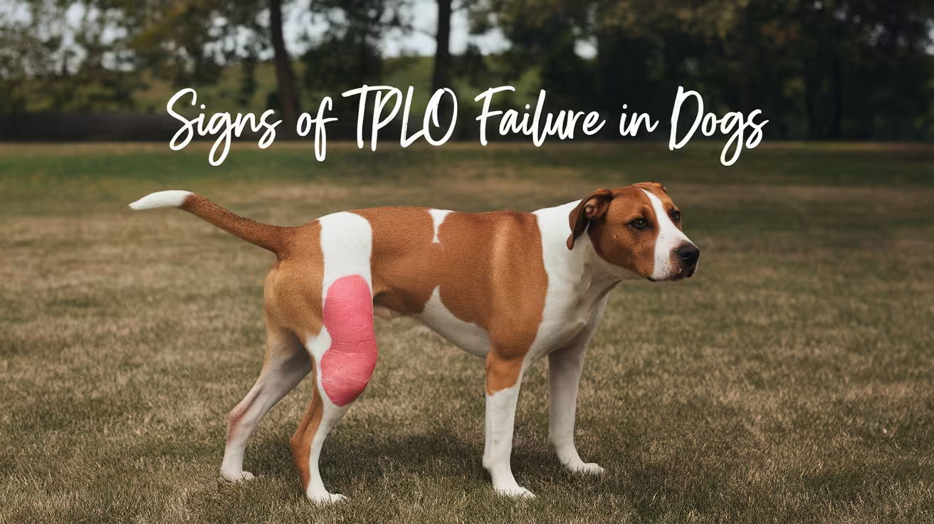
Signs of TPLO Failure in Dogs & When to See a Vet
Learn the key symptoms of TPLO failure in dogs, from limping to implant issues. Spot warning signs early and know when to seek veterinary care
TPLO failure is not a single event -- it is a spectrum of complications that can occur at different stages of recovery. Some signs are emergencies requiring same-day veterinary contact.
Others are patterns that require evaluation at the next scheduled appointment.
Understanding which signs fall into which category -- and what each sign most likely indicates -- allows owners to respond appropriately rather than either ignoring a serious complication or panicking unnecessarily.
Quick answer: Key signs of TPLO failure: persistent or worsening lameness beyond the expected trajectory; sudden acute non-weight bearing; swelling, warmth, or discharge at the plate site; clicking or popping with pain; and radiographic changes at follow-up. Call your vet the same day for sudden acute lameness, spreading infection signs, or systemic illness.
Key takeaways
- The key diagnostic principle is trajectory: normal TPLO recovery shows steady week-over-week improvement; any reversal -- worsening lameness, new swelling, returning pain -- signals a problem
- Persistent lameness beyond expected recovery is the most common sign of TPLO failure: continued limping or inability to bear weight beyond the expected level weeks after surgery is the hallmark symptom
- Sudden acute non-weight bearing after improvement is a same-day emergency call: causes include plate failure, tibial fracture, late meniscal tear, or acute infection; radiographs are required urgently
- Swelling at the plate site weeks after surgery indicates deep implant infection: this is distinct from normal post-surgical swelling and represents a late-developing implant-associated SSI
- Clicking or popping with pain or lameness indicates meniscal injury: isolated early clicking without lameness is usually benign; clicking with pain or worsening lameness indicates meniscal tear
- Radiographic confirmation is required to diagnose most causes of TPLO failure: X-rays confirm the specific mechanism and guide management
The recovery trajectory principle
The single most useful concept for identifying TPLO failure is trajectory.
Recovery from TPLO is not linear -- there are good days and less good days -- but the overall trend over weeks should be consistently improving.
Normal trajectory: each week, the dog uses the leg a little more freely, lameness is slightly less obvious, weight bearing is more consistent.
Abnormal trajectory: lameness worsens after a period of improvement; the dog is using the leg less at week 4 than at week 2; the dog that was consistently toe-touching now holds the leg entirely off the ground.
Trajectory reversal is the key signal. Single-day fluctuations can occur with activity and weather. Week-over-week regression cannot be dismissed.
Signs of TPLO failure: by type
1. Persistent or progressive lameness
What it means: the dog is limping beyond the expected level for the stage of recovery. At 4 weeks, some lameness is expected. At 8 weeks, most dogs should be consistently bearing significant weight. At 12 weeks, most are nearly normal.
Differential: implant loosening, delayed bone healing, non-union, deep infection, late meniscal tear, progressive arthritis.
When to call: if lameness has not improved for 2 or more weeks at any stage, or has worsened compared to the previous week -- contact your vet for evaluation before the next scheduled appointment.
SustainableVet.org confirms: persistent lameness is the hallmark symptom of TPLO failure; continued limping or inability to bear weight weeks after surgery may indicate failure.
2. Sudden acute non-weight bearing
What it means: the dog was bearing weight adequately and suddenly stops using the leg entirely. This is a distinct clinical event, not a gradual worsening.
Differential: acute plate failure (plate bending or screw pullout), acute tibial tuberosity fracture, acute late meniscal tear, acute hematogenous implant infection.
When to call: same-day emergency veterinary contact. Radiographs are required urgently to determine the cause.
SustainableVet.org confirms: a failed TPLO often presents as persistent pain, joint instability, swelling, or difficulty bearing weight; dogs may show progressive lameness or reluctance to move.
3. Swelling at the plate site (not the incision)
What it means: a distinct swelling appearing over the medial tibial surface at the plate location, separate from and later than the normal post-surgical incision swelling. This is a late-developing sign, often appearing weeks to months after surgery.
Differential: deep implant-associated infection (SSI-D) with abscess formation; rarely, a non-infectious reaction to the implant material.
When to call: same-day if accompanied by warmth, redness, or discharge. Within 24 hours if soft and non-painful but new and increasing.
4. Wound discharge or draining tract
What it means: a draining tract opening near (but not necessarily at) the original incision, often appearing weeks or months after surgery in a wound that appeared fully healed. This is a classic sign of deep implant-associated infection with sinus tract formation.
When to call: same-day. Do not wait.
5. Clicking or popping with pain or lameness
What it means: a meniscal click -- the torn meniscal flap being displaced by the femoral condyle during movement. Distinguished from benign joint adjustment clicking by the presence of pain on stifle manipulation and lameness.
Differential: late meniscal tear (2 to 22% incidence after TPLO).
When to call: same-day if acute lameness accompanies the click. Within 24 to 48 hours for new clicking with mild lameness.
See the dedicated meniscal tear article for full detail on diagnosis and treatment.
6. Radiographic findings at follow-up imaging
What it means: the 6-week and 12-week radiographs may show: peri-implant radiolucent halos (implant loosening or infection), widened osteotomy gap without callus formation (delayed union or non-union), plate migration from original position, screw in joint space, or cortical erosion (osteomyelitis).
When to call: your vet interprets these radiographs and will advise on urgency. Do not wait for the next scheduled appointment if the vet identifies a significant radiographic abnormality.
7. Systemic signs of infection
What it means: fever (rectal temperature above 39.5 C / 103.1 F), lethargy, reduced appetite, or behavioral changes. These indicate the infection has systemic involvement.
When to call: same-day emergency contact. Systemic infection from an implant site is a serious condition.
What is not TPLO failure
Not every deviation from ideal recovery represents failure.
Normal: gradual lameness that is slowly improving week over week.
Normal: mild swelling at the incision that peaks at days 2 to 5 and resolves.
Normal: mild stiffness in the morning that improves with movement.
Normal: intermittent toe-touching or three-legged walking during high-activity periods in the first 4 weeks.
Normal: a dog that is more lame on a day after increased activity compared to a day of rest.
The distinguishing feature is trajectory and pattern. Gradual improvement with day-to-day variation is normal. Reversal of the improvement trend is not.
Revision TPLO: when failure requires re-operation
SustainableVet.org confirms: revision TPLO may involve removing or replacing implants, repositioning the tibial cut, or addressing infections; revision surgery is more complex and requires strict post-op care.
The specific revision approach depends on the cause:
- Implant failure without infection: plate replacement or addition of a fixation device
- Non-union: bone grafting, implant revision, and extended restriction
- Deep infection with healed osteotomy: plate removal, surgical debridement, prolonged antibiotics
- Osteomyelitis: plate removal, debridement, 4 to 6 months of culture-directed antibiotics
For the complication list, see 15 common complications after TPLO surgery. For the implant failure guide, see TPLO implant failure signs and causes.
For the infection guide, see TPLO plate infection signs and treatment. For the meniscal tear guide, see dog meniscus tear after TPLO surgery.
Frequently asked questions
My dog is limping at week 6 but the 6-week radiograph was normal. Is this TPLO failure?
Not necessarily.
Normal radiographs at 6 weeks with persistent lameness suggests the issue is soft-tissue rather than bone or implant related -- meniscal injury, muscle atrophy, scar tissue, or rehabilitation lagging behind bone healing.
Discuss with your vet: physiotherapy and pain management are typically the next step for this presentation.
What happens if TPLO failure is not recognized early?
Delayed recognition of implant infection leads to establishment of deeper infection and biofilm, requiring more complex treatment. Delayed recognition of non-union leads to fibrous union or permanent instability.
Delayed recognition of meniscal tear allows progressive joint damage. Early action is consistently associated with better outcomes across all causes of TPLO failure.
Is TPLO failure common?
Overall TPLO complication rates are 10 to 34%, but most complications are minor (superficial infection, seroma). Serious failure requiring revision surgery is uncommon.
The 2013 study confirmed 93% limb function restoration at 1 year -- meaning the vast majority of TPLO dogs recover successfully.
Can TPLO failure happen years after surgery?
Yes. Late implant-associated infection (hematogenous seeding from dental disease, UTI, or skin infection) can occur years after surgery. The hallmark is sudden lameness regression in a dog with previously normal function.
Any unexplained lameness in a TPLO dog warrants radiographic assessment.
Should I ask for radiographs at every sign of lameness?
Not necessarily for every minor fluctuation.
Radiographs are indicated for: sudden acute lameness, lameness that has not improved for 2 or more weeks, new swelling at the plate site, a draining tract, or any systemic sign of infection.
Your vet will advise on imaging timing based on clinical presentation.
Resources
- SustainableVet. TPLO Failure Symptoms. sustainablevet.org
- Clinician's Brief. Common Tibial Plateau-Leveling Osteotomy Complications. cliniciansbrief.com
- SustainableVet. TPLO Failure Rate in Dogs. sustainablevet.org
- ACVS. Cranial Cruciate Ligament Disease. acvs.org
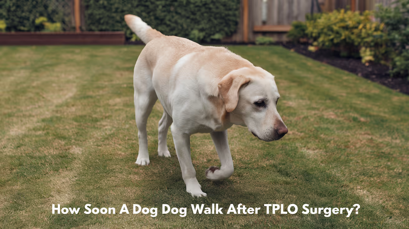
How Soon Can a Dog Walk After TPLO Surgery?
Learn when your dog can safely start walking after TPLO surgery and how to support recovery with proper care and guidance.
TPLO (Tibial Plateau Leveling Osteotomy) surgery is one of the most common and successful procedures performed to stabilize the knee after a cranial cruciate ligament (CCL) tear in dogs.
While many pet parents expect their dog to immediately stop limping after surgery, recovery is a gradual healing process. It requires structure, careful activity restriction, and close adherence to surgical recommendations to achieve the best long-term outcome.
At SustainableVet, we emphasize that recovery is just as important as the surgery itself. The goal after TPLO surgery is not simply to get your dog walking quickly. It is to allow the bone and soft tissues to heal properly while safely rebuilding strength, comfort, and confidence in the limb.
When Can a Dog Start Walking After TPLO Surgery?
One of the most common questions after surgery is how soon dogs can walk again. The short answer: dogs are encouraged to begin controlled limb use early in recovery, but activity must remain extremely restricted while the bone heals.
We want your dog to use the operated leg, but not overuse it.
Most dogs will begin toe-touching on the operated leg within the first several days after surgery. Limping is expected during the early recovery period. What we want to see is gradual improvement over time:
- More consistent weight bearing
- Improvement in muscle mass
- Improved comfort during controlled, regimented leash walks
While early limb use is encouraging, it is crucial to understand that the bone itself is still healing internally. That process typically takes 8 to 12 weeks.
TPLO Recovery Timeline at a Glance
Indoor Recovery: 8 to 12 Weeks
During the entire recovery, exercise restriction is critical. Crate confinement or restriction to a small area with good traction is strongly recommended. Most dogs will slip and slide on hardwood floors and can easily injure themselves inside the house when not confined.
Days 1 to 10: Initial Rest Period
During the first 7 to 10 days after surgery, walking is restricted to short, slow leash walks only for bathroom breaks. These walks should be calm, controlled, and limited to what is necessary.
Days 10 to 14: First Recheck Appointment
By the first recheck appointment, many dogs are partially weight-bearing during a controlled, slow walk.
At this stage, your veterinary team will evaluate the incision, swelling, comfort level, and early limb use. Even if your dog is doing well, activity should still remain very restricted.
Weeks 2 to 8: Gradual Activity Increase
As healing progresses, 10 to 15 minute leash walks may be introduced to build more strength, reduce stiffness, and encourage bony healing without overloading the leg.
Walking activity and physical therapy are gradually increased according to your surgeon's recommendations. This stage is not about testing how much your dog can do. It is about safely rebuilding muscle mass while protecting the healing bone.
Weeks 8 to 12: Final Healing and Recheck
At the final recheck appointment, radiographs (X-rays) are typically performed to assess bone healing.
By this stage, many dogs have minimal or no lameness during controlled activity. If the osteotomy has healed adequately, your surgeon may recommend a gradual transition back toward more normal activity.
Why Strict Rest Is Required After TPLO: The Mechanics
During a TPLO procedure, a precise geometric cut is made in the tibia. The bone is then rotated into a new position and stabilized using a specialized metal plate and screws.
Until the bone heals, the plate and screws are responsible for maintaining the new bone position and protecting the osteotomy during normal controlled movement.
Excessive activity, slipping, jumping, or twisting too early can place forces on the repair that it is not yet ready to tolerate.
Even if your dog appears comfortable and eager to do more, unrestricted activity too early can create significant complications before the bone has fully healed.
Recovery after TPLO surgery is truly a situation where slow and steady wins the race.
Factors That Affect TPLO Walking Recovery Time
Recovery time depends on several individual factors. These influence how quickly walking progresses and how carefully activity must be advanced.
Postoperative Care
Strict environmental management, appropriate pain control, and structured rehabilitation directly support a safer recovery. This includes using a leash at all times outdoors, preventing stairs and jumping, keeping floors non-slip, and following medication and activity instructions closely.
Presence of Complications
Issues such as incision problems, infection, implant failure, delayed bone healing, or fracture can extend the recovery timeline and delay walking progression.
Any sudden worsening in lameness, swelling, pain, or reluctance to use the limb should be reported to your veterinary team immediately.
How to Safely Support Your Dog's Mobility During Recovery
A structured support plan improves long-term outcomes and reduces the risk of setbacks.
Use a Short Leash and Harness at All Times
All walks during recovery must remain on a short leash with a slow, controlled pace. A harness can help control movement and prevent sudden lunging or twisting.
Your dog should not be allowed to run ahead, play, greet other dogs energetically, or move unpredictably during recovery walks.
Stick to Strict Exercise Limits
Follow your veterinarian's recommended walking times and avoid any off-leash activity, running, jumping, or rough play.
Many complications occur not because a dog was walking, but because the dog suddenly slipped, jumped, twisted, or did too much too soon.
Prioritize Pain Management
Administer all prescribed pain medications on schedule. Good pain control keeps your dog comfortable and encourages more even, controlled weight-bearing during brief walks.
Do not stop medications early unless your veterinarian advises you to do so.
Incorporate Professional Physical Therapy
Gentle range-of-motion exercises may help reduce stiffness early in recovery. As healing permits, rehabilitation options such as controlled strengthening exercises, hydrotherapy, or underwater treadmill sessions may help rebuild muscle while reducing stress on the limb.
Working with a certified canine rehabilitation specialist allows for a customized plan that helps restore joint flexibility, rebuild muscle, improve comfort, and support a safe return to normal mobility.
Risks of Walking Too Soon or Too Much After TPLO Surgery
Being cautious protects the surgical repair. Allowing your dog to run, jump, slip, or play too early carries real clinical risks.
Implant Strain or Failure
Excessive weight-bearing, sudden twisting, or impact forces too early can place abnormal stress on the plate and screws. In severe cases, implants can loosen or fail, requiring additional treatment or corrective surgery.
Delayed Bone Healing
Too much motion or repeated stress at the surgical site can slow the bone's ability to heal properly, prolonging the total recovery period.
Increased Pain and Inflammation
Overexertion can cause swelling, soreness, and joint inflammation. This may make your dog less willing to use the leg and can delay functional recovery.
Setbacks in Recovery
Too much activity too soon can cause limping, soft tissue irritation, swelling around the surgical site, or a slower return to comfortable walking. In some cases, setbacks require a longer rehabilitation plan or additional veterinary care.
Frequently Asked Questions
How long should my dog be confined after TPLO surgery?
Your dog needs indoor confinement for the entire two to three months of recovery. Confinement prevents accidental running, jumping, and rough movement.
Can I let my dog run or jump after TPLO surgery?
No. Running, jumping, stair use, and off-leash activity are restricted for approximately 8 to 12 weeks. The exact timeline depends on your dog's healing and X-ray findings.
What signs show my dog is recovering well and ready for longer walks?
Positive signs include reduced swelling, steady comfort levels, consistent toe-touching, and gradual improvement in walking quality. Always confirm with your veterinary team before increasing walk duration.
Is physical therapy necessary after TPLO surgery?
Some dogs recover well with structured home care alone, but physical therapy is always an excellent adjunct to build muscle strength and improve use of the operated leg.
When can my dog return to full, normal activity?
Most dogs achieve adequate bone and soft tissue healing between 8 and 12 weeks after surgery. Full activity should only resume gradually after follow-up X-rays confirm the osteotomy has healed and your surgeon provides formal approval.
Final Thoughts
Dogs are often ready to do more before their bone is ready to tolerate more. Dogs will not moderate their own activity. That is why TPLO recovery requires a balance between early controlled limb use and strict protection of the surgical repair.
With appropriate rest, controlled leash activity, pain management, follow-up care, and rehabilitation when needed, most dogs return to comfortable, active lives after TPLO surgery.
About this Collaboration: Successful healing happens both inside and outside the operating room. This article combines structured veterinary rehabilitation guidelines with direct clinical insights from veterinary orthopedic surgeons to support pet parents through every stage of the recovery journey.
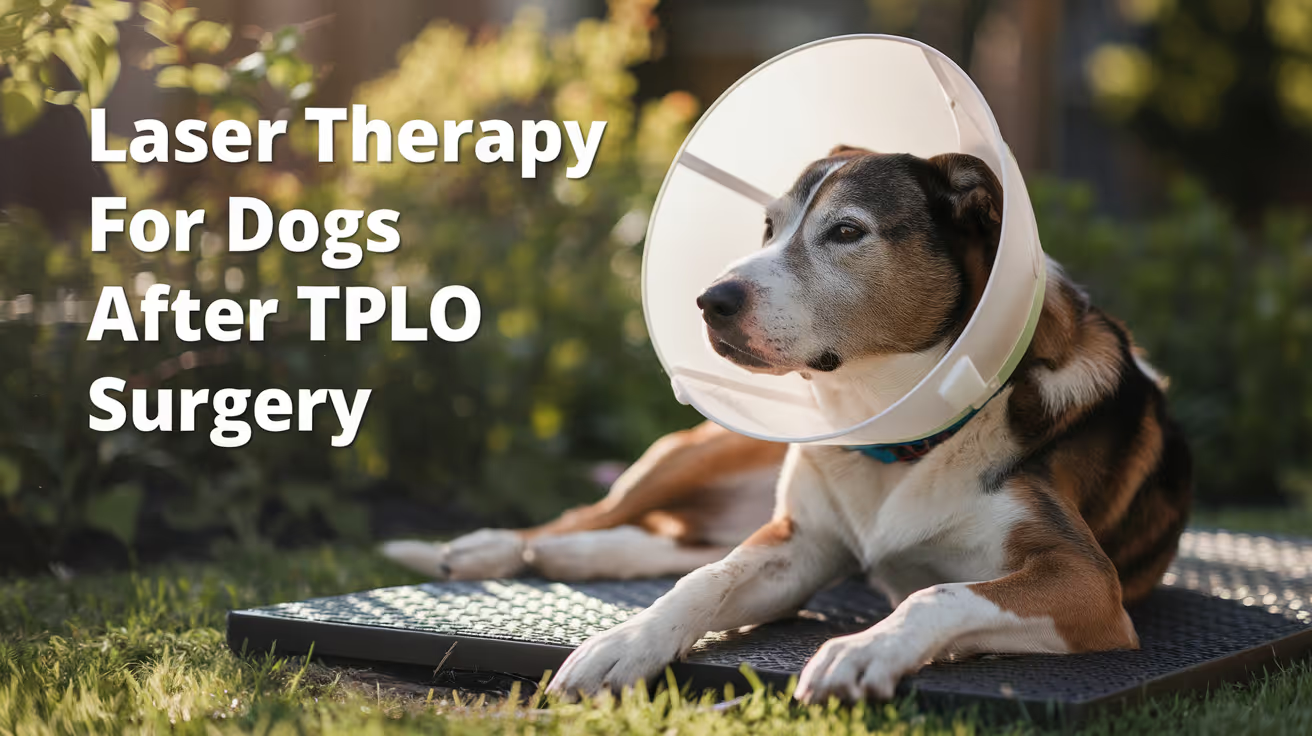
Laser Therapy for Dogs After TPLO Surgery
Learn how laser therapy helps dogs recover faster and with less pain after TPLO surgery for cruciate ligament repair.
Laser therapy also called photobiomodulation (PBMT) or low-level laser therapy (LLLT) is one of the most commonly offered adjunct treatments during TPLO recovery. Many specialist and rehabilitation centres include it routinely.
But the evidence for its benefits is more nuanced than the marketing suggests, and owners deserve an honest picture of what it does and does not reliably achieve.
Quick answer: Laser therapy after TPLO uses specific light wavelengths to reduce inflammation and support tissue healing. Evidence for early pain reduction is moderately supported; evidence for improved radiographic bone healing is weak. It is a safe adjunct but should not replace rehabilitation exercises, pain medication, or activity restriction.
Key takeaways
- Laser therapy reduces postoperative inflammation and may improve gait scores: a TPLO study found better hindlimb function at 8 weeks in treated dogs
- Evidence for improving radiographic bone healing is weak: three controlled studies found no statistically significant difference in healing time
- The 2024 AVMA randomized trial found no significant difference in CRP, weight bearing, pain scores, or SSI rates between PBMT and sham groups
- Sessions typically begin within the first few days of surgery and continue through the rehabilitation phase
- Laser therapy is safe with few contraindications: avoid eyes and active tumour sites; safe over the TPLO incision once closed
- It works best as part of a multimodal plan: exercise therapy and pain medication carry stronger evidence than laser alone
What laser therapy does
Laser therapy for dogs, also known as photobiomodulation, involves using specific wavelengths of light to penetrate tissues and promote cellular regeneration and healing. The laser light stimulates the production of ATP (adenosine triphosphate), enhancing cell repair and growth, reducing inflammation, and increasing blood circulation.
Photobiomodulation therapy has been shown to decrease inflammation, and increase analgesia, vascularization, and tissue healing after musculoskeletal injury or surgery.
The mechanism is photochemical: light energy at specific wavelengths (typically 630 to 980 nm) is absorbed by mitochondria.
This increases ATP production, modulates reactive oxygen species, and influences gene expression related to inflammation and healing.
The effects are local confined to the tissue depth the light reaches.
For post-TPLO use, the targets are: the surgical incision, the osteotomy site in the proximal tibia, and the surrounding periarticular soft tissues.
What the clinical evidence shows
Pain and function
Research following TPLO surgery showed that dogs receiving LLLT had better hindlimb function and gait scores at 8 weeks compared to controls. This is especially valuable in orthopedic recovery, where early weight-bearing can prevent muscle atrophy and joint stiffness.
In a controlled veterinary study, dogs with surgical incisions treated with laser therapy exhibited significantly less inflammatory cell infiltration and tissue necrosis within the first week post-op compared to untreated controls.
The 2024 randomized trial
54 client-owned dogs with CCL rupture undergoing unilateral TPLO surgery were enrolled. The study population was randomly assigned to either a treatment group receiving PBMT (24 dogs) or a control group (30 dogs). PBMT was performed immediately after induction, and at 6 hours, 24 hours, 48 hours, and 8 weeks postoperatively. Evaluation of CRP, pain scores, evidence of SSI, and percentage weight bearing were assessed at all time points.
The trial found the therapy showed promise but no statistically significant difference between groups on any primary outcome measure.
Bone healing
Three studies compared LLLT to a control and concluded that LLLT treatment did not make a significant difference in improving radiographic bone healing. The studies collectively provide weak evidence for this outcome.
This is an important distinction: laser therapy may support soft tissue healing, pain, and early function but it does not appear to accelerate the osteotomy healing visible on radiographs.
When to start and how often
Laser therapy uses focused light energy on the surgical site to support healing.
Most rehabilitation programmes begin laser therapy within the first 1 to 3 days after TPLO surgery, often at the surgical centre before discharge or at the first rehabilitation visit.
Typical post-TPLO laser protocol:
- Frequency: 3 to 5 sessions per week in the first 2 to 3 weeks
- Frequency: 1 to 2 sessions per week from weeks 3 to 8
- Session duration: 5 to 15 minutes depending on the laser system and dosing protocol
- Total sessions: typically 6 to 12 in the first 8-week recovery phase
The protocol varies by laser system, power output, and the individual patient's response.
Realistic expectations
Laser therapy is a useful adjunct in TPLO recovery. It is not a substitute for the treatments with stronger evidence: pain medication, activity restriction, and structured rehabilitation exercises.
Laser therapy could be particularly helpful for dogs with weight-bearing and gait issues while recovering from TPLO surgery after a cruciate injury.
Dogs with significant early swelling, wound sensitivity, or slow initial weight-bearing progress may benefit most. Dogs recovering well with standard multimodal analgesia and rehabilitation may show less measurable difference.
For the bone healing timeline that laser therapy supports during recovery, see TPLO bone healing time in dogs explained.
For the full recovery plan that laser therapy fits into, see 10 essential TPLO recovery tips for pet owners.
For the physical therapy that is the primary evidence-based adjunct, see when to start physical therapy after TPLO surgery.
For swelling management in the recovery period, see how long does swelling last after TPLO surgery.
Frequently asked questions
Is laser therapy safe over the TPLO incision?
Yes, once the incision is closed. Laser therapy is safe over sutured incisions and can be applied at the surgical site from the first post-operative day in most protocols.
Avoid direct application over open wounds or actively infected tissue.
How many laser sessions does a dog need after TPLO?
Typically 6 to 12 sessions across the first 8 weeks.
Start at 3 to 5 sessions per week for the first 2 to 3 weeks, tapering to 1 to 2 per week through the rehabilitation phase.
The exact protocol depends on the laser system and rehabilitation plan.
Does laser therapy replace pain medication after TPLO?
No. Laser therapy is an adjunct to pharmaceutical pain management, not a replacement. Post-TPLO pain management requires NSAIDs, and often gabapentin or other analgesics.
Laser therapy may reduce the pain burden and support earlier mobility but does not provide sufficient analgesia on its own.
Can I do laser therapy at home with a consumer device?
Consumer-grade red light therapy devices exist but operate at lower power densities than veterinary therapeutic lasers. The clinical evidence discussed in this article relates to veterinary-grade PBMT devices.
Home devices may offer some benefit but cannot replicate the dosing of professional equipment. Discuss with your rehabilitation veterinarian before purchasing.
Will laser therapy prevent my dog from needing more medication?
Possibly. If laser therapy reduces post-operative inflammation and pain, some dogs may need lower doses of pain medication or taper off sooner.
This should be guided by your veterinarian based on your dog's individual recovery trajectory.
Resources
- AVMA Journal. Photobiomodulation Therapy in Dogs Undergoing TPLO After Cranial Cruciate Ligament Rupture. avmajournals.avma.org
- Veterinary Evidence. Does LLLT Improve Radiographic Healing for Dogs with CCL Rupture Undergoing TPLO Surgery? veterinaryevidence.org
- AKC. Laser Therapy For Dogs: Uses, Side Effects, and Alternatives. akc.org
- Erchonia. Laser Therapy for Post-Surgical Recovery in Pets. erchonia.com

Redness After TPLO Surgery in Dogs: Causes & Care
Learn why redness occurs after TPLO surgery in dogs, how to recognize normal healing versus complications, and when to seek veterinary care.
Some redness around a TPLO incision is a completely normal part of healing.
The skin and underlying tissues respond to any surgical trauma with an inflammatory response redness is one of the visible signs of that response.
The question is always: is this normal healing redness, or is this infection?
Quick answer: Mild redness at the TPLO incision is normal for 2 to 3 days, fading over 1 to 2 weeks. Call your vet if redness spreads, is warm, produces discharge, worsens after day 3, or your dog develops fever.
Key takeaways
- Mild redness at the incision is normal for 2 to 3 days post-surgery: it should be fading by day 3 to 5
- Normal redness is localized to the incision line: redness spreading outward from the wound edge is not normal
- Infection redness is spreading, warm, and accompanied by discharge: these features together require veterinary evaluation
- Suture reaction redness is localized and itchy: the pattern is different from infection and typically resolves after suture removal
- Licking is the most preventable wound complication: saliva introduces bacteria; the E-collar must stay on until suture removal
- Redness after week 2 warrants veterinary evaluation: new redness after day 14 suggests a complication
Normal redness after TPLO surgery
The inflammatory phase of wound healing is the body's first response to surgical trauma. Blood flow increases to the wound, bringing immune cells and clotting factors.
This increased blood flow makes the skin appear pink or faintly red. This is expected and necessary for healing.
SustainableVet.org confirms: normal redness after TPLO surgery is usually mild and limited to the incision area. It should improve daily as the wound heals.
Characteristics of normal healing redness:
- Confined to the immediate edges of the incision not spreading outward
- Mild pale pink to light red, not deep red or purple
- Decreasing over time improving from day 3 onward
- Not warm to the touch (normal healing generates mild local warmth, but not the significant heat of infection)
- No discharge from the wound other than possibly a small amount of serum in the first 24 to 48 hours
- The dog is not bothered by gentle examination of the area
The redness typically peaks at 48 to 72 hours post-surgery and should be noticeably fading by the end of the first week.
Abnormal redness: signs of infection
Infection redness is different from normal healing redness in several characteristic ways:
Spreading: the redness extends outward from the incision margin into the surrounding skin. This cellulitis pattern spreading red discoloration in surrounding tissue is a sign that bacteria are spreading through the tissue planes.
Warmth: the infected area feels significantly warmer than surrounding skin or the opposite leg. Infection drives significant local heat through the inflammatory cytokine response.
Discharge: infected wounds produce discharge that may be clear-to-yellow early, progressing to yellow-green, cloudy, or frankly purulent. Foul odor is a hallmark of anaerobic bacterial involvement.
Worsening, not improving: normal redness fades. Infected redness intensifies. If redness is worse at day 5 than at day 2, infection is strongly suspected.
Systemic signs: fever (rectal temperature above 102.5 F), lethargy, loss of appetite, and reluctance to bear weight alongside redness suggest the infection is causing systemic effects.
SustainableVet.org confirms: infection risk includes bacteria entering the incision, causing redness that worsens along with discharge or odor.
The TPLO incision infection guide confirms: infection risk occurs in approximately 3 to 10% of TPLO cases. Signs of infection include redness, swelling, warmth, discharge, and delayed healing.
Other causes of post-TPLO redness
Suture or staple reaction
Some dogs develop a local inflammatory reaction to the suture material more commonly with older suture types or in dogs with sensitive skin.
This produces redness specifically along the suture line, often with mild swelling and sometimes itching (prompting the dog to lick or chew at the site).
It is localized, does not produce the spreading pattern of infection, and typically resolves after suture removal.
Contact dermatitis
Redness from a reaction to bandage material, antiseptic solution applied to the wound, or topical medications can mimic early infection. The pattern follows the contact area rather than the wound edge.
Seroma formation
A seroma that presses against the overlying skin may cause mild skin discoloration and the appearance of redness over the fluid-filled swelling. The area is soft, fluctuant, and typically not warm.
For the full seroma guide, see seroma in dogs after TPLO surgery.
What to do if you notice redness
First 3 days: normal range
Monitor daily. Take a photograph to track progression. Ensure the E-collar is on at all times. Check that no discharge or spreading is occurring.
Days 3 to 10: should be improving
If redness is not clearly fading by day 5 to 7, contact your veterinarian.
Photograph the wound to share with the vet by phone or telehealth before deciding whether an in-person visit is needed.
Do not apply any topical products to the wound without veterinary guidance antiseptics can delay healing and worsen skin reaction.
After day 10: any new or persistent redness needs evaluation
By day 10 to 14, most surgical incisions are well into the proliferative healing phase.
Persistent redness at this stage, or new-onset redness that was not present before, warrants a veterinary appointment rather than home monitoring.
Incision care during recovery
- Keep the incision dry: no bathing, no swimming, no wet grass
- E-collar on at all times until sutures are removed (typically day 10 to 14)
- Check the wound twice daily: look at it, do not touch it unless instructed
- No ointments or antiseptics unless specifically prescribed
- Contact your vet before applying anything to the wound
For the incision infection guide with full symptom detail, see TPLO incision infection symptoms and prevention. For the full complications reference, see 15 common complications after TPLO surgery in dogs.
For the seroma guide (fluid that can cause discoloration), see seroma in dogs after TPLO surgery. For the full TPLO recovery guide, see what to expect after TPLO surgery in dogs.
Frequently asked questions
How do I know if my dog's incision redness is normal?
Normal redness is mild, pink, confined directly to the wound edges, and fading by day 3 to 5.
Abnormal redness is spreading outward from the incision, deepening in color, warm to the touch, or accompanied by discharge or odor. If you are unsure, photograph the wound and contact your veterinarian.
My dog's incision looks red and my dog keeps licking it. What should I do?
Replace or refit the E-collar immediately if the dog has been reaching the wound. Even a few minutes of licking introduces bacteria.
Examine the wound for any discharge, spreading redness, or wound breakdown. If you see any of these, contact your vet the same day.
If the wound looks intact, continue monitoring closely with the E-collar secured.
Can redness appear weeks after surgery?
Yes. Late-onset redness appearing 2 to 6 weeks post-surgery often signals a developing infection rather than normal healing.
Common causes include low-grade suture infection, developing seroma, or a bacterial infection that has been slowly progressing. Any new redness appearing after the initial 2-week healing phase warrants veterinary evaluation.
Is red skin around the TPLO staples normal?
A small amount of mild pinkness immediately at the staple insertion points is normal.
Spreading redness between staple sites, significant swelling along the staple line, or discharge from staple holes is not normal and requires veterinary evaluation before the scheduled staple removal visit.
What happens if redness from infection is left untreated?
Superficial infections can progress to deeper tissue infections, then to implant-associated infections and osteomyelitis.
Laguna Hills Animal Hospital confirms that even superficial soft tissue infections have been shown to progress to implant infections and bone infections when not treated promptly and appropriately.
Resources
- SustainableVet. TPLO Incision Infection Symptoms and Prevention. sustainablevet.org
- Dial A Vet. Redness and Swelling Around TPLO Incision. dialavet.com
- Laguna Hills Animal Hospital. TPLO Surgery Complications. lhah.com
- VCA Animal Hospitals. Tibial Plateau Leveling Osteotomy (TPLO) in Dogs. vcahospitals.com
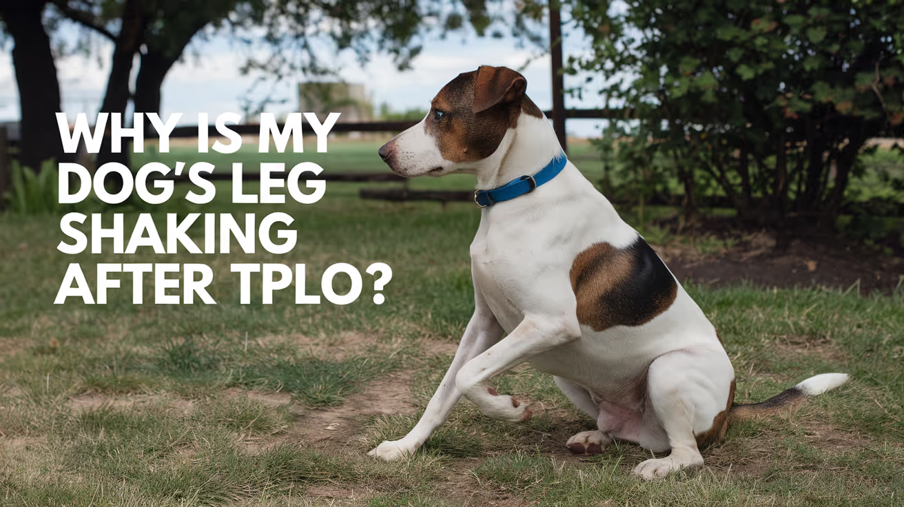
Dog Leg Shaking After TPLO Surgery: Causes & Care
Learn why your dog may shake its leg after TPLO surgery, what to expect during recovery, and how to support healing safely.
Seeing your dog's leg shake or tremble after TPLO surgery is alarming. In most cases, it is a completely normal part of recovery.
In some cases, it signals a problem that needs attention. Knowing the difference and knowing what to watch for reduces unnecessary worry and helps you catch real complications early.
Quick answer: Leg shaking after TPLO is usually from muscle weakness, nerve recovery, pain, or residual anesthesia. TPLO Info confirms this is normal. Call your vet if shaking worsens, comes with fever or swelling, or persists beyond 2 to 3 weeks.
Key takeaways
- Leg shaking immediately after TPLO is most often from residual anesthesia, cold, or pain; TPLO Info confirms this is normal
- Muscle weakness is the most common ongoing cause: muscles around the knee need weeks of rehab to recover strength
- Nerve recovery causes tremors as disrupted signals re-establish; this resolves over weeks to months
- Inadequate pain control is a key contributing factor: shaking that worsens or persists often means current pain medication is insufficient
- Cold and hypothermia after surgery cause shivering that can mimic leg-specific shaking small dogs and lean breeds are most susceptible
- Warning signs: shaking with fever, surgical site swelling, discharge, worsening lameness, or refusal to bear weight warrants a vet call
Why do dogs' legs shake after TPLO surgery?
Residual anesthesia effects (first 24 to 48 hours)
The most common reason dogs shake immediately after surgery is residual anesthesia. As anesthetic drugs wear off, the body compensates by increasing muscle activity to generate heat this produces shivering and trembling.
TPLO Info explains that once anesthesia wears off, the dog's body induces heat by contracting muscles.
This side effect typically resolves within 12 to 24 hours, though some dogs may tremble for a few days.
Nausea is another anesthesia side effect that produces shaking. A dog that is shaking and yawning or repeatedly swallowing is likely experiencing post-anesthetic nausea.
Muscle weakness and atrophy
TPLO surgery involves cutting through and retracting the muscles surrounding the tibial osteotomy site. These muscles are weak immediately after surgery and require weeks of progressive rehabilitation to regain their pre-surgical strength.
Weak muscles produce involuntary tremors when asked to support body weight or during movement especially on an uneven surface, during transitions between positions, or after brief walks.
This type of shaking is most visible when the dog is standing or trying to move, and improves progressively as rehabilitation exercises build muscle mass.
Dr. Altman on JustAnswer confirms: muscles are disrupted during the TPLO procedure, and there will be a period of healing and weakness that can include muscle spasms.
Massage and warm compresses help, and PROM exercises are beneficial.
Nerve recovery
The surgical procedure disrupts nerves around the tibial osteotomy site and the surrounding soft tissues.
As these nerves regenerate and re-establish normal signal pathways, they can produce involuntary muscle twitches and tremors similar to the sensation of a leg "falling asleep" in humans.
Nerve-related shaking typically affects the area distal to the surgical site and may be more noticeable at certain times of day or after periods of rest.
It is a normal healing process that can continue for weeks to months as nerve regeneration proceeds.
Pain and discomfort
Pain is a significant driver of leg trembling after TPLO. The body responds to pain by increasing muscle tension and guarding the affected area, which can manifest as visible shaking.
Pets Care notes that if shaking is accompanied by shallow breathing, reluctance to move, a distant or glazed look, or loss of appetite, inadequate pain control is likely.
Contact your veterinarian promptly if you suspect your dog's prescribed medications are not adequately controlling pain.
Cold and hypothermia
Small dogs and lean breeds lose body heat rapidly after surgery. Both the surgical room and recovery ward are cooler than most homes, and sedated dogs cannot thermoregulate effectively.
Whole-body or leg-specific shivering after surgery is often simply the dog being cold.
Ensure your dog has a warm, draft-free recovery area. Soft bedding and a light blanket (not heated pads, which risk contact burns on sedated dogs) help restore body temperature.
Anxiety and stress
Major surgery is a stressful experience.
Being in an unfamiliar environment, wearing an E-collar, restricted movement, and the general disruption of normal routine all cause stress that can manifest as trembling or restlessness.
This type of shaking is typically whole-body rather than isolated to the leg, and improves as the dog settles into a home routine.
How to help a dog with post-TPLO leg shaking
Warm, quiet recovery space: create a confined, comfortable, draft-free area with soft bedding away from household noise and activity.
Ensure pain medication is given on schedule: pain medications prescribed by your surgeon should be given at the prescribed times. Do not skip doses consistent blood levels prevent pain spikes that trigger shaking.
Gentle massage: massaging the muscles above and below the knee reduces muscle spasm and improves local circulation. Dr. Altman recommends massage to reduce spasms in the post-TPLO leg.
Warm compresses: after the first 5 days (when ice packs are used), warm compresses before PROM exercises relax tight muscles and reduce tremors.
Progressive rehabilitation: muscle weakness resolves with structured rehabilitation. As muscles strengthen through controlled exercises, trembling decreases. See TPLO rehab exercises for dogs for the full rehabilitation guide.
When to contact your veterinarian
Contact your vet if leg shaking is accompanied by:
- Fever or lethargy
- Increased swelling, redness, or discharge at the surgical site
- Worsening lameness or sudden refusal to bear any weight
- Muscle tremors that are severe, rhythmic, or involve the whole body (which could indicate neurological causes unrelated to TPLO)
- No improvement in shaking after 2 to 3 weeks despite adequate pain management
For the complete TPLO recovery guide, see what to expect after TPLO surgery in dogs. For incision and infection warning signs, see TPLO incision infection symptoms and prevention.
For the lameness reference guide that helps distinguish shaking from other post-TPLO problems, see lameness after TPLO surgery in dogs.
For pain management guidance, see dog whining after TPLO surgery: causes and care.
Frequently asked questions
How long does leg shaking last after TPLO surgery?
Anesthesia-related shaking resolves within 12 to 48 hours. Shaking from muscle weakness and nerve recovery typically decreases over 2 to 8 weeks as rehabilitation progresses.
Mild intermittent trembling during activity may persist for up to 3 months in dogs with significant muscle atrophy before surgery.
Is my dog in pain if their leg is shaking?
Possibly. Pain is one of the causes of post-TPLO shaking.
Signs that pain is contributing: the dog refuses to bear weight, has a glazed or distant expression, breathes shallowly at rest, or stops eating.
If you suspect inadequate pain control, contact your veterinarian to discuss adjusting the medication protocol.
My dog's leg shakes only when they stand up. Is that normal?
Yes. Shaking during transitions lying to standing, or during early walking is typical of muscle weakness after TPLO. The muscles are not yet strong enough to support smooth, stable movement against gravity.
This improves progressively with structured rehabilitation.
Should I restrict my dog's activity more if they're shaking?
Strict rest is already required for TPLO recovery. The short controlled leash walks that are prescribed are beneficial they stimulate muscle recovery without stressing the osteotomy.
Do not add more exercise because of shaking, but also do not restrict below the prescribed protocol.
Can I give my dog anything to stop the shaking?
Not without veterinary guidance. The appropriate response to pain-related shaking is to contact your vet about pain medication adjustments. For cold-related shaking, warmth and comfort are the solution.
Never give human pain medications ibuprofen and acetaminophen are toxic to dogs.
Resources
- TPLO Info. What to Do If Your Dog's Leg Is Shaking After ACL Surgery. tploinfo.com
- Pets Care. Understanding Dog Shaking After Surgery. petscare.com
- Simon Veterinary Surgical. Dog TPLO Surgery Recovery Tips. simonvetsurgical.com
- VCA Animal Hospitals. Tibial Plateau Leveling Osteotomy (TPLO) in Dogs. vcahospitals.com

Common Myths About TPLO Surgery Explained
Discover the truth about TPLO surgery for dogs as we debunk common myths and provide evidence-based insights for informed pet care decisions
TPLO is the most commonly performed surgery for CCL rupture in dogs and the most commonly misunderstood.
Misconceptions about TPLO cause unnecessary hesitation, lead some owners to choose inferior alternatives, and create unrealistic expectations about recovery.
This guide addresses the 10 most common myths directly -- with evidence-based clarification for each.
Quick answer: The most persistent TPLO myths: that dogs can manage without surgery, that TPLO is only for large breeds, that recovery takes a few weeks, that the plate must be removed after healing, and that TPLO prevents arthritis. All are false. TPLO has a 90 to 95% success rate.
Key takeaways
- Myth: "My dog can heal without surgery": dog knee anatomy differs fundamentally from human; without surgery, severe OA, meniscal tearing, and chronic lameness are expected in most dogs over 15 kg
- Myth: "TPLO is only for large dogs": SustainableVet.org confirms TPLO is just as effective for small and toy breeds; the biomechanical correction is appropriate regardless of size
- Myth: "Recovery only takes a few weeks": bone healing takes 8 to 12 weeks; full muscle recovery takes 4 to 6 months; 8 to 12 weeks is the minimum restriction period
- Myth: "The plate needs to be removed after healing": the TPLO plate is designed to be permanent; removal is only performed when complications arise
- Myth: "TPLO prevents arthritis": all CCL-affected stifles develop progressive OA regardless of surgical technique; TPLO reduces OA progression but does not prevent it
- Myth: "Dogs are in severe pain after TPLO": SustainableVet.org confirms this is a misconception; modern multi-modal pain management controls post-surgical pain effectively
Myth 1: "My dog can heal without surgery"
The truth: dogs and humans have fundamentally different knee anatomy. In humans, the flat tibial plateau and strong surrounding musculature allow some CCL tears to be managed conservatively with physical therapy and bracing. In dogs, the tibial plateau slopes steeply downward, generating a cranial shear force that the absent CCL cannot resist. Without surgical correction of this force, the joint remains mechanically unstable with every weight-bearing step.
TPLO Info confirms: dogs and humans respond very differently to CCL tears. Without surgery, severe OA can develop, meniscal tearing can occur, and other side effects may be present.
Conservative management (strict rest, weight management, joint supplements, physical therapy) can provide temporary improvement, particularly in small dogs and partial tears.
For most dogs over 15 kg with complete CCL rupture, conservative management does not restore normal joint stability, and progressive lameness, OA, and joint deterioration are the expected outcome.
Who can be managed conservatively?
- Very small dogs (under 10 to 15 kg) with partial tears and low activity levels
- Dogs with significant comorbidities where anesthetic risk outweighs surgical benefit
- Owners who decline surgery after being fully informed of expected outcomes
For all other dogs -- medium and large breeds, young dogs, active dogs, complete ruptures -- surgery produces substantially better outcomes than conservative management.
Myth 2: "TPLO is only for large dogs"
The truth: TPLO was originally developed for large and giant breeds because they were the most severely affected by CCL disease and the most poorly served by lateral suture stabilization. But TPLO corrects the underlying biomechanical problem (excessive tibial plateau angle) in any dog, regardless of size.
SustainableVet.org confirms: TPLO was initially developed for large dogs due to biomechanical challenges, but has been found just as effective for small and toy breeds.
For small dogs, both TPLO and lateral suture stabilization can achieve good outcomes.
The decision for a small dog should be based on the dog's TPA, activity level, and individual assessment -- not a blanket assumption that TPLO is unnecessary.
Myth 3: "TPLO recovery only takes a few weeks"
The truth: bone healing takes 8 to 12 weeks. This is the period of activity restriction. But bone healing and functional recovery are not the same thing. Full muscle rebuilding and joint function restoration takes 4 to 6 months from surgery.
Canada Vet Express confirms: recovery is gradual; most dogs begin walking within a few days and return to normal activity within 3 to 4 months with proper care.
The 8 to 12 week bone healing timeline means:
- Activity restrictions are in place for 8 to 12 weeks minimum
- Radiographic confirmation of healing is required before restrictions are lifted
- Progressive return to full activity follows the radiographic clearance
After restrictions are lifted, the operated leg continues to rebuild strength and coordination for months. For sport dogs and working dogs, the return-to-sport protocol extends to 7 to 9 months from surgery.
Myth 4: "The TPLO plate needs to be removed after the bone heals"
The truth: the plate is designed to remain in place permanently. It does not need to be removed when the osteotomy heals. Routine plate removal is not standard practice in veterinary orthopedics.
SustainableVet.org confirms: these implants are meant to stay in place permanently, but in rare cases they can cause problems; removal is only performed when complications arise.
When plate removal is performed:
- Deep implant-associated infection requiring source removal
- Implant failure (breakage, loosening causing instability)
- Persistent implant irritation producing clinical lameness specifically attributable to the plate
In the vast majority of dogs, the plate is never removed. It remains in place for life with no adverse effects.
Myth 5: "TPLO prevents arthritis"
The truth: TPLO reduces and slows arthritis progression. It does not prevent it. All CCL-affected stifles develop progressive OA regardless of surgical technique or outcome.
SustainableVet.org confirms: mild arthritis is expected even in successful cases.
Published long-term data (TPLO Info, citing long-term retrospective studies) confirms moderate but significant OA progression in all TPLO cohorts with follow-up extending to 6.8 years.
TPLO produces substantially less OA progression than TTA or lateral suture stabilization, but OA progression is universal.
The appropriate expectation: TPLO minimizes OA development and clinical impact. Long-term management (weight management, exercise modification, joint supplements, and periodic NSAID use for flares) is required in all TPLO dogs.
Myth 6: "Dogs are in severe pain after TPLO"
The truth: modern veterinary anesthesia and pain management protocols produce well-controlled post-operative pain in TPLO dogs. Dogs typically begin bearing some weight within 24 to 48 hours. Pain that is not controlled is a clinical problem requiring medication adjustment -- not an expected feature of TPLO recovery.
SustainableVet.org confirms: it is a misconception that dogs experience severe pain after TPLO surgery; modern multi-modal pain management controls post-surgical pain effectively.
Post-operative pain management typically includes: NSAIDs (carprofen, meloxicam), gabapentin, opioids during hospitalization, local anesthetic nerve blocks at surgery.
Dogs in adequate pain control rest comfortably, bear some weight within days, and show appetite by day 3.
Uncontrolled post-operative pain (dog cannot settle, crying, refuses all weight bearing beyond day 3) indicates a need for pain medication adjustment -- contact your vet rather than accepting it as inevitable.
Myth 7: "TPLO is too risky for most dogs"
The truth: TPLO has a 90 to 95% success rate. The complication rate across all studies is 10 to 34%, but the vast majority of complications are minor (superficial infection, seroma, mild swelling). Serious complications requiring revision surgery occur in fewer than 10% of cases.
Dispomed confirms: overall complication rates are 14 to 34%, but a second surgery is required in less than 10% of cases.
No surgery is without risk.
But for a dog with a torn CCL, the risk of TPLO is substantially lower than the certainty of progressive joint deterioration, pain, and reduced quality of life without surgical treatment.
Myth 8: "TPLO is only for show dogs or working dogs"
The truth: TPLO is appropriate for any dog with CCL rupture that will benefit from restored joint stability -- including middle-aged household pets with no competitive goals.
SustainableVet.org confirms: some pet owners mistakenly believe that TPLO surgery is only for show dogs or high-performance athletes; this is not true; TPLO benefits any dog with CCL disease.
The goal of TPLO is freedom from joint pain and restored ability to perform normal daily activities -- walking, playing, using stairs, sitting and rising comfortably.
These are benefits for every dog, not just sport dogs.
Myth 9: "All dogs with CCL tears need TPLO immediately"
The truth: TPLO is not always an emergency. The timing of surgery should be guided by the veterinary assessment. Partial tears in small dogs may be monitored with conservative management. Dogs with significant concurrent illness may benefit from medical optimization before surgery. The appropriate goal is surgical timing that optimizes anesthetic risk and surgical conditions, not surgical urgency at any cost.
SustainableVet.org confirms: while TPLO surgery is the best treatment for CCL tears, it isn't necessary for every case; for mild or partial tears in less active dogs, alternatives can be effective.
However, in dogs with complete rupture, particularly larger breeds, delay generally produces worse outcomes as OA progresses and the meniscus is exposed to ongoing mechanical insult from the unstable joint.
Myth 10: "If TPLO fails, the dog will never recover"
The truth: most TPLO complications are treatable. Superficial infections resolve with antibiotics. Late meniscal tears are treated with partial meniscectomy. Implant failures can be revised. Osteomyelitis, the most challenging complication, requires prolonged treatment but is often successfully managed.
SustainableVet.org confirms: revision TPLO surgery can be performed if the initial procedure fails; this may involve removing or replacing implants, repositioning the tibial cut, or addressing infections.
The critical factor in outcome from a complication is early detection and prompt treatment. Most complications that are identified and treated early resolve without long-term sequelae.
For the full TPLO overview, see what is TPLO surgery in dogs?. For the success rate data, see TPLO failure rate in dogs. For recovery, see what to expect after TPLO surgery.
For alternatives, see alternatives to TPLO surgery for dogs.
Frequently asked questions
Is TPLO actually the best treatment for CCL rupture?
For most dogs over 15 to 20 kg, TPLO is the most evidence-supported surgical option, producing the highest rates of long-term function and owner satisfaction in the published literature.
For small dogs, lateral suture stabilization achieves comparable results at lower cost. The choice depends on the individual dog's size, activity level, TPA, and health status.
Can I wait and see how my dog does without surgery?
You can choose conservative management.
The expected outcome in most dogs over 15 kg with complete CCL rupture who do not have surgery is progressive lameness, rapidly advancing OA, ongoing meniscal damage, and reduced quality of life.
Early surgical intervention consistently produces better long-term outcomes than delayed or non-surgical management.
Is the recovery really as strict as described?
Yes. The restriction protocol exists because the plate holds the osteotomy until new bone consolidates -- a process that takes 8 to 12 weeks regardless of how well the dog appears.
A single running or jumping episode before healing is confirmed can displace the plate and require revision surgery.
Will my dog be the same dog after TPLO?
Most owners report their dog is effectively the same dog -- or better than before the CCL rupture -- within 6 to 12 months of surgery.
The 90 to 95% success rate reflects a high proportion of dogs returning to their previous activity level.
Some dogs develop mild age-related stiffness years later as OA progresses, but this is manageable with appropriate care.
Does TPLO cure the CCL problem permanently?
TPLO permanently corrects the biomechanical cause of CCL-related instability in the operated stifle. It does not repair or replace the CCL -- it makes the CCL unnecessary.
It does not prevent CCL disease in the contralateral stifle.
Resources
- SustainableVet. Common Myths About TPLO Surgery Explained. sustainablevet.org
- TPLO Info. 5 Misconceptions About TPLO Surgery. tploinfo.com
- Dispomed. TPLO Surgery in Dogs: Success Rates and Alternatives. dispomed.com
- ACVS. Cranial Cruciate Ligament Disease. acvs.org

