

Protecting
Pets, People & Planet
Join a group of veterinarians leveraging the latest technologies to deliver excellent care to their patients while being a responsible and positive force for their local and global communities.
100% secure. We do not share your information


Recent Articles
Why Is My Dog's Foot Swollen?
Discover why your dog's foot is swollen, common causes, treatments, and when to see a vet for proper care.
A swollen dog foot is one of the most common reasons owners call their vet. It can be as simple as a bee sting or as serious as a deep infection or bone tumor. The location, speed of onset, and other symptoms all help narrow down the cause.
This guide covers every common cause, how to assess severity at home, and the exact signs that mean your dog needs same-day care.
Quick answer: A swollen dog foot usually has a local cause: injury, insect sting, foreign object, or infection. Warm, increasing, or discharging swelling needs same-day vet care. Multiple swollen paws suggest allergies.
Key takeaways
- Single-paw swelling almost always has a local cause: injury, sting, foreign body, or infection
- Interdigital furunculosis (infected hair follicle cysts between toes) is among the most misidentified causes of recurring paw swelling
- Foreign objects like foxtails and glass splinters can migrate deep into tissue if left untreated
- Pododermatitis is the clinical term for inflamed paw skin; allergies are the most common underlying driver
- Swelling spreading up the leg or paired with face or neck swelling is an emergency requiring immediate care
- Most mild swelling from a sting or minor sprain improves within 24 to 48 hours with rest and monitoring
How to assess your dog's swollen foot before calling the vet
Before looking up causes, do a quick home assessment. This takes two minutes and tells you how urgent the situation is.
Step 1: Which paw?One paw = likely local cause. All four paws = likely allergic or systemic.
Step 2: Feel for heat.Warm or hot tissue means active inflammation or infection.
Step 3: Look between every toe.Redness, swelling between the digits, or visible nodules between the toes points to interdigital furunculosis or a foreign body.
Step 4: Check the pads.Burns appear as red, blistered, or peeling pad tissue. Cuts and punctures are often visible on the pad surface.
Step 5: Look at each nail.A broken nail or swollen nail bed causes localized pain and swelling at the toe tip.
VCA Animal Hospitals: "A deeper infection, like an abscess, will appear as a warm, soft to mildly firm swelling under the skin."
8 common causes of a swollen foot in dogs
1. Injury or trauma
Sprains, cuts, fractured toes, torn nails, and pad burns are the most frequent causes of sudden single-paw swelling. Dogs running on rough, hot, or icy terrain are especially prone.
PetMD notes that pad burns are particularly common in summer: check the pads after any walk on hot pavement. If your palm cannot hold against the pavement for five seconds, it is too hot for your dog's feet.
Signs: sudden lameness after activity, localized swelling, visible wound or bruising.
2. Foreign object
Thorns, glass, foxtails, and grass awns lodge between the toes or penetrate the pad. The body mounts an inflammatory response around the object, causing swelling that worsens over days.
SpectrumCare: "Foxtails and grass awns can lodge between the toes or in the pad and trigger pain, swelling, and infection."
Foxtails are particularly dangerous because they are barbed and can migrate deeper into tissue over days, eventually requiring surgical removal. If you cannot see and safely remove the object, do not probe. See a vet.
3. Insect sting or bite
Bees, wasps, fire ants, and spiders cause rapid single-paw swelling that appears within minutes of the sting. The paw may look puffy and your dog may lick or hold it up.
Dyer Animal Clinic advises watching for anaphylaxis signs: hives, difficulty breathing, excessive swelling spreading beyond the paw. These require emergency care immediately.
Localized sting swelling that stays in the paw and is not worsening can be monitored at home for 24 to 48 hours.
4. Infection: bacterial and fungal
Bacterial or fungal infections enter through cuts, puncture wounds, or damaged skin. Infected paws are typically warm, red, swollen, and often have an odor or discharge. VCA lists the visual signs: "Skin infections on the feet may result in red, moist lesions between the toes."
Pododermatitis (inflammation of the paw skin) is the umbrella clinical term. Common causes include:
- Secondary bacterial infection from chronic licking
- Yeast overgrowth in skin folds between toes
- Fungal infections (ringworm can affect paws)
- Demodectic mange (mite overgrowth)
Pododermatitis does not resolve without treatment. Antibiotics, antifungal medication, or both are typically required.
5. Interdigital furunculosis
This is one of the most commonly missed causes of recurring paw swelling. Interdigital furunculosis occurs when hair follicles between the toes become infected, forming painful reddish-purple nodules that may rupture and drain.
AKC notes that the condition is especially prevalent in short-coated, heavyset breeds: Bulldogs, Labrador Retrievers, and Chinese Shar-Pei are among the most commonly affected. Chronic licking due to allergies is a major driver.
Signs: swollen, painful nodules between the toes, draining tracts, recurrent swelling in the same location.
For how furuncles between the toes connect to abscess formation, see abscess as a cause of limb swelling.
6. Allergic reaction and pododermatitis
Environmental allergens (pollen, grass, lawn chemicals, road salt) and food allergies frequently cause paw inflammation. Dogs with allergies lick their feet chronically, creating secondary infection on top of the allergic reaction.
Wakefield Pet Vet: "Allergic reactions typically cause itching, redness, and sometimes blistering between the toes or on paw pads."
Allergic paw swelling usually affects multiple paws. It tends to recur seasonally or after contact with the trigger substance. Dogs with seasonal allergies often have their worst paw symptoms in spring and fall.
7. Bursitis
Bursae are small fluid-filled sacs that cushion joints. Repeated pressure on bony prominences, especially the elbow and hock, can cause bursitis. Affected joints may appear as soft, fluctuant swellings near a joint.
For how bursitis specifically produces foot and joint swelling, see bursitis as a common cause of foot swelling.
8. Cysts, tumors, and nail bed disease
Cysts, mast cell tumors, and subungual (under-nail) tumors can all present as localized swelling on or near the foot. Petcube notes that toenail tumors are more prevalent in large black-coated breeds such as Standard Poodles, Gordon Setters, and Schnauzers.
A lump that is growing, firm, or pigmented should always be evaluated by a vet rather than monitored at home.
For an overview of lumps that cause swelling on the legs, see lumps that can cause swelling.
Severity triage: what to do right now
Home care for mild cases
For minor swelling without infection signs:
- Keep the dog calm and limit walking
- Soak the paw in warm (not hot) water with Epsom salts for 10 minutes. PetMD recommends this as an excellent short-term measure regardless of cause.
- Gently clean any visible wound with mild soap and warm water
- Apply a cool damp cloth for 10 to 15 minutes to reduce swelling from sprains or stings
- Do not apply antibiotic ointments without vet guidance some formulations are toxic to dogs if licked
For pressure-related paw and leg swelling in dogs that rest in one position for extended periods, see pressure-related swelling in dogs.
Frequently asked questions
How do I treat my dog's swollen paw at home?
For mild swelling with no wound or discharge, rest the dog and soak the paw in warm Epsom salt water for 10 minutes. Check carefully for a foreign object or insect stinger. Do not use human antibiotic creams without vet guidance. If swelling has not reduced within 24 to 48 hours or is getting worse, contact your vet.
When should I be worried about my dog's swollen paw?
Be concerned immediately if the swelling is warm, increasing, or has discharge or odor. Call the vet the same day if your dog refuses to bear weight, if the swelling is spreading up the leg, or if there is any sign of an allergic reaction such as facial swelling or breathing difficulty.
What can I give my dog for a swollen paw?
Do not give human pain medications (ibuprofen, acetaminophen, aspirin) to dogs; many are toxic. For minor swelling, warm Epsom salt soaks are safe and effective for short-term relief. Your vet can prescribe appropriate anti-inflammatory medication if needed after examining the paw.
My dog's foot was normal this morning and is swollen now. What happened?
Sudden single-paw swelling most often means an insect sting, a foreign object picked up during activity, or a minor sprain. Check each toe carefully for a stinger, thorn, or visible wound. If swelling is mild and localized, monitor for 24 hours. If increasing or painful, call the vet the same day.
Can swelling in a dog's foot go away on its own?
Minor swelling from a bee sting or small sprain typically resolves within 24 to 48 hours with rest. Infections, foreign objects, interdigital furunculosis, and bursitis do not resolve without treatment. If swelling has not improved within 48 hours, veterinary assessment is needed.
My dog's foot smells bad and is swollen. Is that serious?
Yes. Odor from a swollen paw strongly indicates infection. A foul smell means bacteria are actively present. Common sources include interdigital furunculosis, a nail bed infection, or an abscess. This requires prompt veterinary treatment and should not be left to resolve on its own.
Resources
- PetMD. Dogs Swollen Paws: Causes and Treatments. petmd.com
- VCA Animal Hospitals. First Aid for Limping Dogs. vcahospitals.com
- AKC. Pododermatitis on Dog Paw: Causes, Symptoms and Treatment. akc.org
- SpectrumCare. Paw Swelling in Dogs. spectrumcare.pet
- Dyer Animal Clinic. Reasons Your Dog's Paw is Swollen. dyeranimalclinic.com
Laser Therapy for Dogs After TPLO Surgery
Learn how laser therapy helps dogs recover faster and with less pain after TPLO surgery for cruciate ligament repair.
Laser therapy also called photobiomodulation (PBMT) or low-level laser therapy (LLLT) is one of the most commonly offered adjunct treatments during TPLO recovery. Many specialist and rehabilitation centres include it routinely.
But the evidence for its benefits is more nuanced than the marketing suggests, and owners deserve an honest picture of what it does and does not reliably achieve.
Quick answer: Laser therapy after TPLO uses specific light wavelengths to reduce inflammation and support tissue healing. Evidence for early pain reduction is moderately supported; evidence for improved radiographic bone healing is weak. It is a safe adjunct but should not replace rehabilitation exercises, pain medication, or activity restriction.
Key takeaways
- Laser therapy reduces postoperative inflammation and may improve gait scores: a TPLO study found better hindlimb function at 8 weeks in treated dogs
- Evidence for improving radiographic bone healing is weak: three controlled studies found no statistically significant difference in healing time
- The 2024 AVMA randomized trial found no significant difference in CRP, weight bearing, pain scores, or SSI rates between PBMT and sham groups
- Sessions typically begin within the first few days of surgery and continue through the rehabilitation phase
- Laser therapy is safe with few contraindications: avoid eyes and active tumour sites; safe over the TPLO incision once closed
- It works best as part of a multimodal plan: exercise therapy and pain medication carry stronger evidence than laser alone
What laser therapy does
Laser therapy for dogs, also known as photobiomodulation, involves using specific wavelengths of light to penetrate tissues and promote cellular regeneration and healing. The laser light stimulates the production of ATP (adenosine triphosphate), enhancing cell repair and growth, reducing inflammation, and increasing blood circulation.
Photobiomodulation therapy has been shown to decrease inflammation, and increase analgesia, vascularization, and tissue healing after musculoskeletal injury or surgery.
The mechanism is photochemical: light energy at specific wavelengths (typically 630 to 980 nm) is absorbed by mitochondria.
This increases ATP production, modulates reactive oxygen species, and influences gene expression related to inflammation and healing.
The effects are local confined to the tissue depth the light reaches.
For post-TPLO use, the targets are: the surgical incision, the osteotomy site in the proximal tibia, and the surrounding periarticular soft tissues.
What the clinical evidence shows
Pain and function
Research following TPLO surgery showed that dogs receiving LLLT had better hindlimb function and gait scores at 8 weeks compared to controls. This is especially valuable in orthopedic recovery, where early weight-bearing can prevent muscle atrophy and joint stiffness.
In a controlled veterinary study, dogs with surgical incisions treated with laser therapy exhibited significantly less inflammatory cell infiltration and tissue necrosis within the first week post-op compared to untreated controls.
The 2024 randomized trial
54 client-owned dogs with CCL rupture undergoing unilateral TPLO surgery were enrolled. The study population was randomly assigned to either a treatment group receiving PBMT (24 dogs) or a control group (30 dogs). PBMT was performed immediately after induction, and at 6 hours, 24 hours, 48 hours, and 8 weeks postoperatively. Evaluation of CRP, pain scores, evidence of SSI, and percentage weight bearing were assessed at all time points.
The trial found the therapy showed promise but no statistically significant difference between groups on any primary outcome measure.
Bone healing
Three studies compared LLLT to a control and concluded that LLLT treatment did not make a significant difference in improving radiographic bone healing. The studies collectively provide weak evidence for this outcome.
This is an important distinction: laser therapy may support soft tissue healing, pain, and early function but it does not appear to accelerate the osteotomy healing visible on radiographs.
When to start and how often
Laser therapy uses focused light energy on the surgical site to support healing.
Most rehabilitation programmes begin laser therapy within the first 1 to 3 days after TPLO surgery, often at the surgical centre before discharge or at the first rehabilitation visit.
Typical post-TPLO laser protocol:
- Frequency: 3 to 5 sessions per week in the first 2 to 3 weeks
- Frequency: 1 to 2 sessions per week from weeks 3 to 8
- Session duration: 5 to 15 minutes depending on the laser system and dosing protocol
- Total sessions: typically 6 to 12 in the first 8-week recovery phase
The protocol varies by laser system, power output, and the individual patient's response.
Realistic expectations
Laser therapy is a useful adjunct in TPLO recovery. It is not a substitute for the treatments with stronger evidence: pain medication, activity restriction, and structured rehabilitation exercises.
Laser therapy could be particularly helpful for dogs with weight-bearing and gait issues while recovering from TPLO surgery after a cruciate injury.
Dogs with significant early swelling, wound sensitivity, or slow initial weight-bearing progress may benefit most. Dogs recovering well with standard multimodal analgesia and rehabilitation may show less measurable difference.
For the bone healing timeline that laser therapy supports during recovery, see TPLO bone healing time in dogs explained.
For the full recovery plan that laser therapy fits into, see 10 essential TPLO recovery tips for pet owners.
For the physical therapy that is the primary evidence-based adjunct, see when to start physical therapy after TPLO surgery.
For swelling management in the recovery period, see how long does swelling last after TPLO surgery.
Frequently asked questions
Is laser therapy safe over the TPLO incision?
Yes, once the incision is closed. Laser therapy is safe over sutured incisions and can be applied at the surgical site from the first post-operative day in most protocols.
Avoid direct application over open wounds or actively infected tissue.
How many laser sessions does a dog need after TPLO?
Typically 6 to 12 sessions across the first 8 weeks.
Start at 3 to 5 sessions per week for the first 2 to 3 weeks, tapering to 1 to 2 per week through the rehabilitation phase.
The exact protocol depends on the laser system and rehabilitation plan.
Does laser therapy replace pain medication after TPLO?
No. Laser therapy is an adjunct to pharmaceutical pain management, not a replacement. Post-TPLO pain management requires NSAIDs, and often gabapentin or other analgesics.
Laser therapy may reduce the pain burden and support earlier mobility but does not provide sufficient analgesia on its own.
Can I do laser therapy at home with a consumer device?
Consumer-grade red light therapy devices exist but operate at lower power densities than veterinary therapeutic lasers. The clinical evidence discussed in this article relates to veterinary-grade PBMT devices.
Home devices may offer some benefit but cannot replicate the dosing of professional equipment. Discuss with your rehabilitation veterinarian before purchasing.
Will laser therapy prevent my dog from needing more medication?
Possibly. If laser therapy reduces post-operative inflammation and pain, some dogs may need lower doses of pain medication or taper off sooner.
This should be guided by your veterinarian based on your dog's individual recovery trajectory.
Resources
- AVMA Journal. Photobiomodulation Therapy in Dogs Undergoing TPLO After Cranial Cruciate Ligament Rupture. avmajournals.avma.org
- Veterinary Evidence. Does LLLT Improve Radiographic Healing for Dogs with CCL Rupture Undergoing TPLO Surgery? veterinaryevidence.org
- AKC. Laser Therapy For Dogs: Uses, Side Effects, and Alternatives. akc.org
- Erchonia. Laser Therapy for Post-Surgical Recovery in Pets. erchonia.com
All Articles
Tightrope vs TPLO Surgery: Which Is Right for Your Dog?
Compare Tightrope and TPLO surgeries to find the best option for your dog's cruciate ligament injury treatment.
Sustainable Vet Group
When a dog tears its cranial cruciate ligament (CCL), the two most commonly discussed surgical options in the US are TPLO (Tibial Plateau Leveling Osteotomy) and the Tightrope procedure.
Both have strong clinical support. The right choice depends on the dog's size, activity level, the surgeon's experience, and the clinical details of the injury.
Quick answer: TPLO and Tightrope achieve similar outcomes: 93.1% and 92.7% functional restoration respectively at over 1 year. Tightrope has shorter surgery time and lower reported complications. TPLO provides more durable stability for large, active dogs. Most orthopedic surgeons recommend TPLO for dogs over 30 to 40 lbs.
Key takeaways
- Both procedures achieve comparable outcomes: 93.1% good function for TPLO vs 92.7% for Tightrope at over 1 year
- Tightrope has shorter surgery and anesthesia times and a lower complication rate than TPLO in comparative studies
- TPLO provides more durable long-term stability for large, active dogs because it changes joint mechanics, not a synthetic material
- Tightrope uses bone tunnels to anchor a synthetic fiber-tape; it is distinct from extracapsular repair despite both being suture-based
- TPLO costs more: $3,000 to $6,000 vs $1,500 to $3,500 for Tightrope depending on location and surgeon type
- Neither procedure is designated superior by the ACVS: choice depends on dog size, activity level, surgeon expertise, and clinical factors
How each procedure works
TPLO (Tibial Plateau Leveling Osteotomy)
TPLO changes the mechanics of the stifle joint by rotating the tibial plateau the top surface of the tibia to a flatter angle.
This eliminates the need for a functioning CCL to resist the cranial thrust that occurs during weight-bearing. The osteotomy is held in place with a bone plate and screws.
TPLO does not replace the torn ligament. Instead, it renders the ligament biomechanically unnecessary by altering joint geometry.
The stability it provides does not depend on any synthetic material that could fail over time.
Tightrope procedure
Tightrope uses a strong synthetic fiber-tape (FiberTape) passed through bone tunnels drilled in the femur and tibia to mimic the function of the torn CCL.
Unlike extracapsular repair (lateral suture), which anchors to bone surfaces, Tightrope anchors through the bone, allowing more accurate anatomic placement.
Dispomed notes that the Tightrope method uses a suture placed into the bone, allowing more accurate placement than standard extracapsular repair. It is a minimally invasive procedure performed through small incisions.
Success rates and clinical evidence
Functional outcomes
A comparative study reviewing TPLO (n=65), Tightrope (n=79), and TTA (n=18) found:
- TPLO: 93.1% good function at over 1 year
- Tightrope: 92.7% good function at over 1 year
- TTA: 89.2% good function (with significantly higher major complications than the other two)
Sunnyside Veterinary Clinic's review of the 2010 comparative study found no difference between Tightrope and TPLO at 6 months for OA progression or client-evaluated function.
Tightrope had shorter anesthesia and surgery times and a lower complication rate.
Complication rates
The same study showed TTA had significantly higher major complication rates than both TPLO and Tightrope. TPLO had higher major complications and meniscal tear rates than Tightrope in the study population.
However, these findings vary across studies, and TPLO has a much larger body of evidence accumulated over 25+ years.
The overall TPLO complication rate across the literature ranges from 14 to 34%, with most complications being minor.
Key differences: head-to-head comparison
Which dogs is each procedure best for?
TPLO is typically recommended for:
- Dogs over 30 to 40 lbs especially large and giant breeds
- Highly active dogs (working dogs, sport dogs, hunting dogs)
- Dogs where long-term mechanical durability is the priority
- Cases where a board-certified orthopedic surgeon is performing the procedure
Most orthopedic surgeons at US veterinary specialty hospitals recommend TPLO as the first-choice procedure for large-breed dogs.
The ACVS lists TPLO as the most commonly recommended procedure for CCL rupture in medium-to-large dogs.
Tightrope is often chosen for:
- Small to medium dogs (under 30 to 40 lbs)
- Dogs in which a shorter procedure and anesthesia time is clinically important
- Cases where cost is a significant factor
- General practice surgeons experienced in the technique
PetMD notes that Tightrope CCL repair involves bone tunnels in both tibia and femur with a strong fiber-tape suture a safe and effective option for many CCL cases.
Cost comparison
TPLO typically costs $3,000 to $6,000 per leg at specialist centres in the US. Tightrope is generally $1,500 to $3,500, with some practices charging as low as $1,500 for smaller dogs.
The lower cost of Tightrope may make it accessible when TPLO is financially out of reach, particularly for dogs where the functional difference between the two procedures is likely to be small.
For the TPLO surgical alternatives comparison, see alternatives to TPLO surgery for dogs. For the complete guide to TPLO pros and cons, see TPLO surgery pros and cons for dogs.
Frequently asked questions
Is Tightrope as good as TPLO for dogs?
For small and medium dogs, the outcomes are comparable. A published comparative study found no significant difference in 6-month outcomes between Tightrope and TPLO.
For large, highly active dogs, most specialists prefer TPLO because of its superior long-term mechanical stability and larger evidence base.
Can a large dog have Tightrope surgery instead of TPLO?
Technically yes, but most board-certified orthopedic surgeons advise against Tightrope for dogs over 30 to 40 lbs.
Concerns center on long-term stability under the high joint forces generated by large, active dogs.
How much cheaper is Tightrope than TPLO?
Typically $1,000 to $3,000 less per leg. Tightrope is generally $1,500 to $3,500 compared to $3,000 to $6,000 for TPLO. The cost difference is meaningful, especially for bilateral cases.
Does Tightrope have fewer complications than TPLO?
Some comparative studies show a lower complication rate for Tightrope than TPLO in the same study population.
However, TPLO has a much larger evidence base, making complication rate comparisons difficult to interpret definitively. Both are considered safe procedures when performed by experienced surgeons.
How quickly does a dog recover from Tightrope compared to TPLO?
Tightrope recovery is generally faster: 6 to 10 weeks of restricted activity compared to 8 to 12 weeks for TPLO.
Since Tightrope does not involve a bone osteotomy, there is no bone healing phase to wait for before increasing activity.
Resources
- Sunnyside Veterinary Clinic. Tightrope Studies. sunnysidevetclinic.com
- Dog Knee Injury. Dog Knee Surgery Success Rates for TPLO, Tightrope and More. dogkneeinjury.com
- Dispomed. TPLO Surgery in Dogs: Success Rates and Alternatives. dispomed.com
- PetMD. Dog ACL (CCL) Surgery: Cost and Recovery Timeline. petmd.com
- ACVS. Cranial Cruciate Ligament Disease. acvs.org
Dog Leg Shaking After TPLO Surgery: Causes & Care
Learn why your dog may shake its leg after TPLO surgery, what to expect during recovery, and how to support healing safely.
Sustainable Vet Group
Seeing your dog's leg shake or tremble after TPLO surgery is alarming. In most cases, it is a completely normal part of recovery.
In some cases, it signals a problem that needs attention. Knowing the difference and knowing what to watch for reduces unnecessary worry and helps you catch real complications early.
Quick answer: Leg shaking after TPLO is usually from muscle weakness, nerve recovery, pain, or residual anesthesia. TPLO Info confirms this is normal. Call your vet if shaking worsens, comes with fever or swelling, or persists beyond 2 to 3 weeks.
Key takeaways
- Leg shaking immediately after TPLO is most often from residual anesthesia, cold, or pain; TPLO Info confirms this is normal
- Muscle weakness is the most common ongoing cause: muscles around the knee need weeks of rehab to recover strength
- Nerve recovery causes tremors as disrupted signals re-establish; this resolves over weeks to months
- Inadequate pain control is a key contributing factor: shaking that worsens or persists often means current pain medication is insufficient
- Cold and hypothermia after surgery cause shivering that can mimic leg-specific shaking small dogs and lean breeds are most susceptible
- Warning signs: shaking with fever, surgical site swelling, discharge, worsening lameness, or refusal to bear weight warrants a vet call
Why do dogs' legs shake after TPLO surgery?
Residual anesthesia effects (first 24 to 48 hours)
The most common reason dogs shake immediately after surgery is residual anesthesia. As anesthetic drugs wear off, the body compensates by increasing muscle activity to generate heat this produces shivering and trembling.
TPLO Info explains that once anesthesia wears off, the dog's body induces heat by contracting muscles.
This side effect typically resolves within 12 to 24 hours, though some dogs may tremble for a few days.
Nausea is another anesthesia side effect that produces shaking. A dog that is shaking and yawning or repeatedly swallowing is likely experiencing post-anesthetic nausea.
Muscle weakness and atrophy
TPLO surgery involves cutting through and retracting the muscles surrounding the tibial osteotomy site. These muscles are weak immediately after surgery and require weeks of progressive rehabilitation to regain their pre-surgical strength.
Weak muscles produce involuntary tremors when asked to support body weight or during movement especially on an uneven surface, during transitions between positions, or after brief walks.
This type of shaking is most visible when the dog is standing or trying to move, and improves progressively as rehabilitation exercises build muscle mass.
Dr. Altman on JustAnswer confirms: muscles are disrupted during the TPLO procedure, and there will be a period of healing and weakness that can include muscle spasms.
Massage and warm compresses help, and PROM exercises are beneficial.
Nerve recovery
The surgical procedure disrupts nerves around the tibial osteotomy site and the surrounding soft tissues.
As these nerves regenerate and re-establish normal signal pathways, they can produce involuntary muscle twitches and tremors similar to the sensation of a leg "falling asleep" in humans.
Nerve-related shaking typically affects the area distal to the surgical site and may be more noticeable at certain times of day or after periods of rest.
It is a normal healing process that can continue for weeks to months as nerve regeneration proceeds.
Pain and discomfort
Pain is a significant driver of leg trembling after TPLO. The body responds to pain by increasing muscle tension and guarding the affected area, which can manifest as visible shaking.
Pets Care notes that if shaking is accompanied by shallow breathing, reluctance to move, a distant or glazed look, or loss of appetite, inadequate pain control is likely.
Contact your veterinarian promptly if you suspect your dog's prescribed medications are not adequately controlling pain.
Cold and hypothermia
Small dogs and lean breeds lose body heat rapidly after surgery. Both the surgical room and recovery ward are cooler than most homes, and sedated dogs cannot thermoregulate effectively.
Whole-body or leg-specific shivering after surgery is often simply the dog being cold.
Ensure your dog has a warm, draft-free recovery area. Soft bedding and a light blanket (not heated pads, which risk contact burns on sedated dogs) help restore body temperature.
Anxiety and stress
Major surgery is a stressful experience.
Being in an unfamiliar environment, wearing an E-collar, restricted movement, and the general disruption of normal routine all cause stress that can manifest as trembling or restlessness.
This type of shaking is typically whole-body rather than isolated to the leg, and improves as the dog settles into a home routine.
How to help a dog with post-TPLO leg shaking
Warm, quiet recovery space: create a confined, comfortable, draft-free area with soft bedding away from household noise and activity.
Ensure pain medication is given on schedule: pain medications prescribed by your surgeon should be given at the prescribed times. Do not skip doses consistent blood levels prevent pain spikes that trigger shaking.
Gentle massage: massaging the muscles above and below the knee reduces muscle spasm and improves local circulation. Dr. Altman recommends massage to reduce spasms in the post-TPLO leg.
Warm compresses: after the first 5 days (when ice packs are used), warm compresses before PROM exercises relax tight muscles and reduce tremors.
Progressive rehabilitation: muscle weakness resolves with structured rehabilitation. As muscles strengthen through controlled exercises, trembling decreases. See TPLO rehab exercises for dogs for the full rehabilitation guide.
When to contact your veterinarian
Contact your vet if leg shaking is accompanied by:
- Fever or lethargy
- Increased swelling, redness, or discharge at the surgical site
- Worsening lameness or sudden refusal to bear any weight
- Muscle tremors that are severe, rhythmic, or involve the whole body (which could indicate neurological causes unrelated to TPLO)
- No improvement in shaking after 2 to 3 weeks despite adequate pain management
For the complete TPLO recovery guide, see what to expect after TPLO surgery in dogs. For incision and infection warning signs, see TPLO incision infection symptoms and prevention.
For the lameness reference guide that helps distinguish shaking from other post-TPLO problems, see lameness after TPLO surgery in dogs.
For pain management guidance, see dog whining after TPLO surgery: causes and care.
Frequently asked questions
How long does leg shaking last after TPLO surgery?
Anesthesia-related shaking resolves within 12 to 48 hours. Shaking from muscle weakness and nerve recovery typically decreases over 2 to 8 weeks as rehabilitation progresses.
Mild intermittent trembling during activity may persist for up to 3 months in dogs with significant muscle atrophy before surgery.
Is my dog in pain if their leg is shaking?
Possibly. Pain is one of the causes of post-TPLO shaking.
Signs that pain is contributing: the dog refuses to bear weight, has a glazed or distant expression, breathes shallowly at rest, or stops eating.
If you suspect inadequate pain control, contact your veterinarian to discuss adjusting the medication protocol.
My dog's leg shakes only when they stand up. Is that normal?
Yes. Shaking during transitions lying to standing, or during early walking is typical of muscle weakness after TPLO. The muscles are not yet strong enough to support smooth, stable movement against gravity.
This improves progressively with structured rehabilitation.
Should I restrict my dog's activity more if they're shaking?
Strict rest is already required for TPLO recovery. The short controlled leash walks that are prescribed are beneficial they stimulate muscle recovery without stressing the osteotomy.
Do not add more exercise because of shaking, but also do not restrict below the prescribed protocol.
Can I give my dog anything to stop the shaking?
Not without veterinary guidance. The appropriate response to pain-related shaking is to contact your vet about pain medication adjustments. For cold-related shaking, warmth and comfort are the solution.
Never give human pain medications ibuprofen and acetaminophen are toxic to dogs.
Resources
- TPLO Info. What to Do If Your Dog's Leg Is Shaking After ACL Surgery. tploinfo.com
- Pets Care. Understanding Dog Shaking After Surgery. petscare.com
- Simon Veterinary Surgical. Dog TPLO Surgery Recovery Tips. simonvetsurgical.com
- VCA Animal Hospitals. Tibial Plateau Leveling Osteotomy (TPLO) in Dogs. vcahospitals.com
Can Dogs Get a Staph Infection?
Learn if dogs can get staph infections, their symptoms, causes, treatment, and prevention tips to keep your pet healthy.
Sustainable Vet Group
Yes, dogs get staph infections, and they're among the most common skin problems seen in veterinary practice.
The bacteria responsible, primarily Staphylococcus pseudintermedius, are already present on healthy dogs. They live on the skin, in the mouth, nose, and gut without causing problems in most cases. Infection happens when the skin barrier breaks down or the immune system weakens, giving bacteria the opportunity to move from normal colonizer to active pathogen.
Quick answer: Yes, dogs get staph infections. Staphylococcus pseudintermedius is the most common cause in dogs and is found on the skin of roughly 50% of healthy dogs as normal flora. Infection typically occurs when the skin is damaged or immunity is compromised. Staph infections in dogs present as skin lesions, pustules, redness, and hair loss, and are treated with topical or systemic antibiotics depending on severity.
Key takeaways
- S. pseudintermedius is the primary staph species in dogs, not S. aureus (which is more common in humans).
- Staph is normal skin flora in about 50% of dogs and only causes infection under specific conditions.
- Skin barrier disruption is the most common trigger: allergies, wounds, and scratching allow bacteria in.
- Pyoderma (staph skin infection) is the most common form dogs develop, accounting for 92% of canine dermatitis.
- MRSP is the resistant form to watch for, particularly in dogs with prior antibiotic exposure.
- Transmission to humans is possible but uncommon with basic hygiene precautions.
Which staph bacteria affects dogs?
The primary staph species in dogs is Staphylococcus pseudintermedius. This is distinct from Staphylococcus aureus, which causes most human staph infections.
MedVet explains: "S. pseudintermedius is a bacterium commonly found on the skin, mouth, nose, or in the gastrointestinal tract of approximately 50% of dogs and cats. It typically causes no problems at all but can cause opportunistic infections."
The key word is opportunistic. This bacterium waits. When conditions shift in its favor, it becomes a pathogen.
Staphylococcus aureus can also affect dogs, though it's less common than in humans. Dogs can also harbor MRSA (methicillin-resistant S. aureus), but veterinarians are more concerned about MRSP (methicillin-resistant S. pseudintermedius) because it's more prevalent in canine infections.
For the full overview of staph infections in dogs including all affected body systems, see full overview of staph infections in dogs.
For the full breakdown of what staph aureus specifically is in dogs, see what staph aureus is specifically.
What triggers a staph infection in dogs?
Staph doesn't establish infection in healthy skin. Something has to compromise the barrier first.
Skin barrier disruption
The most common triggers are anything that breaks the skin or weakens its protective function:
- Allergies: atopic dermatitis causes chronic scratching and licking, creating micro-abrasions where bacteria enter
- Wounds and abrasions: even small cuts allow surface bacteria to penetrate
- Parasites: fleas and mites damage skin integrity and cause scratching-related trauma
- Moisture: skin folds and areas that stay wet create ideal bacterial growth conditions
Immune system compromise
Dogs with conditions that impair immune function are at higher risk:
- Hypothyroidism: reduces immune response and alters skin cell turnover
- Cushing's disease (hyperadrenocorticism): excess cortisol suppresses immune defenses
- Diabetes mellitus: impairs white blood cell function and healing
- Long-term corticosteroid use: similar effect to Cushing's, suppressing local immunity
Age and breed factors
Puppies have immature immune systems and are more susceptible to staph. Elderly dogs have declining immune function. Breeds with skin folds (Bulldogs, Shar-Peis, Basset Hounds) are at higher baseline risk because the warm, moist fold environment supports bacterial overgrowth.
What does a staph infection look like in dogs?
Staph infection in dogs most commonly presents as pyoderma, literally "pus in the skin." The clinical appearance varies by depth and severity.
Surface signs (superficial pyoderma):
- Small round pustules (pimple-like lesions)
- Redness and inflammation around hair follicles
- Circular crusted lesions with a clearing center ("epidermal collarettes")
- Hair loss in affected patches
- Itching and scratching
Deep infection signs:
- Nodules or boils beneath the skin surface
- Discharge from draining tracts
- Significant swelling and pain on palpation
- Fever and lethargy in severe cases
The AKC notes: "Dogs with allergies or diseases that weaken the immune system can develop infections in their skin, ears, and respiratory, reproductive, or urinary tracts."
Staph infections can affect the ears (otitis), urinary tract, and surgical wounds, not just the skin surface.
For how early-stage staph infections look and how they progress, see early signs of staph infection in dogs.
Diagnosis
Your vet will use one or more of these methods to confirm a staph infection:
Skin cytology: tape pressed to the skin collects surface cells and bacteria, then stained and examined under a microscope. Provides rapid results and can identify bacteria and inflammatory cells.
Culture and sensitivity: a swab from the lesion is cultured in a laboratory to identify the specific species and determine which antibiotics it is and isn't resistant to. Takes 5 to 7 days but is essential when MRSP is suspected or initial treatment fails.
Skin biopsy: used in severe or unusual presentations to examine deeper tissue changes.
Treatment
Treatment depends on whether the infection is superficial or deep and whether antibiotic resistance is a concern.
Topical treatment
Superficial staph infections often respond well to topical antibacterial therapy alone:
- Antibacterial shampoos: chlorhexidine-based or benzoyl peroxide shampoos used 2 to 3 times weekly
- Topical sprays or wipes: chlorhexidine applied directly to lesion areas
- Topical antibiotics: mupirocin or fusidic acid for localized infections
Topical treatment is preferred where possible because it avoids systemic antibiotic exposure and reduces resistance pressure.
Systemic antibiotics
More extensive or deep infections require oral antibiotics. The choice must be guided by culture and sensitivity results, particularly when MRSP is possible. Commonly used antibiotics (when sensitivity allows) include:
- Cefalexin (cephalexin)
- Clindamycin
- Doxycycline
- Trimethoprim-sulfamethoxazole
Course length: typically 3 to 6 weeks for superficial infections, longer for deep pyoderma or recurrent cases.
Addressing the underlying cause
Treating the staph infection without addressing what enabled it leads to recurrence. If allergies are the root cause, allergy management must be part of the plan. If hypothyroidism is present, thyroid supplementation is needed alongside antibiotic treatment.
Can staph spread from dogs to humans or other dogs?
Transmission is possible but uncommon with basic precautions. The AKC notes: "Staphylococcus pseudintermedius can be transferred from other dogs and even the people they live with. However, transmission between dogs and humans is uncommon."
Published case reports confirm that S. pseudintermedius can cause skin and soft tissue infections in humans who have close contact with infected dogs, typically through wound exposure or direct skin contact.
Practical hygiene during your dog's staph infection:
- Wash hands after handling the dog or their bedding
- Avoid direct contact between the dog's lesions and your broken skin or mucous membranes
- Wear gloves when applying topical treatments
- Limit face-to-face contact until the infection is resolved
For whether staph spreads between dogs in a household, see whether staph spreads between dogs. For how dogs contract staph infections specifically, see how dogs contract staph infections.
Frequently asked questions
My dog has a small patch of hair loss with some redness. Could that be staph?
Possibly. Circular patches of hair loss with redness and sometimes crusting are classic signs of superficial pyoderma. They can also be caused by ringworm or other conditions. A vet visit and skin cytology will clarify the cause. Don't treat with leftover human antibiotics: appropriate diagnosis matters, and human antibiotic formulations are not appropriate for dogs.
How long does staph treatment take in dogs?
Superficial staph pyoderma typically clears with 3 to 6 weeks of appropriate treatment. Deep pyoderma may require 6 to 12 weeks. Treatment should continue for at least one week beyond complete clinical resolution to prevent relapse. Stopping early is a common cause of recurrence.
Is MRSP the same as MRSA?
No. MRSP is methicillin-resistant Staphylococcus pseudintermedius, the dog-specific staph strain. MRSA is methicillin-resistant Staphylococcus aureus, more associated with humans. Both are drug-resistant staph bacteria, but they're different species. MRSP is the more clinically significant form in dogs because S. pseudintermedius is already the dominant staph species in canine infections.
Staph infections in dogs are common, treatable, and often preventable through management of the conditions that enable them. The bacteria are already there in most dogs. The question is always what allowed them to shift from harmless resident to active infection.
Resources
- MedVet. Methicillin-Resistant Staphylococcus Pseudintermedius (MRSP) in Dogs and Cats. medvet.com
- AKC. Staph Infections in Dogs: Signs, Symptoms, Treatments. akc.org
- Paw Origins. Understanding Staph Infections in Dogs. paworigins.com
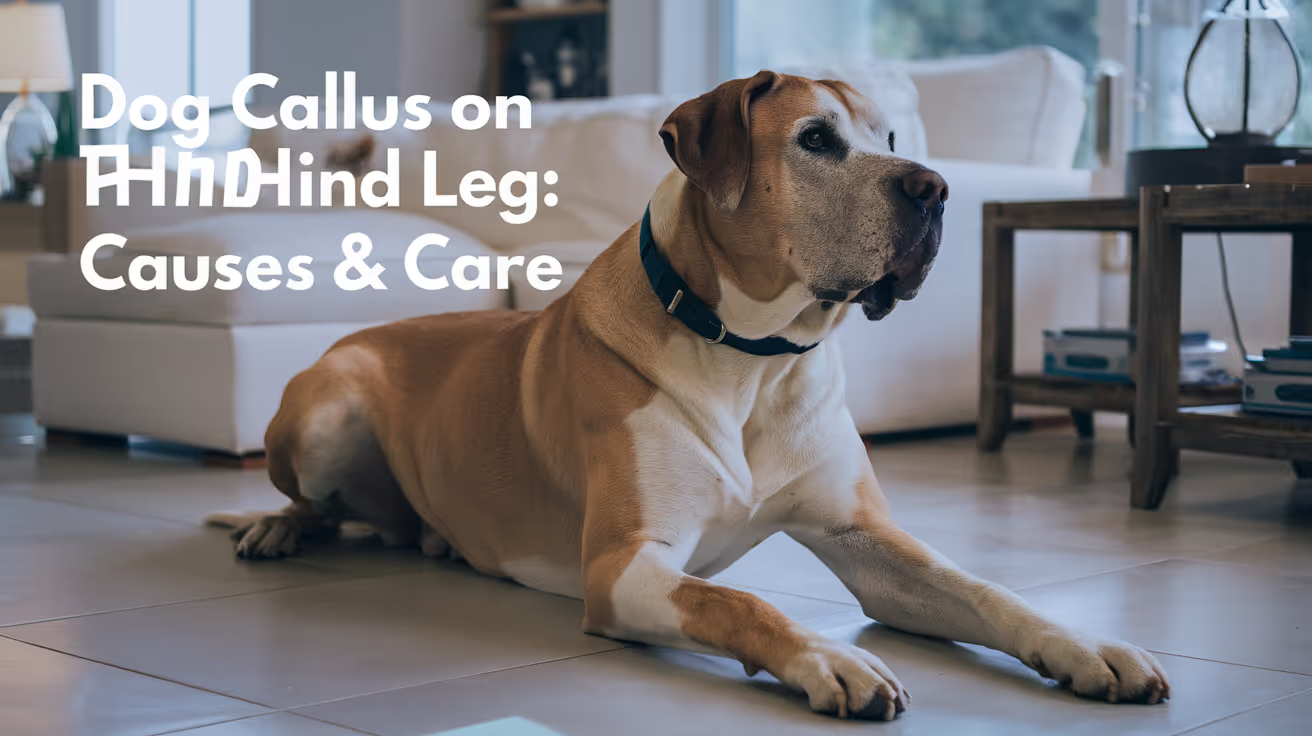
Dog Callus on Hind Leg Causes & Treatment
Learn about dog callus on the hind leg, its causes, symptoms, and effective treatments to keep your pet comfortable and healthy.
Sustainable Vet Group
A callus on your dog's hind leg is a thickened, hairless patch of skin that forms in response to repeated friction or pressure on a bony area. Most are harmless and cosmetic. Some become cracked, infected, or progress to a hygroma a fluid-filled swelling underneath the skin. Knowing the difference matters.
Quick answer: A dog callus is a rough, hairless area of thickened skin that forms over bony pressure points like hocks and hips from repeated lying on hard surfaces. Most are harmless. Cracked, bleeding, or infected calluses need veterinary care.
Key takeaways
- Calluses form over bony prominences where the skin contacts hard surfaces repeatedly: hocks, elbows, hips, and knees
- Large and giant breeds are most affected because more body weight concentrates pressure at these contact points
- A callus is thickened surface skin; a hygroma is a fluid sac below it; each requires different treatment
- Cracked or infected calluses need veterinary assessment; uncomplicated ones can be managed at home with moisturizer and soft bedding
- Orthopedic foam bedding is the most effective prevention measure for at-risk dogs
- Calluses that are warm, draining, or painful have likely become infected and need same-day veterinary attention
What is a callus on a dog's leg?
A callus is a thickened, hardened area of skin that forms as a protective response to repeated mechanical pressure or friction. The body produces extra keratin in the affected area, creating a rough, hairless patch.
Dr. Buzby's ToeGrips describes the appearance: "Calluses are rough, thickened skin that may be hairless." Unlike a hygroma, which forms under the skin and feels squishy, a callus is a surface change it sits on top of the skin and feels firm and rough to the touch.
Calluses are not tumors and are not signs of systemic disease. They are a normal biological response to an abnormal amount of pressure on skin. The clinical term is an elbow callus (or hock callus, hip callus, depending on location), and the condition is formally classified as a grade-based lesion depending on severity.
Common locations on the hind leg
Although elbow calluses get the most attention in veterinary literature, hind leg calluses are equally common:
- Hocks (the ankle joint of the hind leg) the most common hind leg site
- Lateral hip and hip bones especially in dogs that lie on their sides
- Outer stifle (knee) where the outside of the knee contacts the floor
- Lateral thigh in dogs that always sleep on the same side
PetHelpful notes that large or giant breed dogs, thin or underweight dogs, and obese dogs are all at elevated risk: lean dogs have reduced natural padding, while heavy dogs exert greater compressive force on the same points.
Why do dogs develop calluses?
The cause is always the same: repeated pressure or friction on skin overlying a bony prominence, usually from lying on hard surfaces over an extended period.
Merck Veterinary Manual notes that the typical history involves a dog that regularly lies on hard floors tile, hardwood, concrete rather than soft bedding. Large breed dogs that drop heavily to the ground rather than lowering themselves gradually cause repetitive impact injuries at the same pressure points.
Contributing factors:
- Hard flooring surfaces (tile, hardwood, concrete) rather than soft bedding
- Reduced activity and prolonged lying in one position (senior dogs, post-surgical patients)
- Excess body weight (more compressive force at pressure points)
- Loss of muscle mass (less natural padding over bones)
- Breed predisposition (large and giant breeds: Great Danes, Mastiffs, German Shepherds, Saint Bernards, Irish Wolfhounds)
For how long-standing calluses at pressure sites relate to deeper pressure injury, see pressure necrosis in chronic callus cases.
Callus vs hygroma: how to tell the difference
Calluses and hygromas form at the same sites from the same causes but are different conditions requiring different management.
Dr. Buzby's ToeGrips: "On the other hand, hygromas form under the skin, feel squishy, and usually have hair over them unless they are ulcerated or have a callus on top. Your vet can help you determine if your dog has a hygroma, callus, or both."
If you are unsure which one your dog has, see a vet rather than treating at home. Probing or draining what you think is a hygroma that turns out to be infected tissue can worsen the condition.
Stages of callus severity
Not all calluses are equal. PetHelpful grades elbow and hock calluses from mild to severe:
Grade I: superficial hair loss and mild skin thickening. No cracking. Skin intact and not inflamed.
Grade II: more pronounced thickening. Surface is rough and dry. Mild cracking may be present. No infection.
Grade III: deep fissures (cracks) in the callus. Surface is hyperkeratotic (extremely thickened). Risk of secondary bacterial infection.
Grade IV: the callus has become infected or ulcerated. The underlying tissue is involved. Discharge or odor may be present. Bone may be at risk in severe untreated cases.
Grade I and II calluses are manageable at home. Grade III and IV require veterinary assessment.
Treatment options
Home treatment for Grade I and II calluses
Moisturizing: apply a dog-safe moisturizer or balm (shea butter, coconut oil, or veterinary callus cream) to the callus daily. This softens the thickened skin, reduces cracking, and prevents fissures from developing. Do not use human hand creams some contain additives toxic to dogs if licked.
Soft bedding: provide a thick orthopedic foam or memory foam bed and encourage the dog to use it. This reduces the mechanical cause of the callus. VCA Animal Hospitals emphasizes soft bedding as the cornerstone of both callus management and hygroma prevention.
Protective wraps: elbow or hock pads (such as DogLeggs) protect the area from further friction. They are especially useful for dogs that refuse to stay off hard floors.
Veterinary treatment for Grade III and IV calluses
Grade III: a vet may gently debride (remove) the thickest layers of dead skin to allow better moisturizer penetration and reduce fissure depth. Topical antibiotics may be applied if there are early signs of infection.
Grade IV (infected callus): requires culture-guided antibiotics, wound debridement, and potentially surgical removal of the callus and reconstruction with a skin flap. FirstVet: "If the hygroma has a big sore or ulcer... the callus needs to be removed and skin flaps or grafts may be necessary for skin reconstruction."
For how calluses at pressure sites relate to the broader pattern of bed sores in immobile dogs, see bed sores as a related pressure injury.
Prevention
Orthopedic bedding: place thick foam beds in every location the dog rests. For large dogs, memory foam or high-density egg-crate foam is most effective. Check that the foam does not fully compress under the dog's weight.
Encourage bedding use: some dogs prefer hard floors even when soft bedding is available. Placing beds in favored spots, using non-slip mats on hard floors, and feeding or treating the dog near the bed can encourage use.
Weight management: keeping a large breed dog at a healthy weight reduces compressive force on bony prominences. VCA recommends weight control specifically for hygroma and callus prevention.
Protective joint wraps: for dogs already developing early calluses, commercially available elbow pads or hock wraps protect the skin from additional friction.
For how bursitis develops alongside calluses at the same pressure points, see bursitis alongside callus formation.
Frequently asked questions
Are calluses on a dog's hind leg dangerous?
Uncomplicated calluses are not dangerous. They are cosmetic changes that do not cause pain in most dogs. They become concerning when they crack deeply (creating an infection entry point), become infected, or progress to a hygroma. Monitor any callus for changes in size, texture, warmth, or the presence of discharge.
Can I treat my dog's callus at home?
Grade I and II calluses (hair loss, thickening, no cracking or infection) can be managed at home with daily moisturizer and improved bedding. Grade III calluses with deep fissures and Grade IV calluses with infection require veterinary treatment. If you are unsure of the grade, a vet visit to assess severity is worthwhile.
What moisturizer is safe for dog calluses?
Dog-specific balms containing shea butter, beeswax, or coconut oil are safe and effective. Veterinary products like Musher's Secret or Bag Balm are widely used. Avoid human hand creams and lotions, which may contain fragrances, urea, or other additives that can irritate dog skin or cause toxicity if licked.
How long does it take for a dog's callus to go away?
Calluses do not disappear quickly. With consistent daily moisturizing and soft bedding, a Grade I callus may soften noticeably within 4 to 6 weeks. More established calluses may take months to show significant improvement. The callus will often remain visible permanently in dogs that continue to use hard surfaces, even with good management.
My dog's callus is cracked and bleeding. What should I do?
A cracked, bleeding callus is at risk of infection. Clean the area gently with dilute chlorhexidine or warm water, apply a dog-safe antibiotic ointment (such as a veterinary triple antibiotic), and cover loosely with a clean bandage. See your vet within 24 to 48 hours. Deep fissures in calluses are painful and bacteria can enter easily through the broken skin.
Is there a difference between an elbow callus and a hock callus?
The mechanism is identical repeated pressure on a bony prominence but the anatomy and management differ slightly. Hock calluses form at the point of the hock (heel) on the hind leg. They are particularly common in large breeds and dogs with conformational issues that cause the hock to contact the floor during normal sitting or lying. Both respond to the same treatment: moisturizer, soft bedding, and protective padding.
Resources
- Dr. Buzby's ToeGrips. Hygromas in Dogs: Treatment and Prevention. toegrips.com
- VCA Animal Hospitals. Hygroma in Dogs. vcahospitals.com
- PetHelpful. Why Does My Dog Have Swollen or Calloused Elbows? pethelpful.com
- Merck Veterinary Manual. Hygroma in Dogs. merckvetmanual.com
Pustules in Dogs Causes and Treatment
Learn about pustules in dogs, their causes, symptoms, and effective treatments to keep your pet healthy and comfortable.
Sustainable Vet Group
A pustule is a small, pus-filled bump on the skin. In dogs, pustules are one of the most common signs of a bacterial skin infection collectively called pyoderma. They look like pimples: red and raised, with a white or yellow pus-filled center.
Pustules vary widely in severity. Some are superficial and clear with topical treatment. Others signal a deep infection that needs oral antibiotics for weeks, or an underlying disease that must be found and treated to prevent recurrence.
Quick answer: Pustules in dogs are almost always caused by pyoderma (bacterial skin infection), most commonly Staphylococcus pseudintermedius. They appear as red pus-filled bumps on the belly or around hair follicles. Treatment ranges from medicated shampoo to oral antibiotics.
Key takeaways
- Staphylococcus pseudintermedius causes over 90% of canine pyoderma and pustule formation
- Three depth levels exist: surface pyoderma, superficial bacterial folliculitis, and deep pyoderma, each needing different treatment
- Pustules that rupture leave circular crusts called epidermal collarettes, a hallmark sign vets look for
- Allergies, parasites, and hormonal disorders are the most common underlying causes of recurring pustules
- Skin cytology (examining cells from a pustule under a microscope) is the first and most useful diagnostic step
- Full antibiotic courses are essential: stopping treatment early is the most common reason pyoderma returns
What causes pustules in dogs?
The primary cause: bacterial infection (pyoderma)
PetMD confirms that the most common bacterial cause is "Staphylococcus pseudintermedius, which is responsible for more than 90 percent of cases. This type of bacteria normally inhabits the skin, but it can increase in number and cause problems when the skin barrier is damaged or unhealthy."
Other bacteria involved less commonly include Staphylococcus schleiferi, Staphylococcus aureus, E. coli, Pseudomonas, Streptococci, and Proteus. In most cases, these are secondary invaders that take advantage of already-damaged skin.
Why does the skin become infected?
Healthy dry dog skin is highly resistant to bacterial overgrowth. Merck Veterinary Manual explains: "Any skin disease that changes the normally dry, desert-like environment to a more humid environment can cause overcolonization of the skin with bacteria."
Common triggers that damage the skin barrier and allow infection:
- Allergies (environmental allergens, food, contact irritants): the most common trigger; chronic scratching and licking disrupts skin integrity
- External parasites (fleas, mites, mange): bite wounds and inflammatory reactions create entry points
- Hormonal disorders (hypothyroidism, Cushing's disease, diabetes): alter skin physiology and immune response
- Keratinization disorders: abnormal keratin production creates conditions for bacterial overgrowth
- Poor grooming or dirty environments: increased moisture and bacterial load on the skin surface
- Physical trauma: cuts, wounds, or abrasions that open the skin to bacterial colonization
The three types of canine pyoderma
Understanding which type your dog has determines the treatment approach.
1. Surface pyoderma
The infection involves only the very outermost skin layer (stratum corneum). It does not penetrate into hair follicles or deeper tissue.
Examples: puppy impetigo (belly pustules in puppies), skin fold pyoderma (in facial folds, lip folds, tail folds), hot spots (acute moist dermatitis).
Signs: redness, pustules, and crusting on hairless or folded skin areas.
Treatment: topical therapy alone is usually sufficient medicated wipes, chlorhexidine spray, or topical antibiotic.
2. Superficial bacterial folliculitis (SBF)
The infection involves the epidermis and the upper portion of hair follicles. VCA Animal Hospitals describes the visual result: "In short-haired breeds, the coat may appear to protrude or stick up in areas, mimicking hives, or it may appear moth-eaten because of patchy hair loss."
Signs: pustules and papules around hair follicles, circular crusts called epidermal collarettes, hair loss in a patchy pattern, itching. Dr. Buzby's ToeGrips lists the full spectrum: "papules, pustules, collarettes, crusts, and erythema of the armpits and groin."
Treatment: topical therapy plus oral antibiotics in most cases; minimum 3 to 4 weeks of treatment.
3. Deep pyoderma
The infection penetrates into the dermis (deep skin layer) and may cause furunculosis rupture of hair follicles releasing bacteria and keratin into surrounding tissue. This is the most serious form.
Signs: painful, thickened skin; draining tracts; crusts over larger areas; foul odor; systemic signs in severe cases (lethargy, fever).
WebMD Pets: "Deep pyoderma will appear as pain at the infected site, crusting, a bad smell, and oozing of pus and blood."
Treatment: prolonged oral antibiotics (often 6 to 8 weeks or more), guided by culture and sensitivity results; treatment of the underlying cause is essential.
For how pustule-causing infections can progress into deep abscesses, see furuncles as a severe progression of pustules.
What pustules look like at each stage
VCA Animal Hospitals: "The most common clinical signs associated with pyoderma are papules or pustules that form on the skin. These lesions often look similar to pimples in humans. They are most often red and raised, with a white pus-filled center."
How vets diagnose the cause of pustules
A vet will usually start with a physical examination and skin cytology. Further tests are ordered if the infection is severe, recurrent, or unresponsive to initial treatment.
Skin cytology: a glass slide is pressed against a pustule or ruptured lesion and examined under a microscope. Dr. Buzby's ToeGrips explains: "Seeing inflammatory cells and bacteria makes a pyoderma likely. And bacteria identified inside of cells is a slam-dunk for a diagnosis."
Culture and sensitivity: a swab is taken from inside a pustule or lesion and sent to a lab. This identifies the exact bacterial species and which antibiotics will and will not work. Essential for recurrent or non-responsive infections.
Skin scraping: rules out mites (demodex, sarcoptes) which can cause secondary bacterial infections.
Wood's lamp: rules out ringworm (dermatophytosis), which can mimic superficial pyoderma.
Bloodwork: thyroid panel, cortisol testing, or full chemistry panel to screen for hormonal disorders in adult dogs with recurring pustules.
Treatment
Topical treatment
Medicated shampoos containing chlorhexidine (2 to 4%) or benzoyl peroxide are the foundation of topical pyoderma management. Used 2 to 3 times per week, they reduce bacterial load on the skin surface and support antibiotic treatment.
Topical sprays, wipes, and creams are useful for localized lesions. Chlorhexidine solution applied directly to pustule-bearing areas between baths is effective for surface pyoderma.
Oral antibiotics
Required for superficial bacterial folliculitis and all deep pyoderma. Common choices include:
- Cephalexin (first-line for most uncomplicated cases)
- Amoxicillin-clavulanate
- Trimethoprim-sulfamethoxazole
- Clindamycin
Treatment duration matters enormously. Merck Veterinary Manual is explicit: "Even though your dog may seem better after only a few days or a week, it is still very important for you to continue the prescribed treatment program for the full length of time. The bacteria causing pyoderma can still be present and ready to multiply again if the complete course of medication is not given."
Minimum durations: 3 to 4 weeks for superficial folliculitis; 6 to 8 weeks or longer for deep pyoderma. The infection should be resolved for at least 7 days before stopping antibiotics.
Treating the underlying cause
Merck Veterinary Manual identifies the most common reasons pyoderma recurs: "failure to treat underlying causes, use of glucocorticoid drugs, and inappropriate treatment with prescribed antibiotic medications."
If your dog has recurring pustules, the vet will investigate for allergies, parasites, or hormonal disease. Controlling the underlying cause is the only way to prevent recurrence.
For how impetigo specifically presents as pustules in puppies on the belly and groin, see impetigo as a pustule-causing condition.
Home care during treatment
- Give all medications on schedule and complete the full course
- Bathe with the prescribed medicated shampoo 2 to 3 times per week leave it on for 10 minutes before rinsing
- Keep the affected skin dry between baths; moisture encourages bacterial regrowth
- Prevent licking with an E-collar if your dog is bothering the lesions
- Wash bedding weekly to reduce environmental bacterial load
- Do not squeeze or pop pustules this spreads bacteria and may cause scarring
For how skin conditions that cause scabs relate to the pustule cycle, see skin conditions that cause scabs and pustules.
Frequently asked questions
Are pustules in dogs contagious to humans or other pets?
Most canine pyoderma is caused by Staphylococcus pseudintermedius, which does not routinely infect healthy humans. However, Staphylococcus aureus can occasionally transfer between species, including to immunocompromised people. As a precaution, wash hands after handling a dog with active pustules and keep other pets separated if the infection is extensive.
How long does it take for pustules to clear up?
With appropriate antibiotic treatment, pustules typically begin to dry and resolve within 7 to 14 days. However, treatment must continue for the full prescribed course (minimum 3 to 4 weeks for superficial infections) even after the skin looks normal. Stopping early is the most common cause of recurrence.
My dog keeps getting pustules even after treatment. Why?
Recurring pustules almost always indicate an unidentified or untreated underlying cause. Allergies are the most common culprit the chronic itching and skin damage from allergies continuously set up conditions for bacterial infection. Inadequate antibiotic duration and bacterial resistance are other causes. Ask your vet to investigate for allergies, mites, or hormonal disorders if your dog's pustules keep returning.
Can I use human acne products on my dog's pustules?
No. Products containing benzoyl peroxide at human concentrations, salicylic acid, retinoids, or alcohol can irritate or damage dog skin. Veterinary formulations of benzoyl peroxide shampoo (at lower concentrations designed for dog skin) are appropriate. Use only products your vet recommends.
What is an epidermal collarette and should I be concerned about it?
An epidermal collarette is a circular scale or crust ring left behind when a pustule ruptures. It is not itself a sign of active infection but indicates that a pustule was recently present. Your vet will note collarettes during examination as evidence of pyoderma. They typically resolve as the skin heals during treatment.
When should pustules in a dog be treated as an emergency?
Most pyoderma is not an emergency. Seek same-day care if your dog has rapidly spreading skin lesions, swelling, signs of pain, fever, lethargy, or is not eating. These signs suggest deep infection or a systemic response. Also contact your vet urgently if the skin appears dark, necrotic, or has a foul odor signs that deep tissue may be involved.
Resources
- PetMD. Pyoderma in Dogs. petmd.com
- VCA Animal Hospitals. Pyoderma in Dogs. vcahospitals.com
- Merck Veterinary Manual. Pyoderma in Dogs. merckvetmanual.com
- Dr. Buzby's ToeGrips. Pyoderma in Dogs: Signs, Diagnosis, and Treatment. toegrips.com
- WebMD Pets. What Is Pyoderma in Dogs? webmd.com
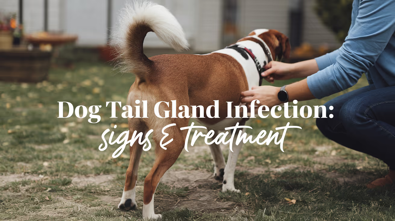
Dog Tail Gland Infection Symptoms and Treatment
Learn about dog tail gland infection symptoms, causes, and effective treatment options to keep your pet healthy and comfortable.
Sustainable Vet Group
A greasy, smelly patch at the base of your dog's tail with some hair loss isn't something most owners immediately connect to a glandular condition. It's easy to assume it's a bite, a rash, or a minor skin problem.
What it often is, is a condition called stud tail, or supracaudal gland hyperplasia, where the oil-producing gland at the top of the tail base becomes overactive, clogs hair follicles, and eventually becomes infected.
Quick answer: Tail gland infections in dogs, also called stud tail or supracaudal gland hyperplasia, occur when the supracaudal gland on the tail base overproduces sebum, blocking follicles and creating conditions for bacterial infection. Symptoms include a greasy, bald, and sometimes odorous patch at the tail base, blackheads, and crusted discharge. Treatment involves medicated antibacterial shampoos, topical or systemic antibiotics, and in intact males, neutering often reduces recurrence.
Key takeaways
- The supracaudal gland sits at the top of the tail base, about 2.5 to 5 cm from the anus.
- Overproduction of sebum clogs follicles, creating blackheads and inviting bacterial infection.
- Intact male dogs are most commonly affected due to androgen-driven gland overactivity.
- Secondary bacterial infection (pyoderma) is the clinical concern, not the gland enlargement itself.
- Antibacterial shampoos are the first-line treatment for mild to moderate cases.
- Neutering reduces recurrence in male dogs by lowering androgen levels.
What is the tail gland?
Every dog has a supracaudal gland, also called the violet gland, on the dorsal (top) surface of the tail, roughly 2.5 to 5 cm from the anus. This gland is a cluster of modified sebaceous glands that produce sebum, pheromones, and other secretions used for scent marking.
In most dogs, the gland functions without incident. When it becomes overactive, typically due to hormonal influence or underlying skin disease, it produces excess sebum that can block the surrounding hair follicles. This leads to:
- Follicular plugging (blackheads or comedones)
- Hair loss (follicles can't support the hair shaft)
- Greasy, malodorous skin
- Secondary bacterial infection (pyoderma)
The condition is more common in intact male dogs, but can occur in females and neutered males. It is not exclusive to dogs: intact male cats are similarly affected.
Symptoms of a tail gland infection in dogs
The visible signs vary depending on whether the condition is at the early (hyperplasia only) or later (infected) stage.
Early stage: no infection yet
- Greasy or oily patch at the top of the tail base
- Slight hair thinning or a small bald spot
- Blackheads visible in the affected area
- Mild odor from excess sebum secretion
- Dog may lick or chew at the tail base
Infected stage (pyoderma present)
- Clearly visible hair loss over a defined patch
- Redness and swelling at the gland site
- Pustules or crusted lesions
- Discharge (yellow or brown)
- Noticeable foul smell
- Increased licking or chewing as the dog responds to discomfort
- Darkening or thickening of the surrounding skin in chronic cases
The distinction matters because infected cases require antibiotic treatment, while early non-infected cases may respond to shampoo therapy alone.
Causes and risk factors
Androgen excess
The most established cause is elevated androgen (testosterone) levels in intact male dogs. Androgens stimulate the sebaceous glands to overproduce sebum. This is why the condition is called "stud tail" and is most common in intact males. Wagwalking describes the mechanism: "Sometimes this gland secretes too much androgen, which can raise levels and cause a bare spot in that area."
Primary seborrhea
Dogs with generalized seborrhea (a skin disorder involving abnormal cell turnover and excessive oiliness) are predisposed because the underlying skin condition already drives gland hyperactivity.
Other contributing factors
- Parasites at the tail base (fleas, mites) that cause scratching and secondary gland irritation
- Allergies causing generalized skin inflammation
- Follicular plugging from anatomical features
Diagnosis
A vet will examine the tail base, assess whether secondary infection is present, and rule out conditions that cause similar signs:
- Perianal adenoma: tumors of the hepatoid glands around the anus and tail base can look similar and require biopsy
- Anal gland disease: anal gland impaction can cause tail-base licking and discomfort
- Hypothyroidism: secondary skin changes including seborrhea can affect the tail area
- Mange or ringworm: parasitic or fungal causes of tail-base hair loss
If infection is present, skin cytology or culture confirms the causative bacteria. MRSP or other resistant staph warrants culture and sensitivity testing before committing to an antibiotic.
For how staph functions as the most common infectious agent in these presentations, see staph as a common cause of tail gland infections.
Treatment
Medicated shampoos (first-line)
For mild to moderate cases with or without early infection, medicated shampoos are the starting point:
- Benzoyl peroxide shampoos: flush follicles, reduce oiliness, antibacterial properties
- Chlorhexidine-based shampoos: strong antibacterial action for infected cases
- Salicylic acid or sulfur-containing shampoos: anti-seborrheic effect, reduces oiliness
Frequency: 2 to 3 times per week during active treatment, then maintained as a preventive routine. The affected area must be rinsed thoroughly to avoid irritation from product buildup.
Topical antibiotics
When infection is confirmed but limited in extent, topical mupirocin or fusidic acid applied to the affected area may be sufficient without systemic antibiotics.
Systemic antibiotics
Established pyoderma at the tail gland site requires oral antibiotics. Severe cases with significant infection may need a 4 to 8 week course. Antibiotic choice should be based on culture and sensitivity if MRSP is suspected or initial treatment fails.
Severe cases with recurrent deep infection may temporarily require systemic antibiotics for 2 months or more, per veterinary dermatology guidance.
Neutering
In intact male dogs, neutering is one of the most effective long-term management strategies. Reducing androgen levels decreases glandular stimulation, which reduces sebum overproduction and the likelihood of recurrent infection. Neutering won't necessarily resolve an active infection, but it significantly reduces the chance of repeated episodes.
Surgery
In rare cases with severe, recurrent deep infection unresponsive to medical treatment, surgical removal of the gland may be performed. This is not a first-line approach and is reserved for cases where all other management has failed.
For how general wound infection signs apply to a tail gland infection site, see general wound infection signs. For broader infection prevention strategies relevant to dogs with skin conditions, see preventing tail gland infections.
What happens if tail gland infection is left untreated?
Untreated, a mild case of gland hyperplasia can progress to established pyoderma. Established pyoderma can develop into deep pyoderma with draining tracts. Chronic infections alter the surrounding skin: thickening, hyperpigmentation, and scarring can occur.
Repeated antibiotic courses without addressing the underlying cause (particularly androgen excess in intact males) also raise the risk of antibiotic resistance, including MRSP.
For how post-surgical infections compare to this type of glandular infection, see post-surgical infections that can affect the tail gland area.
Frequently asked questions
Is stud tail the same as an anal gland infection?
No. Stud tail involves the supracaudal gland on the top of the tail, not the anal glands. Anal gland impaction or infection causes scooting, licking at the back end, and discomfort around the anus. Stud tail causes a patch of hair loss and skin changes on the top of the tail base. Both can cause tail-base licking, so the two are sometimes confused.
Can a female dog get a tail gland infection?
Yes. While stud tail is most common in intact males due to androgen influence, it can occur in spayed females and neutered males. In these cases, underlying seborrhea, allergies, or other skin conditions are more likely to be the driver. Diagnosis and treatment are the same regardless of sex.
How long does treatment take?
Mild cases often improve within 2 to 4 weeks of shampoo therapy. Established bacterial infections require antibiotic treatment for 3 to 8 weeks depending on depth and severity. Recurrent cases in intact males typically don't fully resolve without neutering as part of the management plan.
Tail gland infections in dogs are straightforward to manage when caught early. The challenge is recognizing that a greasy, thinning patch at the tail base isn't a minor cosmetic issue: it's the first stage of a condition that progresses predictably if the underlying glandular overactivity isn't addressed.
Resources
- DogTime. Stud Tail in Dogs: Symptoms, Causes, Treatments. dogtime.com
- Wagwalking. Stud Tail in Dogs. wagwalking.com
- PetEdge. Tail Gland Hyperplasia in Dogs. petedge.com
Modified Maquet Procedure vs TPLO Surgery
Compare Modified Maquet Procedure and TPLO surgery for canine cruciate ligament rupture treatment, including benefits, risks, and recovery.
Sustainable Vet Group
The Modified Maquet Procedure (MMP) is an evolution of the Tibial Tuberosity Advancement (TTA) that uses a titanium foam wedge instead of a cage to advance the tibial tuberosity.
It has been positioned as a less invasive, faster-to-perform alternative to TPLO for dogs with CCL rupture.
Published clinical evidence shows both procedures produce good outcomes, with TPLO demonstrating a slight advantage in objective gait analysis at 3 months.
Quick answer: A prospective randomized study found TPLO achieves 93.9% of normal peak vertical force vs 89.4% for MMP at 6 months — no significant difference in complications or OA progression. MMP is less invasive, faster, and generally less expensive. Both are appropriate for medium-to-large breed dogs.
Key takeaways
- TPLO achieves 93.9% of normal ground reaction force at 6 months vs 89.4% for MMP — numerically superior but not statistically significant
- No significant difference in major complications or OA progression was found between TPLO and MMP in the comparative study
- MMP is less invasive and faster to perform: a titanium foam wedge and partial osteotomy instead of TPLO's full proximal tibial cut
- MMP is generally less expensive than TPLO: shorter surgical time and different implant costs reduce the procedure price
- Early partial weight-bearing may occur sooner after MMP due to less invasive fixation and the partial osteotomy
- TPLO has a much larger evidence base: 25+ years of data vs limited MMP evidence
How each procedure works
TPLO (Tibial Plateau Leveling Osteotomy)
TPLO addresses CCL rupture by making a curved osteotomy through the proximal tibia and rotating the tibial plateau to approximately 5 degrees.
This changes joint biomechanics so the stifle is stable during weight-bearing without a functioning CCL. The osteotomy is held in place with a bone plate and screws.
Modified Maquet Procedure (MMP)
MMP is an adaptation of TTA in which the tibial tuberosity is advanced cranially by making a partial osteotomy in the tibial crest.
A porous titanium foam wedge is inserted into the gap and a fixation staple holds the construct in position.
The cranial advancement of the tuberosity repositions the patellar tendon to neutralize the cranial thrust that occurs in the CCL-deficient stifle the same biomechanical principle as TTA, with different implants.
Animal Works Veterinary Surgery describes MMP as using improved implants and surgical techniques designed to reduce operating time and minimize complications compared to standard TTA.
The titanium foam wedge promotes bone ingrowth and healing without the need for a cage or plate system. Bone healing in the MMP osteotomy site is typically complete at 90 days radiographically.
Clinical evidence: TPLO vs MMP
Primary comparative study
PubMed (prospective randomized controlled study, 61 dogs, 76 joints): dogs were treated with TPLO (n=30, 41 joints) or MMP (n=31, 35 joints) and compared with healthy controls.
Outcomes were assessed by clinical examination, radiography, and treadmill force plate gait analysis at 6 weeks, 3 months, and 6 months.
Results at 6 months:
- TPLO: 93.9% of normal peak vertical force (PVF), 85.9% of normal vertical impulse (VI)
- MMP: 89.4% of normal PVF, 79.9% of normal VI
At 3 months, significantly more TPLO patients were within the reference range for healthy dogs on peak vertical force. By 6 months, the difference was not statistically significant.
No significant difference was found in major complications or OA progression at any time point.
Conclusion from the study: "Although no significant differences were found between the surgical methods, TPLO patients showed superiority with regard to clinical outcome."
MMP standalone study
PMC (35 dogs, unilateral CCL rupture, MMP): significant improvement in ground reaction forces at all time intervals. At 90 days, 54.2% of patients had a Symmetry Index suggesting normal gait.
Complete bone healing on radiographs at 90 days. Major complication rate: 8.5% (3 of 35), minor complication rate: 2.8%.
Practical comparison
Which dogs are best suited to each?
MMP is often chosen for:
- Medium and large breed dogs where a less invasive approach is preferred
- Cases where surgical time is a clinical consideration
- Practices experienced in MMP technique
- Dogs where cost is a factor
TPLO is typically recommended for:
- Large and giant breed dogs with high joint loads
- Dogs with steep tibial plateau angles where TPLO provides more precise biomechanical correction
- Cases where the largest available evidence base is preferred
- Surgeons with established TPLO experience
Animal Hospital of Adel notes that both surgeries are effective, with MMP being less invasive, less expensive, and quicker to perform.
Walkerville Vet's evidence review concludes that TPLO gives slightly better objective results and that MMP is expected to outperform standard TTA, though more evidence is needed.
For the full TPLO pros and cons, see TPLO surgery pros and cons for dogs. For the full alternatives overview, see alternatives to TPLO surgery for dogs.
Frequently asked questions
Is MMP as effective as TPLO for large dogs?
The evidence suggests TPLO has a slight advantage at 3 months, with equivalent outcomes by 6 months in the primary comparative study.
Most surgeons still prefer TPLO for large and giant breeds due to the larger evidence base and slightly superior short-term objective results. MMP is appropriate for many large dogs in experienced hands.
How much cheaper is MMP than TPLO?
MMP is generally less expensive than TPLO due to shorter surgical time and different implant costs. The specific difference varies by practice, region, and surgeon.
Ask your veterinarian for a direct cost comparison for your dog's specific case.
What is the complication rate for MMP compared to TPLO?
The MMP standalone study reported an 8.5% major complication rate. TPLO complication rates across the literature range from 14 to 34%.
However, these figures come from different study populations and settings, making direct comparison difficult. Neither procedure is free of risk.
How long does MMP recovery take compared to TPLO?
Both procedures require 8 to 12 weeks of restricted activity, though MMP's less invasive partial osteotomy may allow earlier partial weight-bearing in some dogs.
Full activity return for both procedures is typically at 4 to 6 months.
Can a dog have TPLO if MMP fails?
Yes. TPLO can be performed as a revision if MMP results in ongoing instability or poor function. The converse is less common.
Discuss revision options with an orthopedic specialist if your dog is not progressing as expected after MMP.
Resources
- PubMed. Outcome after Tibial Plateau Levelling Osteotomy and Modified Maquet Procedure in Dogs with CCL Rupture. pubmed.ncbi.nlm.nih.gov
- PMC. Outcome after Modified Maquet Procedure in Dogs: Force Plate Gait Analysis. ncbi.nlm.nih.gov
- Animal Works Veterinary Surgery. MMP vs TPLO. animalworksvets.com
- Dog Knee Injury. Modified Maquet Procedure (MMP Surgery) for Dogs. dogkneeinjury.com
- Walkerville Vet. Which ACL Surgery Is Best for Dogs? walkervillevet.com.au

Bed Sores on Dogs Causes Treatment and Prevention
Learn about bed sores on dogs, including causes, treatments, and prevention tips to keep your pet healthy and comfortable.
Sustainable Vet Group
Bed sores in dogs also called pressure sores or decubitus ulcers develop when prolonged pressure on a bony area cuts off blood supply, causing the tissue to break down. They are the same condition as bedsores in hospitalized humans, and they are just as serious when they reach advanced stages.
The good news is that early-stage bed sores are very treatable at home. The bad news is that they can progress from a small red patch to a deep infected wound within days if not caught and addressed.
Quick answer: Bed sores form over bony prominences like elbows and hips when sustained pressure blocks blood flow. Most common in senior, large-breed, and immobile dogs. Early stages respond to soft bedding and repositioning; advanced stages need veterinary care.
Key takeaways
- Bed sores form over bony prominences: elbows, hips, hocks, stifles, and sides of the knees
- Four stages exist: from mild redness to deep infected wounds reaching muscle and bone
- Senior dogs, large breeds, and post-surgical dogs are at highest risk
- Stage 1 sores reverse with home management: soft bedding and repositioning every 2 to 4 hours
- Stage 3 and 4 sores need veterinary debridement and often systemic antibiotics
- Thick orthopedic foam bedding is the single most effective prevention measure for at-risk dogs
What causes bed sores in dogs?
Bed sores develop when the weight of a dog's body continuously presses a bony prominence against a hard surface. Walkin' Pets explains the mechanism: "Decubital ulcers form when repeated pressure on an affected area constricts the blood vessels, decreasing the area's blood supply and as the skin is deprived of oxygen, it begins to die."
Once the tissue starts to die, it creates a wound that bacteria readily colonize. This is why advanced bed sores are almost always infected.
The process requires two things to come together: sustained mechanical pressure and skin that is not being repositioned or protected. Any dog that lies in the same position for hours without shifting is at risk.
Additional factors that increase risk
Incontinent dogs: urine-soaked skin breaks down much faster under pressure. Walkin' Pets advises that "reducing skin moisture from urine is important to help prevent the bacterial growth and skin degradation that contribute to pressure sores."
Thin or underweight dogs: less muscle and fat over bony prominences means the bone contacts the surface with less cushioning.
Obese dogs: more body weight concentrates greater compressive force at each contact point.
Dogs on hard floors: concrete, hardwood, and tile dramatically increase the pressure at contact points compared to soft surfaces.
Which dogs get bed sores?
Dr. Buzby's ToeGrips identifies the most at-risk groups:
- Senior dogs: reduced muscle mass, reduced mobility, longer periods in one position
- Large and giant breed dogs: more body weight, greater force at bony contact points
- Post-surgical patients: strict rest requirements limit natural repositioning
- Paralyzed or paraplegic dogs: unable to shift position at all
- Dogs with arthritis: pain limits movement and encourages sustained resting in one position
- Short-coated breeds: less coat padding between skin and hard surfaces
DogCancer.com notes that bed sores can also be a concern for dogs in late-stage cancer, where weakness and pain significantly reduce their ability to reposition.
The 4 stages of bed sores
Recognizing the stage determines the treatment approach.
Stage 1: redness and intact skin
The skin over the bony prominence is red, warm, and may have some hair loss. The surface is not broken. This is the ideal intervention point.
What to do at home: add thick orthopedic padding under the affected area, increase repositioning to every 2 hours, and monitor the site twice daily. Most Stage 1 sores reverse completely with these measures.
Stage 2: shallow open wound
The skin has partially broken down. A shallow blister or open area appears. The surrounding tissue remains inflamed and warm.
WagWalking: "Stage 1 ulcers may improve with padding, regular repositioning of the dog, and a quality moisture-wicking dog bed. Vets may instruct pet parents to regularly apply moist dressings to open pressure sores."
What to do: contact your vet. Stage 2 requires wound cleaning, appropriate dressings, and assessment for infection. Antibiotics may be started if signs of infection are present.
Stage 3: full-thickness wound
The sore extends through the full skin thickness into subcutaneous tissue. VetMobile describes what you see: "The sore becomes deeper and it may extend into the underlying tissue, such as the muscle layer. Pus or discharge may be present. At this stage, the area is surrounded by dead or dying tissue."
What to do: veterinary care is required. Treatment involves debridement of dead tissue, wound culture, and systemic antibiotics based on culture results.
Stage 4: bone or muscle involvement
The wound extends to muscle, tendon, or bone. This is a serious and potentially life-threatening stage. Bacteria can enter the bloodstream from a Stage 4 wound, causing sepsis.
What to do: emergency veterinary care. Hospitalization, IV antibiotics, surgical debridement, and reconstructive surgery may be necessary.
Treatment by stage
Home wound care for Stage 1 and 2
For Stage 1 and early Stage 2 sores, clean the area daily with dilute chlorhexidine solution (not hydrogen peroxide, which damages healing tissue). Apply a non-adherent wound dressing and change it once or twice daily. Keep the area dry between dressing changes.
Puainta recommends: "Pressure Relief: Encourage the dog to change positions frequently. Soft Bedding: Provide cushioned surfaces to reduce pressure. Cleanliness: Keep the area clean and dry."
For incontinent dogs, clean and dry the skin after every urination. Use a waterproof mattress cover that can be washed daily. Sheepskin pads are effective at wicking moisture away from skin while providing cushioning.
For how bed sores relate to callus formation at the same pressure sites, see calluses from similar pressure points.
When to call the vet
Call your vet within 24 hours if:
- The wound is open, moist, or has any discharge
- The area is warm and the dog shows signs of pain when the area is touched
- The sore is growing despite home management
- Your dog is lethargic, off food, or running a fever
These signs suggest infection is establishing. Early antibiotic treatment is significantly more effective than treating advanced infection.
WagWalking notes: "Antibiotics may be necessary if the wound is infected or at risk of becoming infected. Antibiotics have minimal risks and a high success rate when used early on."
For how the progression of pressure necrosis connects to bed sore development, see pressure necrosis in severe bed sore cases.
Prevention
Orthopedic bedding
The most effective single intervention. Memory foam or high-density egg-crate foam distributes weight across a larger surface area, reducing peak pressure at bony contact points. Petful emphasizes that "you might be more likely to notice pressure sores in dogs if you have a large breed pup or a short-haired breed" both groups where bedding choice is critical.
The foam must not fully compress under the dog's full weight. Test by pressing firmly: if it bottoms out quickly, it is not thick enough. For large dogs, minimum 4 inches of high-density foam is recommended.
Repositioning schedule
Reposition immobile dogs every 2 to 4 hours, alternating which side bears weight. Walkin' Pets advises: "Try not to let the dog rest on the hip bone on one side for very long, reposition your pet every 2 to 3 hours."
If you are not home during the day, consider whether a pet sitter or midday check can provide repositioning support for a dog with high risk.
Daily inspection
Check every bony prominence every day. Run your hand along the elbows, hips, hocks, and knees. Hair loss is the first visible sign act at Stage 1, before any skin breakdown occurs.
Moisture management for incontinent dogs
Wet skin under pressure breaks down dramatically faster than dry skin. Change diapers and male wraps frequently. Use raised mesh beds for overnight rest in incontinent dogs urine drains through rather than pooling under the dog. Walkin' Pets recommends sheepskin pads specifically for their wicking properties.
For how swelling associated with bed sores can extend to the feet and lower legs, see swelling associated with pressure injuries.
Frequently asked questions
How do I know if my dog has a bed sore starting?
The first sign is a small patch of hair loss over a bony area, sometimes with mild redness and warmth. The skin is still intact. Run your hand over the elbows, hips, and hocks daily in at-risk dogs. Acting at this point before the skin opens is far easier than treating an open wound.
Can bed sores in dogs heal on their own?
Stage 1 sores can reverse with prompt management: improved bedding, repositioning, and keeping the area dry. Stage 2 and above rarely resolve without veterinary intervention. The wound environment actively promotes bacterial growth, which prevents natural healing without treatment.
How long does a bed sore take to heal in a dog?
Stage 1 sores managed early typically show improvement within 1 to 2 weeks with good home care. Stage 2 wounds typically heal in 3 to 6 weeks with consistent veterinary wound care. Stage 3 and 4 wounds may take months and may not fully close in all cases.
My senior dog keeps getting bed sores despite soft bedding. What else can I do?
Check whether the foam has compressed over time and needs replacing. Add protective wraps (elbow pads or hock wraps) over at-risk sites. Increase repositioning frequency to every 2 hours. Consult your vet about whether the dog's underlying mobility level has declined and whether additional pain management or physiotherapy could help. Nutritional support including protein and vitamin C promotes skin healing.
Are bed sores painful for dogs?
Stage 1 sores are usually not painful. Stage 2 onward becomes progressively more painful as the wound deepens and infection sets in. Dogs with painful bed sores may lick or bite at the area, avoid lying on that side, or show behavioral changes. Pain management from your vet is an important part of treatment from Stage 2 onward.
Can bed sores become life-threatening in dogs?
Yes. Stage 3 and 4 sores can allow bacteria to enter deeper tissues and the bloodstream, causing sepsis. A dog with an advanced bed sore that is also lethargic, not eating, or running a fever is potentially septic and needs emergency veterinary care the same day.
Resources
- Dr. Buzby's ToeGrips. Pressure Sores on Dogs: Treatment and Prevention. toegrips.com
- WagWalking. Pressure Sores in Dogs. wagwalking.com
- Walkin' Pets. Pressure Sores in Pets: Treatment and Prevention. walkinpets.com
- Petful. Pressure Sores on Dogs: What You Need to Know. petful.com
- DogCancer.com. Bed Sores on Dogs: Preventing and Treating Pressure Sores. dogcancer.com
DeAngelis vs TPLO Surgery for Dogs
Compare DeAngelis and TPLO surgery for dogs, exploring benefits, risks, recovery, and costs to help pet owners choose the best treatment.
Sustainable Vet Group
DeAngelis surgery also called lateral suture, extracapsular repair, or ELSS (extracapsular lateral suture stabilization) is one of the oldest and most widely performed CCL repair techniques.
TPLO is the most commonly recommended procedure by the ACVS for dogs over 60 lbs.
Decades of research strongly favor TPLO for larger, more active dogs while supporting extracapsular repair as a cost-effective option for smaller dogs.
Quick answer: TPLO produces superior outcomes in medium and large breed dogs: better limb loading at 8 weeks, higher 1-year function, and slower arthritis progression. DeAngelis surgery is effective for small dogs under 15 to 20 lbs and costs $800 to $1,500 vs $3,000 to $6,000 for TPLO.
Key takeaways
- TPLO produces superior outcomes at 8 weeks and 1 year in size-matched studies: more symmetric loading, higher vertical force, higher owner satisfaction
- DeAngelis surgery is effective in small dogs under 15 to 20 lbs: success rates above 90% reported; significantly worse in larger dogs
- Extracapsular repair relies on periarticular fibrosis for stability; the suture eventually stretches or fails in larger dogs
- DeAngelis surgery costs significantly less: $800 to $1,500 vs $3,000 to $6,000 for TPLO
- Arthritis progression is faster after extracapsular repair than TPLO in multiple studies; TPLO slows OA more effectively
- 10 to 20% of extracapsular repair dogs will require additional treatment for lameness during their lifetime per Today's Veterinary Practice
How each procedure works
DeAngelis surgery (lateral suture / extracapsular repair)
DeAngelis surgery uses a heavy monofilament nylon suture (or, in the TightRope variation, a fiber-tape through bone tunnels) placed outside the joint to mimic the function of the torn CCL.
The suture runs from the lateral fabella behind the femur around to the tibial crest, restraining the cranial drawer movement that occurs with CCL loss.
The suture is not a permanent replacement.
DVM360 confirms that extracapsular stabilization relies on periarticular fibrosis for long-term stability the suture eventually stretches or fails, and joint stability depends on scar tissue formed around the joint during recovery.
DeAngelis surgery does not require bone cutting, specialized equipment, or the steep learning curve associated with TPLO. It is technically accessible to general practice surgeons with appropriate training.
TPLO (Tibial Plateau Leveling Osteotomy)
TPLO addresses CCL rupture by changing joint geometry rotating the tibial plateau so the stifle is mechanically stable without any functioning CCL.
The stability provided by TPLO is bone-based and does not depend on scar tissue or suture integrity over time.
TPLO requires specialized equipment, a radiolucent table, intraoperative radiographs, and surgeon training in the procedure. It is primarily performed at specialty hospitals and by board-certified orthopedic surgeons.
What the clinical evidence shows
Short-term outcomes
DVM360's evidence review found TPLO dogs bear more weight sooner; extracapsular repair dogs hold the leg up for 1 to 2 weeks.
A force plate gait study (ECR n=23, TPLO n=15) found more symmetric limb loading at 8 weeks in TPLO patients compared to extracapsular repair patients at both walk and trot.
Long-term outcomes
Today's Veterinary Practice's evidence-based review reports:
- A randomized blinded clinical trial of 80 dogs found TPLO resulted in higher peak vertical force and higher owner satisfaction at 1 year post-surgery compared to lateral fabellar suture stabilization
- A more recent study showed TPLO outcomes were always better than lateral suture outcomes at any time point up to 1 year
DVM360 notes one early study found no difference at 2 years, but more recent studies consistently show TPLO outperforming extracapsular repair.
Arthritis progression
Today's Veterinary Practice confirms that extracapsular stabilization may result in increased progression of radiographic osteoarthritis compared to TPLO.
Restoring joint mechanics (TPLO) slows cartilage degradation more effectively than temporary stabilization with subsequent fibrosis (extracapsular repair).
Long-term failure
Today's Veterinary Practice reports that 10 to 20% of dogs with extracapsular stabilization will require treatment or reoperation for lameness during their lifetimes, sometimes years after original surgery.
This rate is higher than the expected rate of reoperation-requiring complications after TPLO.
Head-to-head comparison
Which dogs are best suited to each?
DeAngelis surgery is appropriate for:
- Small dogs under 15 to 20 lbs with CCL rupture
- Dogs with significant health conditions making a longer anesthetic or more invasive procedure higher risk
- Cases where cost is the primary determining factor
- Older, less active dogs with lower functional demands
TPLO is recommended for:
- Dogs over 30 lbs, especially active and large breeds
- Dogs where long-term joint function and minimal OA progression are the goals
- Working dogs, sport dogs, or highly active pets
- Dogs with steep tibial plateau angles that make extracapsular repair less reliable
For the full surgical alternatives overview, see alternatives to TPLO surgery for dogs. For the TPLO pros and cons, see TPLO surgery pros and cons for dogs.
For the lateral suture detailed comparison, see lateral suture vs TPLO: what's the difference?.
Frequently asked questions
Is DeAngelis surgery as good as TPLO?
For small dogs under 15 to 20 lbs, DeAngelis surgery can be as effective as TPLO.
For medium and large breed dogs, the evidence clearly favors TPLO: better short-term function, better 1-year outcomes, slower arthritis progression, and lower long-term reoperation rates.
How much less does DeAngelis surgery cost than TPLO?
Typically $2,000 to $4,500 less per leg. DeAngelis surgery costs approximately $800 to $1,500 at general practices vs $3,000 to $6,000 for TPLO at specialist centres.
For bilateral cases (both knees), the cost difference is doubled.
Does DeAngelis surgery fail more often than TPLO?
In large dogs, yes. The suture stretches and fails in larger, more active patients because the periarticular fibrosis cannot compensate for the higher joint forces.
Today's Veterinary Practice estimates 10 to 20% of extracapsular repair dogs will need further treatment for lameness during their lifetime.
Can my large dog have DeAngelis surgery instead of TPLO?
Technically yes, but the evidence does not support it as the primary recommendation for dogs over 30 lbs.
DVM360 confirms it is possible that larger dogs treated with lateral suture technique may have significantly worse outcomes than those treated with TPLO.
If DeAngelis surgery fails, can TPLO be done afterward?
Yes. TPLO is a standard revision option when extracapsular repair fails or when the dog develops ongoing instability and lameness.
Discuss this with a board-certified orthopedic surgeon if your dog's original repair has not produced adequate results.
Resources
- DVM360. Cranial Cruciate Ligament Repair: One Size Does NOT Fit All. dvm360.com
- Today's Veterinary Practice. Canine Cranial Cruciate Disease: An Evidence-Based Look at Current Treatment Modalities. todaysveterinarypractice.com
- DVM360. Journal Scan: Ruptured CCL Surgery: TPLO vs Extracapsular Repair. dvm360.com
- Walkerville Vet. Cruciate Surgery: TPLO, TTA, MMP, DeAngelis. walkervillevet.com.au
- ACVS. Cranial Cruciate Ligament Disease. acvs.org
What Causes TPLO Surgery to Be Needed in Dogs
Learn what causes TPLO surgery to be needed in dogs, including common injuries and conditions leading to this important orthopedic procedure.
Sustainable Vet Group
TPLO surgery is not performed because of a single moment of bad luck.
In the vast majority of cases, the cruciate ligament has been quietly degenerating for months or years before the day it finally fails.
Understanding what drives that degeneration and which dogs are most vulnerable is the first step toward recognizing early warning signs and making informed decisions about your dog's care.
Quick answer: TPLO is needed when a dog's CCL ruptures from progressive degeneration, causing stifle instability and lameness. Key risk factors are breed (Labs, Rottweilers, Mastiffs), obesity, steep tibial plateau angle, and neutering. CCL rupture costs US owners over $1 billion annually.
Key takeaways
- CCL rupture is the most common cause of hind limb lameness in dogs: over $1 billion spent annually in the US on treatment
- Most CCL ruptures result from degeneration, not trauma: sudden injury to a healthy ligament is rare in dogs
- Breed is the strongest single risk factor: Labs, Rottweilers, Mastiffs, and Newfoundlands most commonly affected
- Obesity significantly increases CCL rupture risk: excess weight increases mechanical load on the stifle and systemic inflammation
- Steep tibial plateau angle is a mechanical risk factor: extra CCL strain with every stride; TPLO corrects this directly
- Neutered dogs have higher CCL rupture rates than intact dogs across multiple studies, particularly in high-risk breeds
Why CCL rupture leads to TPLO
When the CCL ruptures, the stifle joint becomes unstable. With each step, the tibia slides forward under the femur a motion called cranial tibial thrust that normally the CCL prevents.
This sliding causes:
- Pain with every weight-bearing step
- Damage to the articular cartilage
- Progressive osteoarthritis
- Meniscal injury (the meniscus acts as a shock absorber and is frequently torn by the unstable joint)
Laguna Hills Animal Hospital explains that a dog with a torn CCL cannot walk normally, and the instability caused by the tear damages the bone and cartilage surrounding the joint.
TPLO surgery addresses this by rotating the tibial plateau to approximately 5 degrees a position at which cranial tibial thrust no longer occurs during normal weight-bearing.
The joint becomes stable without a functioning CCL.
The primary cause: CCL degeneration
The crucial distinction between canine CCL rupture and human ACL tears is the mechanism.
Virginia Veterinary Centers states directly: CCL rupture can result from activity-related trauma, but is most commonly caused by progressive degeneration of the ligament.
Laguna Hills Animal Hospital adds: unlike in humans, the CCL rarely ruptures from sudden trauma in an otherwise healthy ligament.
The degeneration process involves progressive deterioration of collagen fibers within the CCL weakening that begins long before any clinical signs appear.
By the time a dog presents lame, the CCL has typically been failing at a microscopic level for months.
The rupture event a step off a curb, a jump in the yard is the endpoint of this process, not the cause.
This degenerative process explains why partial tears almost always progress to full rupture.
NCBi (2014) confirms 22 to 54% of dogs rupture the other CCL within 6 to 17 months.
Risk factors for CCL rupture
Breed
Breed is the most powerful predictor of CCL rupture. MedVet lists Labs, Golden Retrievers, German Shepherds, Boxers, Pit Bulls, American Bulldogs, Mastiffs, Rottweilers, and Newfoundlands as most commonly affected.
Laguna Hills Animal Hospital confirms a genetic component has been established specifically in Newfoundlands and Labrador Retrievers.
The genetic risk likely involves inherited factors in ligament collagen quality, joint conformation, and inflammatory susceptibility.
Obesity
Excess body weight increases the compressive and shear forces on the CCL with every step.
Overweight dogs are at significantly higher risk of CCL rupture, and obesity is the most modifiable risk factor.
MetLife Pet Insurance identifies weight as a key contributing factor, noting that overweight dogs place additional stress on their joints, increasing rupture risk.
Maintaining a lean body condition score is one of the few actionable CCL prevention strategies available to owners.
Tibial plateau angle (TPA)
Dogs with a steeper-than-average tibial plateau angle experience higher cranial tibial thrust with each stride, increasing the load on the CCL throughout the dog's life.
MetLife Pet confirms that some dogs have a naturally steep tibial plateau angle, which puts extra strain on the CCL.
TPLO surgery addresses this directly by reducing the TPA to approximately 5 degrees it not only treats the rupture but corrects the underlying mechanical vulnerability.
Neutering
Multiple studies have found that neutered dogs both spayed females and castrated males have higher rates of CCL rupture than intact dogs.
PMC (agility risk factors study) lists neuter status as a previously identified risk factor alongside age, sex, breed, and body weight.
Proposed mechanisms include earlier closure of growth plates (affecting long bone development), changes in muscle mass distribution, and the absence of sex hormones that influence ligament maturation and quality.
Age
Older dogs have ligaments with more cumulative degeneration. The risk of CCL rupture increases with age, particularly in at-risk breeds.
Rancho Palos Verdes Vets lists age, obesity, poor conformation, breed, and lack of fitness as the main risk factors for CCL tears.
Physical conditioning
Poor physical fitness reduces the muscle mass that supports and offloads the stifle joint.
Well-conditioned muscles act as dynamic stabilizers; dogs with poor muscle mass rely more heavily on their passive stabilizers including the CCL increasing ligament load.
Clinical signs that TPLO is needed
When the CCL ruptures or significantly tears, dogs show:
- Sudden hind limb lameness often appearing after exercise or normal activity
- Toe-touching or non-weight-bearing on the affected leg
- Sitting with the leg extended to the side rather than folded under the body
- Stiffness after rest that may temporarily improve with movement
- Swelling or thickening around the stifle joint (medial buttress)
- Muscle atrophy of the affected thigh over time
- Positive cranial drawer sign or tibial compression test on examination
Rancho Palos Verdes Vets notes that sitting with one leg extended to the side is one of the most obvious signs that a CCL injury requiring evaluation is present.
For the full CCL causes article, see what causes cruciate ligament tears in dogs. For the TPLO surgery overview, see what is TPLO surgery in dogs?.
For the signs your dog needs TPLO, see 13 signs your dog may need TPLO surgery. For surgical alternatives, see alternatives to TPLO surgery for dogs.
For the long-term outcomes of TPLO, see long-term outcomes of TPLO surgery.
Frequently asked questions
Can a dog rupture its CCL just from running in the yard?
Yes but the run did not cause the rupture. In almost all cases, the ligament was already significantly degenerated before it failed.
The activity is the final stressor on an already compromised structure, not the cause. This is one of the most important things to understand about canine CCL disease.
What breeds are most likely to need TPLO surgery?
Labrador Retrievers, Golden Retrievers, Rottweilers, Boxers, Mastiffs, Newfoundlands, Staffordshire Terriers, and German Shepherds are most commonly affected. Genetic inheritance of CCL disease has been specifically confirmed in Labs and Newfoundlands.
Smaller breeds are less commonly affected and often managed with less invasive procedures when rupture does occur.
Does fixing the CCL prevent the other knee from tearing?
No. TPLO stabilizes the operated knee but does not prevent degeneration in the other knee, which is affected by the same systemic risk factors.
NCBi research shows 22 to 54% of dogs develop contralateral CCL rupture within 6 to 17 months. Weight management and controlled exercise are the main strategies for reducing the risk.
Can obesity cause CCL rupture?
Yes. Obesity increases joint load and systemic inflammation, both of which accelerate CCL degeneration. Weight management is the single most impactful lifestyle intervention for reducing CCL disease risk in predisposed breeds.
Is CCL rupture preventable?
Not entirely genetic factors are outside owner control. However, maintaining a lean body condition, providing consistent moderate exercise, and avoiding obesity are the most effective risk-reduction strategies.
Discuss the role of neutering timing with your veterinarian if you own a high-risk breed.
Resources
- Virginia Veterinary Centers. What Dog Owners Should Know About TPLO Surgery. virginiaveterinarycenters.com
- Laguna Hills Animal Hospital. Why Does My Dog Need TPLO Surgery? lhah.com
- MedVet. Dogs and Knee Surgery: Understanding TPLO Surgery. medvet.com
- MetLife Pet Insurance. TPLO Surgery for Dogs. metlifepetinsurance.com
- PMC. Radiographic Risk Factors for Contralateral Rupture in Dogs with Unilateral CCL Rupture. ncbi.nlm.nih.gov
Dog Incontinence After TPLO Surgery: Causes & Care
Learn about dog incontinence after TPLO surgery, its causes, treatment options, and recovery tips for your pet’s comfort and health.
Sustainable Vet Group
Urinary incontinence after TPLO surgery is not a common complication, but it does occur and is understandably alarming for owners.
In most cases it is a temporary consequence of anesthesia, pain medication, stress, or reduced mobility not a sign of a serious surgical problem.
Knowing the causes, the timeline, and when to call your vet helps you respond appropriately.
Quick answer: Incontinence after TPLO is usually temporary from anesthesia, pain medications, reduced mobility, or stress. Most cases resolve within days to weeks. Call your vet if it persists beyond 2 to 4 weeks or comes with bloody urine or fever.
Key takeaways
- Immediate post-TPLO incontinence is most often from anesthesia or nerve irritation: both are temporary and resolve within days to weeks
- Opioid pain medications cause urinary retention or reduced bladder awareness in some dogs this resolves as medications are tapered
- Postoperative swelling may temporarily irritate nerves involved in bladder control; this resolves with swelling
- Reduced mobility is a practical contributor: a confined, painful dog may not signal in time or reach the door quickly
- Stress after major surgery can cause accidents in previously house-trained dogs; this is behavioral, not neurological
- Genuine nerve damage from TPLO is rare: incontinence persisting beyond 6 weeks warrants neurological evaluation
Why dogs may become incontinent after TPLO surgery
Anesthesia effects
General anesthesia temporarily suppresses normal nerve function throughout the body, including the neural pathways that control bladder filling awareness and sphincter tone.
As anesthetic drugs are metabolized over the 12 to 24 hours after surgery, some dogs have reduced bladder control.
This is the most common cause of immediate post-surgical incontinence. It resolves as anesthesia fully clears, typically within 24 to 48 hours.
Nausea, disorientation, and general sedation from anesthesia also mean the dog may not signal clearly when they need to go outside.
Scheduled outdoor bathroom trips every 2 to 4 hours in the first 24 to 48 hours post-surgery help manage this.
Opioid and sedative medications
Opioid pain medications commonly prescribed after TPLO affect bladder control in two ways: they reduce awareness of bladder fullness, and in some dogs cause urinary retention.
Both effects resolve as medications are tapered in the days following surgery.
Retention and incontinence are opposite problems retention is not urinating at all, while incontinence is involuntary urination but both can occur at different times as different medications have their effects.
As pain medications are tapered in the days after surgery, these effects resolve for most dogs.
Postoperative swelling and nerve irritation
The proximal tibia is surrounded by neurovascular structures. Postoperative swelling around the surgical site may temporarily compress or irritate nerves that travel to the bladder and urethra.
This is more likely in the first week after surgery when swelling is at its peak.
This type of incontinence improves as swelling resolves and typically does not indicate permanent nerve damage.
Reduced mobility
A dog recovering from TPLO is confined, in pain, and may not reach the door quickly enough when the bladder signals urgency.
This is a practical mobility problem, not a neurological one.
Frequent scheduled outdoor trips every 2 to 4 hours in the first week reduce accidents significantly. Waterproof mattress covers and washable bedding help manage the recovery period hygienically.
Stress and behavioral response
Major surgery is a significant stressor. Pain, an unfamiliar environment, an E-collar, restricted movement, and disrupted routine can cause previously house-trained dogs to have accidents unrelated to neurological dysfunction.
This type of incontinence typically resolves as the dog settles into a recovery routine at home, usually within 1 to 2 weeks.
Pre-existing urinary conditions
Dogs with pre-existing urinary sphincter mechanism incompetence (common in middle-aged spayed females), bladder infections, or weak bladder control may show worsened incontinence after surgery.
Anesthesia, medications, and reduced mobility all unmask or worsen underlying conditions.
If your dog was occasionally leaking urine before surgery, report this to your vet during the pre-surgical assessment so appropriate management can be planned for the recovery period.
Types of post-TPLO incontinence by timing
Immediate (within 24 to 48 hours): almost always anesthesia effects or opioid medication. Expected and self-limiting.
Early post-operative (days 3 to 14): medication effects, swelling-related nerve irritation, mobility limitation, or stress. Usually self-limiting with appropriate management.
Delayed onset (weeks 2 to 6): may indicate a developing urinary tract infection (UTI), which can occur after catheterization during surgery or from reduced immune response. A urinalysis is warranted if incontinence appears or worsens after initial improvement.
Chronic (beyond 6 weeks): uncommon. If incontinence persists beyond 6 weeks without improvement, veterinary evaluation for nerve damage or underlying urinary tract disease is appropriate.
Home management during recovery
- Scheduled outdoor bathroom trips every 2 to 4 hours for the first 1 to 2 weeks
- Waterproof mattress protectors and washable bedding in the recovery area
- Non-slip surfaces to reduce the effort required to stand and move toward the door
- Monitor urine color and odor cloudy, bloody, or foul-smelling urine suggests UTI
- Keep the genital area clean to prevent skin irritation from urine contact
- Use a belly band (for males) or absorbent dog diapers if incontinence is significant
When to contact your veterinarian
Call your vet if:
- The dog cannot urinate at all for more than 12 to 24 hours after surgery
- Urine is bloody, cloudy, or has a strong unusual odor (UTI signs)
- Incontinence is accompanied by straining, crying during urination, or frequent squatting without producing urine
- Incontinence worsens after initial improvement
- The dog has persistent incontinence beyond 2 to 4 weeks without any improvement
- Incontinence is combined with significant worsening lameness or systemic signs
For the not-peeing guide that covers urinary retention after TPLO, see dog not peeing after TPLO surgery: causes and care.
For the full TPLO complications reference, see 15 common complications after TPLO surgery in dogs.
For TPLO plate infection signs that may be contributing to systemic effects, see TPLO plate infection signs and treatment.
For post-op whining that may relate to the same pain driving accidents, see dog whining after TPLO surgery: causes and care.
Frequently asked questions
Is incontinence normal after TPLO surgery?
Temporary incontinence particularly in the first 24 to 48 hours is a recognized post-surgical finding related to anesthesia and opioid pain medications.
It is not an expected routine outcome, but it is not rare. If it resolves within a few days, it was almost certainly anesthesia-related.
Could my dog have nerve damage from TPLO surgery?
True nerve damage causing persistent incontinence from TPLO is rare. TPLO is performed at the proximal tibia, which is some distance from the pelvic nerves controlling bladder function.
Temporary nerve irritation from swelling is much more common than actual nerve injury and resolves as swelling subsides.
My dog was continent before TPLO and is now leaking at night. Why?
Nighttime leaking specifically is typical of urinary sphincter mechanism incompetence (USMI) a condition where the urethral sphincter relaxes during sleep or rest.
If this is new after surgery, anesthesia and opioid medications may have temporarily worsened underlying borderline sphincter function.
If it continues beyond 2 to 3 weeks, a vet visit for urinalysis and discussion of phenylpropanolamine (PPA) or other treatment is appropriate.
How do I know if my dog has a UTI after TPLO?
Signs of UTI include: cloudy or bloody urine, strong or unusual odor, frequent small amounts of urination, straining to urinate, and licking at the genital area more than normal.
Some dogs with UTI also show lethargy and decreased appetite. Urinalysis and culture confirm the diagnosis.
Should I limit my dog's water intake to reduce accidents?
No. Do not restrict water. Hydration is important for recovery and kidney function. Restricting water can concentrate the urine and increase UTI risk.
Instead, manage accidents through scheduled outdoor trips and waterproof bedding.
Resources
- VCA Animal Hospitals. Tibial Plateau Leveling Osteotomy (TPLO) in Dogs. vcahospitals.com
- ACVS. Cranial Cruciate Ligament Disease. acvs.org
- PetMD. Urinary Incontinence in Dogs. petmd.com
- Midwest Veterinary Specialists. Post-Operative Care Following TPLO. midwestveterinaryspecialists.com

TPLO Plate Rejection Symptoms in Dogs
Learn to recognize TPLO plate rejection symptoms in dogs, including signs, causes, and treatment options for better recovery.
Sustainable Vet Group
TPLO plate rejection is one of the less common but more serious complications that can follow tibial plateau leveling osteotomy surgery.
The term "rejection" is sometimes used loosely distinguishing between true immune-mediated rejection, infection, and implant loosening matters because the treatment differs significantly for each.
Quick answer: True TPLO plate rejection is rare — implants are highly biocompatible. It produces persistent swelling, heat, pain over the plate, and sometimes a draining tract. Infection produces identical symptoms. Contact your vet if lameness worsens or discharge appears weeks to months after surgery.
Key takeaways
- True immune-mediated plate rejection is rare: TPLO plates are made from highly biocompatible titanium or stainless steel
- Symptoms overlap significantly with infection: pain, swelling, heat, and discharge occur in both; bacterial culture distinguishes them
- Infection is the most common implant complication: TPLO infection rates range from 0.8 to 14.3%; true rejection is a small subset
- Signs typically appear weeks to months after surgery: worsening lameness after initial improvement is the classic red flag
- A draining tract (fistula) over the plate is a hallmark sign of implant-associated infection or rejection
- True rejection requires plate removal: once the osteotomy heals (at least 12 weeks), the plate is removed; bone heals without it
What is TPLO plate rejection?
The TPLO plate and screws are made from medical-grade titanium or stainless steel.
These materials are designed to be biologically inert the body should recognize them as neutral foreign material and wall them off without reaction.
Laguna Hills Animal Hospital explains that in some cases, the TPLO plate is rejected by the dog's body because it is perceived as a foreign object.
True immune-mediated rejection occurs when the immune system mounts an inflammatory response against the implant material itself. This is distinct from:
- Periprosthetic infection: bacteria colonize the implant surface and cause chronic infection around the plate; this is far more common than true rejection
- Mechanical failure: the plate bends, breaks, or screws loosen due to excessive force; this is a structural problem, not an immune reaction
- Normal post-surgical inflammation: expected in the first 2 to 3 weeks; should be improving, not worsening
In clinical practice, the distinction between true rejection and low-grade infection is often made by bacterial culture of the fluid or tissue at the site.
True aseptic rejection produces inflammation without bacterial growth; periprosthetic infection produces the same symptoms with positive cultures.
Symptoms of TPLO plate rejection
Dog Discoveries' veterinary reference describes plate rejection as: the knee becomes inflamed and sore around the plate, the dog feels constant pain and irritation, and the dog gets worse rather than better.
Signs of trouble are generally seen weeks to months after surgery rather than in the immediate post-operative period.
Key symptoms to monitor:
Worsening or persistent lameness: a dog that was improving and then regresses, or one whose lameness fails to improve beyond the expected baseline, is showing a key red flag. This is distinct from the expected early post-TPLO lameness that should be progressively improving week by week.
Localized swelling over the plate: visible or palpable swelling directly over the plate site (proximal medial tibia) that does not resolve with rest and is not reducing over time.
Warmth and heat at the surgical site: warmth beyond the first 2 to 3 post-operative weeks suggests ongoing inflammation.
Draining tract (fistula): a small hole in the skin near the surgical site that produces persistent clear, bloody, or purulent discharge. This is a hallmark of implant-associated complications either infection or reaction. Vet Playas confirms persistent drainage or discharge, especially with a foul odor, may indicate infection or plate rejection.
Pain on palpation: the dog reacts to direct pressure over the plate site.
Failure to heal: the dog is not progressing as expected at radiographic rechecks; osteotomy bridging is delayed or absent.
How plate rejection is diagnosed
Clinical examination: systematic palpation of the plate site, assessment of lameness grade, and comparison to expected recovery trajectory.
Radiographs: assess osteotomy healing, implant position, screw integrity, and periimplant bone quality. Periprosthetic bone loss suggests infection or chronic reaction.
Culture and sensitivity: fluid or tissue from the surgical site is cultured to determine whether bacteria are present and which antibiotics are effective. Culture-negative inflammation supports true rejection; culture-positive results confirm infection.
Blood work: elevated inflammatory markers (C-reactive protein, white blood cell count) confirm systemic involvement.
Treatment
For infection (most common implant complication)
Culture-directed antibiotic therapy is initiated. If the osteotomy is not yet healed, the plate must remain in place removing the plate before the osteotomy heals causes the bone segments to lose support.
Targeted antibiotics and local wound management may control infection until healing is sufficient.
If the infection does not resolve with antibiotics, surgical debridement or implant removal (if the bone has healed) may be required.
For the full infection management guide, see TPLO plate infection signs and treatment.
For true aseptic rejection
Plate removal is the definitive treatment. Laguna Hills Animal Hospital confirms that the bone plate must remain in place for at least 12 weeks to allow the bone to heal.
Once healing is confirmed radiographically typically at 10 to 12 weeks the plate can be safely removed.
The bone does not require the plate long-term; it is a scaffold to hold the osteotomy while it heals.
When to contact your veterinarian
Contact your vet if:
- Lameness worsens or plateaus instead of progressively improving
- You notice new swelling, heat, or discharge at the surgical site weeks after surgery
- A draining tract appears at or near the incision
- Your dog shows signs of pain at the plate site on palpation
- Radiographs at rechecks show unexpected findings around the plate or screws
For the implant failure guide, see TPLO implant failure signs and causes. For the plate removal recovery guide, see TPLO plate removal recovery guide.
For the full complications reference, see 15 common complications after TPLO surgery in dogs. For the plate infection article with antibiotic treatment details, see TPLO plate infection signs and treatment.
For when the plate needs to come out due to infection, see when does a TPLO implant need to be removed due to infection.
Frequently asked questions
How common is TPLO plate rejection in dogs?
True immune-mediated rejection is uncommon. TPLO plates are manufactured from highly biocompatible titanium or stainless steel.
The more common implant complication is infection (0.8 to 14.3% of TPLO cases depending on the study), which produces similar symptoms to rejection.
Metal allergy or true rejection accounts for a small subset of these cases.
How do I know if it's plate rejection or infection?
The symptoms overlap significantly. Bacterial culture of fluid from the site distinguishes them: culture-positive means infection; culture-negative inflammation in the right clinical context suggests true rejection.
Both require veterinary evaluation do not attempt to manage either at home.
Can the plate stay in if my dog is rejecting it?
No. If true rejection is diagnosed and the plate is causing ongoing inflammation, it must be removed once the osteotomy has healed sufficiently.
Most dogs do not require the plate after 10 to 12 weeks once the bone has consolidated. Plate removal is a less complex procedure than the original TPLO.
Will my dog need another surgery if the plate is rejected?
Plate removal surgery is typically required if true rejection is confirmed and the osteotomy has healed. This is less invasive than the original TPLO.
In most cases the dog recovers well after plate removal, and long-term outcomes are good.
Can rejection be prevented?
Pre-surgical screening for metal sensitivities can help select the best implant material. Strict aseptic technique during surgery minimizes infection that can mimic rejection.
Activity restriction during recovery reduces mechanical stress on the implant that increases complication risk.
Resources
- Dog Discoveries. Signs of TPLO Plate Rejection in Dogs. dogdiscoveries.com
- Vetplayas. Unveiling TPLO Plate Rejection Symptoms. vetplayas.com
- Laguna Hills Animal Hospital. TPLO Surgery Complications. lhah.com
- ACVS. Cranial Cruciate Ligament Disease. acvs.org

Sustainable Vet | Copyright ©2026 | All Rights Reserved

