

How to Keep an Energetic Dog Calm After Surgery
Learn effective ways to keep your energetic dog calm after surgery with practical tips and expert advice for smooth recovery.

This article is for informational purposes only and is not a substitute for professional veterinary advice. Every case is unique, so always consult your veterinarian for guidance specific to your pet.
This content is intended for veterinary professionals for educational purposes. It does not replace clinical judgment or tailored advice. Always rely on your training, expertise, and the specific context of your patients.

After surgery, energetic dogs can be a challenge to manage because their natural activity may interfere with healing. Keeping your dog calm is crucial to prevent complications and ensure a smooth recovery. This article explains how to handle your dog's energy safely after surgery.
You will learn practical strategies to reduce your dog's activity, create a comfortable environment, and support healing. These tips help you balance your dog's need for rest with their natural energy levels.
Why is it important to keep an energetic dog calm after surgery?
After surgery, your dog's body needs time to heal. Excessive activity can cause pain, reopen wounds, or delay recovery. Energetic dogs may struggle to stay still, increasing the risk of injury.
Understanding the importance of calmness helps you take steps to protect your dog's health during this vulnerable time.
- Prevents wound damage: Limiting movement reduces the chance of stitches tearing or wounds reopening, which can cause infections or require more surgery.
- Reduces pain and swelling: Rest helps control inflammation and discomfort, making recovery smoother and faster.
- Supports medication effectiveness: Calm behavior ensures your dog responds well to pain relief and antibiotics, improving healing outcomes.
- Prevents complications: Overexertion can lead to bleeding, bruising, or internal injuries, which are dangerous after surgery.
Keeping your dog calm is essential to avoid setbacks and promote a healthy recovery process.
What are the best ways to restrict your dog's activity safely?
Restricting activity after surgery requires careful planning. You want to limit movement without causing stress or anxiety. Using physical barriers and controlled routines helps manage your dog's energy safely.
Here are effective methods to keep your dog calm while preventing injury.
- Use a crate or small room: Confine your dog to a safe, comfortable space to limit running and jumping while allowing rest.
- Leash walks only: Take short, slow walks on a leash to allow bathroom breaks without overexertion or excitement.
- Remove toys and distractions: Avoid giving your dog balls or chew toys that encourage active play during recovery.
- Provide comfortable bedding: A soft, supportive bed encourages your dog to lie down and relax instead of moving around.
These steps help control your dog's activity level while keeping them safe and comfortable.
How can you create a calming environment for your dog after surgery?
A calm environment reduces stress and helps your dog rest better. Noise, bright lights, and busy areas can excite your dog and increase activity. Creating a peaceful space supports healing.
Consider these tips to make your dog's recovery area soothing and quiet.
- Choose a quiet room: Place your dog in a low-traffic area away from loud noises and household activity to minimize stimulation.
- Dim the lights: Soft lighting helps your dog relax and signals it is time to rest.
- Use calming scents: Lavender or chamomile diffusers can soothe anxiety and promote sleep in dogs.
- Play soft music: Gentle classical or specially designed pet relaxation music can reduce stress and encourage calmness.
A peaceful environment helps your dog stay calm and recover faster after surgery.
What role does diet and hydration play in calming an energetic dog after surgery?
Proper nutrition and hydration support healing and can influence your dog's energy levels. Feeding the right diet and ensuring water intake helps your dog feel comfortable and less restless.
Here are ways diet and hydration contribute to calming your dog post-surgery.
- Feed easily digestible food: Soft, bland diets reduce stomach upset and encourage eating during recovery.
- Maintain hydration: Fresh water availability prevents dehydration, which can cause irritability and restlessness.
- Limit treats: Avoid high-energy or sugary treats that may increase hyperactivity during healing.
- Follow vet dietary advice: Use prescribed diets or supplements that support tissue repair and immune function.
Balanced nutrition and hydration help your dog stay calm and heal efficiently after surgery.
How can you use mental stimulation to keep your dog calm without physical activity?
Mental stimulation can tire your dog’s mind and reduce restlessness without physical exertion. This approach helps energetic dogs stay calm while respecting activity restrictions.
Try these mental activities to keep your dog engaged safely.
- Use puzzle feeders: Food-dispensing toys challenge your dog’s brain and slow eating, providing calm focus.
- Teach simple commands: Short training sessions with basic commands keep your dog mentally active without movement.
- Offer scent games: Hide treats for your dog to find using smell, which stimulates the brain gently.
- Provide chew toys: Safe chew toys satisfy natural urges and promote relaxation without running or jumping.
Mental exercises help reduce boredom and energy, supporting calmness during recovery.
When should you contact your vet about your dog's activity after surgery?
Monitoring your dog’s behavior after surgery is important. If your dog is too active or shows signs of pain or complications, contact your vet promptly. Early intervention prevents serious problems.
Watch for these warning signs and know when to seek veterinary advice.
- Excessive licking or biting: Persistent attention to the surgical site may indicate pain or irritation needing vet evaluation.
- Swelling or bleeding: Any unusual swelling, redness, or bleeding around the wound requires immediate veterinary care.
- Restlessness or whining: Signs of discomfort or anxiety that do not improve with medication should be reported.
- Difficulty walking or limping: Changes in mobility may signal complications needing professional assessment.
Timely communication with your vet ensures your dog’s recovery stays on track and complications are minimized.
How can medication help keep an energetic dog calm after surgery?
Medications prescribed by your vet play a key role in managing pain and anxiety after surgery. Proper use of these drugs helps keep your dog comfortable and less active, aiding recovery.
Understand how medication supports calmness and what to expect.
- Pain relief drugs: Analgesics reduce discomfort, making your dog less likely to move excessively due to pain.
- Anti-anxiety medications: In some cases, vets prescribe mild sedatives to help overly energetic dogs relax safely.
- Follow dosage instructions: Administer medications exactly as directed to avoid side effects or underdosing.
- Monitor for reactions: Watch for adverse effects like drowsiness or vomiting and report concerns to your vet promptly.
Medication is a valuable tool for controlling activity and ensuring your dog’s comfort during healing.
Conclusion
Keeping an energetic dog calm after surgery is vital for a smooth and safe recovery. By restricting activity, creating a calming environment, and using mental stimulation, you can help your dog heal without stress or injury.
Following veterinary advice on diet, medication, and monitoring signs of complications ensures your dog stays comfortable and recovers well. With patience and care, your energetic dog can return to normal activity safely after surgery.
What should I do if my dog refuses to rest after surgery?
Try increasing mental stimulation with puzzle toys and short training sessions. If restlessness continues, consult your vet about possible anxiety medications or pain management adjustments.
Can I let my dog go outside to play after surgery?
Limit outdoor time to short, slow leash walks for bathroom breaks only. Avoid running, jumping, or playing until your vet confirms it is safe.
How long does it usually take for a dog to calm down after surgery?
Most dogs begin to calm within 7 to 14 days, depending on the surgery type and individual energy levels. Follow your vet’s recovery timeline closely.
Are there natural remedies to help calm my dog after surgery?
Calming scents like lavender and chamomile, as well as gentle massage, can help reduce anxiety. Always check with your vet before using supplements or herbal remedies.
What signs indicate my dog is too active after surgery?
Signs include excessive licking of the wound, swelling, bleeding, restlessness, whining, or difficulty walking. Contact your vet immediately if you notice these symptoms.

Get a Free Poster
Enhance your workspace with a high-quality radiographs reference poster, designed for veterinary professionals. This free physical poster will be shipped directly to you—just fill out the form to request your copy.









Taking Great TPLO Radiographs
Click Below to Watch Live Video Demos


We'll send you a Free Wall Poster with all the steps
Step #1
Getting Ready
Ensuring a clean surgical field starts with proper skin preparation. This video demonstrates the best practices for:
- Shaving the patient – Achieving a close, even shave while minimizing skin irritation
- The Dirty Scrub – The initial skin prep step to remove surface debris and reduce bacterial load before the sterile scrub.
Following these techniques helps reduce infection risk and improve surgical outcomes. Watch the video to see how it’s done effectively!


Step #2
Reduce Your Risks
Many surgeons are shocked to find out that their patients are not protected from biofilms and resistant bacteria when they use saline and post-op antibiotics.
That’s Where Simini Comes In.
Why leave these risks and unmanaged? Just apply Simini Protect Lavage for one minute. Biofilms and resistant bacteria can be removed, and you can reduce two significant sources of infection.
Step #3
Take the Course
Preventing surgical infections is critical for patient safety and successful outcomes. This course covers:
- Aseptic techniques – Best practices to maintain a sterile field.
- Skin prep & draping – Proper methods to minimize contamination.
- Antibiotic stewardship – When and how to use perioperative antibiotics effectively.
Stay up to date with the latest evidence-based protocols. Click the link to start learning and earn CE credits!

Things to know
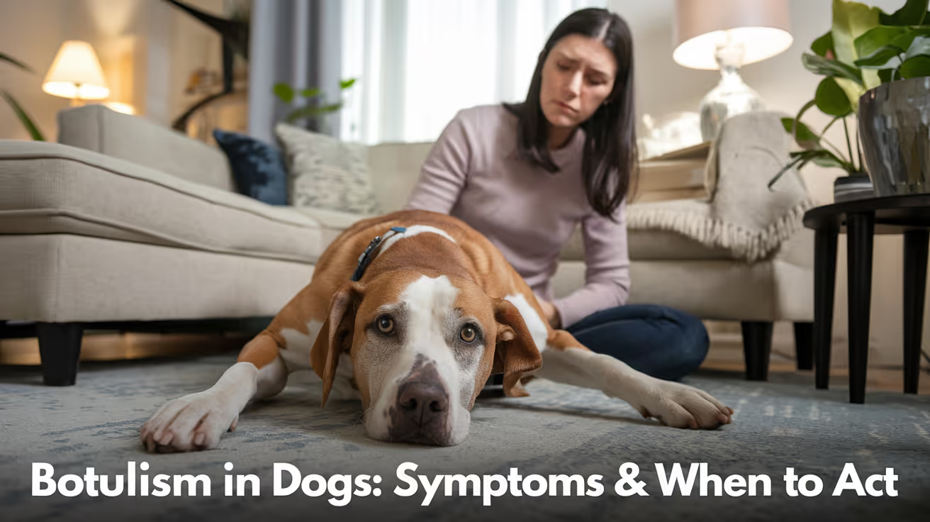
Botulism Symptoms in Dogs and When to Seek Help
Learn to recognize botulism symptoms in dogs and know when to seek urgent veterinary help to protect your pet's health.
Botulism is a rare but potentially fatal condition in dogs. It is not an infection it is a poisoning. The botulinum toxin produced by Clostridium botulinum bacteria blocks the nerve signals that control muscle movement, causing a progressive paralysis that can reach the breathing muscles.
Most dogs that develop botulism have eaten a dead animal or contaminated raw meat. The condition requires immediate veterinary attention the window for antitoxin treatment is narrow.
Quick answer: Botulism causes ascending paralysis in dogs, starting in the hind legs and progressing to breathing muscles. It is caused by ingesting botulinum toxin from decomposing carcasses or meat. Call your vet immediately.
Key takeaways
- Botulism is a poisoning, not a bacterial infection: the dog ingests preformed toxin, not live bacteria
- Symptoms begin 12 to 72 hours after ingestion and can be delayed up to 6 days
- Ascending paralysis is the hallmark: weakness starts in the hind legs and progresses forward to breathing muscles
- The dog remains mentally alert throughout, unlike many other neurological conditions
- Death can occur if the breathing muscles or heart are paralyzed
- Antitoxin is most effective before paralysis sets in early veterinary contact is essential
What is botulism in dogs?
Botulism is caused by the botulinum toxin, produced by the anaerobic bacterium Clostridium botulinum. VCA Animal Hospitals explains what happens after ingestion: "The botulinum toxin is then carried throughout the body, where it binds to nerve cells, resulting in paralysis."
The toxin works by blocking acetylcholine the neurotransmitter that triggers muscle contraction. Without acetylcholine signaling, muscles cannot contract. The result is flaccid (floppy) paralysis: muscles that are not stiff or spastic, but simply limp and non-functional.
Merck Veterinary Manual emphasizes that "botulism is a poisoning, not a true infection with bacteria." The bacteria themselves do not spread through the dog's body the damage is entirely from the preformed toxin that was ingested.
How do dogs get botulism?
The most common route is eating a contaminated carcass or spoiled meat. PetMD: "Dogs become affected by eating decomposing animal carcasses or spoiled vegetation where the bacterium grows."
Specific exposure scenarios include:
- Eating road kill or carrion found during walks (the most common cause)
- Consuming spoiled or improperly stored meat even cooked meat can harbor toxin if contaminated before cooking; AnimalWised notes that "even boiling doesn't necessarily destroy" the toxin once formed
- Access to garbage or compost containing decomposing organic material
- Rural and hunting dogs with access to wildlife carcasses are at higher risk
Botulism is most common in wild waterfowl and only rarely occurs in dogs. Merck Veterinary Manual notes: "Botulism is most common in wild water birds, like ducks, and only rarely occurs in dogs." This rarity is why many vets may not immediately consider it in the differential diagnosis.
Symptoms of botulism in dogs
VCA Animal Hospitals lists the clinical timeline: "Clinical signs of botulism typically develop 12 to 72 hours after ingesting contaminated meat, though the onset can be delayed up to six days."
Early symptoms (12 to 48 hours after ingestion)
- Vomiting (may occur very early as the toxin begins affecting the gut)
- Weakness in the hind legs the dog appears wobbly or reluctant to stand
- Difficulty rising from lying down
- Reduced muscle tone throughout
Progressive symptoms (as paralysis ascends)
- Ascending weakness: hind legs first, then forelimbs
- Difficulty swallowing (dysphagia) the dog may drool or be unable to eat or drink
- Changes in bark or voice (laryngeal muscle involvement)
- Vision problems (eye muscle weakness)
- Dropped jaw or facial droop
Vetster: "Symptoms of botulism generally develop within 24 to 48 hours after ingestion and include vomiting, difficulty swallowing, and progressive ascending motor paralysis that starts in the rear limbs."
Critical symptoms seek emergency care immediately
- Inability to breathe normally (labored breathing, open-mouth breathing)
- Complete inability to move
- Collapse
- Rapid heart rate
Merck Veterinary Manual: "Death can occur if breathing muscles or the heart is paralyzed."
The key distinguishing feature: the dog stays alert
A dog with botulism appears mentally aware throughout the paralysis. The toxin affects motor nerves (which control muscle movement) but not sensory perception or consciousness. WagWalking: "Should you notice that your dog appears alert but is unable to move, it is important to rush him to your veterinarian."
This alertness-with-paralysis combination is one of the strongest clinical clues pointing toward botulism rather than other neurological causes.
Conditions that look like botulism
The symptoms of botulism overlap significantly with several other conditions. Diagnosis is difficult and requires ruling out alternatives.
PetsVetCheck: "The diagnosis relies on the medical history and the exclusion of other causes, as specific tests are time-consuming and not always available."
How vets diagnose botulism
Diagnosis is primarily clinical based on the pattern of signs and the history of possible exposure.
History: the most important element. Has your dog had access to carcasses, garbage, or raw meat in the past 1 to 6 days?
Physical examination: assessing the pattern of weakness (ascending, flaccid), the level of alertness, and the presence of cranial nerve signs (swallowing, vision, jaw tone).
Toxin testing: blood, feces, vomit, and food samples can be tested for botulinum toxin. Vetster confirms this but notes that tests are time-consuming and not universally available treatment should not be delayed while waiting for results.
Rule-out testing: tick check (to rule out tick paralysis), chest X-ray (to assess respiratory function), bloodwork to rule out metabolic or endocrine causes.
Treatment
Antitoxin
Antitoxin binds and neutralizes circulating toxin that has not yet attached to nerve cells. VCA: "There is no vaccine available, although an antitoxin is available if the condition is identified before signs develop."
Important limitation: antitoxin only works on toxin still in circulation. Once the toxin has bound to nerve cells, it cannot be removed. This is why early diagnosis matters enormously.
Antibiotic warning: Vetster advises: "Contact a veterinarian before administering antibiotics as they can cause an increase in botulism toxin concentration, and some kinds of antibiotics can cause a worsening in neuromuscular function." Antibiotics that kill Clostridium in the gut can release additional toxin, worsening the condition.
Decontamination
If exposure is very recent and the dog is not yet showing signs, the vet may induce vomiting or administer activated charcoal or an enema to remove unabsorbed toxin from the digestive tract.
Supportive care
Once paralysis is established, treatment is primarily supportive:
- IV fluids to maintain hydration
- Assisted feeding soft or liquid diet; syringe feeding if swallowing is impaired
- Bladder management manual emptying if the dog cannot urinate independently
- Turning and repositioning every 2 to 4 hours to prevent bed sores in paralyzed dogs
- Respiratory support oxygen supplementation or mechanical ventilation if breathing muscles are compromised
- Prevention of aspiration pneumonia a major complication in dogs with swallowing difficulties
AnimalWised: "Dogs which have a more serious clinical picture will need help being moved. They may be given intravenous fluid to help their system work. If the dog fails to urinate on their own, the dog will have their bladder emptied manually."
Prognosis and recovery
Vetster: "With prompt and long-term, intensive nursing care, most paralyzed dogs recover within 1 to 3 weeks." The prognosis depends on two main factors: the dose of toxin ingested and the speed of treatment.
Dogs that receive antitoxin before significant paralysis develops have the best outcomes. Dogs with respiratory compromise have a significantly worse prognosis respiratory failure is the primary cause of death in severe cases.
Nerve regeneration after toxin binding is a natural process that occurs over weeks. The nerve terminals regrow and acetylcholine signaling is restored the dog essentially has to wait for the toxin's effects to dissipate as new nerve terminals form.
PetsVetCheck: "Long-term complications can include muscle weakness and, in some cases, permanent neurological deficits, depending on the duration and severity of the paralysis."
Prevention
- Prevent access to carcasses and carrion during walks, especially in rural or wildlife-rich areas
- Supervise dogs in areas with road kill or dead animals
- Store meat and food properly never feed dogs meat that has been improperly stored or smells off
- Keep dogs away from garbage, compost bins, and waste containing organic material
- Recall and leash train to prevent opportunistic scavenging
Frequently asked questions
My dog ate something dead on a walk. Should I go to the vet now?
Yes, contact your vet immediately. Eating a dead animal is the primary exposure route for botulism in dogs. Even if your dog shows no symptoms, your vet may recommend inducing vomiting or monitoring closely. Early decontamination before symptoms develop is significantly more effective than treatment after paralysis has begun.
What does botulism look like in dogs compared to a stroke?
A stroke typically affects one side of the body (head tilt, circling, one-sided weakness) and the dog may be disoriented or have altered consciousness. Botulism produces symmetric, ascending weakness with the dog remaining fully alert and mentally aware. A dog that cannot move but responds normally to voice and touch, and does not appear confused, is a stronger candidate for botulism than stroke.
Can humans get botulism from a dog that has it?
Botulism is not contagious it is a toxin poisoning, not a transmissible infection. You cannot get botulism from contact with an affected dog. However, if a shared food source caused the dog's illness, that same food could potentially affect humans. Identify and safely discard any suspected contaminated food.
How long does recovery from botulism take in dogs?
Most dogs that survive botulism and receive appropriate supportive care recover within 1 to 3 weeks. Recovery depends on regrowing the nerve terminals that the toxin has damaged this is a biological process that cannot be accelerated, only supported. Dogs with mild toxin exposure recover faster than those with severe paralysis.
Is there a vaccine for botulism in dogs?
No vaccine is currently available for dogs against botulism. Vaccines exist for livestock species in some countries, but canine botulism is rare enough that vaccination is not a standard recommendation. Prevention relies on limiting exposure to the toxin through management and supervision.
Why do vets say to avoid antibiotics with botulism?
Certain antibiotics (particularly aminoglycosides like gentamicin and neomycin) interfere with neuromuscular junction function and can worsen paralysis. Additionally, killing Clostridium bacteria in the gut can cause them to release additional stored toxin. If antibiotics are needed for a secondary complication (such as aspiration pneumonia), the vet will choose an antibiotic class that does not worsen neuromuscular function.
Resources
- VCA Animal Hospitals. Botulism in Dogs. vcahospitals.com
- PetMD. Botulism in Dogs. petmd.com
- Merck Veterinary Manual. Botulism in Dogs. merckvetmanual.com
- Vetster. Botulism (Clostridium Botulinum Toxin) in Dogs. vetster.com
- WagWalking. Botulism in Dogs. wagwalking.com

Pressure Necrosis in Dogs: Causes, Symptoms & Care
Learn about pressure necrosis in dogs, including causes, symptoms, and effective care to protect your pet's health.
Pressure necrosis in dogs also called decubital ulcers or bedsores is the progressive death of skin and underlying tissue caused by sustained pressure cutting off blood supply. It follows the same biological mechanism as bedsores in bedridden humans, and it is a real and preventable risk for any dog that spends long periods lying in one position.
The condition is deceptive: it starts as a small area of hair loss and can advance to a deep open wound reaching muscle or bone within days. Recognizing it at Stage 1 is the goal.
Quick answer: Pressure necrosis occurs when sustained pressure on bony prominences like elbows and hips cuts off blood flow, killing tissue. It is most common in senior, immobile, or post-surgical dogs. Early signs are hair loss and redness.
Key takeaways
- Prolonged pressure on bony prominences blocks blood flow and causes tissue death from the inside out
- Senior, large-breed, paralyzed, and post-surgical dogs are at highest risk
- Common sites include the elbows, hips, hocks, and the outside of the knees
- Stage 1 (hair loss and redness) is the ideal intervention point; the wound has not yet opened
- Stage 3 and 4 wounds require surgical debridement and often systemic antibiotics
- Orthopedic foam bedding plus repositioning every 2 to 4 hours is the most effective prevention combination
What causes pressure necrosis in dogs
Walkin' Pets explains the mechanism clearly: "Decubital ulcers form when repeated pressure on an affected area constricts the blood vessels, decreasing the area's blood supply and as the skin is deprived of oxygen, it begins to die."
This process concentrates at bony prominences where bone is close to the skin surface with little muscle or fat as a buffer. In dogs, the most commonly affected sites are:
- Elbows the most frequent site, especially in large and giant breeds
- Lateral hips and hip bones
- Hocks (the ankle joint of the hind leg)
- Stifles (outer surface of the knee)
- Sides of the ribs in dogs that always lie on one side
The damage does not require continuous lying. Ortocanis notes that high pressure over a short period is as damaging as lower pressure sustained over longer periods. A heavy dog that drops onto a hard floor with force can create the same tissue trauma as extended immobility.
Which dogs are most at risk
Any dog can develop pressure necrosis if the conditions are right, but several factors increase risk substantially:
Senior dogs: muscle mass decreases with age, reducing the natural padding over bony prominences. Reduced mobility means longer uninterrupted periods in the same position.
Large and giant breeds: more body weight means greater compressive force at contact points. Dr. Buzby's ToeGrips notes that large breeds are disproportionately affected even when they are healthy and mobile.
Post-surgical patients: strict rest requirements, post-anesthesia sedation, and pain-related reluctance to move all increase time in one position.
Paralyzed or paraplegic dogs: no ability to self-reposition at all. These dogs require active owner repositioning to prevent pressure injury.
Obese dogs: higher body weight amplifies compressive force at every contact point.
Short-coated breeds: less coat means less cushioning between skin and hard surfaces.
For how callus formation at the same pressure sites relates to early pressure damage, see calluses as chronic pressure injuries.
The 4 stages of pressure necrosis
Stage 1: redness and hair loss
The skin over the bony prominence loses hair and becomes pink or red. The area may feel slightly warm but the skin surface is intact. There is no open wound.
What to do: act immediately. Add thick orthopedic padding under the affected area. Increase position changes to every 2 hours. Monitor the site daily. Stage 1 reverses with proper management.
Stage 2: surface skin breakdown
The skin partially breaks down. A shallow blister or open area appears. The wound is painful and the dog will likely lick or chew it, worsening the damage.
What to do: veterinary wound care is needed at this stage. Home management alone is not sufficient. The vet will clean the wound, assess for infection, and prescribe appropriate dressings and possibly antibiotics.
Stage 3: full-thickness wound
The sore extends through the full skin thickness into subcutaneous tissue. WagWalking: "Pressure sores can progress quickly, leading to deep wounds that cause the muscle and bone to become visible."
Infection is common at Stage 3. The wound has discharge and often a foul odor. Bacteria can include Staphylococcus, Pseudomonas, and Enterococcus species.
What to do: aggressive veterinary management including wound debridement, culture-guided antibiotics, and advanced wound dressings.
Stage 4: deep tissue involvement
The wound extends to muscle, tendon, or bone. This is a potentially life-threatening stage. Sepsis (systemic bacterial infection) becomes a risk when deep tissue infection spreads.
What to do: emergency veterinary care. Surgical debridement, hospitalization, IV antibiotics, and possibly reconstructive surgery are required.
Treatment by stage
For Stage 1 and 2 home wound care, clean the area gently with dilute chlorhexidine or mild antiseptic solution. Apply a non-adherent wound dressing and change it daily. Do not use hydrogen peroxide, which damages healing tissue. Keep the dog away from hard surfaces at all times until the wound has closed and resolved.
For how bed sores and pressure necrosis overlap and how prevention differs, see bed sores as a related pressure injury.
Prevention: what actually works
Orthopedic bedding
Thick memory foam or egg-crate foam beds distribute body weight more broadly, reducing peak pressure at bony points. The bedding must not fully compress under the dog's weight to be effective. Check the foam by pressing down firmly: if it bottoms out immediately, it offers little protection for a large dog.
Waterproof covers that can be washed frequently are important for dogs at risk of incontinence, which is common in paralyzed patients.
Repositioning schedule
Reposition an immobile dog every 2 to 4 hours, alternating which side bears weight. This mimics the natural unconscious repositioning healthy dogs do throughout the day.
Ortocanis: "This problem occurs mainly in immobile animals: quadriplegic or paraplegic dogs, elderly dogs or those with serious illnesses."
Daily inspection
Check every bony prominence daily. Run your hand over the elbows, hips, hocks, and knees. Hair loss is the first visible sign act at that point rather than waiting for skin breakdown.
Protective wraps and donut pads
Elbow pads and donut-shaped protective wraps can be placed over at-risk sites to prevent direct contact with hard surfaces. These are particularly useful for dogs that regularly rest on hard floors despite having soft bedding available.
For how swelling from pressure injuries can extend into the feet and legs, see swelling associated with pressure injuries.
Frequently asked questions
How do I know if my dog has a pressure sore starting?
The first sign is a patch of hair loss over a bony prominence such as the elbow or hip, sometimes with mild redness. The skin is still intact at this point. Run your hand along these sites daily in at-risk dogs. If you feel a bare or reddened area, increase padding and position changes immediately and monitor closely for any skin breakdown.
My dog is recovering from surgery and lying down constantly. How do I prevent pressure sores?
Reposition every 2 to 4 hours, alternating sides. Use thick orthopedic foam padding rather than thin blankets or bare floors. Check the elbows, hips, and hocks each time you reposition. Ask your vet at discharge whether there are specific at-risk sites based on your dog's size, breed, and surgical procedure.
The skin over my dog's elbow looks bare and red but is not open. Is this a pressure sore?
Yes, this is Stage 1 pressure necrosis. The tissue is under sustained pressure but has not yet broken down. This is exactly the time to act: add thick padding, protect from friction, and monitor daily. If the skin opens, see your vet within 24 to 48 hours.
Can pressure necrosis be fully healed?
Stage 1 and 2 lesions heal well with proper wound care and pressure relief. Stage 3 and 4 wounds can heal but often leave permanent scar tissue, and may involve months of treatment. Full wound closure at advanced stages is not always achievable and managing infection becomes the primary goal.
What is the best bedding to prevent pressure sores?
Thick memory foam or high-density egg-crate foam is most effective. The padding needs to be dense enough not to fully compress under the dog's full weight. Standard flat beds offer minimal protection for large dogs. Waterproof removable covers allow frequent washing, which reduces infection risk at the skin surface.
Can a pressure sore become life-threatening?
Yes. Stage 3 and 4 wounds can allow bacteria to spread from the wound into deeper tissues or the bloodstream, causing sepsis. A dog with a deep infected ulcer that is also lethargic, off food, or running a fever may be systemically ill and needs emergency veterinary care that day.
Resources
- Dr. Buzby's ToeGrips. Pressure Sores on Dogs: Treatment and Prevention. toegrips.com
- WagWalking. Pressure Sores in Dogs. wagwalking.com
- Walkin' Pets. Pressure Sores in Pets: Treatment and Prevention. walkinpets.com
- Ortocanis. Pressure Ulcers in Convalescent Dogs. ortocanis.com
- DogCancer.com. Bed Sores on Dogs: Preventing and Treating Pressure Sores. dogcancer.com
How Long After Neutering Is Testosterone Gone in Dogs
Learn how long testosterone stays in dogs after neutering and what to expect during recovery.
Neutering removes the testicles, which produce most of a dog's testosterone. But the hormone does not vanish from the bloodstream on surgery day.
Testosterone clears gradually as the body metabolizes what remains. Behavior follows the same curve. This is why owners who expect an immediate behavioral shift are often disappointed, and why patience in the weeks after surgery is not just advisable but necessary.
Here is the complete, evidence-based picture of the testosterone clearance timeline and what it means for your dog.
Quick answer: Testosterone levels begin declining within 24 to 72 hours of neutering. Significant reduction occurs within 2 to 3 weeks. Most testosterone is cleared from the bloodstream within 4 to 6 weeks. Full behavioral stabilization typically takes 2 to 3 months as the body and brain adjust to the new hormonal environment. Some habits shaped by testosterone may persist beyond this window and require training to address.
Key takeaways
- Testosterone production stops on surgery day: The testicles are removed, eliminating the primary production source immediately.
- Clearance from the bloodstream takes 4 to 6 weeks: Residual testosterone circulates and is metabolized gradually.
- Behavioral changes follow the hormonal decline: Most testosterone-driven behavioral improvements appear over 2 to 6 weeks.
- Full behavioral stabilization takes 2 to 3 months: Some behaviors fade slowly as hormone levels drop.
- Habits shaped by testosterone may persist: Learned behaviors continue regardless of hormone levels and require training.
- Fertility persists for up to 6 weeks: Residual sperm in the reproductive tract can still result in pregnancy.
What testosterone does in male dogs
Testosterone is produced primarily by the testicles. It drives a specific set of behaviors and physical characteristics in intact male dogs:
- Urine marking: The drive to mark territory with urine is heavily testosterone-dependent.
- Mounting behavior: Sexual and status-related mounting is driven by testosterone.
- Roaming: The drive to escape and find a mate is strongly testosterone-mediated.
- Inter-male aggression: Competition-based aggression with other intact males is often testosterone-linked.
- Dominance behaviors: Some dominant social behaviors decrease after neutering.
Beyond behavior, testosterone influences muscle mass, certain aspects of bone density, and the dog's general physical development.
The testosterone clearance timeline
Understanding this timeline is the most important practical takeaway from this article.
Hours 0 to 72: production stops, clearance begins
The testicles are removed during surgery. Testosterone production essentially stops at that moment.
However, the hormone that was already circulating in the bloodstream at the time of surgery does not disappear immediately. The body metabolizes and clears it through normal hormonal processing.
Within the first 24 to 72 hours, testosterone levels begin their decline. This is measurable but does not immediately translate into behavioral change.
Days 7 to 10: significant early reduction
By one to two weeks post-surgery, testosterone levels have dropped substantially from their pre-surgery baseline. Most of the actively circulating hormone is being metabolized.
This is still not the point at which behavioral changes are reliable. The hormonal environment is in transition, and the brain and behavior patterns are adjusting.
Weeks 4 to 6: functional clearance
By four to six weeks post-neutering, testosterone has reached very low or undetectable levels in most dogs. This is the point that veterinarians generally refer to when asked how long it takes for testosterone to "go away."
Some sources cite two weeks for functional clearance; others cite six weeks. The variation reflects individual differences in age, health, and metabolism. Younger dogs tend to clear residual testosterone faster.
Months 2 to 3: full behavioral stabilization
Even after testosterone levels have dropped to baseline, the brain and learned behavioral patterns continue adjusting to the new hormonal environment.
By two to three months post-neutering, most testosterone-driven behavioral changes have stabilized. This is the point at which you can accurately assess whether neutering has produced the behavioral improvements you were hoping for.
Timeline summary table
What behaviors improve as testosterone declines
These are the behaviors most reliably reduced as testosterone levels fall:
High likelihood of improvement:
- Urine marking indoors and outdoors
- Mounting behavior toward people, other dogs, or objects
- Roaming and escape attempts to seek females in heat
- Some forms of inter-male aggression driven by competition
Variable or uncertain improvement:
- Dominance-related behaviors that have become habitual
- Aggression toward familiar people (testosterone may contribute but is not the sole driver)
- General excitability (energy level is more breed and fitness-driven than hormonal)
Behaviors that will not improve:
- Fear-based aggression (not testosterone-driven)
- Separation anxiety (not testosterone-driven)
- Learned behaviors from training history (habits, regardless of their origin)
- Resource guarding (not testosterone-driven)
Why some behaviors persist after testosterone is gone
This is the part of the conversation that surprises many owners.
Some behaviors that appeared to be testosterone-driven become habitual through repetition. The neural pathways associated with the behavior have been reinforced many times, and they do not disappear when the hormonal fuel is removed.
A dog that has been marking territory for three years before neutering has established a behavioral habit. The testosterone-driven urge may be gone, but the habit of going to specific spots and marking may continue. This requires training to address, not just time.
The general rule: behaviors that have been performed repeatedly over time are more likely to persist after neutering than behaviors that are recent or infrequent.
For how testosterone decline affects dog behavior in the context of the full behavioral landscape after sterilization, that guide covers what the evidence says about which changes are most reliable. For the equivalent question in females, the guide on whether hormone changes calm female dogs walks through which female dogs see the calming effect and which do not.
The fertility window: keep intact females away
One important practical point that owners sometimes overlook: a neutered male can still get an unspayed female pregnant for up to six weeks after surgery.
Sperm are stored in the reproductive tract beyond the testicles, in the vas deferens and associated structures. These residual sperm remain viable for several weeks after the testicles are removed.
Keep your neutered male dog away from intact females for the full six weeks after surgery. This is not a precaution against unlikely events. It is a necessary management step.
Factors that affect how quickly testosterone clears
The 4 to 6 week timeline is a general guide. Individual variation exists.
Age: Younger dogs tend to metabolize and clear testosterone faster than older dogs. Their overall metabolic rate is higher.
Health status: Dogs with liver or kidney impairment may metabolize hormones more slowly.
Pre-surgery testosterone levels: A dog with very high testosterone levels before surgery may take longer to reach baseline.
Individual physiology: As with most biological processes, individual variation exists between dogs of the same age and breed.
None of these factors change the approach. Patience through the full two to three month window remains appropriate regardless.
How testosterone decline affects female dogs after spaying
While this article focuses on testosterone in male dogs, it is worth noting the parallel process in females.
Spaying removes the ovaries, which produce estrogen and progesterone. These hormones do not vanish on surgery day either. They decline over a similar 4 to 6 week clearance window, with behavioral stabilization taking two to three months.
The behavioral changes driven by the female hormonal cycle, including heat restlessness, roaming, false pregnancy behaviors, and cyclical mood shifts, follow the same general clearance curve as testosterone in males.
For how hormonal behavior changes in female dogs specifically, including which behaviors are most reliably reduced and over what timeline, that guide provides the female-specific detail.
Frequently asked questions
How long does it take for testosterone to fully disappear after neutering?
Most testosterone is cleared from the bloodstream within 4 to 6 weeks of neutering in healthy adult dogs. Behavioral stabilization, reflecting the brain's adjustment to the new hormonal environment, typically takes 2 to 3 months. Some habits may persist beyond this window and require training.
My dog was neutered two weeks ago and is still marking. Is something wrong?
No. Two weeks is within the normal clearance window. Testosterone is still declining. Most owners see improvements in marking behavior over 4 to 8 weeks. If marking continues at the same level beyond 3 months, it may have become a learned habit that requires training intervention.
Can my neutered dog still get a female pregnant?
Yes, for up to 6 weeks after surgery. Residual sperm in the reproductive tract remain viable after the testicles are removed. Keep your recently neutered male away from intact females for the full 6-week window.
Will my dog's behavior change immediately after neutering?
No. The first two weeks are recovery from surgery. Behavioral changes related to testosterone decline begin appearing in weeks 3 to 6, with full stabilization at 2 to 3 months. Expecting behavioral change within days of surgery leads to frustration and misattribution.
Does the age at neutering affect how quickly testosterone clears?
Somewhat. Younger dogs tend to metabolize hormones faster due to higher metabolic rates. However, the practical difference in clearance timeline is small compared to the individual variation between dogs. The 4 to 6 week clearance timeline remains a reasonable guide regardless of age. For whether lower testosterone calms dogs down and how the behavioral changes of testosterone decline manifest, that guide addresses the calming question across different dog types. For recovery timeline alongside hormonal changes, that guide places the hormonal adjustment within the broader post-surgical recovery picture.
Understanding the testosterone clearance timeline reframes the post-neutering period from a frustrating wait into an understandable process. The behavior changes are coming, but they follow the hormone. Give it four to six weeks for the hormonal shift and two to three months for the behavioral picture to stabilize. What you see at month three is an accurate reflection of what surgery has and has not changed.
Resources
The following sources were used as reference and background for this article:
- Small Dogs Heaven. Understanding the Timeline: How Long After Neutering a Dog is Testosterone Gone? smalldogsheaven.com
- The Beloved Dog. Your Dog's Testosterone: When It's Gone After Neutering. thebeloveddog.com
- Dog Hutt. How Long After Dog Neutering Is Testosterone Gone? doghutt.com
- Dogo App. How Long After Neutering Dog Is Testosterone Gone? dogo.app
- American Veterinary Medical Association (AVMA). Spaying and Neutering. avma.org
- Embrace Pet Insurance. Dog Neuter and Spay Recovery. embracepetinsurance.com
Pustules in Dogs Causes and Treatment
Learn about pustules in dogs, their causes, symptoms, and effective treatments to keep your pet healthy and comfortable.
A pustule is a small, pus-filled bump on the skin. In dogs, pustules are one of the most common signs of a bacterial skin infection collectively called pyoderma. They look like pimples: red and raised, with a white or yellow pus-filled center.
Pustules vary widely in severity. Some are superficial and clear with topical treatment. Others signal a deep infection that needs oral antibiotics for weeks, or an underlying disease that must be found and treated to prevent recurrence.
Quick answer: Pustules in dogs are almost always caused by pyoderma (bacterial skin infection), most commonly Staphylococcus pseudintermedius. They appear as red pus-filled bumps on the belly or around hair follicles. Treatment ranges from medicated shampoo to oral antibiotics.
Key takeaways
- Staphylococcus pseudintermedius causes over 90% of canine pyoderma and pustule formation
- Three depth levels exist: surface pyoderma, superficial bacterial folliculitis, and deep pyoderma, each needing different treatment
- Pustules that rupture leave circular crusts called epidermal collarettes, a hallmark sign vets look for
- Allergies, parasites, and hormonal disorders are the most common underlying causes of recurring pustules
- Skin cytology (examining cells from a pustule under a microscope) is the first and most useful diagnostic step
- Full antibiotic courses are essential: stopping treatment early is the most common reason pyoderma returns
What causes pustules in dogs?
The primary cause: bacterial infection (pyoderma)
PetMD confirms that the most common bacterial cause is "Staphylococcus pseudintermedius, which is responsible for more than 90 percent of cases. This type of bacteria normally inhabits the skin, but it can increase in number and cause problems when the skin barrier is damaged or unhealthy."
Other bacteria involved less commonly include Staphylococcus schleiferi, Staphylococcus aureus, E. coli, Pseudomonas, Streptococci, and Proteus. In most cases, these are secondary invaders that take advantage of already-damaged skin.
Why does the skin become infected?
Healthy dry dog skin is highly resistant to bacterial overgrowth. Merck Veterinary Manual explains: "Any skin disease that changes the normally dry, desert-like environment to a more humid environment can cause overcolonization of the skin with bacteria."
Common triggers that damage the skin barrier and allow infection:
- Allergies (environmental allergens, food, contact irritants): the most common trigger; chronic scratching and licking disrupts skin integrity
- External parasites (fleas, mites, mange): bite wounds and inflammatory reactions create entry points
- Hormonal disorders (hypothyroidism, Cushing's disease, diabetes): alter skin physiology and immune response
- Keratinization disorders: abnormal keratin production creates conditions for bacterial overgrowth
- Poor grooming or dirty environments: increased moisture and bacterial load on the skin surface
- Physical trauma: cuts, wounds, or abrasions that open the skin to bacterial colonization
The three types of canine pyoderma
Understanding which type your dog has determines the treatment approach.
1. Surface pyoderma
The infection involves only the very outermost skin layer (stratum corneum). It does not penetrate into hair follicles or deeper tissue.
Examples: puppy impetigo (belly pustules in puppies), skin fold pyoderma (in facial folds, lip folds, tail folds), hot spots (acute moist dermatitis).
Signs: redness, pustules, and crusting on hairless or folded skin areas.
Treatment: topical therapy alone is usually sufficient medicated wipes, chlorhexidine spray, or topical antibiotic.
2. Superficial bacterial folliculitis (SBF)
The infection involves the epidermis and the upper portion of hair follicles. VCA Animal Hospitals describes the visual result: "In short-haired breeds, the coat may appear to protrude or stick up in areas, mimicking hives, or it may appear moth-eaten because of patchy hair loss."
Signs: pustules and papules around hair follicles, circular crusts called epidermal collarettes, hair loss in a patchy pattern, itching. Dr. Buzby's ToeGrips lists the full spectrum: "papules, pustules, collarettes, crusts, and erythema of the armpits and groin."
Treatment: topical therapy plus oral antibiotics in most cases; minimum 3 to 4 weeks of treatment.
3. Deep pyoderma
The infection penetrates into the dermis (deep skin layer) and may cause furunculosis rupture of hair follicles releasing bacteria and keratin into surrounding tissue. This is the most serious form.
Signs: painful, thickened skin; draining tracts; crusts over larger areas; foul odor; systemic signs in severe cases (lethargy, fever).
WebMD Pets: "Deep pyoderma will appear as pain at the infected site, crusting, a bad smell, and oozing of pus and blood."
Treatment: prolonged oral antibiotics (often 6 to 8 weeks or more), guided by culture and sensitivity results; treatment of the underlying cause is essential.
For how pustule-causing infections can progress into deep abscesses, see furuncles as a severe progression of pustules.
What pustules look like at each stage
VCA Animal Hospitals: "The most common clinical signs associated with pyoderma are papules or pustules that form on the skin. These lesions often look similar to pimples in humans. They are most often red and raised, with a white pus-filled center."
How vets diagnose the cause of pustules
A vet will usually start with a physical examination and skin cytology. Further tests are ordered if the infection is severe, recurrent, or unresponsive to initial treatment.
Skin cytology: a glass slide is pressed against a pustule or ruptured lesion and examined under a microscope. Dr. Buzby's ToeGrips explains: "Seeing inflammatory cells and bacteria makes a pyoderma likely. And bacteria identified inside of cells is a slam-dunk for a diagnosis."
Culture and sensitivity: a swab is taken from inside a pustule or lesion and sent to a lab. This identifies the exact bacterial species and which antibiotics will and will not work. Essential for recurrent or non-responsive infections.
Skin scraping: rules out mites (demodex, sarcoptes) which can cause secondary bacterial infections.
Wood's lamp: rules out ringworm (dermatophytosis), which can mimic superficial pyoderma.
Bloodwork: thyroid panel, cortisol testing, or full chemistry panel to screen for hormonal disorders in adult dogs with recurring pustules.
Treatment
Topical treatment
Medicated shampoos containing chlorhexidine (2 to 4%) or benzoyl peroxide are the foundation of topical pyoderma management. Used 2 to 3 times per week, they reduce bacterial load on the skin surface and support antibiotic treatment.
Topical sprays, wipes, and creams are useful for localized lesions. Chlorhexidine solution applied directly to pustule-bearing areas between baths is effective for surface pyoderma.
Oral antibiotics
Required for superficial bacterial folliculitis and all deep pyoderma. Common choices include:
- Cephalexin (first-line for most uncomplicated cases)
- Amoxicillin-clavulanate
- Trimethoprim-sulfamethoxazole
- Clindamycin
Treatment duration matters enormously. Merck Veterinary Manual is explicit: "Even though your dog may seem better after only a few days or a week, it is still very important for you to continue the prescribed treatment program for the full length of time. The bacteria causing pyoderma can still be present and ready to multiply again if the complete course of medication is not given."
Minimum durations: 3 to 4 weeks for superficial folliculitis; 6 to 8 weeks or longer for deep pyoderma. The infection should be resolved for at least 7 days before stopping antibiotics.
Treating the underlying cause
Merck Veterinary Manual identifies the most common reasons pyoderma recurs: "failure to treat underlying causes, use of glucocorticoid drugs, and inappropriate treatment with prescribed antibiotic medications."
If your dog has recurring pustules, the vet will investigate for allergies, parasites, or hormonal disease. Controlling the underlying cause is the only way to prevent recurrence.
For how impetigo specifically presents as pustules in puppies on the belly and groin, see impetigo as a pustule-causing condition.
Home care during treatment
- Give all medications on schedule and complete the full course
- Bathe with the prescribed medicated shampoo 2 to 3 times per week leave it on for 10 minutes before rinsing
- Keep the affected skin dry between baths; moisture encourages bacterial regrowth
- Prevent licking with an E-collar if your dog is bothering the lesions
- Wash bedding weekly to reduce environmental bacterial load
- Do not squeeze or pop pustules this spreads bacteria and may cause scarring
For how skin conditions that cause scabs relate to the pustule cycle, see skin conditions that cause scabs and pustules.
Frequently asked questions
Are pustules in dogs contagious to humans or other pets?
Most canine pyoderma is caused by Staphylococcus pseudintermedius, which does not routinely infect healthy humans. However, Staphylococcus aureus can occasionally transfer between species, including to immunocompromised people. As a precaution, wash hands after handling a dog with active pustules and keep other pets separated if the infection is extensive.
How long does it take for pustules to clear up?
With appropriate antibiotic treatment, pustules typically begin to dry and resolve within 7 to 14 days. However, treatment must continue for the full prescribed course (minimum 3 to 4 weeks for superficial infections) even after the skin looks normal. Stopping early is the most common cause of recurrence.
My dog keeps getting pustules even after treatment. Why?
Recurring pustules almost always indicate an unidentified or untreated underlying cause. Allergies are the most common culprit the chronic itching and skin damage from allergies continuously set up conditions for bacterial infection. Inadequate antibiotic duration and bacterial resistance are other causes. Ask your vet to investigate for allergies, mites, or hormonal disorders if your dog's pustules keep returning.
Can I use human acne products on my dog's pustules?
No. Products containing benzoyl peroxide at human concentrations, salicylic acid, retinoids, or alcohol can irritate or damage dog skin. Veterinary formulations of benzoyl peroxide shampoo (at lower concentrations designed for dog skin) are appropriate. Use only products your vet recommends.
What is an epidermal collarette and should I be concerned about it?
An epidermal collarette is a circular scale or crust ring left behind when a pustule ruptures. It is not itself a sign of active infection but indicates that a pustule was recently present. Your vet will note collarettes during examination as evidence of pyoderma. They typically resolve as the skin heals during treatment.
When should pustules in a dog be treated as an emergency?
Most pyoderma is not an emergency. Seek same-day care if your dog has rapidly spreading skin lesions, swelling, signs of pain, fever, lethargy, or is not eating. These signs suggest deep infection or a systemic response. Also contact your vet urgently if the skin appears dark, necrotic, or has a foul odor signs that deep tissue may be involved.
Resources
- PetMD. Pyoderma in Dogs. petmd.com
- VCA Animal Hospitals. Pyoderma in Dogs. vcahospitals.com
- Merck Veterinary Manual. Pyoderma in Dogs. merckvetmanual.com
- Dr. Buzby's ToeGrips. Pyoderma in Dogs: Signs, Diagnosis, and Treatment. toegrips.com
- WebMD Pets. What Is Pyoderma in Dogs? webmd.com

Bed Sores on Dogs Causes Treatment and Prevention
Learn about bed sores on dogs, including causes, treatments, and prevention tips to keep your pet healthy and comfortable.
Bed sores in dogs also called pressure sores or decubitus ulcers develop when prolonged pressure on a bony area cuts off blood supply, causing the tissue to break down. They are the same condition as bedsores in hospitalized humans, and they are just as serious when they reach advanced stages.
The good news is that early-stage bed sores are very treatable at home. The bad news is that they can progress from a small red patch to a deep infected wound within days if not caught and addressed.
Quick answer: Bed sores form over bony prominences like elbows and hips when sustained pressure blocks blood flow. Most common in senior, large-breed, and immobile dogs. Early stages respond to soft bedding and repositioning; advanced stages need veterinary care.
Key takeaways
- Bed sores form over bony prominences: elbows, hips, hocks, stifles, and sides of the knees
- Four stages exist: from mild redness to deep infected wounds reaching muscle and bone
- Senior dogs, large breeds, and post-surgical dogs are at highest risk
- Stage 1 sores reverse with home management: soft bedding and repositioning every 2 to 4 hours
- Stage 3 and 4 sores need veterinary debridement and often systemic antibiotics
- Thick orthopedic foam bedding is the single most effective prevention measure for at-risk dogs
What causes bed sores in dogs?
Bed sores develop when the weight of a dog's body continuously presses a bony prominence against a hard surface. Walkin' Pets explains the mechanism: "Decubital ulcers form when repeated pressure on an affected area constricts the blood vessels, decreasing the area's blood supply and as the skin is deprived of oxygen, it begins to die."
Once the tissue starts to die, it creates a wound that bacteria readily colonize. This is why advanced bed sores are almost always infected.
The process requires two things to come together: sustained mechanical pressure and skin that is not being repositioned or protected. Any dog that lies in the same position for hours without shifting is at risk.
Additional factors that increase risk
Incontinent dogs: urine-soaked skin breaks down much faster under pressure. Walkin' Pets advises that "reducing skin moisture from urine is important to help prevent the bacterial growth and skin degradation that contribute to pressure sores."
Thin or underweight dogs: less muscle and fat over bony prominences means the bone contacts the surface with less cushioning.
Obese dogs: more body weight concentrates greater compressive force at each contact point.
Dogs on hard floors: concrete, hardwood, and tile dramatically increase the pressure at contact points compared to soft surfaces.
Which dogs get bed sores?
Dr. Buzby's ToeGrips identifies the most at-risk groups:
- Senior dogs: reduced muscle mass, reduced mobility, longer periods in one position
- Large and giant breed dogs: more body weight, greater force at bony contact points
- Post-surgical patients: strict rest requirements limit natural repositioning
- Paralyzed or paraplegic dogs: unable to shift position at all
- Dogs with arthritis: pain limits movement and encourages sustained resting in one position
- Short-coated breeds: less coat padding between skin and hard surfaces
DogCancer.com notes that bed sores can also be a concern for dogs in late-stage cancer, where weakness and pain significantly reduce their ability to reposition.
The 4 stages of bed sores
Recognizing the stage determines the treatment approach.
Stage 1: redness and intact skin
The skin over the bony prominence is red, warm, and may have some hair loss. The surface is not broken. This is the ideal intervention point.
What to do at home: add thick orthopedic padding under the affected area, increase repositioning to every 2 hours, and monitor the site twice daily. Most Stage 1 sores reverse completely with these measures.
Stage 2: shallow open wound
The skin has partially broken down. A shallow blister or open area appears. The surrounding tissue remains inflamed and warm.
WagWalking: "Stage 1 ulcers may improve with padding, regular repositioning of the dog, and a quality moisture-wicking dog bed. Vets may instruct pet parents to regularly apply moist dressings to open pressure sores."
What to do: contact your vet. Stage 2 requires wound cleaning, appropriate dressings, and assessment for infection. Antibiotics may be started if signs of infection are present.
Stage 3: full-thickness wound
The sore extends through the full skin thickness into subcutaneous tissue. VetMobile describes what you see: "The sore becomes deeper and it may extend into the underlying tissue, such as the muscle layer. Pus or discharge may be present. At this stage, the area is surrounded by dead or dying tissue."
What to do: veterinary care is required. Treatment involves debridement of dead tissue, wound culture, and systemic antibiotics based on culture results.
Stage 4: bone or muscle involvement
The wound extends to muscle, tendon, or bone. This is a serious and potentially life-threatening stage. Bacteria can enter the bloodstream from a Stage 4 wound, causing sepsis.
What to do: emergency veterinary care. Hospitalization, IV antibiotics, surgical debridement, and reconstructive surgery may be necessary.
Treatment by stage
Home wound care for Stage 1 and 2
For Stage 1 and early Stage 2 sores, clean the area daily with dilute chlorhexidine solution (not hydrogen peroxide, which damages healing tissue). Apply a non-adherent wound dressing and change it once or twice daily. Keep the area dry between dressing changes.
Puainta recommends: "Pressure Relief: Encourage the dog to change positions frequently. Soft Bedding: Provide cushioned surfaces to reduce pressure. Cleanliness: Keep the area clean and dry."
For incontinent dogs, clean and dry the skin after every urination. Use a waterproof mattress cover that can be washed daily. Sheepskin pads are effective at wicking moisture away from skin while providing cushioning.
For how bed sores relate to callus formation at the same pressure sites, see calluses from similar pressure points.
When to call the vet
Call your vet within 24 hours if:
- The wound is open, moist, or has any discharge
- The area is warm and the dog shows signs of pain when the area is touched
- The sore is growing despite home management
- Your dog is lethargic, off food, or running a fever
These signs suggest infection is establishing. Early antibiotic treatment is significantly more effective than treating advanced infection.
WagWalking notes: "Antibiotics may be necessary if the wound is infected or at risk of becoming infected. Antibiotics have minimal risks and a high success rate when used early on."
For how the progression of pressure necrosis connects to bed sore development, see pressure necrosis in severe bed sore cases.
Prevention
Orthopedic bedding
The most effective single intervention. Memory foam or high-density egg-crate foam distributes weight across a larger surface area, reducing peak pressure at bony contact points. Petful emphasizes that "you might be more likely to notice pressure sores in dogs if you have a large breed pup or a short-haired breed" both groups where bedding choice is critical.
The foam must not fully compress under the dog's full weight. Test by pressing firmly: if it bottoms out quickly, it is not thick enough. For large dogs, minimum 4 inches of high-density foam is recommended.
Repositioning schedule
Reposition immobile dogs every 2 to 4 hours, alternating which side bears weight. Walkin' Pets advises: "Try not to let the dog rest on the hip bone on one side for very long, reposition your pet every 2 to 3 hours."
If you are not home during the day, consider whether a pet sitter or midday check can provide repositioning support for a dog with high risk.
Daily inspection
Check every bony prominence every day. Run your hand along the elbows, hips, hocks, and knees. Hair loss is the first visible sign act at Stage 1, before any skin breakdown occurs.
Moisture management for incontinent dogs
Wet skin under pressure breaks down dramatically faster than dry skin. Change diapers and male wraps frequently. Use raised mesh beds for overnight rest in incontinent dogs urine drains through rather than pooling under the dog. Walkin' Pets recommends sheepskin pads specifically for their wicking properties.
For how swelling associated with bed sores can extend to the feet and lower legs, see swelling associated with pressure injuries.
Frequently asked questions
How do I know if my dog has a bed sore starting?
The first sign is a small patch of hair loss over a bony area, sometimes with mild redness and warmth. The skin is still intact. Run your hand over the elbows, hips, and hocks daily in at-risk dogs. Acting at this point before the skin opens is far easier than treating an open wound.
Can bed sores in dogs heal on their own?
Stage 1 sores can reverse with prompt management: improved bedding, repositioning, and keeping the area dry. Stage 2 and above rarely resolve without veterinary intervention. The wound environment actively promotes bacterial growth, which prevents natural healing without treatment.
How long does a bed sore take to heal in a dog?
Stage 1 sores managed early typically show improvement within 1 to 2 weeks with good home care. Stage 2 wounds typically heal in 3 to 6 weeks with consistent veterinary wound care. Stage 3 and 4 wounds may take months and may not fully close in all cases.
My senior dog keeps getting bed sores despite soft bedding. What else can I do?
Check whether the foam has compressed over time and needs replacing. Add protective wraps (elbow pads or hock wraps) over at-risk sites. Increase repositioning frequency to every 2 hours. Consult your vet about whether the dog's underlying mobility level has declined and whether additional pain management or physiotherapy could help. Nutritional support including protein and vitamin C promotes skin healing.
Are bed sores painful for dogs?
Stage 1 sores are usually not painful. Stage 2 onward becomes progressively more painful as the wound deepens and infection sets in. Dogs with painful bed sores may lick or bite at the area, avoid lying on that side, or show behavioral changes. Pain management from your vet is an important part of treatment from Stage 2 onward.
Can bed sores become life-threatening in dogs?
Yes. Stage 3 and 4 sores can allow bacteria to enter deeper tissues and the bloodstream, causing sepsis. A dog with an advanced bed sore that is also lethargic, not eating, or running a fever is potentially septic and needs emergency veterinary care the same day.
Resources
- Dr. Buzby's ToeGrips. Pressure Sores on Dogs: Treatment and Prevention. toegrips.com
- WagWalking. Pressure Sores in Dogs. wagwalking.com
- Walkin' Pets. Pressure Sores in Pets: Treatment and Prevention. walkinpets.com
- Petful. Pressure Sores on Dogs: What You Need to Know. petful.com
- DogCancer.com. Bed Sores on Dogs: Preventing and Treating Pressure Sores. dogcancer.com
Red Lump on Dog's Leg: Common Causes Explained
Learn about common causes of a red lump on a dog's leg, symptoms, diagnosis, and treatment options to keep your pet healthy.
A red lump on a dog's leg is alarming to find, but the range of possible causes stretches from entirely harmless to requiring prompt surgery. The color alone does not tell you which one you are dealing with a bright red histiocytoma (benign, self-resolving) looks very similar to a low-grade mast cell tumor (potentially malignant). Only a vet examination and cell sampling provides a reliable answer.
This guide covers every common cause of red leg lumps in dogs, what distinguishes each one, and what the timeline to veterinary assessment should be.
Quick answer: Red leg lumps in dogs are most commonly histiocytomas (benign, self-resolving), abscesses (infected, warm, painful), or mast cell tumors (must always be tested). Any lump that grows rapidly, ulcerates, or persists over 4 weeks needs vet evaluation.
Key takeaways
- Histiocytomas are the most common red leg lump in young dogs: bright red, button-shaped, self-resolving within 2 to 3 months
- Mast cell tumors are the great imitators and the most common malignant skin tumor; always require testing
- Abscesses are infected pus pockets: warm, painful, often following a bite wound or puncture; always require veterinary treatment
- FNA (fine needle aspiration) is the standard first test for any unidentified leg lump; quick and usually definitive
- A lump that changes size rapidly is a mast cell tumor hallmark (histamine-driven) and needs urgent evaluation
- Most lumps found on routine vet checks are benign but cytology, not appearance, is the only reliable test
Why red lumps appear on a dog's leg specifically
The legs are among the most common sites for several specific tumor types and infections because:
- Legs have constant friction and impact from movement trauma creates entry points for bacteria and stresses existing skin
- Histiocytomas favor the head, ears, and limbs specifically (Merck Veterinary Manual)
- Mast cell tumors frequently occur on the limbs, particularly the upper thigh area
- Bite wounds and puncture injuries that cause abscesses are most common on the extremities
- Foreign body reactions (grass awns, splinters) typically affect feet and lower legs
Common causes of a red lump on a dog's leg
1. Histiocytoma
The most reassuring diagnosis for a red leg lump, particularly in a young dog. PetMD: "Sometimes referred to as button tumors in dogs, histiocytomas appear as single red bumps that are generally less than an inch wide. They most commonly grow on a dog's head, face, ears, or legs."
Key features:
- Bright red or pink, smooth, dome-shaped, hairless
- Usually under 2.5 cm (1 inch) wide
- Appears suddenly and grows quickly in the first few weeks then spontaneously regresses
- The dog is typically not bothered by it unless it becomes ulcerated from licking
Who gets them: primarily dogs under 3.5 years old. Merck: "English Bulldogs, Scottish Terriers, Greyhounds, Boxers, and Boston Terriers are most at risk." Labrador Retrievers, Staffordshire Terriers, and Dachshunds also have elevated incidence.
Why they resolve: VIN's Veterinary Partner explains the mechanism: "The histiocytoma is detected by the immune system, attacked and removed by T-lymphocytes. This process generates some inflammation and requires 2 to 3 months to complete."
Treatment: watchful waiting. The lesion should be monitored for 2 to 3 months. If it has not regressed, is growing, or the dog is bothering it, veterinary removal is appropriate.
The critical caveat: PetMD: "It is difficult to identify the type of skin mass simply by appearance, because mast cell tumors and histiocytomas may look similar. The only way to diagnose a skin mass in dogs is for a veterinarian to perform an FNA or a biopsy." Never assume a red lump is a histiocytoma without cytology.
2. Mast cell tumor (MCT)
The most important diagnosis to rule out for any new skin lump in a dog. Allan Veterinary Clinic: "Mast cell tumors are the most common form of skin cancer in dogs and are known among veterinarians as 'the great imitators' they can look like almost anything."
Key features:
- Highly variable appearance: firm or soft, raised or flat, ulcerated or smooth, red or skin-colored
- PetMD: "In some cases, mast cell tumors can also cause severe allergic (anaphylactic) reactions in dogs"
- The defining clinical feature: they change size. A lump that is visibly larger one week and smaller the next is strongly suggestive of MCT histamine release from mast cells causes local swelling
- Most common in middle-aged dogs; Boxers, Pugs, Boston Terriers at elevated risk
Where they appear: Merck notes that leg tumors "located near mucous membranes, feet, prepuce, or on the lower surface of the body are more likely to spread than mast cell tumors in other areas." Leg location therefore carries more prognostic significance than trunk location.
Diagnosis: fine needle aspiration mast cells have distinctive granular cytoplasm visible under microscopy. Staging (ultrasound, lymph node aspiration, blood work) follows for confirmed MCTs.
Treatment: surgical removal with wide margins; grade determines whether chemotherapy or radiation is needed.
For a broader guide on when cysts and tumors require surgical removal, see when red lumps need to be removed.
3. Abscess
An abscess is a pocket of pus formed when bacteria become walled off in tissue. On the leg, abscesses typically follow a bite wound, puncture injury, or embedded foreign object.
Allan Veterinary Clinic: "On the leg, abscesses can swell surprisingly quickly and become warm, tender, and visibly discolored. Unlike lipomas or cysts, abscesses are uncomfortable and always require veterinary treatment."
Key features:
- Warm, painful, fluctuant (soft and fluid-filled) swelling
- Red, discolored overlying skin
- Dog limps or guards the leg
- May rupture spontaneously and discharge pus
- Often follows a specific traumatic event (dog fight, outdoor activity)
Treatment: veterinary drainage, flushing, antibiotics, and pain management. Do not attempt to drain at home inadequate drainage leads to recurrence.
For the full abscess management guide including home care, see abscess as a cause of red lumps on the leg.
4. Infected or inflamed cyst
Sebaceous cysts and follicular cysts are normally benign and slow-growing. When they rupture or become infected, the overlying skin becomes red and the lump becomes warm and painful resembling an abscess.
Key features:
- History of a pre-existing lump at the site
- Sudden increase in redness and pain at a previously stable mass
- May discharge a thick, paste-like material
Treatment: veterinary assessment; antibiotics if infected; surgical removal if recurrent or growing.
5. Interdigital furunculosis
The reddish-purple swellings that form between the toes are technically a type of furuncle rather than a "lump on the leg," but owners frequently describe them as a red lump near the lower leg or paw.
Key features:
- Located in the webbing between toes
- Painful; dog licks obsessively
- May discharge blood or pus
- Driven by allergies or short-coat breed predisposition
6. Vaccine reaction / injection site sarcoma
A rare but important cause in dogs: a firm, growing lump at the site of a previous injection. Most vaccine-site lumps within 4 weeks of injection are benign inflammatory reactions. Lumps that are still present and growing 4 weeks post-injection, or that are larger than 2 cm, require biopsy.
Diagnostic triage: what to do based on what you see
When to call the vet same-day
- The lump has doubled in size within days
- It is ulcerated (open wound) or actively bleeding
- Your dog is limping or cannot use the affected leg
- The lump is hot and the dog seems systemically unwell (fever, not eating)
- You notice anaphylactic signs: facial swelling, difficulty breathing (rare MCT reaction)
For how benign and malignant diagnoses differ in terms of surgical recommendations, see whether a red lump is benign or malignant. For all the types of cysts and growths that can appear on a dog's leg, see cysts and tumors as causes of red lumps.
Frequently asked questions
My dog has a bright red lump on her leg that appeared overnight. Should I be worried?
A sudden red lump in a young dog is often a histiocytoma benign and self-resolving. However, mast cell tumors can also appear very suddenly. The only way to know which one you are dealing with is FNA cytology at a vet visit. Book an appointment within a week; same-day if the lump is growing rapidly, ulcerated, or the dog is lame.
Can a red lump on a dog's leg go away on its own?
Histiocytomas typically resolve completely within 2 to 3 months without treatment. Small infected cysts may resolve if the cyst ruptures and drains naturally, though antibiotic treatment is still prudent. Mast cell tumors and abscesses do not resolve without treatment. Never assume a lump will resolve without first confirming via FNA what type it is.
How do vets tell the difference between a histiocytoma and a mast cell tumor?
By fine needle aspiration (FNA). Histiocyte cells and mast cells look very different under a microscope. PetMD: "The only way to diagnose a skin mass in dogs is for a veterinarian to perform an FNA or a biopsy." Clinical appearance alone is not reliable even experienced veterinary dermatologists cannot reliably distinguish them without cytology.
Is a red lump on a dog's leg always cancer?
No. Most red lumps found on routine vet examinations are benign. Allan Veterinary Clinic: "The majority of lumps discovered during routine vet checks turn out to be benign." However, the only way to be confident is cytology not appearance or feel.
My dog's red leg lump keeps getting bigger and smaller. What does that mean?
This is the hallmark feature of mast cell tumors: histamine release from mast cells causes fluctuating local swelling. A lump that is noticeably different in size from one examination to the next larger with pressure or excitement, smaller at rest should be evaluated urgently as a potential MCT.
Can I squeeze or drain a red lump on my dog's leg at home?
No. Squeezing a cyst can rupture it internally, spreading inflammatory material into surrounding tissue and causing a severe reaction. Draining an abscess without proper technique leaves bacteria behind and leads to recurrence. Manipulating a mast cell tumor releases histamine and can trigger a local or systemic allergic reaction. All of these are better addressed with veterinary drainage and diagnosis.
Resources
- PetMD. Histiocytomas in Dogs. petmd.com
- PetMD. Mast Cell Tumor (Mastocytoma) in Dogs. petmd.com
- Merck Veterinary Manual. Tumors of the Skin in Dogs. merckvetmanual.com
- VIN Veterinary Partner. Histiocytoma is a Benign Skin Growth in Dogs. veterinarypartner.vin.com
- Allan Veterinary Clinic. Lump on Dog's Leg: Causes, Symptoms and When to Worry. allanveterinaryclinic.com
Dog Callus on Hind Leg Causes & Treatment
Learn about dog callus on the hind leg, its causes, symptoms, and effective treatments to keep your pet comfortable and healthy.
A callus on your dog's hind leg is a thickened, hairless patch of skin that forms in response to repeated friction or pressure on a bony area. Most are harmless and cosmetic. Some become cracked, infected, or progress to a hygroma a fluid-filled swelling underneath the skin. Knowing the difference matters.
Quick answer: A dog callus is a rough, hairless area of thickened skin that forms over bony pressure points like hocks and hips from repeated lying on hard surfaces. Most are harmless. Cracked, bleeding, or infected calluses need veterinary care.
Key takeaways
- Calluses form over bony prominences where the skin contacts hard surfaces repeatedly: hocks, elbows, hips, and knees
- Large and giant breeds are most affected because more body weight concentrates pressure at these contact points
- A callus is thickened surface skin; a hygroma is a fluid sac below it; each requires different treatment
- Cracked or infected calluses need veterinary assessment; uncomplicated ones can be managed at home with moisturizer and soft bedding
- Orthopedic foam bedding is the most effective prevention measure for at-risk dogs
- Calluses that are warm, draining, or painful have likely become infected and need same-day veterinary attention
What is a callus on a dog's leg?
A callus is a thickened, hardened area of skin that forms as a protective response to repeated mechanical pressure or friction. The body produces extra keratin in the affected area, creating a rough, hairless patch.
Dr. Buzby's ToeGrips describes the appearance: "Calluses are rough, thickened skin that may be hairless." Unlike a hygroma, which forms under the skin and feels squishy, a callus is a surface change it sits on top of the skin and feels firm and rough to the touch.
Calluses are not tumors and are not signs of systemic disease. They are a normal biological response to an abnormal amount of pressure on skin. The clinical term is an elbow callus (or hock callus, hip callus, depending on location), and the condition is formally classified as a grade-based lesion depending on severity.
Common locations on the hind leg
Although elbow calluses get the most attention in veterinary literature, hind leg calluses are equally common:
- Hocks (the ankle joint of the hind leg) the most common hind leg site
- Lateral hip and hip bones especially in dogs that lie on their sides
- Outer stifle (knee) where the outside of the knee contacts the floor
- Lateral thigh in dogs that always sleep on the same side
PetHelpful notes that large or giant breed dogs, thin or underweight dogs, and obese dogs are all at elevated risk: lean dogs have reduced natural padding, while heavy dogs exert greater compressive force on the same points.
Why do dogs develop calluses?
The cause is always the same: repeated pressure or friction on skin overlying a bony prominence, usually from lying on hard surfaces over an extended period.
Merck Veterinary Manual notes that the typical history involves a dog that regularly lies on hard floors tile, hardwood, concrete rather than soft bedding. Large breed dogs that drop heavily to the ground rather than lowering themselves gradually cause repetitive impact injuries at the same pressure points.
Contributing factors:
- Hard flooring surfaces (tile, hardwood, concrete) rather than soft bedding
- Reduced activity and prolonged lying in one position (senior dogs, post-surgical patients)
- Excess body weight (more compressive force at pressure points)
- Loss of muscle mass (less natural padding over bones)
- Breed predisposition (large and giant breeds: Great Danes, Mastiffs, German Shepherds, Saint Bernards, Irish Wolfhounds)
For how long-standing calluses at pressure sites relate to deeper pressure injury, see pressure necrosis in chronic callus cases.
Callus vs hygroma: how to tell the difference
Calluses and hygromas form at the same sites from the same causes but are different conditions requiring different management.
Dr. Buzby's ToeGrips: "On the other hand, hygromas form under the skin, feel squishy, and usually have hair over them unless they are ulcerated or have a callus on top. Your vet can help you determine if your dog has a hygroma, callus, or both."
If you are unsure which one your dog has, see a vet rather than treating at home. Probing or draining what you think is a hygroma that turns out to be infected tissue can worsen the condition.
Stages of callus severity
Not all calluses are equal. PetHelpful grades elbow and hock calluses from mild to severe:
Grade I: superficial hair loss and mild skin thickening. No cracking. Skin intact and not inflamed.
Grade II: more pronounced thickening. Surface is rough and dry. Mild cracking may be present. No infection.
Grade III: deep fissures (cracks) in the callus. Surface is hyperkeratotic (extremely thickened). Risk of secondary bacterial infection.
Grade IV: the callus has become infected or ulcerated. The underlying tissue is involved. Discharge or odor may be present. Bone may be at risk in severe untreated cases.
Grade I and II calluses are manageable at home. Grade III and IV require veterinary assessment.
Treatment options
Home treatment for Grade I and II calluses
Moisturizing: apply a dog-safe moisturizer or balm (shea butter, coconut oil, or veterinary callus cream) to the callus daily. This softens the thickened skin, reduces cracking, and prevents fissures from developing. Do not use human hand creams some contain additives toxic to dogs if licked.
Soft bedding: provide a thick orthopedic foam or memory foam bed and encourage the dog to use it. This reduces the mechanical cause of the callus. VCA Animal Hospitals emphasizes soft bedding as the cornerstone of both callus management and hygroma prevention.
Protective wraps: elbow or hock pads (such as DogLeggs) protect the area from further friction. They are especially useful for dogs that refuse to stay off hard floors.
Veterinary treatment for Grade III and IV calluses
Grade III: a vet may gently debride (remove) the thickest layers of dead skin to allow better moisturizer penetration and reduce fissure depth. Topical antibiotics may be applied if there are early signs of infection.
Grade IV (infected callus): requires culture-guided antibiotics, wound debridement, and potentially surgical removal of the callus and reconstruction with a skin flap. FirstVet: "If the hygroma has a big sore or ulcer... the callus needs to be removed and skin flaps or grafts may be necessary for skin reconstruction."
For how calluses at pressure sites relate to the broader pattern of bed sores in immobile dogs, see bed sores as a related pressure injury.
Prevention
Orthopedic bedding: place thick foam beds in every location the dog rests. For large dogs, memory foam or high-density egg-crate foam is most effective. Check that the foam does not fully compress under the dog's weight.
Encourage bedding use: some dogs prefer hard floors even when soft bedding is available. Placing beds in favored spots, using non-slip mats on hard floors, and feeding or treating the dog near the bed can encourage use.
Weight management: keeping a large breed dog at a healthy weight reduces compressive force on bony prominences. VCA recommends weight control specifically for hygroma and callus prevention.
Protective joint wraps: for dogs already developing early calluses, commercially available elbow pads or hock wraps protect the skin from additional friction.
For how bursitis develops alongside calluses at the same pressure points, see bursitis alongside callus formation.
Frequently asked questions
Are calluses on a dog's hind leg dangerous?
Uncomplicated calluses are not dangerous. They are cosmetic changes that do not cause pain in most dogs. They become concerning when they crack deeply (creating an infection entry point), become infected, or progress to a hygroma. Monitor any callus for changes in size, texture, warmth, or the presence of discharge.
Can I treat my dog's callus at home?
Grade I and II calluses (hair loss, thickening, no cracking or infection) can be managed at home with daily moisturizer and improved bedding. Grade III calluses with deep fissures and Grade IV calluses with infection require veterinary treatment. If you are unsure of the grade, a vet visit to assess severity is worthwhile.
What moisturizer is safe for dog calluses?
Dog-specific balms containing shea butter, beeswax, or coconut oil are safe and effective. Veterinary products like Musher's Secret or Bag Balm are widely used. Avoid human hand creams and lotions, which may contain fragrances, urea, or other additives that can irritate dog skin or cause toxicity if licked.
How long does it take for a dog's callus to go away?
Calluses do not disappear quickly. With consistent daily moisturizing and soft bedding, a Grade I callus may soften noticeably within 4 to 6 weeks. More established calluses may take months to show significant improvement. The callus will often remain visible permanently in dogs that continue to use hard surfaces, even with good management.
My dog's callus is cracked and bleeding. What should I do?
A cracked, bleeding callus is at risk of infection. Clean the area gently with dilute chlorhexidine or warm water, apply a dog-safe antibiotic ointment (such as a veterinary triple antibiotic), and cover loosely with a clean bandage. See your vet within 24 to 48 hours. Deep fissures in calluses are painful and bacteria can enter easily through the broken skin.
Is there a difference between an elbow callus and a hock callus?
The mechanism is identical repeated pressure on a bony prominence but the anatomy and management differ slightly. Hock calluses form at the point of the hock (heel) on the hind leg. They are particularly common in large breeds and dogs with conformational issues that cause the hock to contact the floor during normal sitting or lying. Both respond to the same treatment: moisturizer, soft bedding, and protective padding.
Resources
- Dr. Buzby's ToeGrips. Hygromas in Dogs: Treatment and Prevention. toegrips.com
- VCA Animal Hospitals. Hygroma in Dogs. vcahospitals.com
- PetHelpful. Why Does My Dog Have Swollen or Calloused Elbows? pethelpful.com
- Merck Veterinary Manual. Hygroma in Dogs. merckvetmanual.com
Botulism Symptoms in Dogs and When to Seek Help
Learn to recognize botulism symptoms in dogs and know when to seek urgent veterinary help to protect your pet's health.
Botulism is a rare but potentially fatal condition in dogs. It is not an infection it is a poisoning. The botulinum toxin produced by Clostridium botulinum bacteria blocks the nerve signals that control muscle movement, causing a progressive paralysis that can reach the breathing muscles.
Most dogs that develop botulism have eaten a dead animal or contaminated raw meat. The condition requires immediate veterinary attention the window for antitoxin treatment is narrow.
Quick answer: Botulism causes ascending paralysis in dogs, starting in the hind legs and progressing to breathing muscles. It is caused by ingesting botulinum toxin from decomposing carcasses or meat. Call your vet immediately.
Key takeaways
- Botulism is a poisoning, not a bacterial infection: the dog ingests preformed toxin, not live bacteria
- Symptoms begin 12 to 72 hours after ingestion and can be delayed up to 6 days
- Ascending paralysis is the hallmark: weakness starts in the hind legs and progresses forward to breathing muscles
- The dog remains mentally alert throughout, unlike many other neurological conditions
- Death can occur if the breathing muscles or heart are paralyzed
- Antitoxin is most effective before paralysis sets in early veterinary contact is essential
What is botulism in dogs?
Botulism is caused by the botulinum toxin, produced by the anaerobic bacterium Clostridium botulinum. VCA Animal Hospitals explains what happens after ingestion: "The botulinum toxin is then carried throughout the body, where it binds to nerve cells, resulting in paralysis."
The toxin works by blocking acetylcholine the neurotransmitter that triggers muscle contraction. Without acetylcholine signaling, muscles cannot contract. The result is flaccid (floppy) paralysis: muscles that are not stiff or spastic, but simply limp and non-functional.
Merck Veterinary Manual emphasizes that "botulism is a poisoning, not a true infection with bacteria." The bacteria themselves do not spread through the dog's body the damage is entirely from the preformed toxin that was ingested.
How do dogs get botulism?
The most common route is eating a contaminated carcass or spoiled meat. PetMD: "Dogs become affected by eating decomposing animal carcasses or spoiled vegetation where the bacterium grows."
Specific exposure scenarios include:
- Eating road kill or carrion found during walks (the most common cause)
- Consuming spoiled or improperly stored meat even cooked meat can harbor toxin if contaminated before cooking; AnimalWised notes that "even boiling doesn't necessarily destroy" the toxin once formed
- Access to garbage or compost containing decomposing organic material
- Rural and hunting dogs with access to wildlife carcasses are at higher risk
Botulism is most common in wild waterfowl and only rarely occurs in dogs. Merck Veterinary Manual notes: "Botulism is most common in wild water birds, like ducks, and only rarely occurs in dogs." This rarity is why many vets may not immediately consider it in the differential diagnosis.
Symptoms of botulism in dogs
VCA Animal Hospitals lists the clinical timeline: "Clinical signs of botulism typically develop 12 to 72 hours after ingesting contaminated meat, though the onset can be delayed up to six days."
Early symptoms (12 to 48 hours after ingestion)
- Vomiting (may occur very early as the toxin begins affecting the gut)
- Weakness in the hind legs the dog appears wobbly or reluctant to stand
- Difficulty rising from lying down
- Reduced muscle tone throughout
Progressive symptoms (as paralysis ascends)
- Ascending weakness: hind legs first, then forelimbs
- Difficulty swallowing (dysphagia) the dog may drool or be unable to eat or drink
- Changes in bark or voice (laryngeal muscle involvement)
- Vision problems (eye muscle weakness)
- Dropped jaw or facial droop
Vetster: "Symptoms of botulism generally develop within 24 to 48 hours after ingestion and include vomiting, difficulty swallowing, and progressive ascending motor paralysis that starts in the rear limbs."
Critical symptoms seek emergency care immediately
- Inability to breathe normally (labored breathing, open-mouth breathing)
- Complete inability to move
- Collapse
- Rapid heart rate
Merck Veterinary Manual: "Death can occur if breathing muscles or the heart is paralyzed."
The key distinguishing feature: the dog stays alert
A dog with botulism appears mentally aware throughout the paralysis. The toxin affects motor nerves (which control muscle movement) but not sensory perception or consciousness. WagWalking: "Should you notice that your dog appears alert but is unable to move, it is important to rush him to your veterinarian."
This alertness-with-paralysis combination is one of the strongest clinical clues pointing toward botulism rather than other neurological causes.
Conditions that look like botulism
The symptoms of botulism overlap significantly with several other conditions. Diagnosis is difficult and requires ruling out alternatives.
PetsVetCheck: "The diagnosis relies on the medical history and the exclusion of other causes, as specific tests are time-consuming and not always available."
How vets diagnose botulism
Diagnosis is primarily clinical based on the pattern of signs and the history of possible exposure.
History: the most important element. Has your dog had access to carcasses, garbage, or raw meat in the past 1 to 6 days?
Physical examination: assessing the pattern of weakness (ascending, flaccid), the level of alertness, and the presence of cranial nerve signs (swallowing, vision, jaw tone).
Toxin testing: blood, feces, vomit, and food samples can be tested for botulinum toxin. Vetster confirms this but notes that tests are time-consuming and not universally available treatment should not be delayed while waiting for results.
Rule-out testing: tick check (to rule out tick paralysis), chest X-ray (to assess respiratory function), bloodwork to rule out metabolic or endocrine causes.
Treatment
Antitoxin
Antitoxin binds and neutralizes circulating toxin that has not yet attached to nerve cells. VCA: "There is no vaccine available, although an antitoxin is available if the condition is identified before signs develop."
Important limitation: antitoxin only works on toxin still in circulation. Once the toxin has bound to nerve cells, it cannot be removed. This is why early diagnosis matters enormously.
Antibiotic warning: Vetster advises: "Contact a veterinarian before administering antibiotics as they can cause an increase in botulism toxin concentration, and some kinds of antibiotics can cause a worsening in neuromuscular function." Antibiotics that kill Clostridium in the gut can release additional toxin, worsening the condition.
Decontamination
If exposure is very recent and the dog is not yet showing signs, the vet may induce vomiting or administer activated charcoal or an enema to remove unabsorbed toxin from the digestive tract.
Supportive care
Once paralysis is established, treatment is primarily supportive:
- IV fluids to maintain hydration
- Assisted feeding soft or liquid diet; syringe feeding if swallowing is impaired
- Bladder management manual emptying if the dog cannot urinate independently
- Turning and repositioning every 2 to 4 hours to prevent bed sores in paralyzed dogs
- Respiratory support oxygen supplementation or mechanical ventilation if breathing muscles are compromised
- Prevention of aspiration pneumonia a major complication in dogs with swallowing difficulties
AnimalWised: "Dogs which have a more serious clinical picture will need help being moved. They may be given intravenous fluid to help their system work. If the dog fails to urinate on their own, the dog will have their bladder emptied manually."
Prognosis and recovery
Vetster: "With prompt and long-term, intensive nursing care, most paralyzed dogs recover within 1 to 3 weeks." The prognosis depends on two main factors: the dose of toxin ingested and the speed of treatment.
Dogs that receive antitoxin before significant paralysis develops have the best outcomes. Dogs with respiratory compromise have a significantly worse prognosis respiratory failure is the primary cause of death in severe cases.
Nerve regeneration after toxin binding is a natural process that occurs over weeks. The nerve terminals regrow and acetylcholine signaling is restored the dog essentially has to wait for the toxin's effects to dissipate as new nerve terminals form.
PetsVetCheck: "Long-term complications can include muscle weakness and, in some cases, permanent neurological deficits, depending on the duration and severity of the paralysis."
Prevention
- Prevent access to carcasses and carrion during walks, especially in rural or wildlife-rich areas
- Supervise dogs in areas with road kill or dead animals
- Store meat and food properly never feed dogs meat that has been improperly stored or smells off
- Keep dogs away from garbage, compost bins, and waste containing organic material
- Recall and leash train to prevent opportunistic scavenging
Frequently asked questions
My dog ate something dead on a walk. Should I go to the vet now?
Yes, contact your vet immediately. Eating a dead animal is the primary exposure route for botulism in dogs. Even if your dog shows no symptoms, your vet may recommend inducing vomiting or monitoring closely. Early decontamination before symptoms develop is significantly more effective than treatment after paralysis has begun.
What does botulism look like in dogs compared to a stroke?
A stroke typically affects one side of the body (head tilt, circling, one-sided weakness) and the dog may be disoriented or have altered consciousness. Botulism produces symmetric, ascending weakness with the dog remaining fully alert and mentally aware. A dog that cannot move but responds normally to voice and touch, and does not appear confused, is a stronger candidate for botulism than stroke.
Can humans get botulism from a dog that has it?
Botulism is not contagious it is a toxin poisoning, not a transmissible infection. You cannot get botulism from contact with an affected dog. However, if a shared food source caused the dog's illness, that same food could potentially affect humans. Identify and safely discard any suspected contaminated food.
How long does recovery from botulism take in dogs?
Most dogs that survive botulism and receive appropriate supportive care recover within 1 to 3 weeks. Recovery depends on regrowing the nerve terminals that the toxin has damaged this is a biological process that cannot be accelerated, only supported. Dogs with mild toxin exposure recover faster than those with severe paralysis.
Is there a vaccine for botulism in dogs?
No vaccine is currently available for dogs against botulism. Vaccines exist for livestock species in some countries, but canine botulism is rare enough that vaccination is not a standard recommendation. Prevention relies on limiting exposure to the toxin through management and supervision.
Why do vets say to avoid antibiotics with botulism?
Certain antibiotics (particularly aminoglycosides like gentamicin and neomycin) interfere with neuromuscular junction function and can worsen paralysis. Additionally, killing Clostridium bacteria in the gut can cause them to release additional stored toxin. If antibiotics are needed for a secondary complication (such as aspiration pneumonia), the vet will choose an antibiotic class that does not worsen neuromuscular function.
Resources
- VCA Animal Hospitals. Botulism in Dogs. vcahospitals.com
- PetMD. Botulism in Dogs. petmd.com
- Merck Veterinary Manual. Botulism in Dogs. merckvetmanual.com
- Vetster. Botulism (Clostridium Botulinum Toxin) in Dogs. vetster.com
- WagWalking. Botulism in Dogs. wagwalking.com

Can Dogs Get Keloid Scars?
Learn if dogs can develop keloid scars, how to identify them, treatment options, and prevention tips for your pet's skin health.
Owners frequently notice a raised, thickened area near a healed wound or surgical incision and worry about keloid scarring. The answer is nuanced: true keloid scars as seen in humans are extremely rare in dogs, but dogs do form abnormal raised scars and scar-like growths that need veterinary attention for a different reason.
Understanding the distinction matters because some raised scars in dogs are not scars at all they are tumors that can mimic scar tissue.
Quick answer: True keloid scars are extremely rare in dogs. Dogs most commonly form hypertrophic scars (raised, within wound boundary). Any growing raised growth at a scar site needs vet evaluation; some are fibromas or fibrosarcomas, not true scars.
Key takeaways
- True keloid scars are extremely rare in dogs: very few cases documented in veterinary literature
- Dogs most commonly form hypertrophic scars: raised, firm scars that stay within the wound boundary
- Dog skin heals differently from human skin: collagen remodeling differences make keloid formation far less likely
- A raised growth near a scar site is not always a scar: granulomas, fibromas, and fibrosarcomas can mimic scar tissue
- Intralesional corticosteroids are used to treat hypertrophic scars in dogs when they are uncomfortable or growing
- Any growing raised growth at a wound or incision site warrants veterinary evaluation
What is a keloid scar?
In humans, keloid scars are an abnormal healing response where collagen production continues far beyond the wound boundary. Cuteness explains: "A keloid scar, also called a hypertrophic scar, is a thick, raised overgrowth of collagen tissue. They are often shiny and firm to the touch and may have a flesh-colored or dark brown pigment."
More precisely, veterinary and human dermatology literature distinguishes two types of abnormal scarring:
Hypertrophic scars: raised, firm, often red or pink scars that remain within the original wound boundary. They may be uncomfortable but do not invade surrounding tissue. They can improve spontaneously over time.
Keloid scars: raised scars that grow beyond the original wound boundary, extending into surrounding normal tissue. They do not regress spontaneously and tend to recur after removal. Far more common in humans than in dogs.
PetsVetCheck: "Keloids, which are rarer in dogs, are a more extreme form of scar tissue formation where the scar tissue extends beyond the boundaries of the original wound."
Why true keloids are rare in dogs
Dog skin heals through a fundamentally different biological process than human skin. SustainableVet notes: "Dog skin has different collagen remodeling, reducing the chance of keloid formation."
The key differences:
- Collagen organization: dog skin produces and organizes collagen in ways that are less prone to the dysregulated overproduction seen in human keloids
- Wound contraction: dogs rely more heavily on wound contraction (the skin literally pulling together) as a primary healing mechanism, rather than the collagen proliferation that dominates in humans
- Immune response: the inflammatory cascade that drives keloid formation in susceptible humans appears to play a less prominent role in canine wound healing
The Veterinary Dermatology journal (Avallone et al., 2011) published one of the few documented cases of hypertrophic scarring in a dog, noting that the histological findings were similar to hypertrophic scars in humans, and concluding: "Even if uncommon, hypertrophic scar should be included among the differential diagnoses of spindle cell tumours in dogs."
What dogs actually develop: the normal scar process
When a dog's skin is injured or sutured after surgery, the healing process follows predictable stages:
Days 1 to 5 (inflammatory phase): redness, swelling, warmth at the wound site. The body sends inflammatory cells to the area and begins collagen production.
Days 5 to 21 (proliferative phase): new collagen is deposited and the wound edges are pulled together. A slightly raised, pink scar forms.
Weeks 3 onward (remodeling phase): collagen fibers are reorganized and the scar gradually flattens, softens, and fades. This process continues for months.
Dial A Vet: "It's normal for a dog's surgical incision site to appear slightly raised or bumpy during the healing process. The skin contracts as it heals, which can create a little bumpiness."
Most post-surgical or post-wound scars in dogs follow this normal course. A slightly raised, firm scar 2 to 6 weeks after surgery is not a keloid it is normal healing.
When a raised scar needs veterinary attention
Not every raised growth near a wound is a healing scar. Cuteness identifies an important concern: "Keloid scars can also be potentially malignant (cancerous) tumors. Keloidal scarring can indicate tumors, known as keloidal fibromas or fibrosarcomas, that require surgical removal and monitoring."
Call your vet if a scar or raised growth at a wound site:
- Is still growing after 6 to 8 weeks (normal scars plateau and begin to flatten)
- Is growing beyond the original wound boundary into surrounding normal tissue
- Becomes painful or causes the dog to scratch, bite, or lick at it
- Changes surface appearance becomes ulcerated, bleeds, or develops a different texture
- Feels different from surrounding tissue (harder, rubbery, or attached to underlying structures)
- Was preceded by injection at that site (injection-site sarcomas are an important consideration at previous vaccination or injection sites)
For how raised lumps at wound or scar sites compare to cysts and tumors, see keloids vs other skin growths.
Diagnosis
When a raised growth at a wound site needs investigation, a vet will:
Fine needle aspiration (FNA): a sample of cells from the lesion is examined under a microscope. This distinguishes scar tissue from tumor cells in most cases.
Biopsy: if FNA is inconclusive or the growth has atypical features, a tissue core biopsy or surgical excision with histopathology provides a definitive diagnosis.
The Avallone et al. (2011) case in Veterinary Dermatology illustrates this: a dog presented with a raised growth that was histologically confirmed as a hypertrophic scar. On initial presentation, the differential diagnosis included spindle cell tumors a reminder that scar-like growths require pathological confirmation before assuming benignity.
Treatment options for abnormal canine scars
Monitoring
For hypertrophic scars that are not growing, not painful, and are showing signs of flattening over time, watchful waiting is appropriate. Most normal and hypertrophic scars in dogs improve without intervention over 3 to 12 months.
Intralesional corticosteroid injection
The Avallone et al. (2011) case documented successful treatment of a canine hypertrophic scar with intralesional methylprednisolone acetate. The lesion regressed within 10 days of injection, though it recurred after self-trauma, highlighting that preventing licking and scratching is a critical component of scar management.
Corticosteroids reduce collagen production and inflammatory activity in the scar, allowing it to soften and flatten.
Surgical removal
If a growth is confirmed as a scar-related fibroma or is causing significant discomfort, surgical excision is appropriate. Cuteness: "Keloidal fibromas or fibrosarcomas require surgical removal and monitoring." For true fibrosarcomas (malignant), wider margins and oncological follow-up are needed.
Laser therapy and topical treatments
PetsVetCheck notes that "treatments including laser therapy, special creams, and in some cases surgical procedures" are available to reduce scar appearance. These are adjuncts to primary treatment rather than first-line options.
Nutritional support
PetsVetCheck: "A diet rich in vitamins, minerals, and omega-3 fatty acids can promote wound healing and potentially reduce scar tissue formation." Omega-3 fatty acids (found in fish oil) are anti-inflammatory and may support healthier collagen remodeling during wound healing.
For how keloid-like growths on the legs appear alongside other types of lumps that may need removal, see whether keloid scars need removal. For the range of raised lumps that appear on dogs that can be mistaken for scars, see keloid scars as a cause of lumps.
Preventing poor wound healing and scar formation
- Treat wounds promptly: wounds that become infected or heal slowly are more likely to develop abnormal scar tissue
- Prevent self-trauma: E-collar use after surgery prevents licking that disrupts healing and may drive hypertrophic scar formation
- Follow post-surgical care instructions: proper wound cleaning, dressing changes, and activity restriction support smooth healing
- Nutrition: adequate protein intake supports collagen quality during wound healing
Frequently asked questions
My dog had surgery and the incision looks raised and bumpy. Is this a keloid?
Almost certainly not. A raised, slightly bumpy incision at 1 to 6 weeks post-surgery is normal healing. The skin contracts during repair, creating a firm ridge. This should progressively flatten over the following weeks and months. If the ridge continues to grow after 6 to 8 weeks or extends beyond the original incision line, contact your vet for assessment.
Can a keloid scar in a dog become cancerous?
True keloid scars themselves are not cancerous. However, some raised growths at wound sites that owners identify as keloids are actually fibromas or fibrosarcomas tumors that arise from fibroblast cells and can resemble scar tissue. Fibrosarcomas are malignant. This is why any growing, unusual, or persistent raised growth at a wound site should be evaluated by FNA or biopsy rather than assumed to be a benign scar.
Is there anything I can apply to my dog's scar to help it heal?
Vitamin E oil is sometimes suggested by owners, but evidence for its efficacy is mixed. Petroleum-based products (plain Vaseline) keep wound surfaces moist and may improve the cosmetic outcome of small scars. Do not apply any product to a wound that has not fully closed. For any raised, abnormal, or growing scar, consult a vet before applying anything some products can cause contact dermatitis or interfere with diagnosis.
Do certain dog breeds scar more than others?
Breed predisposition to abnormal scarring in dogs is not well characterized in veterinary literature, unlike in humans where dark-skinned individuals have higher keloid rates. The Avallone case was in a mixed-breed dog. Some large breed dogs (Great Danes are mentioned in some NF literature) may have different wound healing characteristics, but breed-specific keloid predisposition in dogs remains undocumented.
How long does it take for a dog's scar to fully heal?
The visible scar typically stops changing significantly by 3 to 6 months after the original wound, though remodeling continues for up to 12 months. Scars over joints or areas of high movement may take longer to mature. During this time, the scar should progressively flatten, soften, and fade from pink or red to white or skin-colored.
My dog keeps licking at a healed scar. Is that a problem?
Yes. Chronic licking at a scar causes repeated mechanical trauma that can drive abnormal collagen deposition and hypertrophic scar formation. The Avallone et al. case documented a recurrence specifically linked to "severe self-trauma." An E-collar should be used if your dog is repeatedly accessing a scar site. If the dog is licking because the area is itchy or uncomfortable, a vet visit is appropriate to assess the scar and potentially treat with intralesional corticosteroids.
Resources
- Cuteness. Keloid Scars In Dogs. cuteness.com
- Veterinary Dermatology. Hypertrophic Scar in a Dog: Histological and Clinical Features. (Avallone et al., 2011) onlinelibrary.wiley.com
- PetsVetCheck. Hypertrophic Scarring Following Skin Trauma in Dogs. petsvetcheck.de
- Dial A Vet. Can Dogs Get Keloids or Any Type of Scarring After Surgery? dialavet.com
Treating Dog Wounds Naturally: Safe and Unsafe Methods
Learn safe and natural ways to treat dog wounds at home, including what remedies to avoid for your pet's health and healing.
The internet is full of natural wound remedies for dogs. Some have genuine evidence behind them. Others cause real harm to healing tissue.
Knowing which is which could mean the difference between a wound that heals well and one that becomes infected, chemically damaged, or toxic.
Quick answer: Saline solution, medical-grade Manuka honey, and dilute chlorhexidine are the safest and most effective natural or gentle home wound treatments. Hydrogen peroxide, rubbing alcohol, tea tree oil, and undiluted iodine all cause tissue damage and must be avoided. Natural treatment works for minor wounds only. Any wound with pus, spreading redness, deep tissue involvement, or systemic signs in your dog needs veterinary care, not home remedies.
Key takeaways
- Saline is the safest cleaning solution for dog wounds: Gentle, effective, no tissue damage.
- Medical-grade Manuka honey has genuine antibacterial evidence: Raw grocery store honey does not.
- Hydrogen peroxide is not safe for dog wounds: It destroys healthy cells and delays healing.
- Tea tree oil is toxic to dogs: Never use it on or near any wound.
- Natural does not mean safe: Several natural substances are harmful or toxic to dogs.
- No home remedy treats infection: Established infection requires veterinary antibiotics.
When home treatment is and is not appropriate
Before reaching for any remedy, assess the wound honestly.
Minor wounds appropriate for home care:
- Small surface scrapes and abrasions with no debris
- Shallow cuts under half an inch that have stopped bleeding
- Minor skin irritation around a healing area
Wounds that require veterinary attention regardless of home remedy interest:
- Any wound showing pus, foul odor, spreading redness, or heat
- Bite wounds and puncture wounds of any size
- Wounds that gape, are deep, or are over a joint
- Any wound accompanied by lethargy, reduced appetite, or fever
- Wounds that have not improved after 48 hours of appropriate home care
For identifying wounds that need veterinary care not home treatment, that guide covers the full range of signs that distinguish a manageable wound from one requiring professional assessment.
Safe natural and gentle wound treatments
1. Saline solution
Evidence level: Strong. Recommended by veterinarians universally.
Sterile saline (0.9% sodium chloride in water) is the gold standard for wound irrigation at home. It is gentle enough not to damage healthy tissue, effective at removing debris and reducing surface bacterial load, and widely available as pre-made wound wash.
How to use:
- Irrigate directly into the wound using gentle syringe pressure
- Do not scrub or dab: irrigation is more effective and less damaging
- Use at each wound cleaning session (typically two to three times daily for active wounds)
DIY option: 1 teaspoon of table salt dissolved in 2 cups of clean water approximates 0.9% saline. Use within 24 hours.
2. Medical-grade Manuka honey
Evidence level: Good. Multiple studies confirm antibacterial and wound-healing properties.
Manuka honey is produced from the Leptospermum scoparium plant in New Zealand and Australia. It contains methylglyoxal (MGO), a compound with potent antibacterial activity against a wide range of bacteria, including antibiotic-resistant strains such as MRSA and MRSP.
Research has confirmed Manuka honey inhibits Staphylococcus pseudintermedius, the most common cause of dog skin infections, including antibiotic-resistant strains.
Key distinctions:
How to use:
- Apply a thin layer to the wound surface
- Cover with a clean, non-stick dressing
- Change daily
- Do not use on deep wounds or established infections without veterinary guidance
- Prevent licking (most dogs find honey highly appealing)
3. Dilute chlorhexidine solution
Evidence level: Strong. Used in veterinary practice worldwide.
Chlorhexidine is technically a pharmaceutical antiseptic, not a "natural" remedy. However, it is frequently recommended as a safe home antiseptic for dog wounds when diluted correctly.
At 0.05% concentration (50 times more dilute than the standard 2.5% concentrate), chlorhexidine is safe for wound contact and effective against bacteria, yeasts, and some viruses.
How to use:
- Ask your vet for the correct dilution and product
- Do not use concentrated chlorhexidine directly on wounds
- Use diluted chlorhexidine scrub only on surrounding skin, not in open wounds
4. Coconut oil
Evidence level: Moderate. Useful as a surface moisturizer; not a primary antiseptic.
Coconut oil contains lauric acid and medium-chain fatty acids with mild antimicrobial properties. It is non-toxic, well-tolerated by most dogs, and can prevent dry skin around a healing wound from cracking.
Where it helps:
- Keeping the skin around a healing wound from drying out
- Minor superficial scratches and abrasions on intact skin
Limitations:
- Not appropriate for open, infected, or discharging wounds
- Most dogs will lick it off immediately: only useful when the wound is inaccessible to the dog
- Does not replace antiseptic cleaning
5. Chamomile tea rinse
Evidence level: Limited but low-risk for minor surface irritation.
Cooled chamomile tea applied gently to the skin around a wound may reduce mild inflammation and redness. German chamomile contains apigenin, a compound with documented anti-inflammatory properties.
Use only for:
- Minor surface redness around a nearly-healed wound
- Not for open wounds, infected wounds, or wounds with discharge
Natural treatments to avoid: what causes harm
Hydrogen peroxide
This is the most commonly misused wound treatment in both human and dog wound care.
Hydrogen peroxide feels like it should work because it bubbles, and the bubbling looks like cleaning. What it actually does:
- Destroys healthy cells: The oxidative action that kills bacteria also kills fibroblasts (healing cells) and disrupts granulation tissue
- Delays healing: Each application sets back tissue repair
- Pushes debris deeper: The bubbling action can drive contamination into deeper tissue layers rather than removing it
Verdict: Do not use hydrogen peroxide on dog wounds. At all. For any reason.
Rubbing alcohol (isopropyl alcohol)
- Causes significant pain and burning on contact with open tissue
- Destroys healing cells just as hydrogen peroxide does
- Dries wound tissue and creates a hostile environment for cell migration
- Provides no benefit over saline for wound cleaning
Verdict: Never use on dog wounds.
Tea tree oil
This is the most dangerous "natural remedy" on this list.
Tea tree oil (melaleuca oil) is toxic to dogs whether applied topically or ingested. Toxicity signs include:
- Weakness and muscle tremors
- Loss of coordination
- Drooling and vomiting
- Skin redness and irritation at application site
- In severe cases, liver damage
Tea tree oil should never be used on dogs in any concentration. This includes products marketed as pet-safe that contain any percentage of tea tree oil.
Verdict: Never, under any circumstances.
Undiluted iodine (Betadine at full concentration)
Betadine at its standard 10% concentration is too strong for open wound contact. At full concentration it:
- Causes chemical burns to exposed tissue
- Delays wound healing
- Can be absorbed and cause systemic issues if used extensively
Diluted povidone-iodine at 0.1 to 0.5% (a pale tea color when mixed with water) is safe for wound use and is sometimes recommended by vets. The undiluted product from the bottle is not.
Essential oils (most types)
Many essential oils are toxic or irritating to dogs:
- Tea tree oil: Toxic (covered above)
- Eucalyptus oil: Toxic to dogs
- Pennyroyal oil: Highly toxic
- Clove oil: Irritating and potentially toxic
- Lavender oil: Low-level concern; safer than others but still not recommended for open wounds
Verdict: Avoid all essential oils on or near dog wounds unless a veterinarian specifically recommends a particular product.
Safe vs. unsafe: quick reference table
What natural methods cannot do
This is the most important point in this guide.
Natural treatments can support the conditions for healing in minor wounds. They cannot:
- Clear an established bacterial infection
- Replace systemic antibiotics for wounds with significant infection
- Treat abscess formation or deep tissue infection
- Prevent spreading infection from reaching the bloodstream
For when natural methods are insufficient and veterinary treatment is required, that guide covers the full treatment picture including when antibiotics are necessary and what the treatment protocol looks like.
The role of licking prevention in natural wound care
No natural wound treatment works if the dog can access the wound.
Saliva introduces bacteria. Physical licking removes whatever was just applied. The mechanical trauma from the tongue disrupts healing tissue.
E-collar or recovery suit use is not optional regardless of which wound treatment approach you use.
For why licking is not a natural remedy despite the instinct behind it, that guide explains the biological mechanisms through which licking worsens infections specifically.
Tracking healing when using natural methods
If you are managing a minor wound at home with natural methods, daily monitoring determines whether you are on the right track.
Signs the wound is responding:
- Redness reducing each day
- No discharge developing beyond day one
- Wound shrinking or closing
- Your dog less interested in the area
Signs you need veterinary care:
- Discharge appears (especially yellow, green, or thick)
- Redness is spreading beyond the wound margin
- Wound is not visibly improving after 48 hours of care
- Your dog is showing systemic signs: lethargy or reduced appetite
- Any foul or unusual odor from the wound
For tracking healing when using natural methods and understanding which stage a wound is in and what progression should look like, that guide provides the stage-by-stage framework for monitoring.
Frequently asked questions
Can I use Neosporin on my dog's wound?
Use with caution. Some formulations of Neosporin contain polymyxin B, which is generally safe for topical use on dogs. However, Neosporin is designed for human skin, dogs will lick it off immediately (requiring a cone), and it provides minimal benefit over saline cleaning for most minor wounds. Ask your vet before using.
Is coconut oil antibacterial enough to use on infected wounds?
No. Coconut oil has mild surface antibacterial properties but is not strong enough to address an established wound infection. It is appropriate as a surface moisturizer around healing wounds, not as a treatment for infected ones.
What can I put on a dog wound to help it heal faster?
The most evidence-backed home approach: saline irrigation two to three times daily, E-collar to prevent licking, and monitoring. Medical-grade Manuka honey can be added for minor wounds. Everything else is less supported and some options cause harm.
My dog ate some of the honey I applied. Is that dangerous?
Medical-grade Manuka honey in small amounts is not toxic to dogs. However, honey is high in sugar. If your dog is diabetic or weight-sensitive, contact your vet. This also illustrates why E-collar use is essential when applying any topical wound treatment.
Can I use diluted apple cider vinegar on a dog wound?
No. Apple cider vinegar is acidic and causes irritation and tissue damage when applied to open wounds. Despite being widely suggested online, it is not appropriate for wound care in dogs.
The natural wound care options that genuinely help dogs are simple: saline, Manuka honey for minor wounds, and clean, consistent monitoring. The ones that harm are often the most popular. Hydrogen peroxide belongs in the "never use" category, and tea tree oil belongs nowhere near a dog. When in doubt, saline is safe. When infection is present, call your vet.
Resources
The following sources were used as reference and background for this article:
- Whole Dog Journal. Natural Antiseptics for Dog Wounds. whole-dog-journal.com
- PetMD. Dog Wound Care: How to Clean and Treat Dog Wounds at Home. petmd.com
- American Kennel Club. Should Dogs Lick Wounds? How Saliva Affects Wound Healing. akc.org
- Institute for Environmental Research and Education. What Is the Best Natural Antiseptic for Dog Wounds? iere.org
- Can Dogs Eat It. 3 Natural Antiseptics for Dog Wounds for Safe Healing. candogseatit.com
- The Natural Dog Store. Treating Dog Wounds Naturally at Home. thenaturaldogstore.com
Dog Tail Gland Infection Symptoms and Treatment
Learn about dog tail gland infection symptoms, causes, and effective treatment options to keep your pet healthy and comfortable.
A greasy, smelly patch at the base of your dog's tail with some hair loss isn't something most owners immediately connect to a glandular condition. It's easy to assume it's a bite, a rash, or a minor skin problem.
What it often is, is a condition called stud tail, or supracaudal gland hyperplasia, where the oil-producing gland at the top of the tail base becomes overactive, clogs hair follicles, and eventually becomes infected.
Quick answer: Tail gland infections in dogs, also called stud tail or supracaudal gland hyperplasia, occur when the supracaudal gland on the tail base overproduces sebum, blocking follicles and creating conditions for bacterial infection. Symptoms include a greasy, bald, and sometimes odorous patch at the tail base, blackheads, and crusted discharge. Treatment involves medicated antibacterial shampoos, topical or systemic antibiotics, and in intact males, neutering often reduces recurrence.
Key takeaways
- The supracaudal gland sits at the top of the tail base, about 2.5 to 5 cm from the anus.
- Overproduction of sebum clogs follicles, creating blackheads and inviting bacterial infection.
- Intact male dogs are most commonly affected due to androgen-driven gland overactivity.
- Secondary bacterial infection (pyoderma) is the clinical concern, not the gland enlargement itself.
- Antibacterial shampoos are the first-line treatment for mild to moderate cases.
- Neutering reduces recurrence in male dogs by lowering androgen levels.
What is the tail gland?
Every dog has a supracaudal gland, also called the violet gland, on the dorsal (top) surface of the tail, roughly 2.5 to 5 cm from the anus. This gland is a cluster of modified sebaceous glands that produce sebum, pheromones, and other secretions used for scent marking.
In most dogs, the gland functions without incident. When it becomes overactive, typically due to hormonal influence or underlying skin disease, it produces excess sebum that can block the surrounding hair follicles. This leads to:
- Follicular plugging (blackheads or comedones)
- Hair loss (follicles can't support the hair shaft)
- Greasy, malodorous skin
- Secondary bacterial infection (pyoderma)
The condition is more common in intact male dogs, but can occur in females and neutered males. It is not exclusive to dogs: intact male cats are similarly affected.
Symptoms of a tail gland infection in dogs
The visible signs vary depending on whether the condition is at the early (hyperplasia only) or later (infected) stage.
Early stage: no infection yet
- Greasy or oily patch at the top of the tail base
- Slight hair thinning or a small bald spot
- Blackheads visible in the affected area
- Mild odor from excess sebum secretion
- Dog may lick or chew at the tail base
Infected stage (pyoderma present)
- Clearly visible hair loss over a defined patch
- Redness and swelling at the gland site
- Pustules or crusted lesions
- Discharge (yellow or brown)
- Noticeable foul smell
- Increased licking or chewing as the dog responds to discomfort
- Darkening or thickening of the surrounding skin in chronic cases
The distinction matters because infected cases require antibiotic treatment, while early non-infected cases may respond to shampoo therapy alone.
Causes and risk factors
Androgen excess
The most established cause is elevated androgen (testosterone) levels in intact male dogs. Androgens stimulate the sebaceous glands to overproduce sebum. This is why the condition is called "stud tail" and is most common in intact males. Wagwalking describes the mechanism: "Sometimes this gland secretes too much androgen, which can raise levels and cause a bare spot in that area."
Primary seborrhea
Dogs with generalized seborrhea (a skin disorder involving abnormal cell turnover and excessive oiliness) are predisposed because the underlying skin condition already drives gland hyperactivity.
Other contributing factors
- Parasites at the tail base (fleas, mites) that cause scratching and secondary gland irritation
- Allergies causing generalized skin inflammation
- Follicular plugging from anatomical features
Diagnosis
A vet will examine the tail base, assess whether secondary infection is present, and rule out conditions that cause similar signs:
- Perianal adenoma: tumors of the hepatoid glands around the anus and tail base can look similar and require biopsy
- Anal gland disease: anal gland impaction can cause tail-base licking and discomfort
- Hypothyroidism: secondary skin changes including seborrhea can affect the tail area
- Mange or ringworm: parasitic or fungal causes of tail-base hair loss
If infection is present, skin cytology or culture confirms the causative bacteria. MRSP or other resistant staph warrants culture and sensitivity testing before committing to an antibiotic.
For how staph functions as the most common infectious agent in these presentations, see staph as a common cause of tail gland infections.
Treatment
Medicated shampoos (first-line)
For mild to moderate cases with or without early infection, medicated shampoos are the starting point:
- Benzoyl peroxide shampoos: flush follicles, reduce oiliness, antibacterial properties
- Chlorhexidine-based shampoos: strong antibacterial action for infected cases
- Salicylic acid or sulfur-containing shampoos: anti-seborrheic effect, reduces oiliness
Frequency: 2 to 3 times per week during active treatment, then maintained as a preventive routine. The affected area must be rinsed thoroughly to avoid irritation from product buildup.
Topical antibiotics
When infection is confirmed but limited in extent, topical mupirocin or fusidic acid applied to the affected area may be sufficient without systemic antibiotics.
Systemic antibiotics
Established pyoderma at the tail gland site requires oral antibiotics. Severe cases with significant infection may need a 4 to 8 week course. Antibiotic choice should be based on culture and sensitivity if MRSP is suspected or initial treatment fails.
Severe cases with recurrent deep infection may temporarily require systemic antibiotics for 2 months or more, per veterinary dermatology guidance.
Neutering
In intact male dogs, neutering is one of the most effective long-term management strategies. Reducing androgen levels decreases glandular stimulation, which reduces sebum overproduction and the likelihood of recurrent infection. Neutering won't necessarily resolve an active infection, but it significantly reduces the chance of repeated episodes.
Surgery
In rare cases with severe, recurrent deep infection unresponsive to medical treatment, surgical removal of the gland may be performed. This is not a first-line approach and is reserved for cases where all other management has failed.
For how general wound infection signs apply to a tail gland infection site, see general wound infection signs. For broader infection prevention strategies relevant to dogs with skin conditions, see preventing tail gland infections.
What happens if tail gland infection is left untreated?
Untreated, a mild case of gland hyperplasia can progress to established pyoderma. Established pyoderma can develop into deep pyoderma with draining tracts. Chronic infections alter the surrounding skin: thickening, hyperpigmentation, and scarring can occur.
Repeated antibiotic courses without addressing the underlying cause (particularly androgen excess in intact males) also raise the risk of antibiotic resistance, including MRSP.
For how post-surgical infections compare to this type of glandular infection, see post-surgical infections that can affect the tail gland area.
Frequently asked questions
Is stud tail the same as an anal gland infection?
No. Stud tail involves the supracaudal gland on the top of the tail, not the anal glands. Anal gland impaction or infection causes scooting, licking at the back end, and discomfort around the anus. Stud tail causes a patch of hair loss and skin changes on the top of the tail base. Both can cause tail-base licking, so the two are sometimes confused.
Can a female dog get a tail gland infection?
Yes. While stud tail is most common in intact males due to androgen influence, it can occur in spayed females and neutered males. In these cases, underlying seborrhea, allergies, or other skin conditions are more likely to be the driver. Diagnosis and treatment are the same regardless of sex.
How long does treatment take?
Mild cases often improve within 2 to 4 weeks of shampoo therapy. Established bacterial infections require antibiotic treatment for 3 to 8 weeks depending on depth and severity. Recurrent cases in intact males typically don't fully resolve without neutering as part of the management plan.
Tail gland infections in dogs are straightforward to manage when caught early. The challenge is recognizing that a greasy, thinning patch at the tail base isn't a minor cosmetic issue: it's the first stage of a condition that progresses predictably if the underlying glandular overactivity isn't addressed.
Resources
- DogTime. Stud Tail in Dogs: Symptoms, Causes, Treatments. dogtime.com
- Wagwalking. Stud Tail in Dogs. wagwalking.com
- PetEdge. Tail Gland Hyperplasia in Dogs. petedge.com

Furuncle in Dogs: Causes, Symptoms & Treatment
Learn about furuncles in dogs, including causes, symptoms, and effective treatments to keep your pet healthy and comfortable.
A furuncle is a deep, infected boil in the skin a more serious and painful version of a pustule that penetrates into the hair follicle and surrounding tissue. In dogs, furuncles most commonly develop between the toes (interdigital furunculosis), though they can appear anywhere short-bristled hair follicles are subject to repeated trauma or infection.
Most owners first notice them as swollen, reddish-purple nodules between the toes that their dog licks obsessively. Left untreated, they rupture, drain, and recur often for months without addressing the underlying cause.
Quick answer: Furuncles are deep infected boils from ruptured hair follicles. The most common type, interdigital furunculosis, causes painful nodules between toes. Allergies and breed predisposition are the main drivers. Treatment needs up to 12 weeks of antibiotics.
Key takeaways
- Furuncles are deeper and more serious than pustules: they involve the dermis and surrounding tissue, not just the skin surface
- Interdigital furunculosis is the most common type painful nodules in the webbing between toes
- Short-bristled breeds (Labrador Retrievers, English Bulldogs, Chinese Shar-Peis) are disproportionately affected
- Allergies are the most common underlying trigger for recurring interdigital furunculosis
- Treatment requires oral antibiotics for up to 12 weeks shorter courses lead to recurrence
- Underlying cause must be identified and treated antibiotics alone without addressing the trigger lead to chronic recurring disease
What is a furuncle?
Merck Veterinary Manual provides the clinical definition: "An abscess, or localized infection of the skin, between the toes is also called an interdigital furuncle. It is similar to a severely infected pimple or boil on the face. These painful, pus-filled blisters often occur in the webbing between a dog's toes."
The key distinction from a superficial pustule: a furuncle extends deep into the dermis and the hair follicle wall has ruptured, releasing keratin and hair shaft material into the surrounding tissue. The body treats this material as foreign, mounting a pyogranulomatous (pus-and-inflammatory cell) response. Dr. Buzby's ToeGrips explains: "Furunculosis in dogs is a painful skin condition that involves inflammation and infection of the deep layers of the skin surrounding the hair follicles."
This depth is why furuncles are more painful, take longer to treat, and recur more readily than superficial skin infections.
Types of furuncles in dogs
1. Interdigital furunculosis (between the toes)
The most common type. Occurs in the webbing between the toes when short, bristly hair shafts are forced backward into hair follicles during walking, rupturing them and triggering deep infection.
Appearance: reddish-purple, swollen nodules in the interdigital webbing; may be single or affect multiple toes; often rupture and drain a bloody or purulent discharge.
Common triggers: Unleashed Veterinary Dermatology explains the key mechanism: "When the dog is exposed to something it is allergic to, the skin becomes inflamed. Inflammation of the feet causes them to swell, though this swelling can range from subtle to severe. Subsequently, walking on the swollen feet can cause short bristly hairs to break off inside the skin due to frictional forces. The immune system then treats the broken off hair follicle as foreign material, much like a wood splinter."
Note: Merck Veterinary Manual clarifies a common misconception: "Interdigital furuncles are areas of deep pyoderma and are often incorrectly referred to as interdigital cysts. These lesions represent areas of nodular pyogranulomatous inflammation they are almost never cystic."
2. Post-grooming furunculosis
A specific syndrome that develops days after a dog is bathed or groomed. Bacteria are introduced into hair follicles during the grooming process, leading to widespread furuncle formation on the back, neck, or sides.
My Pet Nutritionist describes the typical presentation: "Usually starting with small red lumps on the skin. These often progress into large, boil-like, usually pus-filled furuncles. It's highly likely your dog will show signs of pain when touched in these areas, and they may even have a fever."
3. Anal furunculosis (perianal fistulas)
Painful ulceration and furuncle formation around the anus. Pure Pet Food notes this type "differs from other furuncle conditions because recent studies have indicated it may be an immune-mediated condition rather than a simple infection." It is most common in German Shepherds and is treated differently, often requiring immunosuppressive medication alongside antibiotics.
Which dogs are most affected?
Breed predisposition
Merck Veterinary Manual identifies specific breeds: "Chinese Shar-Peis, Labrador Retrievers, and English Bulldogs are predisposed to the condition because they have short, bristly hairs on the webbing between the toes and/or prominent amounts of webbing. The short hair shafts are easily forced backward into the hair follicles while the dog is walking."
Other commonly affected short-coated breeds: Bull Terriers, Boxers, Great Danes, Pit Bull Terriers, and Dachshunds.
Dogs with allergies
Allergic dogs have chronically inflamed skin, including their feet. This creates the swollen tissue environment that allows short hairs to break off inside follicles with normal walking. Bark & Whiskers: "The constant moisture and friction can eventually cause furuncles to form. In these cases, the skin infection is really a symptom of an allergic reaction that needs to be addressed from the inside out."
Overweight dogs
Obesity increases the weight and pressure on the paw webbing with every step, worsening friction and predisposing to follicular trauma. Bark & Whiskers: "Dogs that are overweight or have flat, wide paws may experience extra friction and pressure on the webbing between their toes."
Dogs with hormonal disorders
Hypothyroidism and Cushing's disease impair skin immune defenses. Bark & Whiskers: "Diseases like hypothyroidism or Cushing's disease can weaken a dog's immune system and skin defenses, making infections more likely."
Dogs kept on wire or concrete surfaces
Merck Veterinary Manual: "Furuncles in confined dogs are likely to recur unless the dog is removed from wire or concrete surfaces."
Symptoms
Interdigital furuncles:
- Reddish-purple swollen nodule(s) between the toes
- Pain the dog limps, licks the foot obsessively, or is reluctant to walk
- Discharge (blood or pus) if the furuncle has ruptured
- Recurring in the same location or spreading to other feet over time
Post-grooming furuncles:
- Multiple raised red lumps on the back, neck, or sides appearing 1 to 5 days after grooming
- Fever
- Pain when the skin is touched
All types:
- The dog may be reluctant to allow the affected area to be handled
- Secondary bacterial skin infection (pyoderma) often present in the surrounding tissue
How vets diagnose furuncles
Because interdigital furunculosis looks similar to abscesses, cysts, and even tumors, a vet examination is required. Bark & Whiskers: "Because interdigital furunculosis can look similar to other skin issues like cysts, abscesses, or even cancerous growths, a veterinary diagnosis is essential."
Diagnostic steps:
- Skin cytology: examination of cells from the lesion confirms bacteria and inflammatory cells consistent with deep pyoderma
- Culture and sensitivity: identifies the specific bacteria and antibiotic sensitivities essential for recurrent cases or cases not responding to initial treatment
- Skin scraping: rules out Demodex mites, which can cause secondary furunculosis
- Bloodwork: rules out hypothyroidism and Cushing's disease if these are suspected
- Biopsy: may be needed to confirm the diagnosis in atypical presentations or when neoplasia cannot be excluded
For how furuncles relate to abscesses and the progression of deep skin infection, see abscess as a related deep skin infection.
Treatment
Oral antibiotics: the cornerstone
Dr. Buzby's ToeGrips: "Since the infection is so deep in the dog's skin and hair follicles, the vet will typically prescribe a long course of systemic (oral) antibiotics potentially for up to 12 weeks."
This duration is non-negotiable. Shorter courses allow the deep infection to persist and recur. Culture and sensitivity results should guide antibiotic selection in all but the most straightforward initial presentations.
Topical treatment
- Medicated shampoos (chlorhexidine 2 to 4%, or benzoyl peroxide) applied to the affected feet 2 to 3 times per week
- Foot soaks in warm dilute chlorhexidine solution for 10 to 15 minutes
- Topical antimicrobial sprays or ointments applied directly between the toes
Unleashed Veterinary Dermatology: "Topical therapy to reduce inflammation and fight infection is often helpful."
Anti-inflammatory medication
Steroids (prednisone) significantly reduce the inflammation that drives follicular rupture in allergic dogs. Unleashed Veterinary Dermatology: "Prednisone is the best anti-inflammatory medication and most powerful treatment at inducing remission of furunculosis." Long-term use is reserved for severe cases due to systemic side effects.
Allergy management
If allergic dermatitis is driving the furunculosis, treating the allergy is essential to prevent recurrence. Options include:
- Oclacitinib (Apoquel) or lokivetmab (Cytopoint) for environmental allergies
- Allergen-specific immunotherapy (allergy shots)
- Elimination diet trial for food allergies
Unleashed Veterinary Dermatology: "If allergic dermatitis is suspected, anti-inflammatory allergy medications are the mainstay of treatment."
Surgical treatment
Merck: "In some long-term cases, surgical excision or surgical correction of the webbing may be needed." For dogs with severe, chronic, treatment-resistant interdigital furunculosis, surgical removal of the affected interdigital tissue or laser ablation of the lesions may be considered.
For how pustules precede furuncle formation as a progression of skin infection, see pustules that precede furuncle formation. For how severe skin infections like necrotizing fasciitis can develop from untreated deep infections, see severe skin infection progression.
Home care
- Soak the affected foot in warm dilute chlorhexidine solution for 10 to 15 minutes once or twice daily
- Keep the foot dry between soaks
- Prevent licking with an E-collar at all times chronic licking perpetuates the infection
- Trim the fur between the toes to reduce moisture retention (with vet guidance)
- Apply prescribed topical medications as directed after soaking
- Give all oral medications on schedule and complete the full course
Frequently asked questions
How do I know if my dog has a furuncle between the toes or just a cyst?
A furuncle is typically painful, red or purple, may have discharge, and the dog actively bothers it. A true cyst between the toes is usually less painful, smooth, and does not have an inflammatory color. However, Merck Veterinary Manual notes that interdigital furuncles are "often incorrectly referred to as interdigital cysts" the distinction matters for treatment. Only a vet examination with cytology can reliably confirm which one you are dealing with.
Why does my dog keep getting furuncles between the toes despite antibiotic treatment?
Recurring furuncles after antibiotic courses almost always mean the underlying trigger is not being managed. Allergies are the most common culprit. If your dog is allergic and you treat the infection without treating the allergy, the inflamed feet will continue to cause follicular trauma and the cycle will restart. Ask your vet about allergy testing and long-term allergy management.
Are furuncles between the toes contagious to other dogs?
No. Interdigital furunculosis is not contagious. The condition arises from the dog's own skin bacteria and an individual predisposition (breed conformation, allergies, hormonal disorders). A dog sharing a home with an affected dog will not develop furuncles from that exposure.
Can I treat a furuncle at home without going to the vet?
Home care (foot soaks, E-collar, keeping the area clean) provides comfort but does not treat the underlying deep infection. Furuncles require oral antibiotics for weeks to months, which are prescription-only. Attempting to manage furuncles without veterinary treatment typically results in chronic recurring disease and long-term discomfort for the dog.
How long does it take for a furuncle to heal in dogs?
With appropriate antibiotic treatment (typically 4 to 12 weeks), furuncles usually resolve within the treatment period. Without addressing the underlying cause (allergies, breed conformation, hormonal disease), recurrence is the rule rather than the exception. Some dogs manage their furunculosis as a chronic condition requiring ongoing allergy treatment and topical maintenance.
My dog licks between the toes constantly but I don't see any obvious swelling. Could it still be furunculosis?
Yes. Early-stage interdigital furunculosis and the allergic inflammation that drives it can cause intense itching and licking before visible nodules form. Chronic licking itself worsens follicular trauma and sets up conditions for furuncle development. A vet examination at this stage before visible nodules appear is worthwhile, as early allergy management can prevent the cycle from fully establishing.
Resources
- Merck Veterinary Manual. Abscesses Between the Toes (Interdigital Furunculosis) in Dogs. merckvetmanual.com
- Dr. Buzby's ToeGrips. Furunculosis in Dogs: 3 Types That Can Affect Your Dog. toegrips.com
- Unleashed Veterinary Dermatology. Interdigital Furunculosis in Dogs. unleashedvetderm.com
- Bark & Whiskers. Interdigital Furunculosis in Dogs: Causes, Signs and Care. barkandwhiskers.com

