Acrochordons and Plaque Lesions in Dogs
Learn about acrochordons and plaque lesions in dogs, their causes, symptoms, diagnosis, and treatment options for pet owners.

This article is for informational purposes only and is not a substitute for professional veterinary advice. Every case is unique, so always consult your veterinarian for guidance specific to your pet.
This content is intended for veterinary professionals for educational purposes. It does not replace clinical judgment or tailored advice. Always rely on your training, expertise, and the specific context of your patients.

Acrochordons and plaque lesions are common skin conditions in dogs that can cause concern for pet owners. These growths often appear as small, soft bumps or raised plaques on the skin and can vary in size and number. Understanding what these lesions are and how they affect your dog is important for timely diagnosis and treatment.
This article explains the causes, signs, diagnosis, and treatment options for acrochordons and plaque lesions in dogs. You will learn how to recognize these skin changes and when to seek veterinary care to keep your dog comfortable and healthy.
What are acrochordons and plaque lesions in dogs?
Acrochordons, also known as skin tags, are benign skin growths that appear as small, soft, and often pedunculated bumps on a dog's skin. Plaque lesions are raised, flat-topped areas of thickened skin that may result from chronic irritation or inflammation.
Both types of lesions are usually non-cancerous but can sometimes indicate underlying health issues. Recognizing these lesions early helps in managing them effectively.
- Acrochordon characteristics: These are soft, flesh-colored or slightly pigmented skin tags that hang from the skin by a narrow stalk and are usually painless.
- Plaque lesion features: Plaques are flat or slightly raised, thickened areas of skin that can be scaly or crusty, often caused by chronic skin irritation.
- Common locations: Both lesions commonly appear on areas like the neck, chest, abdomen, and near the limbs where skin folds or friction occur.
- Benign nature: Most acrochordons and plaques are harmless but should be monitored for changes in size, color, or ulceration.
Understanding these lesions helps pet owners distinguish between harmless growths and those needing veterinary attention.
What causes acrochordons and plaque lesions in dogs?
The exact causes of acrochordons and plaque lesions in dogs are not always clear but often relate to skin irritation, genetics, or underlying diseases. Several factors can contribute to their development.
Knowing these causes can help in preventing or managing the lesions effectively.
- Chronic skin irritation: Repeated rubbing or friction on certain skin areas can lead to the formation of acrochordons and plaques over time.
- Genetic predisposition: Some dog breeds may be more prone to developing these skin lesions due to inherited skin characteristics.
- Hormonal influences: Hormonal imbalances, especially in older dogs, can promote the growth of skin tags and plaques.
- Underlying skin diseases: Conditions like allergies, infections, or autoimmune disorders can cause skin changes that result in plaque formation.
Identifying and addressing these causes is key to managing your dog's skin health and preventing lesion recurrence.
How can you recognize acrochordons and plaque lesions in your dog?
Recognizing these skin lesions early allows for prompt veterinary evaluation. Owners should regularly check their dog's skin for any new or changing growths.
Knowing the typical appearance and symptoms of acrochordons and plaques helps in distinguishing them from other skin problems.
- Appearance of acrochordons: Look for small, soft, skin-colored or slightly pigmented bumps that hang from the skin by a narrow stalk.
- Appearance of plaques: Identify raised, flat-topped, thickened skin areas that may be scaly, crusty, or discolored.
- Symptoms to watch: Most lesions are painless, but watch for itching, bleeding, or ulceration which require veterinary attention.
- Changes over time: Monitor any rapid growth, color changes, or ulceration as these may indicate complications.
Regular skin checks and noting any changes help in early detection and treatment of these lesions.
How are acrochordons and plaque lesions diagnosed in dogs?
Diagnosis involves a veterinary examination and may include additional tests to rule out other skin conditions or malignancies. Accurate diagnosis ensures appropriate treatment.
Your veterinarian will assess the lesions and may recommend diagnostic procedures based on the lesion's appearance and your dog's health history.
- Physical examination: The vet will visually and physically examine the lesions and surrounding skin for signs of infection or malignancy.
- Skin scrapings or cytology: Samples from the lesion may be taken to check for infections or abnormal cells under a microscope.
- Biopsy and histopathology: A small tissue sample may be surgically removed and analyzed to confirm the lesion type and rule out cancer.
- Blood tests: These may be done to check for underlying systemic diseases that could contribute to skin changes.
Proper diagnosis helps differentiate benign lesions from more serious conditions and guides treatment decisions.
What treatment options are available for acrochordons and plaque lesions in dogs?
Treatment depends on the lesion type, size, location, and whether it causes discomfort or complications. Many lesions do not require treatment unless problematic.
Your veterinarian will recommend the best approach based on your dog's specific condition and overall health.
- Surgical removal: Small acrochordons or plaques causing irritation can be removed surgically under local or general anesthesia.
- Cryotherapy: Freezing the lesion with liquid nitrogen can be effective for some skin tags and plaques.
- Topical treatments: Medicated creams or ointments may reduce inflammation or secondary infections associated with plaques.
- Monitoring without treatment: If lesions are benign and not causing issues, regular monitoring may be advised instead of immediate removal.
Choosing the right treatment minimizes discomfort and prevents lesion recurrence or complications.
How can you prevent acrochordons and plaque lesions in your dog?
While not all lesions can be prevented, certain measures can reduce the risk or severity of acrochordons and plaque lesions. Good skin care and health maintenance are essential.
Implementing preventive strategies helps keep your dog's skin healthy and reduces the chance of lesion development.
- Maintain skin hygiene: Regular grooming and cleaning reduce dirt and bacteria that can irritate the skin and cause lesions.
- Manage allergies: Controlling environmental or food allergies prevents chronic skin inflammation that leads to plaques.
- Reduce friction: Avoid tight collars or harnesses that cause rubbing and skin irritation in vulnerable areas.
- Regular veterinary check-ups: Routine exams help detect early skin changes and address underlying health issues promptly.
Consistent care and attention to your dog's skin condition support overall health and comfort.
What are the potential complications of untreated acrochordons and plaque lesions?
Although often benign, untreated lesions can sometimes lead to complications such as infection, discomfort, or rarely, malignant transformation. Understanding these risks helps in deciding when to seek treatment.
Monitoring and timely veterinary care reduce the chance of complications and improve your dog's quality of life.
- Secondary infections: Lesions that ulcerate or are scratched can become infected, causing pain and requiring antibiotics.
- Discomfort and itching: Large or irritated lesions may cause itching or discomfort, affecting your dog's behavior and wellbeing.
- Interference with movement: Lesions near joints or skin folds can restrict movement or cause irritation during activity.
- Rare malignancy: Though uncommon, some lesions may develop into cancer, making early diagnosis critical.
Prompt veterinary evaluation and treatment prevent these complications and ensure your dog remains healthy and comfortable.
Conclusion
Acrochordons and plaque lesions in dogs are common skin growths that are usually benign but require attention to avoid complications. Recognizing these lesions early and understanding their causes help you provide the best care for your dog.
Regular skin checks, good hygiene, and timely veterinary visits are essential to manage these conditions effectively. If you notice any new or changing skin lesions on your dog, consult your veterinarian for proper diagnosis and treatment to keep your pet healthy and comfortable.
What causes acrochordons in dogs?
Acrochordons in dogs are caused mainly by chronic skin irritation, genetics, and hormonal changes, often appearing in areas with skin folds or friction.
Are plaque lesions in dogs dangerous?
Most plaque lesions are benign but can cause discomfort or infection if untreated; rare cases may require veterinary evaluation to rule out malignancy.
How are skin tags removed from dogs?
Skin tags can be removed surgically or by cryotherapy under veterinary care, especially if they cause irritation or grow in size.
Can acrochordons turn into cancer in dogs?
Acrochordons are usually benign and rarely become cancerous, but any rapid changes in size or appearance should be checked by a vet.
How often should I check my dog’s skin for lesions?
It is recommended to check your dog's skin monthly for any new or changing lesions and consult a vet if you notice abnormalities.

Get a Free Poster
Enhance your workspace with a high-quality radiographs reference poster, designed for veterinary professionals. This free physical poster will be shipped directly to you—just fill out the form to request your copy.


Taking Great TPLO Radiographs
Click Below to Watch Live Video Demos


We'll send you a Free Wall Poster with all the steps
Step #1
Getting Ready
Ensuring a clean surgical field starts with proper skin preparation. This video demonstrates the best practices for:
- Shaving the patient – Achieving a close, even shave while minimizing skin irritation
- The Dirty Scrub – The initial skin prep step to remove surface debris and reduce bacterial load before the sterile scrub.
Following these techniques helps reduce infection risk and improve surgical outcomes. Watch the video to see how it’s done effectively!


Step #2
Reduce Your Risks
Many surgeons are shocked to find out that their patients are not protected from biofilms and resistant bacteria when they use saline and post-op antibiotics.
That’s Where Simini Comes In.
Why leave these risks and unmanaged? Just apply Simini Protect Lavage for one minute. Biofilms and resistant bacteria can be removed, and you can reduce two significant sources of infection.
Step #3
Take the Course
Preventing surgical infections is critical for patient safety and successful outcomes. This course covers:
- Aseptic techniques – Best practices to maintain a sterile field.
- Skin prep & draping – Proper methods to minimize contamination.
- Antibiotic stewardship – When and how to use perioperative antibiotics effectively.
Stay up to date with the latest evidence-based protocols. Click the link to start learning and earn CE credits!

Things to know

Pros and Cons of Luxating Patella Surgery in Dogs
Explore the pros and cons of luxating patella surgery in dogs, including benefits, risks, recovery, and long-term outcomes.
Making a surgical decision for your dog is one of the harder things pet ownership asks of you. The stakes feel high, the information is complex, and the outcome is uncertain.
This guide gives you a clear, honest breakdown of what surgery for luxating patella genuinely offers and what it genuinely carries as risk, so you can make a decision based on the full picture rather than either the most optimistic or most alarming version of it.
The bottom line up front: For dogs with Grade 3 or 4 MPL, or Grade 2 with significant symptoms, the benefits of surgery substantially outweigh the risks for most dogs. For Grade 1 and mild Grade 2, conservative management is a reasonable path. The risks are real but manageable with a skilled surgeon and proper recovery. The biggest risk of surgery is usually lower than the long-term risk of leaving significant MPL untreated.
Key takeaways
- The primary benefit is structural correction: surgery fixes the cause, not just the symptoms.
- Arthritis risk is reduced but not eliminated by successful surgical correction.
- Complication rates range from 18 to 37% across published studies, most complications are minor.
- Overall success rate is approximately 90 to 93%, with "success" meaning acceptable or full return to function.
- Recurrence affects 5 to 21% of cases depending on grade; most recurrences are lower-grade.
- Recovery is 8 to 12 weeks of restricted activity: this is a genuine commitment for the owner.
- The cons of not treating are also real for higher-grade dogs: progressive arthritis, cruciate risk, declining quality of life.
The pros of luxating patella surgery
1. It fixes the structural problem
Conservative management manages symptoms. Surgery corrects anatomy. This is the central distinction.
Exercises, braces, and weight management reduce the mechanical load on the joint and build supporting muscle, but they don't change the shallow groove or misaligned bone that allows the kneecap to slip. Surgery does.
For Grade 3 and 4 dogs especially, this distinction determines whether the dog has a reasonable long-term quality of life. Conservative management cannot indefinitely hold back a Grade 3 joint from deteriorating.
2. Pain relief and improved mobility
The most immediately meaningful outcome for most owners: the dog stops skipping, stops limping, and begins using the leg normally again. For many dogs, the change from pre-surgery to 8 to 12 weeks post-surgery is dramatic.
Dogs that were reluctant to exercise, jump, or play often return to full normal activity levels. This is the outcome most owners describe when they reflect on the decision.
3. Reduced long-term arthritis risk
Every time the kneecap slips, it damages the cartilage lining the groove. Over time, this produces secondary arthritis that causes chronic, progressive pain independent of whether the kneecap is currently in position.
Surgical correction, by eliminating or substantially reducing luxation events, slows this cartilage damage. Dogs who have surgery before significant arthritis develops have meaningfully better long-term joint health than those who wait until secondary changes are established.
4. Reduced cruciate ligament risk
Long-standing MPL significantly increases the risk of cranial cruciate ligament (CCL) rupture. The altered mechanics from an unstable kneecap place abnormal stress on the cruciate ligament over time. CCL rupture is a separate, more serious injury requiring its own surgical repair.
Studies confirm that MPL and CCL disease coexist in approximately 25% of affected cases. Correcting the MPL earlier reduces the cumulative mechanical stress that contributes to this risk.
5. A one-time intervention rather than lifelong management
Successful surgery resolves the structural problem. Conservative management, by contrast, requires ongoing consistency in weight, exercise, and monitoring indefinitely. For owners who find this difficult to maintain, surgery may actually be the more practical long-term path.
The cons of luxating patella surgery
1. Complication rates are real and worth understanding
This is where honest information matters most.
Published complication rates for MPL surgery vary widely across studies:
- A 2019 UK study found complications in 37 of 100 surgeries in dogs under 20 kg
- A 2006 study found an 18% complication rate
- Other studies report complication rates between 22% and 51%
The variation reflects differences in how "complication" is defined (minor vs. major vs. catastrophic), the grades included, the surgical technique used, and follow-up duration.
Most complications are minor: seroma formation, suture reaction, temporary lameness, minor implant issues. Major complications (requiring revision surgery) occur in approximately 8 to 25% of cases depending on grade. Catastrophic complications (permanent impairment or limb loss) are rare.
The Dogs Naturally review notes that many veterinarians don't proactively discuss complication rates with owners. Ask specifically: "What complication rate do you see in your practice for this procedure at this grade?"
2. Recurrence
The kneecap can slip again after surgery. Recurrence rates range from 5 to 21%, with Grade 4 carrying the highest recurrence risk. Most recurrences produce a lower-grade luxation than the original, and many don't require revision surgery.
For more on what recurrence rates look like by grade, see surgery success rates to weigh.
3. Cost
MPL surgery costs $1,000 to $5,000 per knee. Bilateral cases roughly double this. Post-operative care adds $500 to $2,000 more. This is a significant financial commitment, and the cost alone is a legitimate factor in the decision.
For the full cost breakdown and financial planning options, see cost as a factor in the decision.
4. Recovery demands significant owner commitment
8 to 12 weeks of strict activity restriction is genuinely demanding. For the first two weeks, your dog needs crate rest, leash-only bathroom trips, and an E-collar at all times. The gradual return to activity over the following 6 to 10 weeks requires consistency, daily exercise management, and at least 2 to 3 vet rechecks.
Dogs don't understand why they can't run. Keeping an energetic dog at rest for two weeks requires real effort and logistics. This isn't a reason to avoid surgery if surgery is indicated, but it's a genuine consideration for timing.
5. Surgery doesn't guarantee zero arthritis
Correcting the structural problem slows arthritis development. It doesn't always prevent it entirely, particularly in dogs who already had secondary joint changes at the time of surgery, or those with higher-grade luxation.
Long-term joint care (weight management, joint-supportive supplements, appropriate exercise) remains appropriate even after successful surgery.
What the research actually says
The overall picture from published evidence:
- Overall success rate: Approximately 90 to 93% across grades. Total.vet (2025) cites the JAVMA Grade 4 study finding 93% overall success.
- Complication rate: 18 to 51% across studies, with most complications minor and most dogs achieving acceptable function regardless.
- Recurrence: 5 to 21% depending on grade and technique.
- Owner satisfaction: The ACVS reports over 90% of owners satisfied with outcomes following MPL surgery.
The complication statistics and the success statistics come from the same studies. A complication doesn't automatically mean a failure. Many dogs have minor complications and still achieve full recovery.
The cons of not operating for dogs who need surgery
An honest pros and cons article has to include the risks of the alternative for dogs who genuinely need surgery.
For Grade 3 and 4 dogs managed conservatively:
- Progressive arthritis develops as repeated luxation continues to damage cartilage. This arthritis is permanent and cumulative.
- Cruciate ligament risk rises with each passing month of altered joint mechanics.
- Muscle atrophy progresses as the dog uses the leg less.
- Surgical complexity increases as bone deformity worsens and secondary changes accumulate. Surgery done earlier is simpler than surgery done after years of damage.
- Quality of life declines as pain and limitation worsen.
For higher-grade dogs, the question isn't "surgery vs. no consequences." It's "surgery now vs. surgery later under more difficult conditions, or progressive decline."
For a detailed comparison of non-surgical alternatives, see non-surgical alternatives to consider.
For the comparison between bracing and surgery specifically, see braces vs surgery comparison.
The grade 2 decision: where the pros and cons are most balanced
For Grade 3 and 4, the pros of surgery clearly outweigh the cons for most dogs. For Grade 1, the cons of surgery outweigh the pros for most dogs. Grade 2 is where the balance is genuinely uncertain and individual.
For the specific decision framework for Grade 2 cases, see grade 2 specific decision guide.
Questions that help you weigh the decision for your specific dog
- What is my dog's grade, and what are the X-ray findings?
- How frequent and severe are the current symptoms?
- How likely is progression if we wait?
- What is the surgeon's personal complication rate for this procedure at this grade?
- What does recovery realistically involve given my work schedule and living situation?
- What happens to my dog's joint over the next 2 to 3 years if we manage conservatively?
- Is the cost manageable, and what financing options exist?
These questions won't give you a universal answer, but they'll give you the information specific to your dog's case.
For the full surgical decision framework, see when surgery is actually necessary.
For what post-surgery recovery involves, see what post-surgery recovery involves.
Frequently asked questions
Should I get a second opinion before deciding?
For any significant orthopedic surgery, a second opinion is entirely reasonable, particularly if the recommendation is for surgery and you have doubts, or if the grade is 2 and the recommendation surprises you. A board-certified veterinary surgeon's opinion (DACVS) is particularly valuable for complex cases.
Is surgery safer in younger dogs?
Generally yes. Younger dogs have less secondary joint damage at the time of surgery, heal faster, and adapt more readily. Older dogs carry higher anesthetic risk and may have more established arthritis, though surgery remains appropriate and beneficial for many older dogs with significant MPL.
How do I find a surgeon experienced in MPL correction?
Board-certified veterinary surgeons (DACVS) specialize in orthopedics and typically perform MPL surgery regularly. You can search the ACVS's online directory by location. General practitioners with MPL experience are also appropriate for straightforward Grade 2 cases in small breeds; ask about their specific experience with the procedure before proceeding.
The pros and cons of luxating patella surgery don't resolve into a clean answer. They resolve into: this is appropriate for this dog at this grade with these symptoms and this owner's capacity to support recovery. That's the real question. Get the grade, get the X-rays, understand the complication rates, and make the decision based on your dog's actual situation rather than a general rule.
Resources
- American College of Veterinary Surgeons. Patellar Luxations. acvs.org
- Total.vet. Luxating Patella in Dogs: Painful but Treatable. total.vet
- Bestie Paws Hospital. Luxating Patella Surgery for Dogs: Pros, Cons, and Expert Advice. bestiepaws.com
Treating Dog Wounds Naturally: Safe and Unsafe Methods
Learn safe and natural ways to treat dog wounds at home, including what remedies to avoid for your pet's health and healing.
The internet is full of natural wound remedies for dogs. Some have genuine evidence behind them. Others cause real harm to healing tissue.
Knowing which is which could mean the difference between a wound that heals well and one that becomes infected, chemically damaged, or toxic.
Quick answer: Saline solution, medical-grade Manuka honey, and dilute chlorhexidine are the safest and most effective natural or gentle home wound treatments. Hydrogen peroxide, rubbing alcohol, tea tree oil, and undiluted iodine all cause tissue damage and must be avoided. Natural treatment works for minor wounds only. Any wound with pus, spreading redness, deep tissue involvement, or systemic signs in your dog needs veterinary care, not home remedies.
Key takeaways
- Saline is the safest cleaning solution for dog wounds: Gentle, effective, no tissue damage.
- Medical-grade Manuka honey has genuine antibacterial evidence: Raw grocery store honey does not.
- Hydrogen peroxide is not safe for dog wounds: It destroys healthy cells and delays healing.
- Tea tree oil is toxic to dogs: Never use it on or near any wound.
- Natural does not mean safe: Several natural substances are harmful or toxic to dogs.
- No home remedy treats infection: Established infection requires veterinary antibiotics.
When home treatment is and is not appropriate
Before reaching for any remedy, assess the wound honestly.
Minor wounds appropriate for home care:
- Small surface scrapes and abrasions with no debris
- Shallow cuts under half an inch that have stopped bleeding
- Minor skin irritation around a healing area
Wounds that require veterinary attention regardless of home remedy interest:
- Any wound showing pus, foul odor, spreading redness, or heat
- Bite wounds and puncture wounds of any size
- Wounds that gape, are deep, or are over a joint
- Any wound accompanied by lethargy, reduced appetite, or fever
- Wounds that have not improved after 48 hours of appropriate home care
For identifying wounds that need veterinary care not home treatment, that guide covers the full range of signs that distinguish a manageable wound from one requiring professional assessment.
Safe natural and gentle wound treatments
1. Saline solution
Evidence level: Strong. Recommended by veterinarians universally.
Sterile saline (0.9% sodium chloride in water) is the gold standard for wound irrigation at home. It is gentle enough not to damage healthy tissue, effective at removing debris and reducing surface bacterial load, and widely available as pre-made wound wash.
How to use:
- Irrigate directly into the wound using gentle syringe pressure
- Do not scrub or dab: irrigation is more effective and less damaging
- Use at each wound cleaning session (typically two to three times daily for active wounds)
DIY option: 1 teaspoon of table salt dissolved in 2 cups of clean water approximates 0.9% saline. Use within 24 hours.
2. Medical-grade Manuka honey
Evidence level: Good. Multiple studies confirm antibacterial and wound-healing properties.
Manuka honey is produced from the Leptospermum scoparium plant in New Zealand and Australia. It contains methylglyoxal (MGO), a compound with potent antibacterial activity against a wide range of bacteria, including antibiotic-resistant strains such as MRSA and MRSP.
Research has confirmed Manuka honey inhibits Staphylococcus pseudintermedius, the most common cause of dog skin infections, including antibiotic-resistant strains.
Key distinctions:
How to use:
- Apply a thin layer to the wound surface
- Cover with a clean, non-stick dressing
- Change daily
- Do not use on deep wounds or established infections without veterinary guidance
- Prevent licking (most dogs find honey highly appealing)
3. Dilute chlorhexidine solution
Evidence level: Strong. Used in veterinary practice worldwide.
Chlorhexidine is technically a pharmaceutical antiseptic, not a "natural" remedy. However, it is frequently recommended as a safe home antiseptic for dog wounds when diluted correctly.
At 0.05% concentration (50 times more dilute than the standard 2.5% concentrate), chlorhexidine is safe for wound contact and effective against bacteria, yeasts, and some viruses.
How to use:
- Ask your vet for the correct dilution and product
- Do not use concentrated chlorhexidine directly on wounds
- Use diluted chlorhexidine scrub only on surrounding skin, not in open wounds
4. Coconut oil
Evidence level: Moderate. Useful as a surface moisturizer; not a primary antiseptic.
Coconut oil contains lauric acid and medium-chain fatty acids with mild antimicrobial properties. It is non-toxic, well-tolerated by most dogs, and can prevent dry skin around a healing wound from cracking.
Where it helps:
- Keeping the skin around a healing wound from drying out
- Minor superficial scratches and abrasions on intact skin
Limitations:
- Not appropriate for open, infected, or discharging wounds
- Most dogs will lick it off immediately: only useful when the wound is inaccessible to the dog
- Does not replace antiseptic cleaning
5. Chamomile tea rinse
Evidence level: Limited but low-risk for minor surface irritation.
Cooled chamomile tea applied gently to the skin around a wound may reduce mild inflammation and redness. German chamomile contains apigenin, a compound with documented anti-inflammatory properties.
Use only for:
- Minor surface redness around a nearly-healed wound
- Not for open wounds, infected wounds, or wounds with discharge
Natural treatments to avoid: what causes harm
Hydrogen peroxide
This is the most commonly misused wound treatment in both human and dog wound care.
Hydrogen peroxide feels like it should work because it bubbles, and the bubbling looks like cleaning. What it actually does:
- Destroys healthy cells: The oxidative action that kills bacteria also kills fibroblasts (healing cells) and disrupts granulation tissue
- Delays healing: Each application sets back tissue repair
- Pushes debris deeper: The bubbling action can drive contamination into deeper tissue layers rather than removing it
Verdict: Do not use hydrogen peroxide on dog wounds. At all. For any reason.
Rubbing alcohol (isopropyl alcohol)
- Causes significant pain and burning on contact with open tissue
- Destroys healing cells just as hydrogen peroxide does
- Dries wound tissue and creates a hostile environment for cell migration
- Provides no benefit over saline for wound cleaning
Verdict: Never use on dog wounds.
Tea tree oil
This is the most dangerous "natural remedy" on this list.
Tea tree oil (melaleuca oil) is toxic to dogs whether applied topically or ingested. Toxicity signs include:
- Weakness and muscle tremors
- Loss of coordination
- Drooling and vomiting
- Skin redness and irritation at application site
- In severe cases, liver damage
Tea tree oil should never be used on dogs in any concentration. This includes products marketed as pet-safe that contain any percentage of tea tree oil.
Verdict: Never, under any circumstances.
Undiluted iodine (Betadine at full concentration)
Betadine at its standard 10% concentration is too strong for open wound contact. At full concentration it:
- Causes chemical burns to exposed tissue
- Delays wound healing
- Can be absorbed and cause systemic issues if used extensively
Diluted povidone-iodine at 0.1 to 0.5% (a pale tea color when mixed with water) is safe for wound use and is sometimes recommended by vets. The undiluted product from the bottle is not.
Essential oils (most types)
Many essential oils are toxic or irritating to dogs:
- Tea tree oil: Toxic (covered above)
- Eucalyptus oil: Toxic to dogs
- Pennyroyal oil: Highly toxic
- Clove oil: Irritating and potentially toxic
- Lavender oil: Low-level concern; safer than others but still not recommended for open wounds
Verdict: Avoid all essential oils on or near dog wounds unless a veterinarian specifically recommends a particular product.
Safe vs. unsafe: quick reference table
What natural methods cannot do
This is the most important point in this guide.
Natural treatments can support the conditions for healing in minor wounds. They cannot:
- Clear an established bacterial infection
- Replace systemic antibiotics for wounds with significant infection
- Treat abscess formation or deep tissue infection
- Prevent spreading infection from reaching the bloodstream
For when natural methods are insufficient and veterinary treatment is required, that guide covers the full treatment picture including when antibiotics are necessary and what the treatment protocol looks like.
The role of licking prevention in natural wound care
No natural wound treatment works if the dog can access the wound.
Saliva introduces bacteria. Physical licking removes whatever was just applied. The mechanical trauma from the tongue disrupts healing tissue.
E-collar or recovery suit use is not optional regardless of which wound treatment approach you use.
For why licking is not a natural remedy despite the instinct behind it, that guide explains the biological mechanisms through which licking worsens infections specifically.
Tracking healing when using natural methods
If you are managing a minor wound at home with natural methods, daily monitoring determines whether you are on the right track.
Signs the wound is responding:
- Redness reducing each day
- No discharge developing beyond day one
- Wound shrinking or closing
- Your dog less interested in the area
Signs you need veterinary care:
- Discharge appears (especially yellow, green, or thick)
- Redness is spreading beyond the wound margin
- Wound is not visibly improving after 48 hours of care
- Your dog is showing systemic signs: lethargy or reduced appetite
- Any foul or unusual odor from the wound
For tracking healing when using natural methods and understanding which stage a wound is in and what progression should look like, that guide provides the stage-by-stage framework for monitoring.
Frequently asked questions
Can I use Neosporin on my dog's wound?
Use with caution. Some formulations of Neosporin contain polymyxin B, which is generally safe for topical use on dogs. However, Neosporin is designed for human skin, dogs will lick it off immediately (requiring a cone), and it provides minimal benefit over saline cleaning for most minor wounds. Ask your vet before using.
Is coconut oil antibacterial enough to use on infected wounds?
No. Coconut oil has mild surface antibacterial properties but is not strong enough to address an established wound infection. It is appropriate as a surface moisturizer around healing wounds, not as a treatment for infected ones.
What can I put on a dog wound to help it heal faster?
The most evidence-backed home approach: saline irrigation two to three times daily, E-collar to prevent licking, and monitoring. Medical-grade Manuka honey can be added for minor wounds. Everything else is less supported and some options cause harm.
My dog ate some of the honey I applied. Is that dangerous?
Medical-grade Manuka honey in small amounts is not toxic to dogs. However, honey is high in sugar. If your dog is diabetic or weight-sensitive, contact your vet. This also illustrates why E-collar use is essential when applying any topical wound treatment.
Can I use diluted apple cider vinegar on a dog wound?
No. Apple cider vinegar is acidic and causes irritation and tissue damage when applied to open wounds. Despite being widely suggested online, it is not appropriate for wound care in dogs.
The natural wound care options that genuinely help dogs are simple: saline, Manuka honey for minor wounds, and clean, consistent monitoring. The ones that harm are often the most popular. Hydrogen peroxide belongs in the "never use" category, and tea tree oil belongs nowhere near a dog. When in doubt, saline is safe. When infection is present, call your vet.
Resources
The following sources were used as reference and background for this article:
- Whole Dog Journal. Natural Antiseptics for Dog Wounds. whole-dog-journal.com
- PetMD. Dog Wound Care: How to Clean and Treat Dog Wounds at Home. petmd.com
- American Kennel Club. Should Dogs Lick Wounds? How Saliva Affects Wound Healing. akc.org
- Institute for Environmental Research and Education. What Is the Best Natural Antiseptic for Dog Wounds? iere.org
- Can Dogs Eat It. 3 Natural Antiseptics for Dog Wounds for Safe Healing. candogseatit.com
- The Natural Dog Store. Treating Dog Wounds Naturally at Home. thenaturaldogstore.com
Dog Lipoma Removal Cost: Full Price Breakdown
Find out how much dog lipoma removal costs, what factors affect pricing, and when surgery is medically necessary for your pet.
Understanding Dog Lipoma Removal Cost
Lipomas are soft, fatty tumors that develop under a dog’s skin. They are usually benign but can grow large enough to affect comfort or mobility. Many owners choose to remove them when they interfere with daily activity, appearance, or quality of life. Understanding costs helps prepare for both medical and financial planning.
- What Lipomas Are: Fat-based, non-cancerous lumps found mostly in middle-aged and older dogs. They are slow-growing and often appear on the chest, legs, or belly.
- Why Removal Is Considered: Surgery becomes necessary if the lump limits movement, causes irritation, or presses on muscles or organs.
- The Cost Concern: Lipoma removal costs vary depending on the lump’s size, depth, anesthesia needs, and whether advanced imaging is required before surgery.
Knowing what influences lipoma removal pricing allows owners to make informed choices about their dog’s comfort and safety.
Typical Price Range for Lipoma Removal in Dogs
The cost of dog lipoma removal depends on the complexity of the procedure, from a quick surface removal to a more invasive surgery requiring specialized care. Understanding the typical price range helps owners plan both medically and financially before scheduling surgery.
- Simple Lipoma Removal: For small, superficial lipomas under the skin, costs usually range between $200 and $500. These procedures often require local anesthesia and minimal recovery time.
- Moderate to Large Lipomas: Removal of deeper or larger growths, especially near joints or under muscles, typically costs between $600 and $1,200.
- Complex or Infiltrative Lipomas: When a lipoma extends into surrounding tissues or near organs, surgery may require advanced imaging and skilled surgical expertise, raising costs to $1,500–$2,500 or more.
- Additional Medical Fees: Diagnostic tests like biopsies, anesthesia monitoring, or post-op pain management can add $100–$400 depending on clinic standards.
While pricing varies across regions, the total cost mainly reflects surgical complexity, safety measures, and aftercare quality.
Factors Influencing the Cost of Dog Lipoma Surgery
Several factors affect the overall cost of lipoma surgery, from the lump’s size to your dog’s general health. Understanding each helps you anticipate expenses and make informed veterinary decisions.
- Size and Location: Small lipomas under the skin are easier and cheaper to remove than large or deeply seated ones near organs or muscles.
- Depth of Invasion: Surface lipomas need simple excision, while invasive types require longer surgery and possible hospitalization.
- Number of Lipomas: Dogs with multiple fatty lumps often undergo a single surgery to remove several at once, reducing anesthesia cost but extending operating time.
- Age and Health of Dog: Older dogs or those with heart, liver, or metabolic conditions may need additional tests or specialized anesthesia.
- Veterinary Clinic and Surgeon Expertise: Costs differ between general practitioners and board-certified surgeons, with specialty hospitals charging higher fees for complex cases.
- Diagnostic and Aftercare Needs: Pre-surgery imaging, blood tests, and post-op pain management all contribute to total cost.
Each of these factors influences pricing, but also ensures your dog’s safety during and after the procedure.
What’s Included in the Cost: Breakdown of Lipoma Removal Expenses
Dog lipoma removal cost covers multiple stages of medical care — from initial consultation to recovery monitoring. Knowing the breakdown of expenses helps owners understand what they’re paying for and why each step is important.
- Initial Consultation and Diagnosis: Veterinarians start with a physical exam and fine-needle aspiration or biopsy to confirm the lump is a benign lipoma.
- Pre-Surgery Blood Work: Blood chemistry and organ function tests ensure your dog can safely undergo anesthesia.
- Surgery and Anesthesia: The procedure includes anesthesia administration, surgical removal, tissue closure with stitches, and sometimes drain placement for fluid control.
- Post-Operative Care: Costs cover pain relief, antibiotics, wound checks, and protective measures like an e-collar to prevent licking or activity strain.
- Additional Expenses: Imaging such as ultrasound, X-ray, or CT scan may be needed for internal lipomas, and referrals to surgical specialists increase total cost.
This complete breakdown clarifies how veterinary clinics structure pricing, showing that quality care extends beyond the surgery itself to ensure full recovery.
When Lipoma Removal Is Medically Necessary vs Optional
Not every lipoma requires surgery, but some can grow large or cause discomfort that affects your dog’s movement or quality of life. Knowing when removal is necessary helps avoid complications and unnecessary procedures.
- Mobility Limitation: Lipomas near joints or under limbs may interfere with walking or sitting. If your dog struggles to move normally, surgical removal becomes essential.
- Rapid Growth or Size Increase: A fast-growing lump can compress nearby muscles or organs. Such lipomas should be evaluated quickly to rule out malignancy or infiltration.
- Pain or Discomfort: Even benign fatty tumors can cause pain if they press on nerves or skin layers. Removal relieves chronic irritation or tenderness.
- Cosmetic or Preference-Based Removal: Some owners choose surgery for cosmetic reasons or to prevent future discomfort, especially if the lump is large or visible.
- Delaying Surgery Risks: Postponing removal may make surgery more complicated if the lipoma enlarges or invades deeper tissue layers.
Discussing timing with your vet ensures that removal happens for the right medical reasons, not just appearance.
How to Budget and Prepare for Lipoma Removal in Your Dog
Planning financially for lipoma removal helps reduce stress when surgery becomes necessary. Costs can vary widely, so understanding what influences pricing and asking the right questions can help you stay prepared.
- Request Detailed Quotes: Always ask your vet for a written estimate that lists every charge — including anesthesia, diagnostics, and medications.
- Clarify Inclusions and Exclusions: Some clinics quote only surgery fees, while others include follow-up care and pathology testing. Knowing this avoids hidden expenses.
- Explore Payment Options: Many veterinary clinics offer payment plans or third-party financing options to spread out costs.
- Use Pet Insurance: Check if your insurance covers surgical tumor removals; coverage usually excludes pre-existing lipomas.
- Ask Cost-Specific Questions: Discuss whether multiple lipomas can be removed in one session, or if a specialist surgeon will increase the fee.
Budgeting early ensures you’re financially prepared without compromising your dog’s care or surgical safety.
Alternatives and Additional Considerations for Lipoma Management
Surgery isn’t always necessary. In some cases, lipomas can be managed through observation and lifestyle changes, depending on their size and growth behavior. Understanding alternatives helps owners make informed, less invasive decisions.
- Regular Monitoring: Benign, slow-growing lipomas can be left untreated but should be checked during each vet visit for size or texture changes.
- Minimally Invasive Options: Liposuction and laser-assisted removal may work for smaller or superficial lipomas, though recurrence rates can be higher.
- Weight Management: Overweight dogs are more prone to developing multiple lipomas. A lean diet and regular exercise reduce recurrence risk.
- Healthy Lifestyle: Balanced nutrition rich in omega-3 fatty acids supports skin and fat metabolism.
- Pet Insurance Considerations: Lipoma removal may not be covered if the mass existed before the policy started, so confirm details before claiming.
These alternatives are best discussed with your vet to decide whether monitoring or surgical removal offers the safest long-term outcome.
Post-Surgery Care and Hidden Costs of Lipoma Removal
Recovery from lipoma surgery is usually straightforward but requires careful monitoring and follow-up care to ensure healing and prevent complications. Some hidden costs may appear during this phase.
- Recovery Period: Most dogs recover within 10–14 days, but rest and restricted activity are essential to prevent wound strain or reopening.
- Incision Care: Owners must keep the area clean and dry, check daily for redness or swelling, and prevent licking with an e-collar.
- Possible Complications: Minor bleeding, seroma (fluid buildup), or infection may occur, requiring vet rechecks or antibiotics.
- Follow-Up Costs: Suture removal, extra medications, or additional check-ups can add $50–$200 depending on clinic policy.
- Recurrence Management: Though rare, lipomas can regrow in the same spot, and secondary surgeries may increase long-term costs.
Proper aftercare ensures your dog heals quickly and reduces the risk of added medical expenses down the line.
Conclusion
Dog lipoma removal costs vary based on the tumor’s size, complexity, and your clinic’s level of care. While simple removals are affordable, complex cases involving deep or multiple lipomas can be more expensive.
- Major Cost Factors: Size, depth, number of lipomas, anesthesia, diagnostics, and post-op recovery care.
- Decision Balance: Surgery should be based on your dog’s comfort, mobility, and medical need — not only appearance.
- Budget Planning: Request transparent cost breakdowns and explore insurance or payment options to manage expenses.
- Veterinary Discussion: Always consult your vet to assess the best timing, surgical approach, and aftercare plan for your dog.
By evaluating both health priorities and cost considerations, you can ensure your dog receives the right treatment at the right time with full financial clarity.
FAQs
What is the minimum cost I might expect?
For small, simple lipomas located just under the skin, removal may cost as little as $200–$400. This includes basic anesthesia and surgical excision but excludes lab tests or imaging. Prices vary depending on your region, clinic setup, and whether additional diagnostics or medications are required.
Why did the quote for my dog vary so much?
Cost differences depend on the lump’s size, depth, and position, as well as your vet’s experience and the clinic’s surgical setup. Quotes also vary based on whether imaging, biopsy, or post-surgical care is included. Always request a detailed written estimate to compare what each clinic offers.
Will my pet insurance cover lipoma removal?
Most pet insurance policies cover lipoma removal if the lump wasn’t diagnosed before the policy started. Pre-existing lipomas are usually excluded. Always review your plan’s terms for surgical coverage, diagnostic test inclusion, and deductible limits before booking surgery.
Can lipomas come back after removal?
Yes, lipomas can recur, especially if your dog tends to form multiple fatty lumps. New ones may develop in other areas, even after successful removal. Regular check-ups help detect early growths, and maintaining a healthy weight reduces recurrence chances over time.
What if I decide not to remove the lipoma now?
If the lipoma is small and doesn’t affect movement or comfort, your vet may suggest observation instead of immediate surgery. Regular monitoring ensures timely action if the lump changes size, becomes painful, or starts limiting mobility. Non-removal is safe for many stable, slow-growing lipomas.
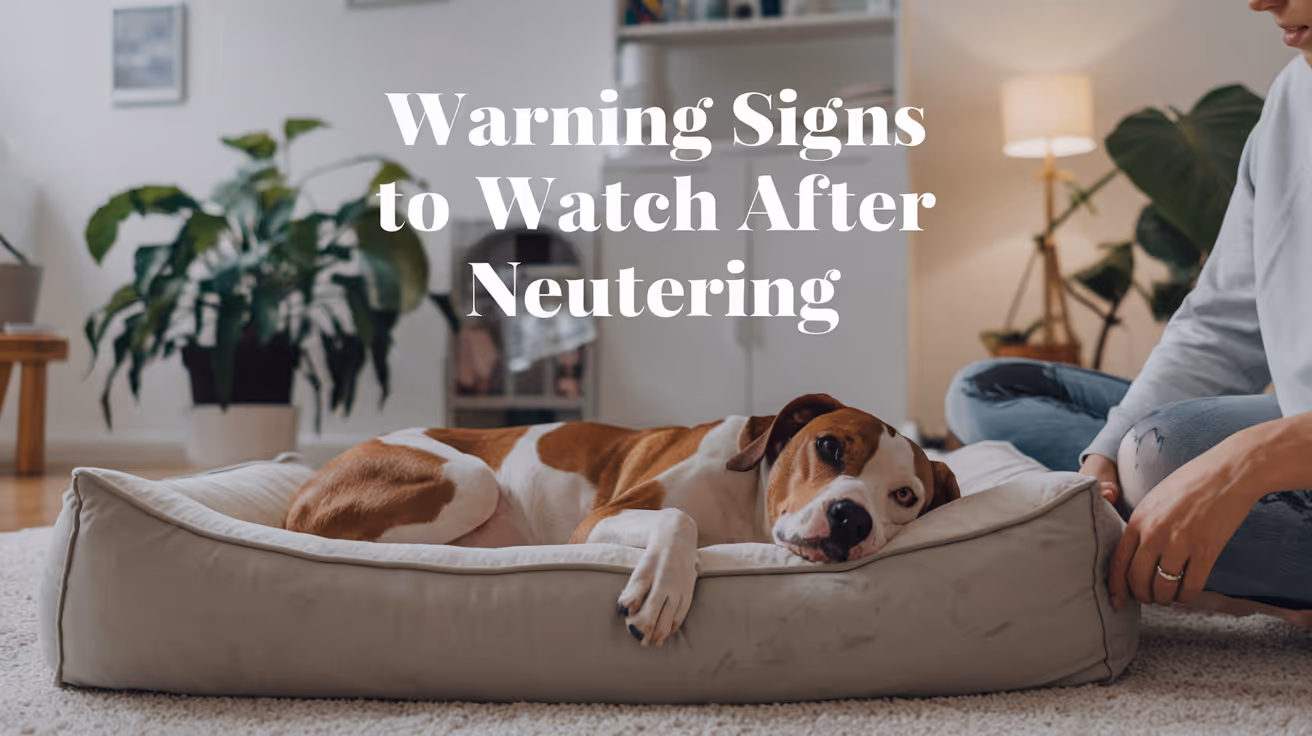
Warning Signs to Watch for After Neutering a Dog
Learn the key warning signs to watch for after neutering your dog to ensure a safe recovery and when to seek veterinary care.
Most dogs recover from neutering without a hitch. But some do not, and the difference between a minor complication and a serious one often comes down to how quickly you notice it.
The tricky part is that some things that look alarming are completely normal, and some things that look minor are not.
This guide gives you a clear, symptom-by-symptom breakdown of what is expected, what to monitor, and what requires an immediate call to your vet.
Quick answer: Mild grogginess, reduced appetite, and slight redness at the incision are all normal in the first 24 to 48 hours. Ongoing bleeding, yellow or green discharge, swelling that grows after day three, lethargy lasting more than 48 hours, or a wound that appears to be opening are all warning signs that need veterinary attention promptly.
Key takeaways
- Normal recovery includes mild grogginess, slight swelling, and reduced appetite for 24 to 48 hours.
- Warning signs that need a vet call include discharge, increasing swelling, continuous bleeding, and persistent lethargy.
- Emergency signs require immediate action: pale gums, collapse, or abdomen that appears bloated.
- Licking is the most preventable complication: the cone prevents most infection and wound problems.
- Daily incision checks catch problems early, when they are still easy to treat.
- When in doubt, call your vet. It is always better to check than to wait.
What normal recovery looks like
Understanding normal first makes it easier to spot abnormal.
In the first 24 to 48 hours after surgery, expect:
- Grogginess, drowsiness, or mild disorientation from anesthesia
- Reduced appetite or no interest in food
- Mild redness or slight puffiness at the incision site
- A small amount of clear or very faintly pink fluid at the wound in the first 24 to 72 hours
- Quieter behavior and preference for rest
- Possible shivering or restlessness as anesthesia wears off
All of these resolve on their own. By day two or three, your dog should be more alert, eating normally, and showing interest in their surroundings again.
Warning signs: incision and wound
The incision site is where most post-surgical problems show up first. Check it morning and evening throughout the recovery period.
Discharge
Swelling
Some swelling in the first two to three days is expected. The body's inflammatory response is part of normal healing.
Swelling that increases after day three, feels warm and firm to the touch, or is accompanied by redness and discharge is not normal. This pattern points to infection or hematoma and needs veterinary evaluation.
In large breed male dogs, scrotal swelling is a specific concern. Moderate scrotal swelling that resolves within a week is often normal. Swelling that grows, becomes hot, or is accompanied by pain or discharge is not.
Wound opening
The incision edges should be staying together, not pulling apart.
If you see any gap in the wound, exposed pink or red tissue beneath the skin, or sutures that appear to have pulled through, contact your vet immediately. This is called dehiscence and requires prompt attention.
Do not attempt to close an open wound yourself. Keep your dog calm, prevent licking, and get to your vet.
Warning signs: behavior and general health
Lethargy
Mild tiredness for the first 24 to 48 hours is expected. Anesthesia and surgery are taxing on the body.
Lethargy that persists beyond 48 hours, or a dog that is completely unresponsive, unable to stand, or does not improve over the first two days, is a warning sign. Call your vet.
Loss of appetite
Not wanting to eat on surgery day and the following morning is normal.
A dog that still refuses food at 48 hours after surgery, particularly if also vomiting, may be experiencing a complication. Call your vet if appetite has not returned by day two.
Vomiting
One or two episodes of vomiting the evening of surgery can be a side effect of anesthesia. This is within normal range.
Vomiting that continues beyond 24 hours after surgery, or that contains blood, requires a vet call. Persistent vomiting can indicate a reaction to pain medication, an obstruction, or another post-surgical issue.
Difficulty urinating or defecating
Some dogs take a day or two to have a bowel movement after surgery. This is usually due to fasting before the procedure and reduced activity afterward.
No bowel movement after three to five days is worth reporting. Difficulty or pain during urination, especially in male dogs, can indicate inflammation near the surgical site and should be checked promptly.
Warning signs: pain
Your dog will have been sent home with pain medication. Use it on schedule.
Signs that pain is not being adequately controlled include:
- Whining, yelping, or crying that persists beyond the first day
- Hunched posture or reluctance to sit or lie down
- Guarding the incision site by flinching or snapping when touched
- Panting excessively without exertion
- Trembling or shivering beyond the first few hours post-anesthesia
Contact your vet if pain seems severe after day three, or if prescribed medication does not appear to be working. Do not give human pain medications. Aspirin, ibuprofen, and acetaminophen are toxic to dogs and can cause serious harm or death.
Emergency warning signs: act immediately
These signs require emergency veterinary care, not a wait-and-see approach.
Call your vet or go to an emergency clinic immediately if you observe:
- Pale, white, or bluish gums
- Sudden collapse or inability to stand
- Abdomen that appears bloated, distended, or hard
- Heavy bleeding that does not stop
- Rapid or labored breathing
- Signs of extreme pain: screaming, freezing in place, inability to move
These can indicate internal bleeding, systemic infection, or other serious complications that are time-sensitive.
The most preventable warning sign: licking
Most of the infection and wound-breakdown cases that end up back at the vet have one thing in common: the cone came off.
Licking introduces bacteria directly into the wound, softens the tissue, and can pull sutures loose. A dog that licks its incision even once can undo days of clean healing.
The E-collar stays on for the full 10 to 14 days. If your dog refuses the hard plastic cone, soft inflatable collars or recovery suits are legitimate alternatives, provided they reliably prevent the dog from reaching the wound site.
For a full guide on what normal recovery looks like day by day and how to distinguish healing progress from early complications, tracking the incision against a clear timeline makes daily monitoring more actionable.
When to call vs. when to go straight to emergency care
Use this as a quick decision guide.
When in doubt, call. Most veterinary practices would rather hear from you about something minor than have you wait too long on something serious.
The role of aftercare in preventing complications
Most warning signs are preventable, not inevitable.
The dogs most likely to develop post-surgical complications are those given too much freedom too soon, or whose cones were removed early. Following a complete aftercare plan covering rest, incision monitoring, medications, and the cone is the most effective way to keep your dog out of trouble during recovery.
For a complete, step-by-step plan on full post-surgery care covering each day of the recovery window, what to feed, how to check the wound, and how to keep your dog calm, having that reference from day one prevents most of the issues covered in this article. Understanding surgical risks that can cause complications beyond the routine is also worth reading before surgery if you have a large breed or older dog.
Frequently asked questions
Is it normal for my dog to be lethargic after neutering?
Yes, for the first 24 to 48 hours. Anesthesia causes genuine fatigue, and your dog needs rest. Lethargy that does not improve by day two or three, or a dog that is completely unresponsive, warrants a call to your veterinarian.
What does an infected incision look like?
An infected incision typically shows yellow or green discharge, increasing redness and swelling after the first few days, warmth around the wound, and often a foul odor. Any of these signs alone justify a same-day call to your vet. For a detailed visual guide to signs of an infected incision, knowing what to look for at each stage of healing helps you act at the right time.
My dog is licking the incision despite wearing a cone. What should I do?
Check that the cone extends at least two inches past your dog's nose. If it is too short, your dog can still reach the incision. Consider switching to a soft inflatable collar or recovery suit if the hard cone is not fitting properly. Contact your vet if there are signs the wound has been compromised.
Should I be worried about scrotal swelling after neutering?
Some scrotal swelling in the first few days is normal. The scrotum fills with fluid as part of the healing response. Swelling that increases after day three, feels hot or very firm, or is accompanied by discharge or pain is abnormal and should be evaluated by your vet.
When should I go to an emergency vet after neutering?
Go immediately if your dog has pale or white gums, collapses, has a bloated or hard abdomen, or is bleeding heavily and continuously. These signs can indicate internal bleeding or systemic infection and are time-sensitive emergencies.
The difference between a smooth recovery and a complication is usually not bad luck. It is usually the cone coming off too early, too much activity on day four, or a wound check that was skipped. Pay attention for two weeks, and most dogs sail through without a single issue.
Resources
The following sources were used as reference and background for this article:
- Ace of Spays. Signs of Complications After Spay/Neuter Surgery: When to Contact Your Veterinarian. aceofspays.com
- Animal Humane Society. Spay/Neuter Post-Surgical Care and Recovery Instructions. animalhumanesociety.org
- Homeaglow. 10 Warning Signs After Spaying or Neutering Your Dog. homeaglow.com
- American Kennel Club. Post-Surgical Care for Dogs Following a Spay or Neuter. akc.org
- Union Hill Animal Hospital. Neutering Recovery Guide for Male Dogs. unionhillvet.com
- Rivergate Veterinary Clinic. Signs of Infection After Spaying or Neutering a Dog. rivergateveterinaryclinic.com
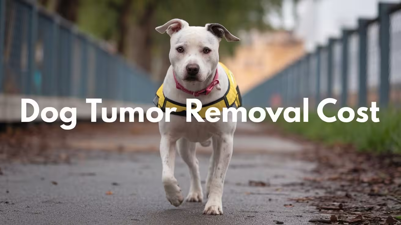
Dog Tumor Removal Cost: What Owners Should Know
Learn how much dog tumor removal costs, what affects the price, and how to plan for surgery, recovery, and vet care expenses.
Understanding Dog Tumor Removal and Its Cost
A tumor in dogs is an abnormal growth of cells that can be either benign (non-cancerous) or malignant (cancerous). Unlike fatty lipomas, some tumors can invade nearby tissues or spread to other organs, making early detection and removal essential. Surgical removal helps diagnose the tumor type and prevents further health complications.
- Benign vs. Malignant Tumors: Benign growths are usually localized, while malignant tumors can spread rapidly.
- Why Removal Is Needed: Surgery may be curative, diagnostic, or preventive, depending on the tumor’s behavior and location.
- Cost Variation: Dog tumor removal costs vary widely from $300 for small skin tumors to several thousand dollars for internal or complex surgeries.
Understanding these basics helps owners plan both medically and financially.
Average Cost of Dog Tumor Removal Surgery
The cost of dog tumor removal varies depending on tumor type, location, and surgical complexity. Some tumors are simple skin growths, while others require advanced procedures involving imaging, specialized anesthesia, or reconstructive surgery.
- Minor Tumor Removal: Small surface tumors on the skin or eyelid usually cost between $300 and $800, including anesthesia and basic pathology.
- Moderate Surgeries: Tumors on the limbs, under the skin, or in sensitive areas like the mouth often cost $1,000 to $2,500 because of deeper tissue involvement and longer surgical time.
- Major or Internal Tumors: Complex cases involving organs such as the spleen, liver, or lungs can range from $3,000 to $7,000 or more, depending on post-op hospitalization.
- National Average Range: Across the U.S., the average cost for tumor removal, including vet consultation and anesthesia, is typically between $800 and $2,500.
This wide range reflects the variation in surgical difficulty, recovery needs, and the diagnostic steps involved.
Factors That Affect Dog Tumor Removal Cost
Tumor removal costs depend on several medical and logistical factors. Each element, from tumor type to the clinic’s expertise, influences both surgical complexity and overall pricing.
- Type of Tumor: Benign tumors like adenomas are easier and cheaper to remove than malignant cancers, which may require wide excision and advanced testing.
- Tumor Location: Growths on the skin surface cost less to treat than internal tumors affecting the abdomen, chest, or organs.
- Size and Number of Tumors: Multiple or large tumors increase anesthesia time, surgical effort, and lab testing costs.
- Diagnostic Needs: Biopsies, X-rays, ultrasounds, or CT scans are often required to evaluate spread, adding $200–$1,000 to the total bill.
- Clinic Type and Expertise: Specialty hospitals or board-certified surgeons typically charge higher fees for complex or high-risk cases.
- Dog’s Health Condition: Dogs with heart, kidney, or respiratory issues may need extra monitoring or tailored anesthesia, raising overall cost.
Each of these factors helps determine the most accurate estimate for your dog’s surgery.
Cost Breakdown: What’s Included in Dog Tumor Removal Surgery
Dog tumor removal involves several stages — from diagnostics to post-operative care. Understanding each cost component helps owners see what their payment truly covers.
- Pre-Surgery Diagnostics: Blood tests, fine-needle aspiration, or imaging confirm the tumor’s nature and assess surgical safety.
- Surgical Procedure: Costs include anesthesia, excision, surgical staff, and necessary monitoring equipment. Deeper tumors may require longer operative times and special tools.
- Lab and Pathology Fees: Removed tissue is sent for biopsy or histopathology to confirm whether the tumor is benign or malignant.
- Post-Operative Care: Pain medication, antibiotics, and wound care supplies are included to ensure proper recovery.
- Follow-Up Visits: Rechecks for healing and suture removal are part of aftercare, and additional testing may be required if malignancy is confirmed.
This breakdown ensures transparency and helps you prepare for both the surgery and follow-up stages without unexpected costs.
When Dog Tumor Removal Is Urgent vs Optional
Not all tumors require immediate surgery. Some grow slowly and can be safely monitored, while others pose urgent medical risks. Recognizing which situation applies helps you make timely, informed decisions.
- Signs of Urgency: Rapid tumor growth, bleeding, ulceration, foul odor, or visible pain when touched indicate the need for prompt removal.
- Location Concerns: Tumors that interfere with movement, breathing, or eating are considered emergencies and should be removed before complications develop.
- Aggressive or Malignant Tumors: If biopsy results show malignancy, early surgery improves prognosis and reduces the chance of spread.
- When Monitoring Is Safe: Small, stable, or benign masses can often be observed with regular vet checkups and measurement tracking.
- Risks of Delay: Waiting too long may allow malignant cells to spread, increasing surgical difficulty and cost later.
Your veterinarian’s evaluation helps determine whether removal is urgent or if observation remains a safe, short-term option.
Post-Surgery Recovery and Aftercare Costs for Dog Tumor Removal
Recovery from tumor removal surgery depends on the tumor’s type, size, and surgical complexity. Post-operative care is crucial to prevent infection, control pain, and promote healing, and it can add to the total cost.
- Typical Recovery Period: Most dogs recover within 10–14 days for small tumors, while major internal surgeries may require 3–4 weeks of restricted activity and monitoring.
- Pain Management: Pain-relief medications and anti-inflammatories usually cost $30–$100 depending on the dosage and duration.
- Antibiotics and Wound Care: Post-surgery antibiotics prevent infection and cost around $20–$60. Owners must keep incisions clean and prevent licking or scratching with an e-collar.
- Hidden Costs: Follow-up appointments, suture removals, and bandage changes can add $50–$200. Additional lab tests or biopsy reviews may increase expenses if complications arise.
- Rehabilitation for Major Surgeries: Some cases benefit from physiotherapy or laser therapy to restore mobility after tumor removal near joints.
Proper aftercare reduces complications and ensures faster recovery while minimizing long-term medical costs.
How to Budget for Dog Tumor Removal Surgery
Financial planning is essential before scheduling tumor removal, as costs can vary widely between general clinics and specialist hospitals. Knowing what to ask and how to prepare helps prevent surprises.
- Request a Detailed Estimate: Ask your vet for a full written quote covering anesthesia, diagnostics, pathology, and aftercare so you understand the total cost.
- Compare Providers: General veterinarians are often more affordable, while board-certified surgeons may charge more for complex or high-risk procedures.
- Pet Insurance Coverage: Most plans cover tumor removals if the mass wasn’t diagnosed before the policy started. Check for deductibles and exclusions.
- Payment Plans and Financing: Many clinics partner with financing companies or offer in-house installment options for expensive surgeries.
- Additional Savings Tips: Combining multiple tumor removals in one procedure can reduce anesthesia costs and overall fees.
A clear financial plan ensures your dog receives timely treatment without financial strain or unexpected costs after surgery.
Alternatives and Long-Term Management of Dog Tumors
Not all tumors require surgery, and some can be managed through observation or supportive care. Long-term management focuses on early detection, lifestyle improvements, and preventive veterinary follow-ups.
- Non-Surgical Options: Benign tumors such as sebaceous adenomas or small lipomas can sometimes be treated with cryotherapy or laser removal at lower costs.
- Lifestyle and Diet: A balanced diet rich in antioxidants and omega-3 fatty acids supports immune health and may slow tumor growth.
- Weight and Exercise: Maintaining ideal body weight reduces inflammatory stress and supports better healing after any surgical intervention.
- Monitoring Guidelines: Regular veterinary exams and at-home checks help detect new growths early, especially in older dogs prone to multiple tumors.
- Owner Awareness: Photograph and measure existing lumps monthly to track changes in size, color, or texture.
Long-term vigilance and proactive lifestyle care help reduce recurrence risk and improve overall well-being for dogs prone to tumors.
Conclusion
Dog tumor removal costs depend on many factors, including the tumor’s size, location, and whether it’s benign or malignant. Early diagnosis can often reduce surgical complexity and overall expense.
- Major Cost Drivers: Diagnostic tests, anesthesia, surgeon expertise, and post-operative care.
- Importance of Timely Action: Treating tumors early prevents spread, lowers costs, and improves recovery outcomes.
- Veterinary Consultation: A trusted veterinarian can assess whether immediate removal or monitoring is appropriate for your dog.
- Balanced Decision-Making: Combine medical priorities with financial readiness by exploring insurance, financing, or low-cost options.
When guided by professional advice and realistic budgeting, tumor removal becomes a manageable step toward protecting your dog’s comfort and long-term health.
FAQs
What is the average cost to remove a dog tumor?
The average cost of tumor removal ranges from $800 to $2,500, depending on the tumor’s size, depth, and location. Small skin tumors are less expensive, while complex surgeries for internal or malignant tumors can cost $3,000 or more, especially if hospitalization and advanced imaging are required.
Why do some tumor removals cost more than others?
Costs rise with surgical difficulty, tumor location, and pre-surgery testing. Internal tumors or those near vital organs need advanced imaging, skilled surgeons, and longer anesthesia time, all of which increase the price. Clinics with specialized facilities may also charge higher fees for complex cases.
Is pet insurance likely to cover tumor surgery?
Yes, if the tumor wasn’t diagnosed before your policy began. Most comprehensive pet insurance plans cover surgery, anesthesia, and pathology tests for tumor removals. However, pre-existing tumors or recurring cases are usually excluded, so review your policy’s coverage limits and waiting periods.
Can tumors come back after removal?
Some tumors can recur, especially malignant or infiltrative types. Even after clean surgical margins, microscopic cancer cells can regrow. Regular post-surgery checkups and imaging help detect any recurrence early and ensure timely intervention to maintain long-term health.
Are there low-cost clinics for tumor surgery?
Yes, many animal welfare organizations, veterinary schools, and community clinics offer discounted surgical programs. While availability varies by region, these options help pet owners manage expenses without compromising on essential care or surgical safety standards.

Torn Meniscus Surgery Cost in Dogs Explained
Learn about torn meniscus surgery cost in dogs, including factors affecting price, procedure details, and recovery tips for your pet's health.
A torn meniscus in dogs is a common injury that affects the knee joint, causing pain and mobility issues. If your dog has this problem, you might wonder about the cost of surgery and what it involves. Understanding the expenses and treatment options can help you prepare for your pet's care.
This article explains the typical cost of torn meniscus surgery in dogs, the factors that influence pricing, and what you can expect during recovery. You will learn how to manage your dog's health and make informed decisions about treatment.
What is torn meniscus surgery in dogs?
Torn meniscus surgery in dogs is a procedure to repair or remove damaged cartilage in the knee joint. The meniscus acts as a cushion between bones, and injury can cause pain and lameness. Surgery aims to restore joint function and reduce discomfort.
The surgery is usually recommended when conservative treatments like rest and medication do not improve the dog's condition. It involves anesthesia and specialized techniques to address the tear.
- Purpose of surgery: To repair or remove the damaged meniscal cartilage to relieve pain and improve knee stability in dogs.
- Common causes: Meniscus tears often result from ligament injuries or trauma during activities like running or jumping.
- Surgical techniques: Options include meniscectomy (removal) or meniscal repair depending on the tear's location and severity.
- Post-surgery goals: Restore normal joint movement, reduce arthritis risk, and help the dog regain mobility.
Understanding the surgery helps you prepare for the treatment and care your dog will need.
How much does torn meniscus surgery cost in dogs?
The cost of torn meniscus surgery in dogs varies widely depending on location, clinic, and the dog's specific needs. On average, prices range from $1,500 to $4,000. This includes pre-surgical exams, anesthesia, surgery, and initial post-op care.
Knowing the cost breakdown can help you budget and discuss options with your veterinarian.
- Base surgery fee: Typically between $1,000 and $3,000, covering the surgical procedure and operating room use.
- Pre-surgical tests: Blood work and X-rays may cost $200 to $500 to assess the dog's health before surgery.
- Anesthesia and monitoring: Usually $300 to $700, essential for safe surgery and pain control.
- Post-operative care: Includes medications, bandages, and follow-up visits costing $200 to $500.
Costs may increase if complications arise or if advanced imaging like MRI is needed.
What factors affect the cost of meniscus surgery in dogs?
Several factors influence the total cost of torn meniscus surgery in dogs. These include the dog's size, the complexity of the injury, and the clinic's location. Understanding these helps you anticipate expenses and plan accordingly.
Discussing these factors with your vet can clarify the expected costs and available options.
- Dog's size and weight: Larger dogs may require more anesthesia and longer surgery time, increasing costs.
- Severity of tear: Complex or multiple tears need more surgical time and skill, raising the price.
- Veterinary clinic location: Urban or specialty clinics often charge more than rural general practices.
- Surgeon's experience: Board-certified surgeons may have higher fees but offer specialized care.
Knowing these factors helps you make informed choices about your dog's treatment plan.
What are the risks and benefits of torn meniscus surgery for dogs?
Surgery for a torn meniscus can improve your dog's quality of life but also carries some risks. Weighing these helps you decide if surgery is the best option for your pet.
The benefits often outweigh the risks when the injury causes significant pain or limits mobility.
- Benefit - Pain relief: Surgery can reduce joint pain and discomfort caused by the torn meniscus.
- Benefit - Improved mobility: Dogs often regain better movement and activity levels after recovery.
- Risk - Infection: Any surgery carries a small risk of infection requiring additional treatment.
- Risk - Anesthesia complications: Though rare, anesthesia can cause adverse reactions in some dogs.
Discussing risks and benefits with your vet ensures the best decision for your dog's health.
How should you prepare your dog for meniscus surgery?
Proper preparation before surgery helps reduce risks and supports a smooth procedure. Your veterinarian will give specific instructions tailored to your dog’s needs.
Following these steps can improve surgery outcomes and reduce stress for your pet.
- Pre-surgery fasting: Your dog should avoid food for 8-12 hours before surgery to prevent anesthesia complications.
- Health evaluation: Complete blood tests and physical exams ensure your dog is healthy enough for surgery.
- Medication review: Inform your vet about all medications or supplements your dog takes to avoid interactions.
- Arrange post-op care: Prepare a quiet, comfortable space and plan for restricted activity during recovery.
Good preparation helps your dog have a safer surgery and faster healing.
What is the recovery process after torn meniscus surgery in dogs?
Recovery after meniscus surgery requires careful management to ensure healing and prevent re-injury. The process usually takes 6 to 12 weeks, depending on the dog's age and health.
Following your vet’s instructions closely improves your dog’s chances of a full recovery.
- Restricted activity: Limit running, jumping, and stairs to protect the healing joint for several weeks.
- Physical therapy: Controlled exercises and rehabilitation help restore strength and flexibility.
- Pain management: Administer prescribed pain medications and monitor for discomfort signs.
- Follow-up visits: Regular check-ups allow your vet to assess healing and adjust care as needed.
Patience and consistency during recovery are key to your dog’s long-term joint health.
Conclusion
Torn meniscus surgery cost in dogs varies but generally ranges from $1,500 to $4,000 depending on many factors. Understanding the procedure, risks, and recovery helps you prepare financially and emotionally for your pet’s care.
With proper preparation and post-operative management, surgery can relieve your dog’s pain and improve mobility. Always consult your veterinarian to choose the best treatment plan for your dog’s torn meniscus injury.
What is the typical recovery time after torn meniscus surgery in dogs?
Recovery usually takes 6 to 12 weeks, during which activity must be restricted and physical therapy may be needed to regain strength and mobility.
Can torn meniscus surgery prevent arthritis in dogs?
While surgery can reduce joint damage and pain, it may not fully prevent arthritis but can slow its progression and improve quality of life.
Are there non-surgical options for treating a torn meniscus in dogs?
Yes, mild tears may be managed with rest, anti-inflammatory medications, and physical therapy, but surgery is often needed for severe cases.
How do I know if my dog needs meniscus surgery?
Your vet will diagnose based on clinical signs, physical exams, and imaging like X-rays or MRI to determine if surgery is necessary.
Is torn meniscus surgery painful for dogs?
Dogs receive anesthesia during surgery and pain medications afterward to minimize discomfort, making the procedure as pain-free as possible.
Why Is My Dog's Foot Swollen?
Discover why your dog's foot is swollen, common causes, treatments, and when to see a vet for proper care.
Seeing your dog's foot swollen can be worrying. Swelling in a dog's foot can happen for many reasons, from injuries to infections. Understanding why this happens helps you act quickly and keep your dog comfortable.
This article explains common causes of swollen dog feet, how to spot serious problems, and what treatments work best. You will learn when to treat at home and when to visit a vet for urgent care.
What Causes Swelling in a Dog's Foot?
Swelling in a dog's foot can come from many different problems. It often shows as puffiness, redness, or heat in the paw area. Knowing the cause helps you decide the right care.
Common causes include injuries, infections, allergies, and insect bites. Each cause needs a different approach to treatment.
- Injury or trauma: A cut, sprain, or broken bone can cause swelling due to inflammation and fluid buildup in the foot tissues.
- Infections: Bacterial or fungal infections can cause swelling, redness, and pain, often needing antibiotics or antifungal treatment.
- Allergic reactions: Allergies to plants, chemicals, or insect stings can cause sudden swelling and itching in the foot.
- Foreign objects: Thorns, splinters, or glass stuck in the paw can cause swelling and discomfort until removed.
Identifying the cause early helps prevent complications and speeds healing.
How Can I Tell If My Dog's Foot Swelling Is Serious?
Not all swelling is an emergency, but some signs mean you should see a vet quickly. Serious swelling can affect your dog's ability to walk or cause severe pain.
Look for symptoms like severe limping, open wounds, or signs of infection. These require prompt veterinary care.
- Severe limping or inability to walk: Indicates pain or serious injury needing urgent veterinary evaluation.
- Open wounds or bleeding: Risk of infection and need for cleaning and possibly stitches.
- Fever or lethargy: Signs that infection may have spread and requires medical treatment.
- Rapidly increasing swelling: Could signal an allergic reaction or deep infection needing emergency care.
When in doubt, it is safer to consult your vet to avoid worsening problems.
What Home Treatments Can Help a Swollen Dog Foot?
For mild swelling without serious signs, you can try some home care steps. These help reduce swelling and keep your dog comfortable.
Always watch your dog closely and stop home treatment if symptoms worsen or do not improve.
- Rest and limit activity: Keep your dog from running or jumping to reduce stress on the swollen foot.
- Cold compress application: Apply a cold pack wrapped in cloth for 10-15 minutes to reduce swelling and pain.
- Clean the paw gently: Use warm water to clean dirt or debris, especially if there are small cuts or irritations.
- Prevent licking or chewing: Use an Elizabethan collar if your dog tries to lick or bite the swollen area, which can worsen irritation.
These steps can help minor swelling but do not replace veterinary care for serious cases.
When Should I Take My Dog to the Vet for a Swollen Foot?
Knowing when to seek professional help is important. Some swelling needs medical treatment to avoid complications.
If your dog's swelling is severe, painful, or lasts more than a day or two, a vet visit is necessary. Early treatment can prevent infections or permanent damage.
- Persistent swelling over 48 hours: Indicates that the problem may not resolve without medical intervention.
- Signs of infection: Pus, foul odor, or heat around the swollen area require antibiotics or cleaning by a vet.
- Suspected broken bone or sprain: Needs X-rays and pain management from a veterinary professional.
- Severe allergic reactions: Swelling with difficulty breathing or collapse needs emergency veterinary care immediately.
Your vet can diagnose the cause and recommend the right treatment plan for your dog's recovery.
How Do Vets Diagnose the Cause of a Swollen Dog Foot?
Veterinarians use several methods to find the cause of swelling. A correct diagnosis is key to effective treatment.
They will examine your dog’s foot carefully and may use tests to look deeper into the problem.
- Physical examination: Checking for wounds, foreign objects, and signs of pain or infection in the foot.
- X-rays: Used to detect fractures, bone infections, or foreign bodies inside the paw.
- Skin scrapings or cultures: To identify infections caused by bacteria, fungi, or parasites.
- Blood tests: To check for systemic infection or allergic reactions affecting the swelling.
These tools help your vet create a treatment plan tailored to your dog's needs.
What Treatments Do Vets Use for Swollen Dog Feet?
Treatment depends on the cause of the swelling. Your vet may use medications, procedures, or supportive care to help your dog heal.
Some treatments can be done at home under vet guidance, while others require clinic visits.
- Antibiotics or antifungals: Prescribed to treat infections causing swelling and prevent spread.
- Anti-inflammatory drugs: Help reduce pain and swelling, improving your dog's comfort.
- Wound care and bandaging: Cleaning and protecting open wounds to promote healing and prevent infection.
- Surgery: May be needed to remove foreign objects or repair fractures causing swelling.
Follow your vet’s instructions carefully to ensure the best outcome for your dog.
How Can I Prevent My Dog's Foot from Swelling?
Preventing foot swelling involves protecting your dog from injuries and infections. Regular care and attention can reduce risks.
Simple habits help keep your dog's paws healthy and avoid painful swelling episodes.
- Regular paw inspections: Check your dog's feet daily for cuts, thorns, or swelling to catch problems early.
- Keep nails trimmed: Prevents nails from breaking or causing injury to the foot pads.
- Avoid walking on rough surfaces: Protect paws from sharp objects or hot pavement that can cause injuries.
- Use protective booties: Especially in harsh weather or rough terrain to shield paws from damage.
Good paw care supports your dog’s overall health and comfort.
Conclusion
Swelling in your dog's foot can have many causes, from minor injuries to serious infections. Understanding why your dog's foot is swollen helps you provide the right care quickly.
Always watch for signs of pain, infection, or worsening symptoms. When in doubt, seek veterinary advice to protect your dog's health and comfort. Early treatment can prevent complications and get your dog back on their feet faster.
Why is my dog's foot swollen after walking?
Your dog's foot may swell after walking due to minor injuries, irritation from rough surfaces, or allergic reactions. Rest and paw care usually help reduce swelling quickly.
Can a swollen dog foot heal without a vet?
Mild swelling from minor injuries or irritations can heal at home with rest and care. However, persistent or severe swelling needs veterinary evaluation to avoid complications.
How long does it take for a dog's swollen foot to go down?
Swelling may reduce within a few days with proper care. If swelling lasts more than 48 hours or worsens, consult a vet for treatment.
Is a swollen dog foot painful?
Yes, swelling often causes pain and discomfort. Your dog may limp, lick, or avoid putting weight on the swollen foot.
Can allergies cause a dog's foot to swell?
Yes, allergies to insect bites, plants, or chemicals can cause sudden swelling and itching in a dog's foot, sometimes requiring veterinary treatment.
Do Dogs Calm Down After Being Spayed?
Learn how dogs behave after being spayed and when to expect them to calm down post-surgery.
It is one of the most common reasons owners choose to spay their female dog. The expectation of a calmer, more settled companion afterward.
Sometimes that is exactly what happens. Sometimes it does not. And in a small subset of cases, anxiety or excitability actually increases.
Understanding why gives you a more accurate picture of what to expect, which makes the outcome less surprising in either direction.
Quick answer: Many dogs do become calmer after spaying, but the effect is specific to hormone-driven behaviors, not overall temperament. Dogs that were restless, anxious, or excitable due to heat cycles typically show significant improvement. Dogs whose energy or anxiety has non-hormonal causes will not reliably calm down. Most behavioral changes, if they occur, appear over two to three months.
Key takeaways
- The calming effect is real but specific: It applies to behaviors driven by reproductive hormones, not all behaviors.
- Heat cycle behaviors disappear: Restlessness, roaming, frequent urination, and cyclical mood shifts all stop after spaying.
- Core energy level and personality do not change: A high-energy dog will still be high-energy after spaying.
- Breed and individual temperament matter: Some breeds retain high energy regardless of hormone status.
- The timeline is weeks to months, not days: Hormone levels decline gradually, so behavioral changes are gradual too.
- A small subset of dogs show increased fearfulness: This is documented in research and worth knowing about before surgery.
What "calming down" actually means after spaying
When people say they want their dog to calm down, they usually mean one of two things:
- They want heat cycle behaviors to stop: the roaming, the restlessness, the vocalization, the constant male attention.
- They want a generally more settled dog overall.
Spaying reliably delivers the first. The second is less predictable.
This distinction is the core of the honest answer to this question.
What spaying removes: the hormonal layer
Spaying removes the ovaries, which are the primary source of estrogen and progesterone. These hormones regulate the reproductive cycle and influence behaviors tied to it.
During heat cycles, which typically occur every six to eight months in adult dogs, estrogen surges create a set of predictable behaviors:
- Restlessness and inability to settle
- Increased vocalization
- Frequent urination in small amounts to signal reproductive status
- Strong drive to escape and find a mate
- Attracting intact male dogs
- Cyclical irritability or anxiety
All of these stop after spaying. The hormonal trigger is permanently removed.
Which dogs calm down most noticeably after spaying
Dogs whose difficult behaviors are primarily or significantly driven by the reproductive cycle show the most consistent improvement after spaying.
Dogs most likely to become calmer:
- Female dogs that showed significant behavior changes during heat cycles, including restlessness, anxiety, and clinginess
- Dogs prone to false pregnancies, with associated nesting, object guarding, and protective behavior
- Dogs that attempted to escape or roam during heat periods
- Dogs whose cyclical mood swings were disruptive to daily life
For these dogs, spaying removes a recurring source of behavioral disruption. The result is often a more predictable, stable dog.
Which dogs may not calm down after spaying
Dogs whose energy level, excitability, or anxiety comes from sources other than reproductive hormones will not reliably settle after spaying.
Dogs less likely to show a calming effect:
- High-energy working or sport breeds where drive is genetic, not hormonal
- Dogs with established anxiety, fearfulness, or reactivity from non-hormonal causes
- Dogs with learned problematic behaviors from their history and environment
- Dogs whose excitability comes from insufficient exercise or mental stimulation
A Border Collie that is highly energetic before spaying will still be highly energetic after. A Labrador that pulls on the leash before spaying will still pull after. These behaviors are not driven by estrogen.
The timeline: when does calming happen?
Behavioral changes from spaying do not happen within days of surgery. The immediate post-surgery period reflects recovery from anesthesia and pain, not hormonal adjustment.
Realistic timeline:
Owners who expect a noticeably calmer dog within days of surgery are working with an inaccurate timeline. The changes are gradual and unfold over weeks to months.
What research says about calming after spaying
The evidence is genuinely mixed, which is worth knowing rather than glossing over.
Supporting the calming effect:
A study of 591 dogs found behavioral problems reduced or eliminated in 59% of spayed females. Heat cycle-related behaviors showed the most consistent improvement. Many owners of females that were particularly difficult during heat cycles report significant positive changes after spaying.
Complicating the picture:
Two large-scale studies using the Canine Behavioral Assessment and Research Questionnaire (C-BARQ) found that sterilization was associated with a roughly 31% increase in fearfulness and a 33% increase in touch sensitivity in some dogs. These findings apply to both males and females.
What does this mean in practice? For a meaningful subset of dogs, spaying does not produce a calmer animal. It may even produce a more anxious one, particularly in dogs that had pre-existing fearfulness or were sterilized at a young age.
This does not mean spaying is wrong for these dogs. It means the behavioral outcome cannot be guaranteed, and expectations should be calibrated accordingly.
For the full picture of all the behavior changes to expect after spaying, including the research on unexpected outcomes, that guide covers the complete behavioral landscape across both sexes.
Does calming depend on the dog's age at spaying?
Yes, and this is one of the most important variables.
Dogs spayed before their first heat cycle:
The hormonal disruption of heat cycles is never established. Many owners report these dogs are consistently more settled than intact females across their lifetime, without ever having the difficult heat cycle period.
Dogs spayed after several heat cycles:
The calming effect from eliminating heat cycles is still real and significant. However, any behaviors that have become habitual through repetition during heat cycles may persist longer or require training to address.
Dogs spayed very young (before 6 months):
Research suggests a higher association with fearfulness in dogs sterilized very early, before hormonal maturity. For whether this applies specifically to puppies, the guide on calming effects specific to female dogs addresses this age-specific question in detail. For the puppy-specific picture, the guide on whether puppies calm down after being spayed covers the developmental nuances unique to younger dogs.
When the expected calming does not happen
If your dog does not seem noticeably calmer after spaying, consider these factors before concluding the surgery did not work.
Check the timeline: Have two to three months passed since surgery? If not, the hormonal adjustment may still be in progress.
Identify the behavior source: Is the behavior you wanted to address actually hormone-driven? If it is not, spaying was never going to address it.
Consider habituation: Some behaviors that started as hormone-driven become habitual over time. The hormone is removed but the habit persists. Consistent training addresses habituated behavior.
Consider an underlying anxiety: If your dog became more fearful or anxious post-surgery, this is a documented outcome in some dogs. A veterinary behaviorist or your regular vet can help evaluate what is happening and what options exist.
Understanding the truth behind the calming effect myth, including why the expectation of calming is sometimes oversold, helps put the variable outcomes in context.
What actually makes dogs calmer long-term
Whether or not spaying produces a calming effect, these factors reliably influence how settled and manageable a dog is:
- Adequate physical exercise: A dog that is genuinely tired is a calmer dog. Exercise needs are determined by breed and individual temperament.
- Mental stimulation: Breed-appropriate mental work, puzzle feeders, training sessions, and enrichment activities reduce restless, problematic behavior.
- Consistent training: Clear, reinforced expectations give dogs a behavioral framework. This is the single most reliable path to a calmer dog.
- Routine and predictability: Dogs settle more easily in stable, predictable environments.
Spaying contributes one element to this picture. It is not a standalone solution.
Frequently asked questions
How long after being spayed will my dog calm down?
If the calming effect is going to occur, most hormone-driven behavioral changes become apparent within two to six weeks as hormone levels decline. Full stabilization typically takes two to three months. Dogs that are still showing challenging behaviors three to four months after surgery are unlikely to improve further from the hormonal change alone.
Will spaying make my high-energy dog less active?
Probably not significantly. Energy level in dogs is primarily determined by breed, genetics, and conditioning. Spaying can cause a modest metabolic slowdown, but it does not reduce drive or enthusiasm. A high-energy breed will still need the same amount of exercise after spaying.
My dog got more anxious after being spayed. Why?
Research has documented increased fearfulness in a subset of dogs after sterilization, associated with a roughly 31% increase in fearfulness in some studies. This appears to occur more often in dogs sterilized at a young age and in dogs with pre-existing anxiety. If your dog shows increased anxiety after spaying, speak to your vet. A referral to a certified veterinary behaviorist may be helpful.
Will spaying help with aggression?
It depends on the type of aggression. Aggression linked to the heat cycle, competition with other female dogs, or false pregnancy guarding often decreases. Fear-based aggression, learned aggression, and resource guarding are not hormone-driven and are not reliably improved by spaying. For whether lower testosterone calms dogs down in the male counterpart to this question, that guide covers the testosterone-behavior link specifically.
Does breed affect whether spaying calms a dog down?
Yes. Breed temperament significantly influences baseline energy and anxiety levels. High-drive working breeds such as Border Collies, Australian Shepherds, and Belgian Malinois have energy and drive that is genetic, not hormonal. Removing reproductive hormones does not change these fundamental breed characteristics. Whether your specific dog falls into a calmer range after spaying depends on what was driving any difficult behavior in the first place.
The calming effect of spaying is real for dogs whose difficult behaviors are hormone-driven. For those dogs, eliminating the heat cycle and its associated hormonal disruption produces a meaningfully more stable animal. For dogs whose energy or anxiety comes from other sources, spaying is still beneficial for health reasons but should not be expected to produce behavioral transformation. The most accurate prediction is not based on "spaying calms dogs" but on "removing a specific hormonal influence changes the behaviors that influence drives."
Resources
The following sources were used as reference and background for this article:
- Stanley Coren, Psychology Today. Are There Behavior Changes When Dogs Are Spayed or Neutered? psychologytoday.com
- PubMed. Changes in the behavior of dogs after castration. pubmed.ncbi.nlm.nih.gov
- ManyPets. Do Dogs Change After Being Neutered or Spayed? manypets.com
- American Veterinary Medical Association (AVMA). Spaying and Neutering. avma.org
- Houndsy. How Spaying Affects Dog Behavior. houndsy.com

Why Does My Dog Have Scabs on Her Back?
Learn why your dog has scabs on her back, common causes, treatments, and when to see a vet for proper care.
Seeing scabs on your dog's back can be worrying. Scabs are signs of skin damage or irritation. They may result from allergies, infections, parasites, or injuries. Understanding why your dog has scabs is important to help her heal quickly and comfortably.
This article explains the common reasons dogs develop scabs on their backs. You will learn about causes, symptoms, treatments, and when to visit a veterinarian. Knowing these details helps you care for your dog better and prevent future skin problems.
What Causes Scabs on a Dog's Back?
Scabs form when the skin is injured or inflamed. Many conditions can cause scabs on a dog's back. Identifying the cause is key to effective treatment.
- Allergic reactions: Dogs can develop scabs due to allergies to food, pollen, or flea bites, causing itching and skin damage from scratching.
- Parasite infestations: Fleas, ticks, and mites bite the skin, leading to irritation, itching, and scab formation from constant scratching.
- Skin infections: Bacterial or fungal infections can cause sores and scabs as the skin tries to heal from the infection.
- Injuries or trauma: Scratches, bites, or wounds from rough play or accidents can scab over as they heal.
Knowing these causes helps you observe your dog’s behavior and environment to find the source of the problem.
How Can Allergies Lead to Scabs on My Dog's Back?
Allergies are a common cause of itchy skin and scabs in dogs. Allergic reactions cause inflammation and discomfort, leading dogs to scratch or bite their skin.
- Food allergies: Certain ingredients in dog food can trigger skin reactions, causing redness and scabs from scratching.
- Environmental allergies: Pollens, dust mites, or mold spores can irritate your dog's skin, especially on the back and neck areas.
- Flea allergy dermatitis: Flea saliva causes intense itching and scabbing even if only a few fleas are present.
- Contact allergies: Chemicals in cleaning products or plants can cause localized skin irritation and scabs.
Managing allergies often requires identifying triggers and using medications or diet changes to reduce symptoms.
What Role Do Parasites Play in Causing Scabs?
Parasites are a frequent cause of skin problems in dogs. Fleas, ticks, and mites irritate the skin and cause itching that leads to scabs.
- Fleas: Flea bites cause itching and allergic reactions, leading to scabs from excessive scratching and biting.
- Ticks: Ticks attach to the skin and can cause localized irritation and scabbing around the bite site.
- Mange mites: Mites burrow into the skin causing mange, which leads to hair loss, redness, and scabs.
- Prevention importance: Regular parasite control helps prevent infestations and the resulting skin damage.
Parasite control products and vet care are essential to stop scabs caused by parasites.
How Do Skin Infections Cause Scabs on Dogs?
Skin infections often develop after injury or from underlying conditions. They cause inflammation, pus, and scabs as the skin tries to heal.
- Bacterial infections: Bacteria enter broken skin causing hot spots, redness, and scabs that may ooze fluid.
- Fungal infections: Yeast or ringworm infections cause flaky, scabby patches often with hair loss.
- Secondary infections: Scratching from allergies or parasites can introduce bacteria, worsening scabs.
- Treatment necessity: Antibiotics or antifungal medications are often needed to clear infections and heal scabs.
Early vet diagnosis and treatment prevent infections from spreading or becoming chronic.
Can My Dog’s Behavior Cause Scabs on Her Back?
Behavioral factors like excessive licking, scratching, or biting can cause scabs. These behaviors often result from discomfort or stress.
- Itchiness response: Dogs scratch or bite itchy areas, causing skin damage and scabs.
- Stress or anxiety: Nervous dogs may lick or chew their backs compulsively, leading to scabs.
- Environmental irritants: Rough surfaces or allergens in bedding can cause irritation and scabbing.
- Observation importance: Watching your dog’s behavior helps identify if self-trauma causes scabs.
Addressing underlying causes and providing enrichment reduces harmful behaviors and skin damage.
When Should I Take My Dog to the Vet for Scabs?
Not all scabs require immediate veterinary care, but some signs mean you should see a vet promptly.
- Persistent scabs: Scabs that do not heal or keep returning need professional evaluation.
- Widespread skin lesions: If scabs cover large areas or spread quickly, vet care is needed.
- Signs of infection: Swelling, pus, bad odor, or pain around scabs indicate infection requiring treatment.
- Behavior changes: Excessive scratching, lethargy, or loss of appetite alongside scabs warrant a vet visit.
Early diagnosis and treatment improve outcomes and prevent complications.
How Can I Treat and Prevent Scabs on My Dog’s Back?
Treatment depends on the cause but generally includes soothing the skin and preventing further damage. Prevention focuses on good hygiene and parasite control.
- Topical treatments: Medicated shampoos and creams help reduce itching and promote healing of scabs.
- Parasite control: Regular flea and tick prevention stops infestations that cause scabs.
- Diet and supplements: Balanced nutrition and omega fatty acids support healthy skin and reduce allergies.
- Environmental care: Keep bedding clean and avoid irritants to prevent skin problems.
Following your vet’s advice and maintaining good skin care routines help your dog stay comfortable and scab-free.
Conclusion
Scabs on your dog's back can result from allergies, parasites, infections, or injuries. Identifying the cause is essential for proper treatment and healing. Watching your dog's behavior and environment helps find the source of scabs.
Timely veterinary care, good hygiene, and parasite control prevent scabs and keep your dog’s skin healthy. If scabs persist or worsen, consult your vet for diagnosis and treatment to ensure your dog’s comfort and well-being.
Why does my dog have scabs on her back?
Scabs on your dog’s back usually come from skin irritation caused by allergies, parasites, infections, or injuries leading to scratching and skin damage.
Can fleas cause scabs on my dog’s back?
Yes, flea bites cause itching and allergic reactions that make dogs scratch and develop scabs on their backs and other areas.
How do I know if my dog’s scabs are infected?
Signs of infection include redness, swelling, pus, bad smell, and pain around scabs. Infections need veterinary treatment to heal properly.
What home care can I do for my dog’s scabs?
Keep the area clean, use vet-recommended shampoos, prevent scratching, and maintain parasite control to support healing at home.
When should I see a vet about my dog’s scabs?
See a vet if scabs persist, spread, show infection signs, or if your dog is very itchy, lethargic, or losing appetite.

How to Keep an Energetic Dog Calm After Surgery
Learn effective ways to keep your energetic dog calm after surgery with practical tips and expert advice for smooth recovery.
After surgery, energetic dogs can be a challenge to manage because their natural activity may interfere with healing. Keeping your dog calm is crucial to prevent complications and ensure a smooth recovery. This article explains how to handle your dog's energy safely after surgery.
You will learn practical strategies to reduce your dog's activity, create a comfortable environment, and support healing. These tips help you balance your dog's need for rest with their natural energy levels.
Why is it important to keep an energetic dog calm after surgery?
After surgery, your dog's body needs time to heal. Excessive activity can cause pain, reopen wounds, or delay recovery. Energetic dogs may struggle to stay still, increasing the risk of injury.
Understanding the importance of calmness helps you take steps to protect your dog's health during this vulnerable time.
- Prevents wound damage: Limiting movement reduces the chance of stitches tearing or wounds reopening, which can cause infections or require more surgery.
- Reduces pain and swelling: Rest helps control inflammation and discomfort, making recovery smoother and faster.
- Supports medication effectiveness: Calm behavior ensures your dog responds well to pain relief and antibiotics, improving healing outcomes.
- Prevents complications: Overexertion can lead to bleeding, bruising, or internal injuries, which are dangerous after surgery.
Keeping your dog calm is essential to avoid setbacks and promote a healthy recovery process.
What are the best ways to restrict your dog's activity safely?
Restricting activity after surgery requires careful planning. You want to limit movement without causing stress or anxiety. Using physical barriers and controlled routines helps manage your dog's energy safely.
Here are effective methods to keep your dog calm while preventing injury.
- Use a crate or small room: Confine your dog to a safe, comfortable space to limit running and jumping while allowing rest.
- Leash walks only: Take short, slow walks on a leash to allow bathroom breaks without overexertion or excitement.
- Remove toys and distractions: Avoid giving your dog balls or chew toys that encourage active play during recovery.
- Provide comfortable bedding: A soft, supportive bed encourages your dog to lie down and relax instead of moving around.
These steps help control your dog's activity level while keeping them safe and comfortable.
How can you create a calming environment for your dog after surgery?
A calm environment reduces stress and helps your dog rest better. Noise, bright lights, and busy areas can excite your dog and increase activity. Creating a peaceful space supports healing.
Consider these tips to make your dog's recovery area soothing and quiet.
- Choose a quiet room: Place your dog in a low-traffic area away from loud noises and household activity to minimize stimulation.
- Dim the lights: Soft lighting helps your dog relax and signals it is time to rest.
- Use calming scents: Lavender or chamomile diffusers can soothe anxiety and promote sleep in dogs.
- Play soft music: Gentle classical or specially designed pet relaxation music can reduce stress and encourage calmness.
A peaceful environment helps your dog stay calm and recover faster after surgery.
What role does diet and hydration play in calming an energetic dog after surgery?
Proper nutrition and hydration support healing and can influence your dog's energy levels. Feeding the right diet and ensuring water intake helps your dog feel comfortable and less restless.
Here are ways diet and hydration contribute to calming your dog post-surgery.
- Feed easily digestible food: Soft, bland diets reduce stomach upset and encourage eating during recovery.
- Maintain hydration: Fresh water availability prevents dehydration, which can cause irritability and restlessness.
- Limit treats: Avoid high-energy or sugary treats that may increase hyperactivity during healing.
- Follow vet dietary advice: Use prescribed diets or supplements that support tissue repair and immune function.
Balanced nutrition and hydration help your dog stay calm and heal efficiently after surgery.
How can you use mental stimulation to keep your dog calm without physical activity?
Mental stimulation can tire your dog’s mind and reduce restlessness without physical exertion. This approach helps energetic dogs stay calm while respecting activity restrictions.
Try these mental activities to keep your dog engaged safely.
- Use puzzle feeders: Food-dispensing toys challenge your dog’s brain and slow eating, providing calm focus.
- Teach simple commands: Short training sessions with basic commands keep your dog mentally active without movement.
- Offer scent games: Hide treats for your dog to find using smell, which stimulates the brain gently.
- Provide chew toys: Safe chew toys satisfy natural urges and promote relaxation without running or jumping.
Mental exercises help reduce boredom and energy, supporting calmness during recovery.
When should you contact your vet about your dog's activity after surgery?
Monitoring your dog’s behavior after surgery is important. If your dog is too active or shows signs of pain or complications, contact your vet promptly. Early intervention prevents serious problems.
Watch for these warning signs and know when to seek veterinary advice.
- Excessive licking or biting: Persistent attention to the surgical site may indicate pain or irritation needing vet evaluation.
- Swelling or bleeding: Any unusual swelling, redness, or bleeding around the wound requires immediate veterinary care.
- Restlessness or whining: Signs of discomfort or anxiety that do not improve with medication should be reported.
- Difficulty walking or limping: Changes in mobility may signal complications needing professional assessment.
Timely communication with your vet ensures your dog’s recovery stays on track and complications are minimized.
How can medication help keep an energetic dog calm after surgery?
Medications prescribed by your vet play a key role in managing pain and anxiety after surgery. Proper use of these drugs helps keep your dog comfortable and less active, aiding recovery.
Understand how medication supports calmness and what to expect.
- Pain relief drugs: Analgesics reduce discomfort, making your dog less likely to move excessively due to pain.
- Anti-anxiety medications: In some cases, vets prescribe mild sedatives to help overly energetic dogs relax safely.
- Follow dosage instructions: Administer medications exactly as directed to avoid side effects or underdosing.
- Monitor for reactions: Watch for adverse effects like drowsiness or vomiting and report concerns to your vet promptly.
Medication is a valuable tool for controlling activity and ensuring your dog’s comfort during healing.
Conclusion
Keeping an energetic dog calm after surgery is vital for a smooth and safe recovery. By restricting activity, creating a calming environment, and using mental stimulation, you can help your dog heal without stress or injury.
Following veterinary advice on diet, medication, and monitoring signs of complications ensures your dog stays comfortable and recovers well. With patience and care, your energetic dog can return to normal activity safely after surgery.
What should I do if my dog refuses to rest after surgery?
Try increasing mental stimulation with puzzle toys and short training sessions. If restlessness continues, consult your vet about possible anxiety medications or pain management adjustments.
Can I let my dog go outside to play after surgery?
Limit outdoor time to short, slow leash walks for bathroom breaks only. Avoid running, jumping, or playing until your vet confirms it is safe.
How long does it usually take for a dog to calm down after surgery?
Most dogs begin to calm within 7 to 14 days, depending on the surgery type and individual energy levels. Follow your vet’s recovery timeline closely.
Are there natural remedies to help calm my dog after surgery?
Calming scents like lavender and chamomile, as well as gentle massage, can help reduce anxiety. Always check with your vet before using supplements or herbal remedies.
What signs indicate my dog is too active after surgery?
Signs include excessive licking of the wound, swelling, bleeding, restlessness, whining, or difficulty walking. Contact your vet immediately if you notice these symptoms.
Warning Signs to Watch for After Neutering a Dog
Learn the key warning signs to watch for after neutering your dog to ensure a safe recovery and when to seek veterinary care.
Most dogs recover from neutering without a hitch. But some do not, and the difference between a minor complication and a serious one often comes down to how quickly you notice it.
The tricky part is that some things that look alarming are completely normal, and some things that look minor are not.
This guide gives you a clear, symptom-by-symptom breakdown of what is expected, what to monitor, and what requires an immediate call to your vet.
Quick answer: Mild grogginess, reduced appetite, and slight redness at the incision are all normal in the first 24 to 48 hours. Ongoing bleeding, yellow or green discharge, swelling that grows after day three, lethargy lasting more than 48 hours, or a wound that appears to be opening are all warning signs that need veterinary attention promptly.
Key takeaways
- Normal recovery includes mild grogginess, slight swelling, and reduced appetite for 24 to 48 hours.
- Warning signs that need a vet call include discharge, increasing swelling, continuous bleeding, and persistent lethargy.
- Emergency signs require immediate action: pale gums, collapse, or abdomen that appears bloated.
- Licking is the most preventable complication: the cone prevents most infection and wound problems.
- Daily incision checks catch problems early, when they are still easy to treat.
- When in doubt, call your vet. It is always better to check than to wait.
What normal recovery looks like
Understanding normal first makes it easier to spot abnormal.
In the first 24 to 48 hours after surgery, expect:
- Grogginess, drowsiness, or mild disorientation from anesthesia
- Reduced appetite or no interest in food
- Mild redness or slight puffiness at the incision site
- A small amount of clear or very faintly pink fluid at the wound in the first 24 to 72 hours
- Quieter behavior and preference for rest
- Possible shivering or restlessness as anesthesia wears off
All of these resolve on their own. By day two or three, your dog should be more alert, eating normally, and showing interest in their surroundings again.
Warning signs: incision and wound
The incision site is where most post-surgical problems show up first. Check it morning and evening throughout the recovery period.
Discharge
Swelling
Some swelling in the first two to three days is expected. The body's inflammatory response is part of normal healing.
Swelling that increases after day three, feels warm and firm to the touch, or is accompanied by redness and discharge is not normal. This pattern points to infection or hematoma and needs veterinary evaluation.
In large breed male dogs, scrotal swelling is a specific concern. Moderate scrotal swelling that resolves within a week is often normal. Swelling that grows, becomes hot, or is accompanied by pain or discharge is not.
Wound opening
The incision edges should be staying together, not pulling apart.
If you see any gap in the wound, exposed pink or red tissue beneath the skin, or sutures that appear to have pulled through, contact your vet immediately. This is called dehiscence and requires prompt attention.
Do not attempt to close an open wound yourself. Keep your dog calm, prevent licking, and get to your vet.
Warning signs: behavior and general health
Lethargy
Mild tiredness for the first 24 to 48 hours is expected. Anesthesia and surgery are taxing on the body.
Lethargy that persists beyond 48 hours, or a dog that is completely unresponsive, unable to stand, or does not improve over the first two days, is a warning sign. Call your vet.
Loss of appetite
Not wanting to eat on surgery day and the following morning is normal.
A dog that still refuses food at 48 hours after surgery, particularly if also vomiting, may be experiencing a complication. Call your vet if appetite has not returned by day two.
Vomiting
One or two episodes of vomiting the evening of surgery can be a side effect of anesthesia. This is within normal range.
Vomiting that continues beyond 24 hours after surgery, or that contains blood, requires a vet call. Persistent vomiting can indicate a reaction to pain medication, an obstruction, or another post-surgical issue.
Difficulty urinating or defecating
Some dogs take a day or two to have a bowel movement after surgery. This is usually due to fasting before the procedure and reduced activity afterward.
No bowel movement after three to five days is worth reporting. Difficulty or pain during urination, especially in male dogs, can indicate inflammation near the surgical site and should be checked promptly.
Warning signs: pain
Your dog will have been sent home with pain medication. Use it on schedule.
Signs that pain is not being adequately controlled include:
- Whining, yelping, or crying that persists beyond the first day
- Hunched posture or reluctance to sit or lie down
- Guarding the incision site by flinching or snapping when touched
- Panting excessively without exertion
- Trembling or shivering beyond the first few hours post-anesthesia
Contact your vet if pain seems severe after day three, or if prescribed medication does not appear to be working. Do not give human pain medications. Aspirin, ibuprofen, and acetaminophen are toxic to dogs and can cause serious harm or death.
Emergency warning signs: act immediately
These signs require emergency veterinary care, not a wait-and-see approach.
Call your vet or go to an emergency clinic immediately if you observe:
- Pale, white, or bluish gums
- Sudden collapse or inability to stand
- Abdomen that appears bloated, distended, or hard
- Heavy bleeding that does not stop
- Rapid or labored breathing
- Signs of extreme pain: screaming, freezing in place, inability to move
These can indicate internal bleeding, systemic infection, or other serious complications that are time-sensitive.
The most preventable warning sign: licking
Most of the infection and wound-breakdown cases that end up back at the vet have one thing in common: the cone came off.
Licking introduces bacteria directly into the wound, softens the tissue, and can pull sutures loose. A dog that licks its incision even once can undo days of clean healing.
The E-collar stays on for the full 10 to 14 days. If your dog refuses the hard plastic cone, soft inflatable collars or recovery suits are legitimate alternatives, provided they reliably prevent the dog from reaching the wound site.
For a full guide on what normal recovery looks like day by day and how to distinguish healing progress from early complications, tracking the incision against a clear timeline makes daily monitoring more actionable.
When to call vs. when to go straight to emergency care
Use this as a quick decision guide.
When in doubt, call. Most veterinary practices would rather hear from you about something minor than have you wait too long on something serious.
The role of aftercare in preventing complications
Most warning signs are preventable, not inevitable.
The dogs most likely to develop post-surgical complications are those given too much freedom too soon, or whose cones were removed early. Following a complete aftercare plan covering rest, incision monitoring, medications, and the cone is the most effective way to keep your dog out of trouble during recovery.
For a complete, step-by-step plan on full post-surgery care covering each day of the recovery window, what to feed, how to check the wound, and how to keep your dog calm, having that reference from day one prevents most of the issues covered in this article. Understanding surgical risks that can cause complications beyond the routine is also worth reading before surgery if you have a large breed or older dog.
Frequently asked questions
Is it normal for my dog to be lethargic after neutering?
Yes, for the first 24 to 48 hours. Anesthesia causes genuine fatigue, and your dog needs rest. Lethargy that does not improve by day two or three, or a dog that is completely unresponsive, warrants a call to your veterinarian.
What does an infected incision look like?
An infected incision typically shows yellow or green discharge, increasing redness and swelling after the first few days, warmth around the wound, and often a foul odor. Any of these signs alone justify a same-day call to your vet. For a detailed visual guide to signs of an infected incision, knowing what to look for at each stage of healing helps you act at the right time.
My dog is licking the incision despite wearing a cone. What should I do?
Check that the cone extends at least two inches past your dog's nose. If it is too short, your dog can still reach the incision. Consider switching to a soft inflatable collar or recovery suit if the hard cone is not fitting properly. Contact your vet if there are signs the wound has been compromised.
Should I be worried about scrotal swelling after neutering?
Some scrotal swelling in the first few days is normal. The scrotum fills with fluid as part of the healing response. Swelling that increases after day three, feels hot or very firm, or is accompanied by discharge or pain is abnormal and should be evaluated by your vet.
When should I go to an emergency vet after neutering?
Go immediately if your dog has pale or white gums, collapses, has a bloated or hard abdomen, or is bleeding heavily and continuously. These signs can indicate internal bleeding or systemic infection and are time-sensitive emergencies.
The difference between a smooth recovery and a complication is usually not bad luck. It is usually the cone coming off too early, too much activity on day four, or a wound check that was skipped. Pay attention for two weeks, and most dogs sail through without a single issue.
Resources
The following sources were used as reference and background for this article:
- Ace of Spays. Signs of Complications After Spay/Neuter Surgery: When to Contact Your Veterinarian. aceofspays.com
- Animal Humane Society. Spay/Neuter Post-Surgical Care and Recovery Instructions. animalhumanesociety.org
- Homeaglow. 10 Warning Signs After Spaying or Neutering Your Dog. homeaglow.com
- American Kennel Club. Post-Surgical Care for Dogs Following a Spay or Neuter. akc.org
- Union Hill Animal Hospital. Neutering Recovery Guide for Male Dogs. unionhillvet.com
- Rivergate Veterinary Clinic. Signs of Infection After Spaying or Neutering a Dog. rivergateveterinaryclinic.com

Does Dog Insurance Cover Spaying or Neutering?
Learn if dog insurance covers spaying or neutering, what policies include, and how to choose the best plan for your pet's care.
Short answer: standard pet insurance does not cover spaying or neutering. The longer answer is more useful.
Most pet insurance policies are designed for unexpected accidents and illnesses. Spaying and neutering are planned, elective procedures, which puts them in a different category entirely. But that does not mean you are on your own.
Several insurers offer optional wellness plans that reimburse part of the cost, and understanding how they work helps you decide whether buying one is actually worth it before your dog's surgery date.
Quick answer: Standard accident and illness pet insurance policies do not cover spaying or neutering. Optional wellness plan add-ons from some insurers do, typically reimbursing $100 to $150 toward the procedure. Given that surgery costs $250 to $800 depending on your dog's size and location, the math on a wellness plan only works if you will use the other covered benefits too.
Key takeaways
- Standard pet insurance excludes spay and neuter: These are classified as elective procedures, not accidents or illnesses.
- Wellness add-ons sometimes cover the procedure: Reimbursement limits typically range from $60 to $150 per procedure.
- One exception exists: If a spay is performed as emergency treatment for pyometra, some standard policies may cover it as a medical procedure.
- The math matters: Wellness plans cost $10 to $30 per month. If spay or neuter coverage is your only goal, paying out of pocket is often cheaper.
- Low-cost alternatives exist independently of insurance: ASPCA mobile clinics, humane societies, and nonprofit clinics offer the same procedure for $50 to $150.
- Buy before the procedure: Wellness plans typically have no waiting period for routine care, but you cannot retroactively claim a surgery that already happened.
Why standard pet insurance does not cover spay or neuter
Pet insurance is built around the concept of unexpected costs. A broken leg, an allergic reaction, a cancer diagnosis: these are the events standard policies are designed for.
Spaying and neutering are the opposite of unexpected. They are planned, elective, and scheduled in advance. Because no insurer treats a scheduled surgery as a surprise event, it falls outside standard accident and illness coverage.
The only exception worth knowing: if a female dog develops pyometra, a life-threatening uterine infection that requires emergency spaying, most standard policies will cover that surgery as a medical treatment for a covered illness. The key distinction is that the spay was performed to treat a disease, not for elective sterilization. Understanding risks that could lead to additional vet bills down the line, including pyometra, helps put the value of insurance coverage in context.
How wellness plan add-ons work
A wellness plan is an optional add-on to a base pet insurance policy. Some insurers also offer wellness plans as standalone products.
These plans cover routine and preventive care: vaccinations, annual exams, dental cleanings, heartworm tests, and in many cases, spaying and neutering. They work differently from standard insurance:
- No deductibles: You are reimbursed the fixed benefit amount directly, not after meeting an annual deductible.
- No waiting periods for routine care: Coverage typically starts immediately or within a very short window.
- Fixed benefit amounts: You receive a set dollar amount per procedure, not a percentage of the bill.
The practical process: you pay the clinic, submit a claim to the insurer, and receive a reimbursement check up to the plan's limit for that service.
Which insurers offer spay and neuter coverage
Coverage amounts and plan availability vary by insurer and sometimes by state. The following reflects current offerings as of 2025.
Note that Embrace is unusual in that it does not impose a sublimit for spaying and neutering specifically. The reimbursement comes from your total annual wellness allowance, which means a $300 plan could cover a larger portion of the procedure than a fixed $150 limit from another insurer.
Some insurers, including Lemonade, only offer wellness plans for puppies and kittens. If your dog is already an adult, check the age eligibility before purchasing.
Is a wellness plan worth buying for spay or neuter coverage
This requires a simple calculation.
A typical wellness add-on costs $10 to $30 per month, or $120 to $360 per year. Most plans reimburse $100 to $150 for a spay or neuter. If spaying your dog costs $455 on average, a $150 reimbursement reduces that to $305 out of pocket.
But here is the key question: will you also use the other benefits the wellness plan covers?
If the plan also reimburses vaccinations, annual exams, heartworm testing, and flea and tick prevention, the combined value of those benefits over a year may make the monthly premium worthwhile. If spay or neuter coverage is the only reason you are buying, the math usually does not work.
A single annual payment to a low-cost nonprofit clinic often costs less than one year of wellness premiums while covering the same procedure. Understanding the full breakdown of what spay or neuter costs at different types of clinics helps you compare options accurately.
Low-cost alternatives that do not require insurance
If cost is the primary concern, wellness insurance is not the only path.
These programs offer the same licensed surgical care at a fraction of private clinic prices:
- ASPCA mobile clinics: $125 or less in select US cities
- SpayUSA (North Shore Animal League America): National referral network connecting owners to reduced-cost providers
- Humane Society and SPCA programs: $0 to $250 depending on income eligibility
- State voucher programs: Many states offer subsidized spay and neuter for income-qualifying pet owners
- Local nonprofit veterinary clinics: High-volume clinics that keep costs low through streamlined processes
Availability varies by location. Contact your local animal shelter or county animal services office for programs in your area.
Before you buy: key questions to ask
If you are considering a wellness add-on for spay or neuter coverage, ask the insurer these questions before purchasing:
- Is spaying or neutering explicitly listed as a covered service?
- What is the reimbursement limit specifically for this procedure?
- Are there age restrictions on which pets qualify?
- Is there a waiting period before spay or neuter coverage activates?
- Does the plan require the surgery to be performed at a specific clinic network?
- What other routine care is covered, and what are the individual limits for each service?
The answers determine whether the plan genuinely saves money or simply shifts the expense to monthly premiums.
Frequently asked questions
Does any standard pet insurance cover spaying or neutering?
No standard accident and illness policy covers elective spaying or neutering. The exception is when the procedure is medically necessary, such as an emergency spay for pyometra. In that case, most standard policies treat it as treatment for a covered illness rather than an elective surgery.
How much do wellness plans typically pay toward a spay or neuter?
Most wellness plan add-ons reimburse between $100 and $150 for a spay or neuter procedure. Embrace is an exception, allowing the full annual wellness allowance to be applied with no sublimit. The average spay costs around $455 nationally, so most reimbursements cover roughly one third of the typical bill.
Is it worth getting a wellness plan just for spay or neuter coverage?
Usually not. Wellness plans cost $120 to $360 per year and typically reimburse $100 to $150 for this procedure. Unless you will also use the plan's other benefits, such as vaccination coverage and annual exam reimbursement, paying out of pocket or using a low-cost clinic is often cheaper overall.
Can I buy a wellness plan after my dog has already been spayed or neutered?
No. Wellness plans reimburse future expenses, not past ones. If your dog's procedure has already occurred, a wellness plan cannot be used to recoup that cost. Purchase the plan before scheduling surgery and confirm whether any waiting period applies.
What is the difference between pet insurance and a pet wellness plan?
Standard pet insurance covers unexpected accidents and illnesses. A wellness plan covers routine and preventive care. They serve different purposes and are typically sold as separate products, though many insurers offer both together. Having what the procedure involves clear in your mind before shopping for coverage helps you ask the right questions about what each plan actually covers.
Whether pet insurance covers your dog's spay or neuter comes down to what kind of plan you have and whether you planned ahead. Standard coverage will not help. A wellness add-on might, but only if the full plan value makes financial sense for your dog's overall care needs. For the procedure itself, a low-cost clinic and a wellness plan that covers other preventive care are often a more effective combination than expensive comprehensive coverage with a small spay or neuter sublimit.
Resources
The following sources were used as reference and background for this article:
- NerdWallet. Does Pet Insurance Cover Spaying or Neutering? nerdwallet.com
- Forbes Advisor. Does Pet Insurance Cover Spaying and Neutering? forbes.com
- Progressive. Does Pet Insurance Cover Spaying and Neutering? progressive.com
- US News. How Much Does It Cost To Spay or Neuter a Pet? usnews.com
- Chewy. Does Pet Insurance Cover Neutering and Spaying? chewy.com
- Spot Pet Insurance. Does Pet Insurance Cover Spaying and Neutering? spotpet.com

