

Protecting
Pets, People & Planet
Join a group of veterinarians leveraging the latest technologies to deliver excellent care to their patients while being a responsible and positive force for their local and global communities.
100% secure. We do not share your information


Recent Articles
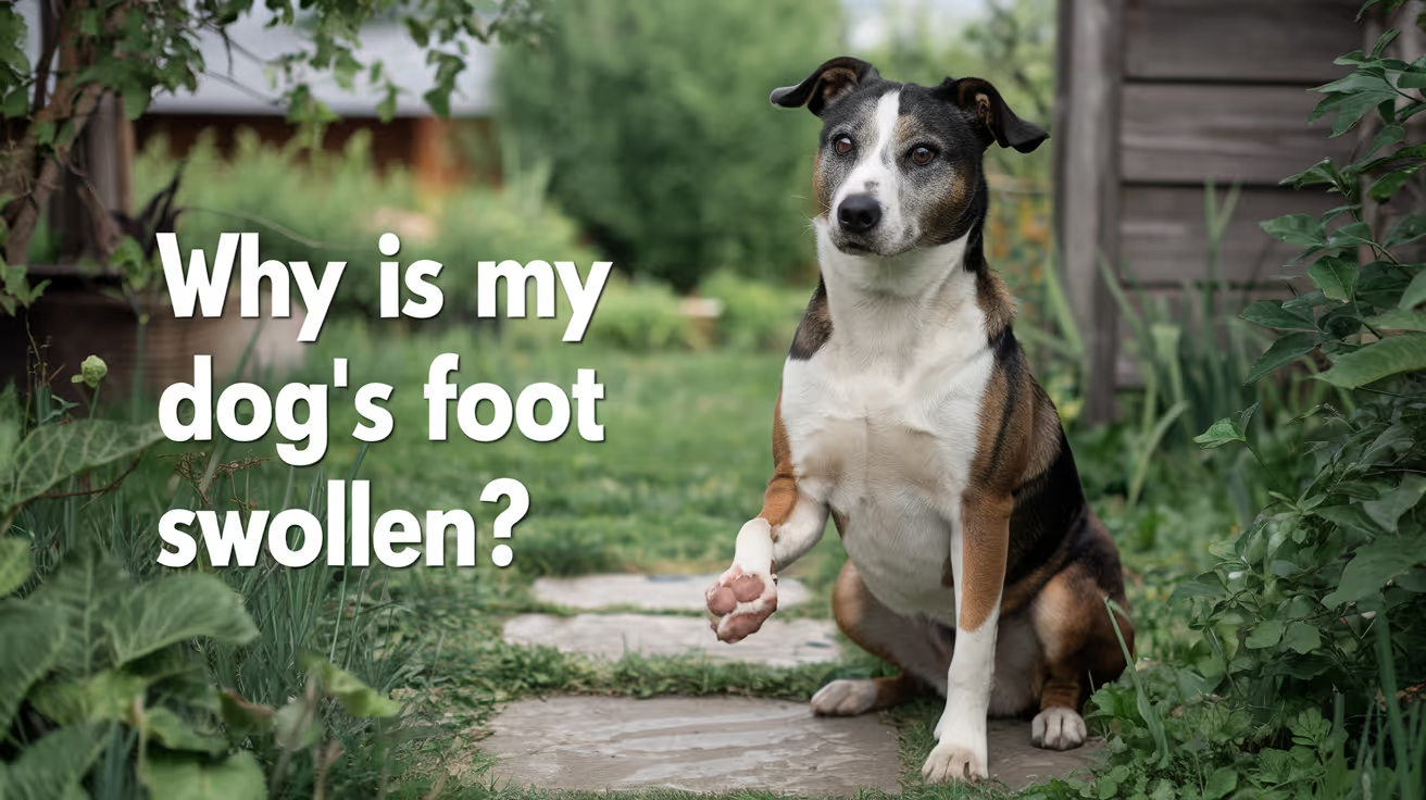
Why Is My Dog's Foot Swollen?
Discover why your dog's foot is swollen, common causes, treatments, and when to see a vet for proper care.
A swollen dog foot is one of the most common reasons owners call their vet. It can be as simple as a bee sting or as serious as a deep infection or bone tumor. The location, speed of onset, and other symptoms all help narrow down the cause.
This guide covers every common cause, how to assess severity at home, and the exact signs that mean your dog needs same-day care.
Quick answer: A swollen dog foot usually has a local cause: injury, insect sting, foreign object, or infection. Warm, increasing, or discharging swelling needs same-day vet care. Multiple swollen paws suggest allergies.
Key takeaways
- Single-paw swelling almost always has a local cause: injury, sting, foreign body, or infection
- Interdigital furunculosis (infected hair follicle cysts between toes) is among the most misidentified causes of recurring paw swelling
- Foreign objects like foxtails and glass splinters can migrate deep into tissue if left untreated
- Pododermatitis is the clinical term for inflamed paw skin; allergies are the most common underlying driver
- Swelling spreading up the leg or paired with face or neck swelling is an emergency requiring immediate care
- Most mild swelling from a sting or minor sprain improves within 24 to 48 hours with rest and monitoring
How to assess your dog's swollen foot before calling the vet
Before looking up causes, do a quick home assessment. This takes two minutes and tells you how urgent the situation is.
Step 1: Which paw?One paw = likely local cause. All four paws = likely allergic or systemic.
Step 2: Feel for heat.Warm or hot tissue means active inflammation or infection.
Step 3: Look between every toe.Redness, swelling between the digits, or visible nodules between the toes points to interdigital furunculosis or a foreign body.
Step 4: Check the pads.Burns appear as red, blistered, or peeling pad tissue. Cuts and punctures are often visible on the pad surface.
Step 5: Look at each nail.A broken nail or swollen nail bed causes localized pain and swelling at the toe tip.
VCA Animal Hospitals: "A deeper infection, like an abscess, will appear as a warm, soft to mildly firm swelling under the skin."
8 common causes of a swollen foot in dogs
1. Injury or trauma
Sprains, cuts, fractured toes, torn nails, and pad burns are the most frequent causes of sudden single-paw swelling. Dogs running on rough, hot, or icy terrain are especially prone.
PetMD notes that pad burns are particularly common in summer: check the pads after any walk on hot pavement. If your palm cannot hold against the pavement for five seconds, it is too hot for your dog's feet.
Signs: sudden lameness after activity, localized swelling, visible wound or bruising.
2. Foreign object
Thorns, glass, foxtails, and grass awns lodge between the toes or penetrate the pad. The body mounts an inflammatory response around the object, causing swelling that worsens over days.
SpectrumCare: "Foxtails and grass awns can lodge between the toes or in the pad and trigger pain, swelling, and infection."
Foxtails are particularly dangerous because they are barbed and can migrate deeper into tissue over days, eventually requiring surgical removal. If you cannot see and safely remove the object, do not probe. See a vet.
3. Insect sting or bite
Bees, wasps, fire ants, and spiders cause rapid single-paw swelling that appears within minutes of the sting. The paw may look puffy and your dog may lick or hold it up.
Dyer Animal Clinic advises watching for anaphylaxis signs: hives, difficulty breathing, excessive swelling spreading beyond the paw. These require emergency care immediately.
Localized sting swelling that stays in the paw and is not worsening can be monitored at home for 24 to 48 hours.
4. Infection: bacterial and fungal
Bacterial or fungal infections enter through cuts, puncture wounds, or damaged skin. Infected paws are typically warm, red, swollen, and often have an odor or discharge. VCA lists the visual signs: "Skin infections on the feet may result in red, moist lesions between the toes."
Pododermatitis (inflammation of the paw skin) is the umbrella clinical term. Common causes include:
- Secondary bacterial infection from chronic licking
- Yeast overgrowth in skin folds between toes
- Fungal infections (ringworm can affect paws)
- Demodectic mange (mite overgrowth)
Pododermatitis does not resolve without treatment. Antibiotics, antifungal medication, or both are typically required.
5. Interdigital furunculosis
This is one of the most commonly missed causes of recurring paw swelling. Interdigital furunculosis occurs when hair follicles between the toes become infected, forming painful reddish-purple nodules that may rupture and drain.
AKC notes that the condition is especially prevalent in short-coated, heavyset breeds: Bulldogs, Labrador Retrievers, and Chinese Shar-Pei are among the most commonly affected. Chronic licking due to allergies is a major driver.
Signs: swollen, painful nodules between the toes, draining tracts, recurrent swelling in the same location.
For how furuncles between the toes connect to abscess formation, see abscess as a cause of limb swelling.
6. Allergic reaction and pododermatitis
Environmental allergens (pollen, grass, lawn chemicals, road salt) and food allergies frequently cause paw inflammation. Dogs with allergies lick their feet chronically, creating secondary infection on top of the allergic reaction.
Wakefield Pet Vet: "Allergic reactions typically cause itching, redness, and sometimes blistering between the toes or on paw pads."
Allergic paw swelling usually affects multiple paws. It tends to recur seasonally or after contact with the trigger substance. Dogs with seasonal allergies often have their worst paw symptoms in spring and fall.
7. Bursitis
Bursae are small fluid-filled sacs that cushion joints. Repeated pressure on bony prominences, especially the elbow and hock, can cause bursitis. Affected joints may appear as soft, fluctuant swellings near a joint.
For how bursitis specifically produces foot and joint swelling, see bursitis as a common cause of foot swelling.
8. Cysts, tumors, and nail bed disease
Cysts, mast cell tumors, and subungual (under-nail) tumors can all present as localized swelling on or near the foot. Petcube notes that toenail tumors are more prevalent in large black-coated breeds such as Standard Poodles, Gordon Setters, and Schnauzers.
A lump that is growing, firm, or pigmented should always be evaluated by a vet rather than monitored at home.
For an overview of lumps that cause swelling on the legs, see lumps that can cause swelling.
Severity triage: what to do right now
Home care for mild cases
For minor swelling without infection signs:
- Keep the dog calm and limit walking
- Soak the paw in warm (not hot) water with Epsom salts for 10 minutes. PetMD recommends this as an excellent short-term measure regardless of cause.
- Gently clean any visible wound with mild soap and warm water
- Apply a cool damp cloth for 10 to 15 minutes to reduce swelling from sprains or stings
- Do not apply antibiotic ointments without vet guidance some formulations are toxic to dogs if licked
For pressure-related paw and leg swelling in dogs that rest in one position for extended periods, see pressure-related swelling in dogs.
Frequently asked questions
How do I treat my dog's swollen paw at home?
For mild swelling with no wound or discharge, rest the dog and soak the paw in warm Epsom salt water for 10 minutes. Check carefully for a foreign object or insect stinger. Do not use human antibiotic creams without vet guidance. If swelling has not reduced within 24 to 48 hours or is getting worse, contact your vet.
When should I be worried about my dog's swollen paw?
Be concerned immediately if the swelling is warm, increasing, or has discharge or odor. Call the vet the same day if your dog refuses to bear weight, if the swelling is spreading up the leg, or if there is any sign of an allergic reaction such as facial swelling or breathing difficulty.
What can I give my dog for a swollen paw?
Do not give human pain medications (ibuprofen, acetaminophen, aspirin) to dogs; many are toxic. For minor swelling, warm Epsom salt soaks are safe and effective for short-term relief. Your vet can prescribe appropriate anti-inflammatory medication if needed after examining the paw.
My dog's foot was normal this morning and is swollen now. What happened?
Sudden single-paw swelling most often means an insect sting, a foreign object picked up during activity, or a minor sprain. Check each toe carefully for a stinger, thorn, or visible wound. If swelling is mild and localized, monitor for 24 hours. If increasing or painful, call the vet the same day.
Can swelling in a dog's foot go away on its own?
Minor swelling from a bee sting or small sprain typically resolves within 24 to 48 hours with rest. Infections, foreign objects, interdigital furunculosis, and bursitis do not resolve without treatment. If swelling has not improved within 48 hours, veterinary assessment is needed.
My dog's foot smells bad and is swollen. Is that serious?
Yes. Odor from a swollen paw strongly indicates infection. A foul smell means bacteria are actively present. Common sources include interdigital furunculosis, a nail bed infection, or an abscess. This requires prompt veterinary treatment and should not be left to resolve on its own.
Resources
- PetMD. Dogs Swollen Paws: Causes and Treatments. petmd.com
- VCA Animal Hospitals. First Aid for Limping Dogs. vcahospitals.com
- AKC. Pododermatitis on Dog Paw: Causes, Symptoms and Treatment. akc.org
- SpectrumCare. Paw Swelling in Dogs. spectrumcare.pet
- Dyer Animal Clinic. Reasons Your Dog's Paw is Swollen. dyeranimalclinic.com
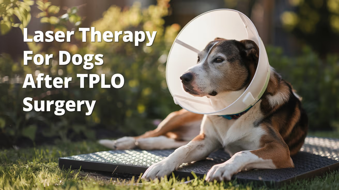
Laser Therapy for Dogs After TPLO Surgery
Learn how laser therapy helps dogs recover faster and with less pain after TPLO surgery for cruciate ligament repair.
Laser therapy also called photobiomodulation (PBMT) or low-level laser therapy (LLLT) is one of the most commonly offered adjunct treatments during TPLO recovery. Many specialist and rehabilitation centres include it routinely.
But the evidence for its benefits is more nuanced than the marketing suggests, and owners deserve an honest picture of what it does and does not reliably achieve.
Quick answer: Laser therapy after TPLO uses specific light wavelengths to reduce inflammation and support tissue healing. Evidence for early pain reduction is moderately supported; evidence for improved radiographic bone healing is weak. It is a safe adjunct but should not replace rehabilitation exercises, pain medication, or activity restriction.
Key takeaways
- Laser therapy reduces postoperative inflammation and may improve gait scores: a TPLO study found better hindlimb function at 8 weeks in treated dogs
- Evidence for improving radiographic bone healing is weak: three controlled studies found no statistically significant difference in healing time
- The 2024 AVMA randomized trial found no significant difference in CRP, weight bearing, pain scores, or SSI rates between PBMT and sham groups
- Sessions typically begin within the first few days of surgery and continue through the rehabilitation phase
- Laser therapy is safe with few contraindications: avoid eyes and active tumour sites; safe over the TPLO incision once closed
- It works best as part of a multimodal plan: exercise therapy and pain medication carry stronger evidence than laser alone
What laser therapy does
Laser therapy for dogs, also known as photobiomodulation, involves using specific wavelengths of light to penetrate tissues and promote cellular regeneration and healing. The laser light stimulates the production of ATP (adenosine triphosphate), enhancing cell repair and growth, reducing inflammation, and increasing blood circulation.
Photobiomodulation therapy has been shown to decrease inflammation, and increase analgesia, vascularization, and tissue healing after musculoskeletal injury or surgery.
The mechanism is photochemical: light energy at specific wavelengths (typically 630 to 980 nm) is absorbed by mitochondria.
This increases ATP production, modulates reactive oxygen species, and influences gene expression related to inflammation and healing.
The effects are local confined to the tissue depth the light reaches.
For post-TPLO use, the targets are: the surgical incision, the osteotomy site in the proximal tibia, and the surrounding periarticular soft tissues.
What the clinical evidence shows
Pain and function
Research following TPLO surgery showed that dogs receiving LLLT had better hindlimb function and gait scores at 8 weeks compared to controls. This is especially valuable in orthopedic recovery, where early weight-bearing can prevent muscle atrophy and joint stiffness.
In a controlled veterinary study, dogs with surgical incisions treated with laser therapy exhibited significantly less inflammatory cell infiltration and tissue necrosis within the first week post-op compared to untreated controls.
The 2024 randomized trial
54 client-owned dogs with CCL rupture undergoing unilateral TPLO surgery were enrolled. The study population was randomly assigned to either a treatment group receiving PBMT (24 dogs) or a control group (30 dogs). PBMT was performed immediately after induction, and at 6 hours, 24 hours, 48 hours, and 8 weeks postoperatively. Evaluation of CRP, pain scores, evidence of SSI, and percentage weight bearing were assessed at all time points.
The trial found the therapy showed promise but no statistically significant difference between groups on any primary outcome measure.
Bone healing
Three studies compared LLLT to a control and concluded that LLLT treatment did not make a significant difference in improving radiographic bone healing. The studies collectively provide weak evidence for this outcome.
This is an important distinction: laser therapy may support soft tissue healing, pain, and early function but it does not appear to accelerate the osteotomy healing visible on radiographs.
When to start and how often
Laser therapy uses focused light energy on the surgical site to support healing.
Most rehabilitation programmes begin laser therapy within the first 1 to 3 days after TPLO surgery, often at the surgical centre before discharge or at the first rehabilitation visit.
Typical post-TPLO laser protocol:
- Frequency: 3 to 5 sessions per week in the first 2 to 3 weeks
- Frequency: 1 to 2 sessions per week from weeks 3 to 8
- Session duration: 5 to 15 minutes depending on the laser system and dosing protocol
- Total sessions: typically 6 to 12 in the first 8-week recovery phase
The protocol varies by laser system, power output, and the individual patient's response.
Realistic expectations
Laser therapy is a useful adjunct in TPLO recovery. It is not a substitute for the treatments with stronger evidence: pain medication, activity restriction, and structured rehabilitation exercises.
Laser therapy could be particularly helpful for dogs with weight-bearing and gait issues while recovering from TPLO surgery after a cruciate injury.
Dogs with significant early swelling, wound sensitivity, or slow initial weight-bearing progress may benefit most. Dogs recovering well with standard multimodal analgesia and rehabilitation may show less measurable difference.
For the bone healing timeline that laser therapy supports during recovery, see TPLO bone healing time in dogs explained.
For the full recovery plan that laser therapy fits into, see 10 essential TPLO recovery tips for pet owners.
For the physical therapy that is the primary evidence-based adjunct, see when to start physical therapy after TPLO surgery.
For swelling management in the recovery period, see how long does swelling last after TPLO surgery.
Frequently asked questions
Is laser therapy safe over the TPLO incision?
Yes, once the incision is closed. Laser therapy is safe over sutured incisions and can be applied at the surgical site from the first post-operative day in most protocols.
Avoid direct application over open wounds or actively infected tissue.
How many laser sessions does a dog need after TPLO?
Typically 6 to 12 sessions across the first 8 weeks.
Start at 3 to 5 sessions per week for the first 2 to 3 weeks, tapering to 1 to 2 per week through the rehabilitation phase.
The exact protocol depends on the laser system and rehabilitation plan.
Does laser therapy replace pain medication after TPLO?
No. Laser therapy is an adjunct to pharmaceutical pain management, not a replacement. Post-TPLO pain management requires NSAIDs, and often gabapentin or other analgesics.
Laser therapy may reduce the pain burden and support earlier mobility but does not provide sufficient analgesia on its own.
Can I do laser therapy at home with a consumer device?
Consumer-grade red light therapy devices exist but operate at lower power densities than veterinary therapeutic lasers. The clinical evidence discussed in this article relates to veterinary-grade PBMT devices.
Home devices may offer some benefit but cannot replicate the dosing of professional equipment. Discuss with your rehabilitation veterinarian before purchasing.
Will laser therapy prevent my dog from needing more medication?
Possibly. If laser therapy reduces post-operative inflammation and pain, some dogs may need lower doses of pain medication or taper off sooner.
This should be guided by your veterinarian based on your dog's individual recovery trajectory.
Resources
- AVMA Journal. Photobiomodulation Therapy in Dogs Undergoing TPLO After Cranial Cruciate Ligament Rupture. avmajournals.avma.org
- Veterinary Evidence. Does LLLT Improve Radiographic Healing for Dogs with CCL Rupture Undergoing TPLO Surgery? veterinaryevidence.org
- AKC. Laser Therapy For Dogs: Uses, Side Effects, and Alternatives. akc.org
- Erchonia. Laser Therapy for Post-Surgical Recovery in Pets. erchonia.com
All Articles
Asepsis During Abscess Drainage
Learn how to maintain asepsis during abscess drainage to prevent infection and ensure safe healing for your pet.
Sustainable Vet Group
Abscess drainage is classified as a dirty-infected wound procedure. It involves the deliberate opening of a pre-existing infection, which creates contamination of the drainage site, instruments, gloves, and surrounding tissues at the moment of incision.
The asepsis goal during abscess drainage is not sterility, which is unachievable in this context. It is contamination control: preventing further spread of the infection to adjacent tissues, other patients, and clinical staff.
What this covers: The asepsis principles and practical protocol for abscess drainage in dogs and cats, including patient preparation, incision site selection, lavage, contamination control, wound management post-drainage, and the specific asepsis requirements for different abscess types.Wound class: Dirty-infected (Class IV). Pre-existing infection is present. SSI rates for dirty-infected procedures range from 18 to 27%+ in the absence of appropriate management.Key principle: Because the procedure opens an infected cavity, the emphasis shifts from preventing contamination of the wound (as in clean surgery) to controlling the spread of contamination from the wound to the patient, the staff, and the clinical environment.
Key takeaways
- Abscess drainage is Class IV (dirty-infected); the wound already contains infection.
- The asepsis goal is contamination control, not wound sterility.
- Wide clip margins prevent hair contamination of the drainage site and surrounding area.
- Lavage after drainage is the most important asepsis step for contamination control.
- Instruments must be treated as contaminated after the abscess opens; do not return to the sterile field.
- Staff PPE (gown, gloves, eye protection) is essential; abscess contents aerosolize during incision.
- Environmental disinfection of the procedure area post-drainage must be thorough.
Pre-procedure preparation
Patient assessment and sedation
Abscess drainage requires adequate analgesia and, in many cases, sedation or general anesthesia. Pain and patient movement during incision compromise contamination control.
Assess for:
- Extent and depth of the abscess (palpation, imaging where indicated)
- Signs of systemic involvement (fever, lethargy, regional lymphadenopathy)
- Identifying the most dependent site for drainage
Bite wound abscesses in cats: Cat bites produce small, deep puncture wounds that frequently develop into deep abscesses. Careful palpation and imaging can reveal the full extent before incision.
Perianal abscesses: Perianal fistulae and anal sac abscesses require specific preparation to minimize fecal contamination during drainage.
Clipping
Clip a generous area around the abscess, extending well beyond the anticipated incision site. University of Minnesota Clinical Skills guidance notes: "Be sure to extend clip below the planned draining stab wound to prevent hair from contaminating the drainage hole, and to keep the area easier to clean of drainage material."
The clip area also needs to extend below the drainage site in the animal's normal standing position, because gravity-assisted drainage will track downward and hair in that path will become contaminated and trap purulent material against the skin.
Skin antisepsis
Apply standard antiseptic scrub to the clipped area around the abscess, using centrifugal technique from the intended drainage site outward. Chlorhexidine-alcohol combination is appropriate.
Important: The abscess wall itself cannot be made sterile by skin antisepsis. The purpose of antisepsis here is to reduce surface contamination adjacent to the drainage site, limiting additional organisms from the skin surface entering the wound.
Staff preparation and PPE for abscess drainage
Abscess drainage requires more protective PPE than clean surgical procedures because:
- The procedure opens a pre-existing infected, often under-pressure cavity
- Purulent material may express at force when the abscess is incised, creating aerosol
- Staff hands, face, and clothing may be exposed to infectious material
Required PPE:
- Gloves: Required throughout; change to a fresh pair after the abscess opens if further sterile field work is needed
- Gown or apron: Protects clothing from purulent material splatter
- Eye protection/face shield: Essential; abscess contents frequently aerosolize at incision
- Mask: Reduces inhalation exposure to aerosolized infectious material
Drainage technique and asepsis
Incision site selection
Select the most dependent site in the abscess when the animal is in normal standing position. This allows passive gravity drainage after the incision is made and minimizes abscess pocket residue.
For bite wound abscesses in cats: locate opposing tooth marks (entry and exit wounds). The abscess often tracks between these points.
Incision
Make a stab incision of sufficient size to allow drainage of thick pus. Too small an incision risks the pocket resealing before the contents fully drain.
After the abscess opens:
- Any instrument that entered the abscess is now contaminated
- Gloves contacting the abscess contents are contaminated
- Do not return contaminated instruments to any sterile field
Lavage after drainage
Lavage is the most important asepsis step after the abscess opens.
Purpose: Mechanically remove purulent material, bacteria, and debris from the abscess pocket and surrounding tissue.
Irrigation solution: Sterile saline. Volume: sufficient to produce clear runoff from the cavity. For large abscesses, 100 to 500+ mL may be required.
Dilute antiseptic lavage: Dilute chlorhexidine (0.05%) or dilute PVI (0.1 to 1%) may be used for the initial lavage of an established abscess. Both reduce bacterial load beyond what saline alone achieves. Do not use concentrated antiseptic solutions within the abscess cavity as they damage granulation tissue.
University of Minnesota guidance: "Flush the abscessed area to remove pus and any gross contamination. Flush solution should be tissue-friendly, aid in removal of bacteria, and ideally isotonic to preserve normal function of cells to promote healing."
Culture
Where clinically appropriate, submit purulent material for aerobic and anaerobic culture and sensitivity testing before lavage. This provides the most reliable identification of causative organisms and guides antimicrobial selection.
In cats with bite wound abscesses, culture is often not performed for straightforward cases where Pasteurella and anaerobes are the expected organisms. For recurrent abscesses, non-responsive cases, or immunosuppressed patients, culture is indicated.
For asepsis principles during wound management, including how abscess drainage fits within the broader wound management asepsis framework and the clean technique standards for ongoing wound care after drainage, that guide covers wound management asepsis in detail.
The intraoperative technique framework for abscess drainage, including sterile field management before the abscess opens and the instrument handling and field abandonment protocol once purulent material is encountered, follows the aseptic technique framework adapted to the dirty-infected context. For core aseptic technique applied during drainage, including the sterile field principles and instrument handling standards that apply before and after the abscess opens, that guide covers the technique framework.
Post-drainage wound management
Open or closed management
Open drainage: Leave the incision open to allow continued drainage. This is appropriate when:
- The abscess is extensive and a single drainage is unlikely to resolve it
- Daily lavage through the open stoma is planned
- Risk of premature closure causing re-accumulation is high
Closed drainage: Primary closure with a drain. Used when:
- The abscess is fully drained and lavaged
- The tissue bed is viable
- Suction or passive drainage can be maintained through a Penrose or closed-suction drain
Closed without drainage: Rarely appropriate for established abscesses. Reserved for small, clean-appearing cavities where the surgeon is confident complete drainage was achieved.
Ongoing wound care
For open-managed abscesses, continued clean technique applies at each dressing change:
- Hand hygiene before any wound contact
- Sterile primary dressing or sterile lavage
- Clean outer bandaging layers
Antimicrobial therapy
Abscess drainage (Class IV wound) warrants antimicrobial therapy, not prophylaxis. Selection should be guided by culture results where available.
For cat bite abscesses without culture: Pasteurella multocida and anaerobic organisms are the primary targets. Amoxicillin-clavulanate provides appropriate coverage for most straightforward cat bite abscesses.
For non-responsive or recurrent cases: culture-guided selection is essential.
Environmental decontamination after abscess drainage
The procedure area requires thorough disinfection after abscess drainage because the environment may be contaminated with purulent material, including organisms that could persist on surfaces and transmit to subsequent patients.
Required steps after abscess drainage:
- Table surface: full disinfection with intermediate-level agent; observe contact time
- Any equipment contacted during the procedure: disinfect per material manufacturer guidelines
- Floor below the drainage site: mop with appropriate disinfectant
- Staff gown and gloves: dispose; do not reuse
- Hand hygiene for all staff involved: thorough wash with soap and water
If significant splatter occurred, extend decontamination to walls, adjacent equipment, and any other surfaces with visible contamination.
For errors to avoid during abscess drainage, including the specific aseptic error categories that are most consequential during dirty-infected wound procedures, that guide covers error prevention.
Specific abscess types: additional considerations
Cat bite abscesses
The most common abscess type in small animal veterinary practice. Cats introduced to multi-cat households or free-roaming outdoors are at highest risk.
Asepsis considerations:
- Explore carefully: bite wound tracts can be deep and tortuous
- Multiple drainage points may be needed if the tract extends far from the visible wound
- Elizabethan collar post-drainage is essential in cats; they will groom the drainage site aggressively
Anal sac abscesses
Close proximity to the anus creates ongoing contamination risk from fecal flora.
Asepsis considerations:
- Clip widely; include adequate area caudal to the abscess for drainage clearance
- Position patient to maximize access while minimizing fecal contamination spread
- Copious lavage essential
- Open management typically preferred to allow continued drainage with daily wound flushing
Perianal fistulae (anal furunculosis)
More complex than simple anal sac abscess; involves extensive sinus tracts. Often managed with a combination of immunosuppressive therapy and surgical debridement rather than simple drainage alone. Aseptic technique for any surgical intervention follows the dirty-infected protocol.
For skin antisepsis before drainage procedures, including the skin prep technique and agent selection applicable when antisepsis of the peri-abscess skin is performed before drainage, that guide covers the antisepsis component.
Frequently asked questions
Do abscesses in cats require general anesthesia for drainage?
In most cases, yes. General anesthesia or deep sedation is required for safe, thorough abscess drainage in cats. Inadequate analgesia results in patient movement that compromises contamination control and prevents adequate exploration and lavage. Topical analgesia alone is insufficient for established abscesses.
Should abscess drainage be performed in the OR or a procedure room?
A clean procedure room with appropriate disinfection before and after is sufficient for most abscess drainage procedures. The OR is not required unless the abscess is extensive and requires general anesthesia with the full surgical infrastructure. After the procedure, the area must receive the same enhanced disinfection protocol regardless of which room was used.
Can antibiotics alone resolve an abscess without drainage?
Rarely. The physical presence of purulent material in an abscess cavity provides a protected environment for bacteria that antibiotics cannot adequately penetrate. Drainage removes the bacterial reservoir. Antibiotics address residual infection after drainage. The combination is more effective than either alone.
Abscess drainage is the procedure where the contamination is already there, and the asepsis goal is to stop it spreading further. Generous clip margins, copious lavage after drainage, full PPE for all personnel involved, and rigorous environmental decontamination after the procedure are the components that distinguish controlled abscess management from a procedure that contaminates the patient, the staff, and the clinical environment.
Resources
The following sources were used as reference and background for this article:
- University of Minnesota Clinical Skills Compendium. Abscess Management in Cat/Dog. open.lib.umn.edu
- Merck Veterinary Manual. Management of Specific Wounds in Small Animals. merckvetmanual.com
- Merck Veterinary Manual. Initial Wound Management in Small Animals. merckvetmanual.com

Asepsis for Urinary Catheterization in Dogs
Learn essential asepsis techniques for urinary catheterization in dogs to prevent infections and ensure safe catheter use.
Sustainable Vet Group
Urinary catheterization in dogs is a common veterinary procedure used to relieve urinary obstruction or collect sterile urine samples. However, improper technique can introduce bacteria into the urinary tract, leading to infections. Understanding asepsis for urinary catheterization in dogs is crucial to protect your pet's health and ensure the procedure's success.
This article provides a clear guide on aseptic techniques during urinary catheterization in dogs. You will learn why asepsis matters, how to prepare equipment and the patient, and best practices to minimize infection risks during catheter insertion and maintenance.
What is asepsis in urinary catheterization for dogs?
Asepsis means preventing contamination by harmful microorganisms during medical procedures. In urinary catheterization, asepsis involves techniques to keep the catheter and urinary tract free from bacteria. This reduces the chance of urinary tract infections (UTIs), which can cause pain and serious complications in dogs.
Maintaining asepsis requires careful preparation, sterile equipment, and clean handling throughout the catheterization process. It is a critical part of veterinary care for dogs needing catheter placement.
- Definition of asepsis: Asepsis is the practice of preventing infection by eliminating bacteria and other microbes during catheterization.
- Importance in catheterization: Proper asepsis lowers the risk of introducing bacteria into the urinary tract, preventing UTIs.
- Common infections prevented: Asepsis helps avoid cystitis, pyelonephritis, and other urinary infections in dogs.
- Role in veterinary practice: Aseptic technique is a standard of care to protect animal health during invasive procedures.
Understanding asepsis sets the foundation for safe urinary catheterization and improves outcomes for your dog.
How do you prepare for aseptic urinary catheterization in dogs?
Preparation is key to maintaining asepsis during catheterization. This includes gathering sterile equipment, cleaning the dog's genital area, and ensuring a clean environment. Proper preparation reduces contamination risks before catheter insertion.
Veterinarians and veterinary technicians follow strict protocols to prepare both the patient and the workspace for aseptic catheterization.
- Equipment sterilization: Use sterile catheters, gloves, lubricant, and drapes to prevent bacterial contamination.
- Patient cleaning: Thoroughly clean the dog's genital area with antiseptic solutions to reduce skin bacteria.
- Environment control: Perform the procedure in a clean, controlled area to limit airborne contaminants.
- Personnel hygiene: Wear sterile gloves and wash hands properly before handling the catheter and patient.
Proper preparation helps create a sterile field and minimizes infection risks during catheter placement.
What are the steps to maintain asepsis during catheter insertion in dogs?
Maintaining asepsis during catheter insertion involves careful technique and attention to detail. Each step must avoid introducing bacteria into the urinary tract. This requires sterile handling of the catheter and minimizing contact with non-sterile surfaces.
Following a step-by-step aseptic protocol ensures safe catheter placement and reduces infection chances.
- Use sterile gloves: Always wear sterile gloves to handle the catheter and avoid touching non-sterile surfaces.
- Lubricate catheter with sterile lubricant: This reduces friction and prevents trauma while maintaining sterility.
- Avoid catheter contamination: Do not let the catheter touch the dog's fur or skin outside the cleaned area.
- Insert catheter gently: Carefully advance the catheter into the urethra without forcing to prevent tissue damage and infection.
Adhering to these steps protects the urinary tract from contamination and supports successful catheterization.
How should you care for the catheter to maintain asepsis after placement?
After catheter placement, ongoing care is essential to maintain asepsis and prevent infections. This includes regular cleaning, monitoring for signs of infection, and proper handling during urine collection or catheter changes.
Good catheter care helps keep the urinary tract sterile and reduces complications.
- Secure catheter placement: Fix the catheter to prevent movement that can introduce bacteria or cause injury.
- Keep insertion site clean: Regularly clean the area around the catheter with antiseptic solutions to prevent bacterial growth.
- Use closed collection systems: Closed urine collection bags reduce exposure to environmental bacteria.
- Monitor for infection signs: Watch for redness, swelling, discharge, or discomfort indicating possible infection.
Proper catheter care after insertion is vital for maintaining asepsis and your dog's comfort.
What are common complications from poor asepsis in dog catheterization?
Poor aseptic technique during urinary catheterization can lead to several complications. The most common is urinary tract infection, which can cause pain, fever, and more serious kidney infections. Other complications include urethral trauma and catheter blockage.
Recognizing these risks highlights why asepsis is critical in catheter procedures.
- Urinary tract infection: Bacteria introduced during catheterization can cause bladder and kidney infections.
- Urethral injury: Improper technique or contamination can cause tissue damage and inflammation.
- Catheter blockage: Infection or debris can block urine flow, requiring catheter replacement.
- Systemic infection risk: Severe infections can spread, leading to sepsis if untreated.
Preventing these complications depends largely on strict aseptic technique throughout catheter use.
How can pet owners support asepsis during and after catheterization?
Pet owners play an important role in supporting asepsis during and after urinary catheterization. Following veterinary instructions carefully and maintaining hygiene at home helps prevent infections and complications.
Understanding your role improves your dog's recovery and comfort.
- Follow veterinary care instructions: Adhere to all cleaning and catheter care guidelines provided by your vet.
- Maintain hygiene: Wash hands before touching the catheter or urinary area to avoid contamination.
- Monitor your dog: Watch for signs of discomfort, swelling, or discharge and report concerns promptly.
- Keep follow-up appointments: Regular veterinary checks ensure the catheter remains clean and functioning properly.
Active owner involvement supports asepsis and promotes your dog's health during catheterization.
What are the best antiseptic agents for urinary catheterization in dogs?
Choosing the right antiseptic agents is important for effective asepsis in urinary catheterization. These agents clean the skin and reduce bacteria without causing irritation. Veterinary professionals select antiseptics based on safety and efficacy.
Using appropriate antiseptics helps maintain a sterile field and protects the urinary tract.
- Chlorhexidine gluconate: A broad-spectrum antiseptic effective against bacteria and safe for canine skin.
- Povidone-iodine: Commonly used antiseptic that kills bacteria but may cause mild skin irritation in some dogs.
- Alcohol-based solutions: Used for rapid disinfection but can be drying and should be used carefully.
- Sterile saline: Used for rinsing after antiseptic application to remove residues and debris.
Proper antiseptic choice and application improve asepsis and reduce infection risk during catheterization.
Conclusion
Asepsis for urinary catheterization in dogs is essential to prevent infections and ensure safe catheter use. Proper preparation, sterile technique during insertion, and careful catheter care reduce the risk of urinary tract infections and other complications.
Pet owners and veterinary professionals must work together to maintain asepsis throughout the catheterization process. Understanding and following aseptic principles protect your dog's health and promote a smooth recovery.
FAQs
How often should the catheter be changed to maintain asepsis?
Catheter change frequency depends on the dog's condition and catheter type but typically occurs every 3 to 7 days to reduce infection risk and maintain sterility.
Can I clean the catheter insertion site at home?
Yes, you can clean the site using sterile wipes or antiseptic solutions recommended by your vet, ensuring your hands are clean before touching the area.
What signs indicate a urinary tract infection in my dog?
Signs include frequent urination, straining, blood in urine, foul odor, licking the genital area, and discomfort. Contact your vet if these occur.
Is it safe to use over-the-counter antiseptics for catheter care?
Only use antiseptics approved or recommended by your veterinarian, as some human products can irritate or harm your dog’s skin.
What should I do if the catheter becomes blocked?
If blockage occurs, do not attempt to fix it yourself. Contact your veterinarian immediately for assessment and safe catheter management.

Suture Size Selection in Small Animal Surgery
Learn how to choose the right suture size for small animal surgery to ensure optimal healing and minimize complications.
Sustainable Vet Group
Every surgical suture comes in multiple sizes, and size matters as much as material. A suture that is too large for the tissue causes unnecessary inflammation and delays healing. One that is too small fails to hold under the mechanical forces the wound faces.
Selecting the right size requires understanding how sutures are sized, what each tissue needs, and how patient weight influences the decision.
Quick answer: Suture sizes follow the USP (United States Pharmacopeia) system, where more zeroes mean smaller diameter so 4-0 is smaller than 2-0, and 2-0 is smaller than 0. The guiding principle is to use the smallest size that provides adequate tensile strength for the tissue. For small animals: linea alba in medium dogs takes 0 to 2-0; subcutaneous layers take 2-0 to 3-0; skin takes 3-0 to 4-0. Cats and small dogs use sizes one unit finer throughout.
Key takeaways
- The USP size system runs from 11-0 (smallest) to 7 (largest) more zeroes means smaller diameter.
- Use the smallest size that provides adequate tensile strength larger sizes cause more inflammation.
- Linea alba in medium dogs: 0 to 2-0 PDS or Biosyn.
- Subcutaneous tissue in dogs: 2-0 to 3-0 Monocryl or Vicryl.
- Skin in dogs and cats: 3-0 to 4-0 for external sutures; 4-0 for intradermal.
- Cats and small dogs use sizes one unit finer than dogs of average size throughout.
Understanding the USP suture sizing system
The United States Pharmacopeia (USP) system is the standard for suture sizing in veterinary and human surgery.
How it works:
- Sutures are assigned a numerical designation based on diameter
- Size 1 is larger than size 0
- Below size 0, additional zeroes are added: 0, 2-0, 3-0, 4-0, 5-0, etc.
- Each additional zero indicates a smaller diameter
- 11-0 is the smallest (microsurgery); 7 is the largest (orthopedic/heavy tissue)
Veterinary Surgery Online explains: "The more zeros, the smaller the material, so 6-0 is actually size 000000, and is pronounced 'six ought' or 'six zero'."
AAHA (Oh, Sew Easy, 2022): "Optimal suture size is determined as the smallest size necessary to achieve a tension-free wound closure."
The core principle: smallest effective size
Veterinary Practice News (Dr. Kendra Freeman, DACVS): "The general principle is to use the smallest suture strong enough for the tissue. This allows for less suture material to be present, which may potentially contribute to inflammation and tissue reaction."
What happens when suture is too large:
- More foreign material in the tissue
- Greater inflammatory response
- Higher infection risk at the suture site
- Possible tissue strangulation if pulled too tight
What happens when suture is too small:
- Suture cuts through the tissue under mechanical load
- Wound dehiscence
- Need for re-closure
Published research (Frontiers in Veterinary Science, 2023): A study comparing USP 2-0, 3-0, and 4-0 PGA sutures in canine abdominal incisions found 4-0 suture had lower inflammatory response markers while maintaining adequate mechanical strength. The authors concluded "USP 4-0 PGA suture has more advantages to suturing canine abdominal surgical incisions."
Size guide by tissue type and patient
Linea alba / fascial closure
Veterinary Practice News: "2-0 or 0 suture is appropriate for the linea alba in medium- to large-size animals."
For why the fascial layer specifically requires larger suture than adjacent muscle, see tissue type as a guide for suture size.
Subcutaneous tissue
Gastrointestinal and urinary tract
Veterinary Practice News: "3-0 or 4-0 suture is generally appropriate for the gastrointestinal tract and urinary bladder."
These organs require fine suture because oversized suture causes excessive lumenal reaction and stenosis risk. Taper-point needles are paired with these sizes to minimize tissue injury.
Skin (external interrupted)
Skin (intradermal)
4-0 is the standard for intradermal (subcuticular) closure across all patient sizes. The dermis is a uniform layer that does not vary as much with patient size as deeper structural layers.
Needle size relationship to suture size
Suture size and needle size are linked the needle is sized to match the suture. Common veterinary needle types:
For how needle selection alongside suture size affects closure quality, see needle size alongside suture size.
Common sizing errors
Too large for the tissue:Most common in the skin layer, where surgeons sometimes default to a larger size "for security." The result is more visible suture marks, more inflammation, and longer healing time.
Too small for the load:More common in the linea alba, where a 3-0 suture is used in a large breed dog that needs 0 or 2-0. Tension exceeds the suture's strength before healing occurs.
Mismatched sizing across layers:Each layer needs its own appropriate size. Using the same suture size throughout all layers (a practice shortcut) results in either oversized subcutaneous sutures or undersized fascial sutures.
For how suture size selection connects to material selection across procedures, see size alongside material selection in dogs. For cats, see size alongside material selection in cats.
Frequently asked questions
My vet used 3-0 suture for my large dog's spay. Is that too small?
It depends on the layer. 3-0 for the subcutaneous and skin layers of a medium-large dog is appropriate. 3-0 for the linea alba in a dog over 25 kg would likely be undersized 0 or 2-0 is standard for that layer. If you are concerned, ask your vet specifically which sizes were used at each layer.
Do smaller sutures dissolve faster?
No. Absorption timeline is determined by the suture material, not by its size. A 3-0 Monocryl and a 2-0 Monocryl absorb over the same timeline approximately 90 to 119 days. The difference is tensile strength, not absorption rate.
Can suture sizes vary between different materials of the same USP designation?
Yes, slightly. Veterinary Surgery Online notes: "It is important to note that the USP dimensions for catgut are different to those of other sutures. For example, 2-0 Catgut is larger than 2-0 PDS." When comparing materials, the USP designation is a starting reference, not an exact interchangeable standard across all material types.
Suture size selection follows one rule applied at every layer: the smallest size that adequately holds the tissue under its expected mechanical load. Getting it right means less inflammation, less reaction, and faster healing. Getting it wrong in either direction creates predictable complications that show up in the first two weeks of recovery.
Resources
- Veterinary Practice News. The Must-Read Guide to Selecting Sutures (Dr. Kendra Freeman, DACVS). veterinarypracticenews.com
- Veterinary Surgery Online. Suture Size. vetsurgeryonline.com
- AAHA. Oh, Sew Easy: A Guide to Sutures. aaha.org
- Frontiers in Veterinary Science (2023). Biomechanical and tissue reaction: the effects of varying suture size on canine abdominal wall stitching. frontiersin.org
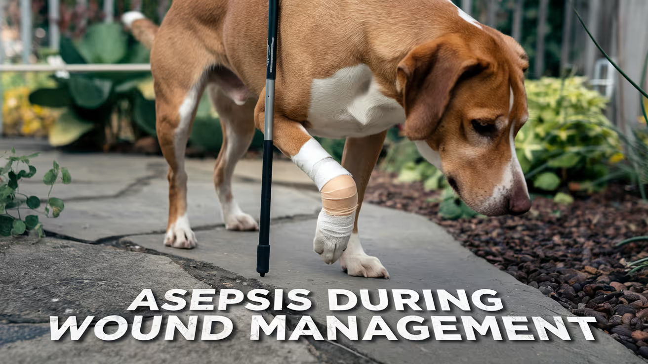
Asepsis During Wound Management
Learn essential asepsis techniques during wound management to prevent infections and promote healing in pets.
Sustainable Vet Group
Wound management in veterinary practice spans a wide contamination spectrum, from simple laceration repair in the clinic to complex open wound management over weeks of repeated bandaging. The asepsis standard applied must match the wound and procedure type.
Not all wound management requires surgical-level sterility. Much of it requires clean technique, which is a defined standard, not simply "fairly clean."
What this covers: The asepsis principles applied during wound assessment, lavage, debridement, dressing, and redressing in small animal veterinary practice, with the distinction between clean technique and sterile technique across different wound types.Scope: Applies to all wound management procedures in dogs and cats, from acute traumatic wounds through chronic open wounds requiring repeated bandage changes.Key distinction: Surgical asepsis (sterile technique) aims to maintain a contamination-free field during invasive procedures. Wound management asepsis uses clean technique for most non-surgical wound care, with sterile technique applied to specific components (irrigation fluids, instruments contacting wound bed).
Key takeaways
- Not all wound management requires sterile technique; clean technique is the standard for most bandage changes.
- Irrigation fluid must always be sterile; non-sterile lavage introduces contamination.
- Instruments contacting the wound bed require sterile-level handling or single use.
- Hand hygiene before any wound contact is non-negotiable regardless of glove use.
- Dressings must be sterile at point of contact with the wound; non-sterile outer layers are acceptable.
- Wound class determines the antimicrobial and closure strategy, not just the asepsis approach.
- Owner-performed home wound care requires explicit asepsis education; compliance reduces SSI risk.
Wound classification and asepsis standard
The same wound classification framework that applies in surgery applies to wound management:
The wound class should be assessed and documented at each evaluation, as wounds may progress from contaminated to dirty-infected without adequate management, or improve toward clean-contaminated with effective treatment.
The clean technique standard for wound management
Clean technique for wound management involves:
- Hand hygiene before any wound contact (minimum: alcohol-based hand rub; soap and water if hands visibly soiled or if Clostridium contamination is possible)
- Clean gloves (not necessarily sterile) for handling the outer wound dressing layers
- Sterile gloves when directly contacting the wound bed or primary dressing layer
- Sterile supplies for any item contacting the wound surface (primary dressings, irrigation fluids, instruments used on the wound)
- Clean but not necessarily sterile outer layers and bandaging materials
This standard differs from sterile surgical technique in that:
- Clean (non-sterile) gloves may be used for outer bandage layers
- The environment does not need to be a sterile OR
- The person managing the wound does not require surgical scrub technique
Clean technique does not mean casual or careless technique. The same attention to preventing contamination events applies; the environment and some personnel requirements are less demanding.
Wound lavage: the highest-priority asepsis component in wound management
Why lavage matters
Wound lavage is the single most effective contamination reduction intervention in wound management. Merck Veterinary Manual states: "Wounds should be irrigated (lavage) with an appropriate solution at appropriate pressure, to remove contamination (bacteria and debris) and improve visibility for wound inspection."
The mechanical action of lavage physically dislodges and removes bacteria, debris, and devitalized material that chemical antiseptics cannot address.
Irrigation fluid
Sterile saline (0.9% sodium chloride): The standard. Isotonic, non-cytotoxic, no antibacterial activity that could delay healing, widely available.
Sterile water: Acceptable where saline is not available; hypotonic, so longer-term use on open wounds may affect healing tissue.
Dilute povidone-iodine (0.1 to 1%): Acceptable for contaminated or infected wounds. Reduces bacterial load including some organisms not addressed by saline alone. Avoid in fresh granulation tissue.
Dilute chlorhexidine (0.05%): As above. Effective against a broad spectrum including biofilm-forming organisms. Avoid in body cavities at concentrations above 0.05%.
Non-sterile tap water: Not acceptable for wound irrigation. Tap water contains microorganisms and mineral contamination that introduce additional bacterial load into the wound.
Irrigation pressure
Wound irrigation should be performed at sufficient pressure to dislodge debris and bacteria:
- Syringe and 18-gauge needle or irrigation catheter: Produces approximately 8 psi of pressure: the range shown to be effective for bacterial removal without tissue damage
- Bulb syringe: Lower pressure; adequate for maintenance irrigation of healing wounds; insufficient for initial contaminated wound lavage
- Pressure irrigation devices: Mechanical wound irrigation at 8 to 15 psi for heavily contaminated wounds
Volume
There is no single correct lavage volume. Volume should be sufficient to visibly clear debris and produce clear runoff. For contaminated traumatic wounds at initial presentation: minimum 200 to 500 mL per site; more for heavily contaminated wounds.
Lavage frequency
For open wounds undergoing repeated management: at each bandage change, lavage before applying the new primary dressing.
For asepsis principles during wound management, including how these principles apply to the specific wound management context of abscess drainage where contamination management is most demanding, that guide covers abscess-specific wound management.
Debridement and asepsis
Debridement (removal of necrotic, contaminated, or non-viable tissue) is performed using sterile instruments: scissors, scalpel, curettes, or forceps.
Asepsis requirements during debridement:
- Sterile instruments for each debridement session; reused instruments between sessions must be re-sterilized or replaced
- Sterile or clean gloves depending on the procedure depth
- Lavage before and after debridement to clear removed material from the wound
- Culture of wound tissue or exudate where infection is suspected or non-responsive
Dressing technique: clean and sterile components
Primary dressing (wound contact layer)
Must be sterile. This layer directly contacts the wound surface and is the contamination-critical interface.
Types of primary dressings:
- Non-adherent sterile dressings (Telfa equivalent)
- Sterile saline-moistened gauze for moist wound healing
- Foam dressings with sterile wound contact surfaces
- Antimicrobial primary dressings (silver-containing, honey-impregnated) where indicated
Opening primary dressings: Use aseptic technique. Handle the wound contact surface using sterile gloves. Do not touch the wound contact layer with clean (non-sterile) gloves.
Secondary and outer layers
Secondary and tertiary bandage layers (padding, conforming gauze, cohesive bandage) do not directly contact the wound. These may be handled with clean gloves.
Changing frequency
Wound type and dressing type determine change interval:
- Heavily exudating wounds: daily or more frequently
- Granulating wounds with low exudate: every 48 to 72 hours
- Follow manufacturer guidelines for specialized dressings (e.g., silver foam, Manuka honey dressings)
Common asepsis errors in wound management
For aseptic errors during wound management, including the broader error taxonomy for all aseptic procedures in small animal practice, that guide covers the error categories and prevention strategies.
Wound management asepsis for specific wound types
Bite wounds
Cat bites (small, deep puncture wounds) frequently become infected and must be treated as potential abscesses. Dog bites have variable presentation from superficial to penetrating.
Asepsis approach:
- Explore under appropriate analgesia or sedation to identify full wound extent
- Generous clip around entry and exit wounds
- Copious lavage with sterile saline at pressure
- Culture of wound content where infection is present or suspected
- Open wound management preferred over primary closure in contaminated bite wounds
Traumatic lacerations
Acute (under 6 hours): Lavage, debridement, primary closure if wound bed is clean and viable. Clean technique throughout.
Delayed presentation (over 6 to 12 hours): Treat as contaminated; open wound management or delayed primary closure after initial lavage and debridement.
Open wounds requiring repeated management
Wounds managed by second-intention healing over days to weeks require consistent clean technique at every bandage change. Each change represents a contamination opportunity. Consistent hand hygiene, sterile primary dressings, and sterile lavage at each change maintain the wound environment conducive to healing.
For aseptic technique applied to wound care, including how the sterile field principles developed for surgical settings adapt to the wound management context, that guide covers the technique framework.
Skin antisepsis before wound management procedures
When a skin incision is required for wound management (drainage, debridement under general anesthesia), standard skin antisepsis applies around the wound margin.
For skin antisepsis before wound management, including the centrifugal scrub technique, agent selection, and contact time requirements that apply when surgical-level preparation is needed around a wound site, that guide covers the skin antisepsis protocol.
Frequently asked questions
Can tap water be used to irrigate wounds in an emergency?
In a true field emergency without access to sterile saline, clean running potable water is preferable to no irrigation at all. However, at any point where sterile saline or sterile water is available, it should be used. Tap water contains bacteria and should not be used for wound irrigation in a clinical setting.
Do bandage changes require the same sterile environment as surgery?
No. Bandage changes use clean technique, not sterile technique. The key distinction is that primary dressings (wound-contact materials) must be sterile, but outer layers and the environment can be clean rather than sterile. The procedure area should be clean and disinfected, but it does not need to meet OR environmental standards.
When should wound cultures be taken for antibiotic guidance?
For any wound that is infected (discharge, odor, local inflammation beyond expected healing response), cultures should be taken before initiating or changing antibiotic therapy. Superficial swabs are less informative than deep tissue or purulent material samples. Submit for aerobic and anaerobic culture with sensitivity testing.
Wound management asepsis is not a scaled-down version of surgical asepsis. It is a parallel discipline with its own standard, clean technique, applied precisely. The critical variables are sterile irrigation fluids, sterile primary dressings, correct lavage pressure, hand hygiene at every contact, and the consistent discipline of treating a non-sterile approach as a patient safety failure, not a minor shortcut.
Resources
The following sources were used as reference and background for this article:
- Merck Veterinary Manual. Initial Wound Management in Small Animals. merckvetmanual.com
- Merck Veterinary Manual. Management of Specific Wounds in Small Animals. merckvetmanual.com
- University of Minnesota Clinical Skills Compendium. Abscess Management in Cat/Dog. open.lib.umn.edu
- WoundSource. Clean Dressing Technique Principles: Clean vs. Asepsis Wound Dressing Change. woundsource.com
Asepsis vs Sterilization in Veterinary Clinics
Learn the key differences between asepsis and sterilization in veterinary clinics to ensure your pet's safety during treatment.
Sustainable Vet Group
Sterilization and asepsis are related but distinct disciplines. Sterilization is a process applied to instruments and materials before a procedure. Asepsis is the ongoing system of practices that protect the sterility of those materials throughout the procedure.
Sterilization is the foundation. Asepsis is the discipline that preserves what sterilization achieved.
What this covers: The formal definitions of asepsis and sterilization, how sterilization supports the aseptic system, the sterilization methods used in veterinary practice, validation requirements, and the relationship between the two in clinical infection control.Core distinction: Sterilization achieves a state: the complete elimination of all microorganisms and spores from an object. Asepsis maintains a state: the prevention of contamination from entering the operative field or patient.Clinical relevance: Sterilization failure is the highest-consequence single point of failure in surgical asepsis. A contaminated instrument delivered via perfect aseptic technique still introduces bacteria directly into the wound. Validating sterilization efficacy is therefore not optional: it is the most critical quality control step in the instrument cycle.
Key takeaways
- Sterilization destroys all microorganisms including spores; disinfection does not: This is the critical distinction between sterilization and lower-level microbial control methods.
- Asepsis depends on sterilization as its starting point: Aseptic technique manages sterile items to prevent re-contamination. If those items were not sterile to begin with, aseptic technique has no valid starting point.
- Chemical indicators confirm exposure, not sterility: A darkened chemical indicator on an instrument pack confirms the pack was exposed to a sterilization cycle. It does not confirm that cycle achieved sterility. Only biological indicators confirm functional sterility.
- Biological indicator testing must be performed regularly: Weekly biological indicator tests are the minimum for active veterinary surgical practices. Any failed test requires removal of all potentially affected packs from use.
- Sterilization methods are not interchangeable: Autoclave (steam sterilization), dry heat, ethylene oxide, and chemical sterilization each have different indications and limitations. Selecting the wrong method for an instrument type risks either sterilization failure or instrument damage.
- Aseptic technique preserves what sterilization achieved: A sterile pack that is opened incorrectly, stored past its expiry date, or handled with non-sterile technique is no longer sterile regardless of the quality of the sterilization cycle it underwent.
Formal definitions
Sterilization
The complete destruction of all microorganisms, including bacterial spores, on an object or material.
Veterian Key defines sterilization as "the destruction of all microorganisms (bacteria, viruses, spores) on an item."
Sterilization achieves an absolute outcome: zero viable microorganisms. This distinguishes it from disinfection, which reduces but does not eliminate all microbial life, and from antisepsis, which reduces microbial load on living tissue.
Sterility assurance level (SAL): In practice, sterilization is described probabilistically. A SAL of 10^-6 means there is a 1 in 1,000,000 probability that any viable microorganism remains after the process. Validated sterilization methods are designed to achieve this standard.
Asepsis
The prevention of contamination of sterile environments, tissues, or materials by pathogenic microorganisms.
Asepsis is not a single action but a system: the combination of sterile technique, physical barriers, environmental controls, and behavioral protocols that maintain contamination-free conditions from the moment of instrument sterilization through wound closure.
The relationship: sterilization creates a sterile item; asepsis keeps it sterile until it reaches the patient.
Sterilization methods in veterinary practice
Steam sterilization (autoclave)
The primary method for metal instruments, textiles, and most reusable surgical supplies.
Mechanism: High-pressure saturated steam denatures proteins and destroys cell membranes across all microorganism types including spores.
Standard parameters:
- Gravity displacement cycle: 121°C at 15 psi for 15 minutes
- Pre-vacuum cycle: 132°C for 4 minutes (faster; requires functional vacuum pump)
Advantages: Reliable, fast, low-cost per cycle, no toxic residues.
Limitations: Cannot be used for heat-sensitive instruments (flexible endoscopes, some electronic components, plastics that melt or warp).
Dry heat sterilization
Mechanism: Oxidation of cellular components at high temperature without moisture.
Parameters: 160°C for 120 minutes; 170°C for 60 minutes.
Indications: Instruments that corrode with moisture (some cutting edges, certain alloys). Powders and oils that steam cannot penetrate.
Limitations: Longer cycle times; higher energy cost; not suitable for heat-sensitive materials.
Chemical sterilization (liquid sterilants)
Mechanism: Alkylation or oxidation of microbial proteins and nucleic acids.
Agents and contact times:
- Glutaraldehyde 2%: 6 to 10 hours for sterilization (versus 20 to 30 minutes for high-level disinfection only)
- 7.5% hydrogen peroxide: 6 hours at 20°C for sterilization
Indications: Heat-sensitive critical devices that cannot be autoclaved (some arthroscopes, fiber-optic equipment).
Limitations: Items must be fully immersed; residual agent must be rinsed before use; sterility is not maintained after removal from solution unless aseptic transfer is used.
Ethylene oxide (EtO) gas sterilization
Mechanism: Alkylation of DNA and proteins; kills all microorganisms including resistant spores.
Indications: Heat-sensitive and moisture-sensitive devices (some electronic implants, complex optics).
Limitations: Requires specialized equipment and aeration period (12 to 24 hours) to remove toxic residues before clinical use. High cost; not practical for most general veterinary practices.
For how sterilization is validated, including biological indicator protocols, chemical indicator interpretation, and what to do when a sterilization cycle fails, that guide covers the validation process in full.
Sterilization validation: the quality control bridge
Sterilization cannot be assumed. It must be confirmed. The three-level monitoring system:
Level 1: Mechanical monitoring
Recording temperature, pressure, and time for each autoclave cycle. Modern autoclaves print cycle records automatically. These records should be filed and reviewed periodically as part of the clinic's quality system.
Mechanical monitoring confirms the autoclave ran a cycle. It does not confirm the cycle achieved sterility.
Level 2: Chemical indicators
Chemical indicator strips and integrators inside and outside instrument packs change appearance when exposed to sterilization conditions.
- External indicators confirm the pack was exposed to a cycle
- Internal indicators confirm the sterilizing agent penetrated the pack interior
- Class 5 and 6 integrating indicators provide the closest chemical approximation to a sterility confirmation
Critical limitation: Chemical indicators confirm exposure to sterilization conditions. They do not confirm the biological kill standard was met.
Level 3: Biological indicators (spore tests)
Biological indicators contain Geobacillus stearothermophilus spores (the most resistant organism to steam sterilization). If the autoclave cycle kills these spores, it confirms the cycle achieved the required sterility standard.
Biological indicators are the only confirmation of functional sterilization efficacy.
Recommended frequency: weekly in active veterinary surgical practices; after any autoclave service or malfunction; when a chemical indicator failure is observed.
For the instrument sterilization protocol, including the step-by-step process from instrument cleaning through packaging, sterilization, storage, and shelf-life management, that guide covers the full instrument reprocessing protocol.
How asepsis preserves sterilization
Sterilization achieves sterility at a point in time. Asepsis is what preserves that sterility from that point through wound closure.
The chain of asepsis after sterilization:
- Pack integrity maintained during storage: No tears, moisture, or compromised seals
- Expiry date respected: Sterility is time-limited; shelf-life management is part of the asepsis system
- Sterile opening technique: Circulating nurse opens the pack without contacting the sterile contents; contents dropped or transferred to the sterile field without touching the non-sterile outer packaging
- Aseptic instrument handling: Instruments handled only by gowned, gloved scrub personnel; not dropped below table level; not contaminated by non-sterile contact
- Sterile field maintenance: Entire operative period managed to prevent any non-sterile item from contacting sterile instruments or the wound
If any step in this chain fails, the sterilization was wasted. An instrument that was sterile when packed but handled with non-sterile technique at the table is contaminated before it enters the wound.
For aseptic technique applied to preserving sterility, including the specific intraoperative behaviors that protect sterile instruments from re-contamination, that guide provides the technique reference that complements sterilization.
Sterilization sits at the top of the microbial control hierarchy, above both disinfection and antisepsis. Understanding where each level applies prevents the common error of applying a lower standard than the clinical situation requires. For disinfection vs. asepsis comparison, including how the three levels of disinfection compare to sterilization and when each applies in veterinary practice, that guide covers the Spaulding classification and the full hierarchy of microbial control.
For antisepsis in the broader asepsis framework, including how antisepsis applied to living tissue relates to sterilization of instruments within the same perioperative infection control system, that guide covers the complementary antisepsis component.
Common failures at the sterilization-asepsis interface
Frequently asked questions
Is sterilization the same as disinfection at a higher level?
No. Disinfection reduces microbial load and kills most pathogens but does not reliably kill all bacterial spores. Sterilization destroys all microorganisms including spores and achieves an absolute sterility standard. High-level disinfection can approach sterilization with extended contact times, but the two are formally distinct levels of microbial control with different validation standards.
How long do sterilized packs remain sterile?
This depends on packaging type and storage conditions. Event-related sterility is the current standard: a pack remains sterile until something happens to compromise it (moisture exposure, torn packaging, improper storage), rather than a fixed time period. Many clinics use a defined shelf life (e.g., 6 months for double-wrapped packs in clean, dry storage) as a practical policy even though the scientific standard is event-related.
What should happen if a biological indicator comes back positive?
All packs sterilized since the last successful biological indicator test should be removed from use immediately. The autoclave should be taken out of service, inspected, and repaired. A new biological indicator should be run before the autoclave returns to service. All clinical cases performed using instruments from potentially non-sterile packs should be flagged for post-operative monitoring. Document the incident and all corrective actions.
Can aseptic technique compensate for a failed sterilization cycle?
No. If an instrument is not sterile, no amount of correct aseptic technique during the procedure prevents the contamination it carries from entering the wound. Sterilization is a prerequisite. Asepsis preserves it. Neither substitutes for the other.
Sterilization and asepsis are not competing approaches or different words for the same thing. Sterilization is the process that creates the starting condition for safe surgery. Asepsis is the system of practices that maintains that condition. Both must function correctly for surgical infection control to hold.
Resources
The following sources were used as reference and background for this article:
- Veterian Key. Sterilization and Disinfection. veteriankey.com
- Today's Veterinary Nurse. Keys to Successful High-Level Disinfection and Sterilization Processes. todaysveterinarynurse.com
- GWU Office of Research Safety. Sterilization, Disinfection, and Decontamination. researchsafety.gwu.edu
- Wiley Online Library. Disinfection and Sterilization, Veterinary Microbiology. onlinelibrary.wiley.com

Asepsis Training for Veterinary Staff
Learn essential asepsis training for veterinary staff to prevent infections and ensure safe animal care in clinics and hospitals.
Sustainable Vet Group
Asepsis training in veterinary practice is not a one-time orientation event. It is a continuous program with defined curriculum, structured competency assessment, and scheduled refresher cycles.
The distinction matters because the evidence is clear: knowledge of correct aseptic technique does not reliably translate into consistent intraoperative behavior without external monitoring and feedback. Training that addresses only knowledge without building the behavioral and cultural components of correct asepsis produces limited improvement.
What this covers: The design, content, delivery, and assessment components of an effective asepsis training program for veterinary surgical staff.Who needs training: All personnel involved in surgical procedures, including surgeons, veterinary nurses, scrub technicians, circulating nurses, and ancillary staff who clean and prepare the OR.Evidence base: AJVR (2025) documented a 46.3% aseptic protocol breach rate during scrubbing, gowning, and gloving in veterinary students, with no association between breach rate and prior experience. This data indicates that experience alone does not maintain aseptic performance without structured reinforcement.Training goal: Not just knowledge of correct technique but consistent performance of correct technique under real surgical conditions, including the ability to recognize and name breaks in the sterile field.
Key takeaways
- Knowledge is necessary but not sufficient: Studies consistently show that personnel who can describe correct aseptic technique still make technique errors in practice. Competency assessment must include observed performance, not just written tests.
- No association exists between experience level and breach rate: AJVR 2025 found no significant difference in protocol breach rates based on prior scrub experience. Experienced staff need refresher programs as much as new staff.
- The cultural component of training is as important as the technical component: Training must address the expectation that breaks will be named and corrected immediately, without hierarchy or blame.
- Competency assessment should be periodic, not only at orientation: A one-time assessed training at hiring does not maintain performance over months and years of practice.
- Different staff roles require different training emphasis: Surgeons, scrub technicians, circulating nurses, and OR cleaners share a common asepsis foundation but have role-specific responsibilities that training should address separately.
- SSI surveillance data should feed back into training: If post-operative infection rates rise, training is one of the first corrective levers to pull.
Training curriculum: what to cover
Core content for all surgical personnel
1. Foundations of surgical asepsis
- Definition and scope: sterility vs. asepsis vs. medical asepsis
- The surgical site infection pathway: how bacteria enter, colonize, and cause SSI
- Wound class and SSI risk stratification (clean, clean-contaminated, contaminated, dirty)
- The five domains of surgical asepsis: patient prep, instrument sterilization, team preparation, OR environment, intraoperative technique
2. Hand hygiene and surgical hand antisepsis
- Correct traditional scrub technique: sequence, surfaces, duration
- Alcohol-based surgical hand rub (ABHR): correct application and contact time
- Common failures: insufficient duration, missed surfaces, incorrect drying technique
- When to re-scrub vs. when ABHR suffices between cases
3. Gowning and gloving
- Closed gloving technique: step-by-step with supervised practice
- Open gloving: when used and critical failure points
- Gown donning without surface contamination
- Double gloving: rationale and technique for orthopedic procedures
4. Patient preparation
- Clipping timing and technique (immediate pre-op; clippers vs. razors)
- Antiseptic agent selection by site and species
- Centrifugal scrub direction and application count
- Common preparation errors and their consequences
5. Sterile field principles
- Boundaries of the sterile field: physical and procedural
- Rules of the sterile field: what belongs, what does not
- Instrument handling: passing, dropping below table level, replacement protocol
- Draping: application, stability, no-reposition rule
6. OR behavior and environment
- Movement in and around the sterile field
- Traffic management during active procedures
- OR door discipline during surgery
- Talking, sneezing, and mask discipline
7. Break recognition and correction
- What constitutes a break vs. an error
- The designated field monitor role
- Non-punitive naming and correction culture
- The correct immediate response to each break category
Role-specific content
Training delivery formats
Initial orientation training
All new surgical personnel should complete a structured orientation before participating in procedures, consisting of:
Didactic component: Lecture or self-directed module covering foundations, technique standards, and break recognition. Duration: 2 to 4 hours depending on role.
Demonstration: Supervised observation of correct scrub, gown, and glove technique by a trained observer.
Supervised practice: Repeated practice of hand antisepsis, gowning, and gloving until technique is correct without correction needed.
Assessed competency: Observed performance of a complete scrub-gown-glove sequence with structured scoring. Personnel should not participate in surgical cases without passing competency assessment.
Ongoing training and refresher cycles
Orientation training alone is insufficient to maintain performance. Refresher programs should include:
- Annual competency re-assessment: Observed scrub, gown, and glove performance scored against a structured rubric
- Case-specific debrief: After any case where an aseptic break is documented, a team debrief reviews what occurred and what the correct response was
- SSI event review: When a post-operative SSI is identified, a structured case review examines all recorded perioperative aseptic practices
- Protocol update training: When antiseptic agents, sterilization methods, or OR procedures change, training must precede implementation
For the checklist staff should follow after training, including the phase-by-phase verification tool that trained personnel use to apply their training consistently during every procedure, that guide provides the operational reference.
Competency assessment: standards and methods
What competency assessment must include
Written tests confirm knowledge. They do not confirm performance. Competency assessment in surgical asepsis must include direct observation of behavior.
Minimum competency assessment components:
Critical errors are those that would result in definite contamination of the sterile field: ungloved skin touching the glove exterior, gown exterior contacting the scrub suit, returning a dropped instrument to the sterile field.
Who conducts competency assessment
Assessment requires a trained observer who knows what correct technique looks like and can distinguish it from incorrect. This is not a self-assessment activity. Peer observation by another trained staff member, supervised by a senior surgeon or clinical director, is the appropriate structure.
Documenting competency
All competency assessments should be dated, signed, and filed. In the event of an SSI, documented competency records demonstrate due diligence and form part of the clinical governance record.
Building a training culture
The most consequential training outcome is not a passing score on a competency assessment. It is a surgical team that names and corrects breaks immediately, regardless of who made them, without judgment or delay.
This outcome requires explicit attention in training, not just technical skill instruction.
Elements of a correction culture that training should address:
The designated field monitor role is a legitimate function, not a policing one: Every team member should understand that the field monitor's job is patient safety, and naming a break is a clinical act, not a personal criticism.
Seniority does not protect against errors: Training should explicitly address the documented tendency for junior staff to avoid correcting senior personnel. The contamination consequences of a missed break are identical regardless of who made it.
Immediate correction is always the right response: There is no scenario in which naming a break later is better than naming it when it occurs.
For the breaks in asepsis that training prevents, including the full taxonomy of break categories, their mechanisms, and the response framework that trained staff should apply, that guide provides the reference material for break-specific training content.
Training scenarios built around specific break categories are more effective than general technique reminders. Simulated break identification exercises, where trainees watch a gowning or gloving sequence and name every deviation, build the observation skills that field monitoring depends on. This kind of scenario-based training also normalizes the act of naming breaks, reducing the cultural hesitation that allows violations to go uncorrected in real procedures.
For the errors addressed through staff training, including the published incidence data for each error category and why experience level does not predict error rate, that guide provides the evidence base for training priority decisions.
Connecting training to audit
Training and audit form a cycle. Training establishes the standard. Audit measures whether the standard is being met. Audit findings drive training refinement.
A clinic that trains without auditing does not know whether training is working. A clinic that audits without training does not have a systematic way to address identified gaps.
For auditing trained staff for compliance, including how to structure observation-based audits, what to measure, and how audit findings should feed back into training program revision, that guide covers the audit component of the training-audit cycle.
Frequently asked questions
How often should asepsis training be repeated?
At minimum annually for all surgical personnel, with additional training triggered by: any SSI event, any documented significant aseptic break, changes to antiseptic agents or sterilization equipment, or new procedures being introduced to the practice. High-volume practices may benefit from semi-annual refreshers.
Should asepsis training be role-specific or universal?
Both. A shared foundational curriculum ensures every person in the OR understands the same principles and the same correction expectations. Role-specific training then addresses the technical responsibilities of each position. Surgeons, scrub technicians, and OR cleaners share the foundation but have different intraoperative responsibilities.
Can online or self-directed training replace observed competency assessment?
No. Online training is an effective delivery format for foundational knowledge. It does not substitute for observed performance assessment. A person who completes an online module on closed gloving technique has not demonstrated that they can perform it correctly. Observed assessment is irreplaceable.
What should happen when a staff member fails a competency assessment?
Additional supervised practice followed by re-assessment. Failing a competency assessment is not a disciplinary event; it is a training event. The appropriate response is remedial practice, not penalty. Personnel should not participate in surgical cases until competency is confirmed.
How should training address MRSP and resistant organisms?
As part of the infection consequence module: explain that antibiotic-resistant organisms including MRSP are increasingly prevalent in veterinary surgical infections, that they are harder to treat when they do occur, and that consistent aseptic technique is the primary prevention strategy. This provides context for why technique rigor matters beyond routine infections.
For the core technique covered in training, including the complete technical reference for every step of aseptic technique that training must cover, that guide serves as the curriculum content source for the technical components.
Asepsis training works when it is designed as a continuous program rather than a one-time event, when competency is assessed through observed performance rather than knowledge tests alone, and when the cultural expectation of immediate, non-judgmental break correction is built into the program from the start. The training that produces the best aseptic outcomes is not the most comprehensive lecture. It is the one that most reliably changes behavior in the OR.
Resources
The following sources were used as reference and background for this article:
- AVMA Journals. Aseptic protocol breaches are common among veterinary students. AJVR, 2025. avmajournals.avma.org
- AVMA Journals. Surgical site infection definitions consensus in veterinary medicine. AJVR, 2026. avmajournals.avma.org
- NIH/PMC. Assessing the effect of a canine surgical-neutering educational programme. ncbi.nlm.nih.gov
- NIH/PMC. The effectiveness of aseptic non-touch technique audit cycle implementation on reducing SSI. ncbi.nlm.nih.gov
- Improve Veterinary Education. VTCert Surgical Nursing module. improveinternational.com
- Veterinary Practice. Infection control in the surgical environment. veterinary-practice.com
Infection Risk and Closure Technique in Dogs
Learn about infection risks and closure techniques in dogs to ensure safe healing after surgery or injury.
Sustainable Vet Group
Every decision made at wound closure affects the infection risk on the other side of it. The suture material used, the number of layers closed, whether dead space is eliminated, the timing of closure after injury all of these create conditions that either favor bacteria or suppress them.
Understanding how closure technique influences infection risk helps you appreciate why your vet makes specific choices and why post-operative care instructions exist.
Quick answer: Infection risk is influenced by four main closure decisions: suture material type (monofilament lower risk than multifilament), dead space elimination (fluid-filled pockets shelter bacteria), wound timing (contaminated wounds older than 6 hours generally should not be closed immediately), and wound classification (clean vs. contaminated vs. infected). Monofilament absorbable sutures for internal layers and layered closure to eliminate dead space are the two most impactful infection-reduction choices a surgeon makes.
Key takeaways
- Monofilament sutures carry lower infection risk than multifilament because their smooth surface resists bacterial adhesion.
- Dead space is a primary infection driver fluid-filled pockets are ideal bacterial growth environments.
- Wound classification (clean/contaminated/infected) determines whether immediate or delayed closure is appropriate.
- Tight sutures cut off blood supply, reducing immune cell delivery to the wound and creating necrotic tissue bacteria colonize.
- Multifilament (braided) sutures trap bacteria between strands, shielding them from the immune response.
- Delayed closure after contamination control produces lower infection rates than immediate closure of dirty wounds.
How suture material affects infection risk
Monofilament vs. multifilament
The surface texture of a suture determines how bacteria interact with it. Monofilament sutures have a smooth, continuous surface. Multifilament (braided) sutures have a textured surface with interstices between strands.
PMC (Review of Barbed Sutures, 2023): "Along with suture knot failure, the major disadvantage of braided monofilament sutures is that they are prone to attract bacteria that proliferate in the interstices between the filaments where they are shielded from the host's inflammatory response, which results in wound infection."
In clinical terms: bacteria sheltered between braided strands are protected from white blood cells and antibiotics. They form a biofilm within the suture material itself.
Practical impact:
- In clean elective wounds: the difference between monofilament and braided sutures is minimal
- In contaminated wounds, bite wounds, or infection-prone cases: monofilament is significantly safer
For the full monofilament vs. multifilament comparison, see monofilament vs multifilament infection risk.
Absorbable vs. non-absorbable in contaminated wounds
Non-absorbable sutures left in contaminated fields remain as permanent foreign material that bacteria can colonize indefinitely. Absorbable sutures, once they have dissolved, are no longer available as a biofilm substrate.
Veterian Key: "Monofilament, nonabsorbable suture material has been associated with suture sinus formation" a chronic infection pathway where bacteria track along a permanent suture.
In infected or high-contamination wounds: absorbable monofilament is the standard choice for all internal layers.
For how absorbable suture selection affects infection risk in different tissue layers, see suture material and infection risk.
How dead space affects infection risk
Dead space any gap left between tissue planes after closure fills with serum. That serum provides an ideal growth medium for bacteria: warm, protein-rich, and largely protected from immune surveillance.
Veterinary Surgery Online: "Any potential space left beneath the wound will fill with tissue fluid and form a seroma that could get infected or prevent adhesion of the tissue layers."
Dead space elimination strategies:
- Subcutaneous closure to bring fat layers into contact
- Walking sutures to anchor skin to underlying fascia
- Drain placement when dead space cannot be fully eliminated by suturing
Each layer of sutures that eliminates dead space removes a potential bacterial growth pocket.
For how dead space management directly reduces infection risk, see infection risk in contaminated wound closure.
How wound timing affects closure decision
Wound age is one of the most important factors in the closure decision. Bacteria multiply exponentially after inoculation. A wound closed too late traps high bacterial counts beneath the skin.
DVM360: "Primary wound closure should be performed when the animal is otherwise in good condition. Additional requirements are a short time lapse (under 6 hours) since injury and a minimal degree of contamination and tissue trauma."
How suture tension affects infection risk
Sutures tied too tightly create tissue ischemia a reduction in blood flow at and around the suture line. Ischemic tissue is highly susceptible to infection because:
- White blood cells and antibodies cannot reach the wound via blood
- Necrotic tissue from ischemia becomes a substrate for bacterial growth
- The tissue cannot mount a local immune response
AAHA (Suture guide): "Optimal suture size is determined as the smallest size necessary to achieve a tension-free wound closure. However, if wound tension is high, smaller-diameter sutures may actually damage tissues by cutting through them."
The suture should appose tissue without strangulating it. The edges should come together, not overlap or be compressed.
For how closure errors related to suture tension increase infection risk, see closure errors that increase infection risk.
Wound classification and closure decision
Surgical wounds are classified by contamination level. This classification directly determines closure strategy:
Today's Veterinary Nurse: "Delayed primary closure occurs 2 to 5 days after injury, before granulation tissue is formed. This process can be used to close clean-contaminated or contaminated wounds."
For the full delayed closure decision process, see delayed closure to reduce infection risk.
Owner-controlled infection risk factors
Several post-operative factors that owners directly control significantly affect infection risk:
Licking: the most common owner-controllable infection source. The dog's oral bacteria include numerous species that readily infect open or healing wounds. E-collar must be used consistently.
Moisture: wet suture lines soften and admit bacteria. No bathing until wound is cleared by vet.
Activity: movement prevents tissue planes from adhering, maintains dead space, and repeatedly stresses the suture line all creating infection opportunity.
Antibiotic compliance: incomplete antibiotic courses select for resistant organisms and allow incomplete infection control.
Frequently asked questions
My dog had surgery and the vet used braided sutures. Should I be worried?
Not necessarily. In clean elective surgeries, braided absorbable sutures (like Vicryl) are commonly used and have very low infection rates. The infection risk difference between monofilament and braided materials is most significant in contaminated or infected wound environments. For most routine spays, tumor removals, and similar procedures, braided sutures in internal layers are well within the standard of care.
Can I do anything to reduce my dog's infection risk after closure?
Yes significantly. E-collar compliance (preventing licking), activity restriction, keeping the wound dry, attending rechecks, and completing prescribed antibiotics are all owner-controlled factors with meaningful impact on whether a wound stays clean.
How do I know if the wound is getting infected?
Early signs: redness extending beyond the wound edge, increased warmth, and mild discharge. Established infection: yellow or green discharge, foul odor, wound edges separating, dog showing systemic signs (fever, lethargy, appetite loss). Contact your vet at the first stage, not the second.
Every element of wound closure is a decision about bacterial access. The material chosen, the tension applied, the dead space left or eliminated, the timing of closure each creates or removes an opportunity for infection. The surgeon controls the intraoperative decisions; the owner controls the post-operative environment. Both halves are needed.
Resources
- PMC (Bioengineering, 2023). A Review of Barbed Sutures Evolution, Applications and Clinical Significance. ncbi.nlm.nih.gov
- Today's Veterinary Nurse. Basic Principles of Wound Care. todaysveterinarynurse.com
- DVM360. Wound Management: Proceedings. dvm360.com
- AAHA. Oh, Sew Easy: A Guide to Sutures. aaha.org

Delayed Primary Closure in Veterinary Surgery
Learn about delayed primary closure in veterinary surgery, its benefits, procedures, and care for your pet's wound healing.
Sustainable Vet Group
Not every wound should be closed immediately. When bacteria are already present in significant numbers, when tissue viability is uncertain, or when contamination cannot be fully cleared at the first visit, closing the wound traps the problem inside.
Delayed primary closure is the planned alternative. The wound is left open, cleaned and bandaged for a defined period, then closed surgically once the wound bed is healthy enough to support suture healing.
Quick answer: Delayed primary closure means closing a wound 3 to 5 days after injury, after open management has reduced bacterial counts and confirmed tissue viability, but before granulation tissue forms. It is used for contaminated wounds that cannot be safely closed at first presentation. The wound is cleaned with daily bandage changes, then closed surgically with standard suture techniques. It produces significantly lower infection rates than immediate closure of contaminated wounds.
Key takeaways
- Delayed primary closure occurs 3 to 5 days after injury, before granulation tissue forms.
- Used for contaminated wounds where immediate closure would trap bacteria.
- Daily wound care is required during the open phase: debridement, lavage, and bandage changes.
- Closure is performed surgically with standard suturing once the wound bed is healthy.
- Wounds closed after 5 days (once granulation tissue forms) are classified as secondary closure, not delayed primary.
- Second intention healing (leaving the wound to close on its own) is different from both no surgical closure is performed.
The four closure options: where delayed primary fits
Veterian Key (Open Wounds chapter) defines four strategies:
Today's Veterinary Practice (Wound Care Principles): "Third intention describes tertiary wound healing or delayed primary closure; it is best for infected or unhealthy wounds that are too contaminated for primary closure, but appear clean and well vascularized after approximately 2 to 5 days."
Wounds appropriate for delayed primary closure
Delayed primary closure is indicated when:
- Wound is 6 to 24 hours old with moderate contamination (too old or dirty for immediate primary closure)
- Bite wounds (animal bites are considered contaminated regardless of appearance see closing bite wounds for species-specific details)
- Traumatic wounds from road accidents, punctures, or foreign body penetration
- Moderately contaminated surgical wounds where complete debridement could not be achieved at first presentation
- Wounds with borderline tissue viability where it is unclear at first presentation which tissue will remain viable
Today's Veterinary Practice: "Wounds that fit in this category are: mildly contaminated wounds that require some debridement and those initially treated by open wound management for a short period of time."
The key qualification: the wound must be manageable within 3 to 5 days. If contamination is too severe or tissue necrosis is extensive, delayed primary closure is not achievable and secondary closure is used instead.
For how contaminated wound management determines whether delayed primary or alternative closure is needed, see contaminated wounds that require delayed closure.
The open wound management phase (days 1 to 5)
Day 1: initial wound assessment and debridement
- Sedate or anesthetize the patient
- Clip hair widely around the wound
- Lavage copiously with sterile saline under pressure to remove gross contamination
- Debride devitalized tissue (scalpel, scissors, or wet-to-dry dressings)
- Assess which tissue is viable and which requires further management
Merck Veterinary Manual: "The time between initial debridement and final closure varies according to the extent of contamination or infection. Minimally contaminated wounds may be closed after 24 to 72 hours. Longer periods may be required for heavily infected wounds."
Days 1 to 5: open wound care
- Bandage changes: daily, or more frequently if the bandage becomes soaked
- Wound lavage: each bandage change includes gentle lavage
- Debridement technique: wet-to-dry dressings remove necrotic tissue mechanically when pulled off; sugar or honey dressings draw fluid and provide antibacterial properties
- Assessment: each bandage change assesses whether the wound is ready for closure or needs continued open management
Veterinary Surgery Online: "Wounds are treated open for a few days with regular lavage, debridement (e.g. sugar, honey, wet-to-dry) and bandage changes to clean them in preparation for closure."
Ready for closure: what "healthy enough" looks like
- No visible purulent discharge
- Healthy granulation tissue beginning but not fully formed (healthy pink-red surface, not yellow or grey)
- Wound edges viable no necrotic margins
- Dog systemically well (no fever, normal appetite)
- Bacterial culture (if performed) shows reduced count
University of Minnesota (Clinical Skills Compendium): Secondary closure applies when delayed primary closure was "not sufficient enough due to persistent inflammation or infection" or "persistence of necrotic tissue that required serial debridement past 5 days."
The surgical closure at day 3 to 5
When the wound bed is ready, closure follows the same principles as any surgical wound:
- Debride wound edges: fresh edges improve healing; remove any epithelium that has begun forming along the wound margins
- Lavage: one final irrigation before closure
- Evaluate for dead space: place drains if needed
- Close in layers: subcutaneous layer, then skin
- Suture material: monofilament absorbable for internal layers; monofilament non-absorbable or absorbable for skin
Note: the wound at this stage is typically less amenable to primary tension-free closure than a fresh wound the edges may have retracted, and tension-relieving patterns may be needed.
For how infection risk reduction is achieved through delayed closure compared to immediate closure of dirty wounds, see infection risk reduction through delayed closure.
Delayed primary closure in emergency contexts
In emergency surgery (GI obstruction, hemoabdomen, uroabdomen), the closure decision is complicated by patient instability and abdominal contamination.
For severely contaminated abdominal cases (fecal peritonitis, bile peritonitis), the abdomen may be left partially open (open abdominal management) for repeated lavage and re-exploration before delayed closure is performed.
For how closure decisions adapt in emergency surgical contexts, see delayed closure in emergency surgery contexts.
Second intention healing vs. delayed primary closure
These are often confused but are fundamentally different:
Delayed primary closure:
- Wound is cleaned and bandaged open for 3 to 5 days
- Surgical closure is performed once wound bed is healthy
- No granulation tissue at time of closure
Second intention healing:
- No surgical closure is performed
- The wound heals on its own by granulation, contraction, and epithelialization
- Used when closure is impossible or not indicated
For the full framework covering how delayed closure fits within wound closure principles, see delayed closure within wound closure principles.
Frequently asked questions
My dog has an open wound with daily bandage changes. When will it be closed?
Your vet is monitoring the wound through each bandage change. Closure is performed when the wound looks healthy no purulent discharge, viable tissue margins, and the wound bed is pink and moist. That typically occurs at day 3 to 5 in uncomplicated cases. More severe contamination may extend this timeline.
Will delayed closure heal as well as immediate closure?
Yes, when the conditions warrant it. Delayed primary closure applied appropriately produces equivalent or better healing outcomes than immediate closure of contaminated wounds. The additional healing time allows bacterial counts to drop to levels the wound can manage.
My dog's wound was left open after surgery. Is that a complication?
Not necessarily. In contaminated wounds, intentional open management is the correct choice. It is a planned step, not a failure of closure. The goal is to clean the wound bed adequately before surgical closure a process that produces better outcomes than forcing premature closure over a contaminated field.
Delayed primary closure is patience applied surgically. The 3-to-5-day window exists because bacterial counts in contaminated wounds fall to manageable levels in that timeframe when the wound is properly managed. Closing too early traps the problem; closing too late allows granulation tissue to form and changes the surgical approach entirely. The window is specific, and the daily wound management within it is what makes delayed closure succeed.
Resources
- Veterian Key. Open Wounds. veteriankey.com
- Veterinary Surgery Online. Wound Closure. vetsurgeryonline.com
- Merck Veterinary Manual. Initial Wound Management in Small Animals. merckvetmanual.com
- Today's Veterinary Practice. Basic Principles of Wound Care and Bandaging Techniques. todaysveterinarypractice.com

Quality Control Measures for Surgical Asepsis
Explore essential quality control measures for surgical asepsis to ensure safe, infection-free surgeries and protect patient health.
Sustainable Vet Group
Quality control in surgical asepsis is the structured system that confirms aseptic standards are being met, not just described.
Training tells the team what to do. Checklists document that it was done. Auditing confirms it was done correctly. SSI surveillance tells the clinic whether the cumulative effect is working. Each component is necessary; none is sufficient alone.
What this covers: The quality control framework for surgical asepsis in small animal veterinary practice, covering the specific QC measures, how they interact, and how quality data should feed back into protocol improvement.Core principle: Quality control in asepsis is a system, not a checklist. The measures described here form an integrated cycle: establish standards, monitor compliance, identify gaps, intervene, and re-assess.Evidence base: AJVR 2026 SSI definitions consensus confirmed the "surveillance effect": the act of participating in an SSI surveillance program increases compliance independently of specific findings. AJVR 2025 breach data confirmed that training alone does not maintain asepsis performance without external monitoring.
Key takeaways
- QC is a cycle, not a checklist: Standards, monitoring, gap identification, intervention, and re-assessment form a continuous loop.
- Auditing is the most sensitive QC tool for technique compliance.
- Biological indicator testing is the most critical QC measure for sterilization.
- SSI surveillance quantifies the patient outcome of all asepsis QC measures combined.
- The surveillance effect is real: monitoring improves compliance independently of findings.
- QC data must feed back into training and protocol revision to close the quality loop.
The quality control framework
Surgical asepsis quality control operates across four interconnected domains:
- Standards: Defined protocols for every asepsis component (patient prep, instrument sterilization, team technique, OR environment)
- Monitoring: Ongoing measurement of compliance against those standards
- Analysis: Identifying gaps between standard and practice, distinguishing individual from systemic failures
- Improvement: Training, protocol revision, and environmental changes that address identified gaps
The framework only works when all four domains are active. Standards without monitoring is assumption. Monitoring without analysis is data collection. Analysis without improvement is documentation of ongoing problems.
QC measure 1: Surgical safety checklists
Checklists convert protocol standards into verified, documented action. They are the most accessible and consistently evidence-supported QC tool for improving surgical safety outcomes.
A Veterinary Practice (2022) publication on infection control in the surgical environment notes that surgical checklists should be used to identify patient risks before surgery and improve team communication, while providing a standardized approach to theatre management.
What checklists quality-control:
- Instrument pack indicator inspection before opening
- Patient prep completion and technique
- Surgical hand antisepsis completion
- Pre-incision time-out including antimicrobial prophylaxis timing
- Instrument count before closure
- Between-case disinfection documentation
For checklists as quality control tools, including the phase-by-phase surgical asepsis checklist and how to implement it as a formal QC document, that guide provides the operational reference.
QC measure 2: Intraoperative auditing
Checklists verify that steps were completed. Auditing verifies that steps were completed correctly.
This distinction is significant. A team can complete a checklist item ("surgical hand antisepsis performed") while the technique used was incorrect. Auditing by a trained observer identifies technique compliance that self-reporting cannot.
Published evidence:
AJVR 2025 found that 46.3% of observed veterinary surgical procedures involved at least one aseptic protocol breach during scrubbing, gowning, and gloving alone. These breaches were largely undetected by the personnel themselves. External observation identified them.
Audit structure:
- Trained observer present during a sample of procedures (minimum 10% of cases per month)
- Standardized observation tool covering hand antisepsis technique, gloving, sterile field maintenance, OR behavior
- Non-punitive debrief after case completion (not during surgery)
- Audit findings documented with date, case type, and specific deviations noted
- Results reviewed monthly to identify patterns
For auditing as a quality control method, including the full audit framework with sampling strategy, observation tools, and feedback protocols, that guide covers intraoperative auditing in comprehensive detail.
QC measure 3: Autoclave validation
Sterilization quality control has its own specific monitoring hierarchy. Instrument sterility is the prerequisite for everything else in surgical asepsis; if sterilization fails, no amount of correct technique downstream compensates.
The three-level autoclave QC system:
- Mechanical monitoring (every cycle): Cycle records confirm temperature, pressure, and time parameters were met
- Chemical indicators (every pack): External and internal indicators confirm the pack was processed and the sterilizing agent penetrated
- Biological indicators (weekly minimum): Spore tests confirm the cycle killed G. stearothermophilus: the only direct confirmation of functional sterility
Class 5 chemical integrating indicators are valuable but cannot substitute for biological indicators. Dispomed (2026): "Class 5 indicators can demonstrate that certain cycle parameters were reached, but they cannot confirm that all microorganisms were killed."
Documentation: Every biological indicator result, positive or negative, must be logged with the date, cycle number, and technician initials. Failed results trigger immediate autoclave removal from service.
For autoclave validation as quality control, including the validation protocol, monitoring schedules, and failed indicator response procedure, that guide covers sterilization QC in full.
QC measure 4: SSI surveillance
SSI surveillance is the outcome measure that validates whether all other QC measures are working. It is also the most powerful single driver of compliance improvement through the surveillance effect.
The surveillance effect:
AJVR 2026 notes that in human healthcare, clear temporal associations between implementation of SSI surveillance programs and decreases in SSI rates have been demonstrated. "Simply the act of participating" in surveillance increases compliance with SSI prevention practices. This surveillance effect operates independently of any specific findings.
SSI surveillance components:
- Standardized SSI definition applied consistently (AVMA Journal 2026 consensus definitions for veterinary use)
- Post-operative follow-up protocol (14-day wound check as minimum)
- SSI data recorded by procedure type, wound class, personnel, and outcome
- Monthly or quarterly SSI rate review
- Trend analysis to identify clusters or increases warranting investigation
Using SSI data:
SSI rate by procedure type provides the clearest signal. A clean-wound SSI rate consistently above 5% in a veterinary practice indicates a systemic asepsis problem requiring investigation across all QC domains.
For errors that quality control aims to prevent, including the most common aseptic error categories with published incidence data and what QC measures are most effective at detecting each, that guide covers the error taxonomy that QC is designed to address.
QC measure 5: Environmental monitoring
OR environmental quality control confirms that the physical environment supports aseptic technique rather than undermining it.
Environmental QC components:
- Surface swabs: Taken from high-contact OR surfaces after between-case disinfection; quarterly baseline with increased frequency after any SSI cluster
- Air sampling: Settle plates or volumetric sampling during occupied OR conditions; provides baseline contamination data
- Positive pressure verification: Smoke pencil or manometer confirmation that the OR maintains positive pressure relative to adjacent spaces
- HVAC maintenance records: Filter replacement, service history, and any pressure differential anomalies documented
Environmental monitoring data should be reviewed alongside SSI rates. An SSI cluster coinciding with a documented HVAC failure or positive pressure loss provides a clear causal relationship requiring investigation.
Connecting QC measures to the improvement cycle
Quality control data has no value unless it drives improvement. The improvement connection requires:
For checklist findings: Any consistently missed checklist item should trigger a review of whether the step is practically achievable within the surgical workflow or requires scheduling/resource changes.
For audit findings: Individual technique deviations → targeted training and observed re-assessment. Systemic pattern across multiple personnel → protocol review or environmental change (e.g., ABHR placement, gloving technique resource).
For autoclave failures: Immediate investigation and repair. Post-repair validation before return to service. Review of all cases in the quarantine window for SSI monitoring.
For SSI rate increases: Structured case review. Cross-reference with audit findings, environmental monitoring, and any protocol changes that preceded the increase. Root cause analysis before intervention.
Documentation of improvement actions: Every identified gap should have a corresponding documented action, responsible person, and follow-up date. Without documentation, quality improvement is anecdotal.
Frequently asked questions
How often should the full QC program be formally reviewed?
Annually at minimum, or whenever a significant SSI event occurs, a new surgical procedure type is introduced, major staff changes occur, or any audit or surveillance data shows a consistent negative trend. An annual comprehensive review covers all four QC domains and assesses whether improvement actions from the prior year produced the expected results.
Should QC findings be shared with all surgical staff?
Yes. Aggregate findings (not individual-level unless performance management is involved) should be shared with the team regularly. Transparency about QC findings builds the culture of quality and accountability that is prerequisite for the surveillance effect to work. Staff who understand QC outcomes and their own contribution to them are more likely to maintain compliance.
Is QC documentation a regulatory requirement?
In many jurisdictions and institutional settings, yes. Autoclave validation records, biological indicator logs, and SSI surveillance data are specifically required by IACUC protocols, veterinary regulatory bodies, and accreditation standards in various countries. Even where not formally required, documentation demonstrates due diligence and supports medicolegal defense if an SSI event results in a complaint or claim.
Quality control in surgical asepsis does not guarantee zero SSI. It guarantees that the practice knows what its aseptic standards are, is actively measuring whether they are being met, and is consistently acting on the gaps it finds. That is the difference between a practice that assumes it is doing well and one that knows it is.
Resources
The following sources were used as reference and background for this article:
- AVMA Journals. Surgical site infection definitions consensus in veterinary medicine. AJVR, 2026. avmajournals.avma.org
- AVMA Journals. Aseptic protocol breaches are common among veterinary students. AJVR, 2025. avmajournals.avma.org
- Dispomed. The Gold Standard of Sterilization: Why Biological Indicators Matter in Veterinary Practice. dispomed.com
- Veterinary Practice. Infection control in the surgical environment. veterinary-practice.com
Surgical Site Infection Prevention in Dogs
Learn effective surgical site infection prevention in dogs with expert tips on hygiene, antibiotics, and wound care for safer surgeries.
Sustainable Vet Group
Surgical site infections are among the most studied complications in veterinary surgery, and the evidence base for preventing them has matured significantly in the last decade.
SSIs are not inevitable. They are the outcome of cumulative failures in preparation, sterile technique, wound management, and post-operative care. Addressing each phase systematically produces the consistently low infection rates that characterize high-quality surgical centers.
Quick answer: Surgical site infection prevention in dogs requires coordinated measures across three phases. Pre-operatively: patient screening and proper skin preparation. Intraoperatively: antibiotic prophylaxis timed 30 to 60 minutes before incision, sterile technique with iodine-impregnated drapes, orthopedic gloves, and intraoperative lavage. Post-operatively: E-collar use, incision monitoring, activity restriction, and follow-up attendance. Extended surgery time is the strongest identified patient-level SSI risk factor in clean surgical procedures.
Key takeaways
- Extended surgery time is the strongest SSI risk factor in clean procedures per published veterinary evidence.
- MRSP carrier status significantly raises SSI risk: one study found a 9x higher odds ratio.
- Perioperative antibiotic prophylaxis without postoperative extension is sufficient for clean procedures.
- Iodophore-impregnated adhesive drapes and orthopedic gloves reduce contamination risk intraoperatively.
- The E-collar at home is the most important owner-controlled variable in post-operative infection prevention.
- SSI rates in canine clean surgery range from 0.8% to 21.3% depending on procedure, center, and protocol quality.
Why SSI prevention matters: the clinical and financial stakes
Surgical site infections in dogs cause measurable harm beyond the immediate complication. A study referenced by Clinician's Brief reported the economic impact of SSIs after TPLO surgery in dogs as ranging from $110.21 to $3,817.12 USD per case, depending on the intervention required. Beyond cost, SSIs extend recovery, increase antibiotic exposure, and in cases involving orthopedic implants, may require hardware removal.
SSI rates in canine surgery vary widely: Clinician's Brief reports published rates ranging from 0.8% to 21.3% of surgical cases depending on procedure type, setting, and protocol quality. This range demonstrates that SSI is not a fixed background risk: it is heavily influenced by practice decisions at every phase of surgical care.
Pre-operative SSI prevention
Patient screening and risk factor management
Not all surgical patients carry equal SSI risk. Identifying and modifying controllable risk factors before surgery reduces baseline risk:
- Active skin infections (pyoderma): must be fully resolved before elective surgery. Skin bacteria can contaminate the surgical field directly.
- MRSP carrier status: a Finnish veterinary teaching hospital study (PMC7495856) identified MRSP carriage as a significant SSI risk factor with an odds ratio of 9.0. Dogs known or suspected to carry MRSP warrant specific pre-operative screening.
- Obesity: impairs blood flow to wound edges and slows healing.
- Metabolic conditions: diabetes, Cushing's disease, and hypothyroidism all compromise immune function and wound repair.
- Higher body temperature: the same Finnish study found higher pre-operative body temperature was associated with increased SSI risk (mean difference of +0.4°C compared to dogs without SSI).
Skin preparation
The skin harbors the microorganisms most likely to contaminate a wound during surgery. Reducing skin flora at the operative site is a fundamental pre-operative measure:
- Hair is clipped, not shaved (shaving creates micro-abrasions that increase bacterial colonization)
- Chlorhexidine gluconate-based prep solution applied in a scrub-then-paint sequence
- The prepared area extends well beyond the planned incision margins
- A minimum skin preparation contact time is observed before draping
The Veterinary Nurse confirms: "Skin preparation and aseptic techniques aim to reduce or eliminate the growth of resident and transient flora at the wound site."
Intraoperative SSI prevention
Antibiotic prophylaxis: timing and duration
The evidence consistently supports perioperative antibiotic prophylaxis without postoperative extension for clean orthopedic and neurosurgical procedures in dogs.
The Finnish study (PMC7495856) found: "Antimicrobial prophylaxis without postoperative antimicrobials is sufficient to maintain the overall rate of SSI at a level similar to published data in canine clean orthopedic and neurosurgeries." Of 406 dogs reviewed, 92.9% received antimicrobial prophylaxis and only 1.1% received postoperative antimicrobials: the SSI rate was comparable to international benchmarks.
The correct protocol: intravenous cefazolin given 30 to 60 minutes before skin incision, repeated every 90 to 120 minutes intraoperatively if surgery exceeds the dosing interval.
For how antibiotic prophylaxis works within the broader SSI prevention framework, see prophylactic antibiotics in SSI prevention.
Sterile field management
Beyond antibiotics, maintaining a sterile intraoperative environment requires:
- Iodophore-impregnated adhesive drapes applied to the skin at the operative field to prevent migration of skin flora into the wound
- Orthopedic surgical gloves for implant procedures to reduce perforation risk
- Double-gloving with glove changes after draping and at the 60-minute mark for extended procedures
- Rigorous instrument handling by all operating room personnel
- Limiting operating room traffic during the procedure
Managing surgery time
Extended surgery time is the only variable identified as a significant SSI risk factor in a large-scale European study of 1,550 dogs undergoing clean procedures (ScienceDirect). Every additional minute of open wound time increases cumulative bacterial exposure. Efficient surgical technique is an infection control measure, not just a quality-of-care issue.
Intraoperative wound lavage
Lavage with sterile isotonic saline before wound closure removes blood, bone debris, and bacteria that accumulate during surgery. This is particularly important in orthopedic procedures where implant surfaces provide an adhesion substrate for bacteria.
For how biofilm forms on implants and why lavage matters, see biofilm as a driver of surgical site infections.
Wound closure technique
Anatomical layer closure without dead space prevents the fluid accumulation that bacteria exploit post-operatively. Antimicrobial suture materials (triclosan-coated) inhibit bacterial colonization of the suture itself. The published TPLO protocol change study (PMID 29878479) demonstrated that replacing staples with antimicrobial intradermal sutures was one component of a bundle that reduced implant-associated infection from 8.5% to 1.3%.
Post-operative SSI prevention
At the hospital before discharge
- Antimicrobial ointment (mupirocin) applied to the wound before bandaging
- Soft-padded bandage protecting the wound during in-hospital recovery
- E-collar placed before the dog recovers from anesthesia
- Single-use gloves for all staff handling the post-operative wound
Owner responsibilities at home
The dog's owner becomes the primary infection control agent once the dog leaves the hospital. The key measures:
E-collar compliance: the most important single owner-controlled factor. Licking delivers oral bacteria directly to the incision and can bypass every intraoperative precaution. The E-collar must remain on whenever the dog is unsupervised.
Incision monitoring: daily visual inspection to identify redness, swelling, discharge, or odor early enough to intervene before deep infection establishes.
Activity restriction: running and jumping create mechanical stress on healing tissue and can open wound layers before they've fully healed, creating dead space and infection opportunity.
Follow-up attendance: post-operative rechecks are not optional. The 10 to 14 day recheck confirms wound healing and removes sutures. The 6 to 8 week recheck (for orthopedic procedures) confirms bone healing and implant stability.
For the practical SSI prevention guide owners use at home, see practical SSI prevention guide. For comparison with SSI prevention in cats, see SSI prevention in cats for comparison. For when infections appear despite prevention, see when surgical site infections typically emerge.
For how SSI prevention in dogs specifically applies to orthopedic cases, see reducing SSI risk in orthopedic cases.
The team culture dimension
The Veterinary Nurse notes: "It is increasingly recognised that team culture can significantly affect the perioperative management of the patient and hence, the SSI risk. A proactive team culture must be cultivated and maintained."
SSI prevention isn't solely a technical matter. Every member of the surgical team must understand their role in maintaining sterile conditions. Compliance with hand hygiene, barrier precautions, and protocol steps must be consistent across all personnel and all cases, not just the highest-profile ones.
Frequently asked questions
What is the most common source of surgical site infections in dogs?
The most common source is translocation of the patient's own skin flora into the wound. The Veterinary Nurse confirms: "Translocation of endogenous microbial flora is the most common route of surgical site infection." This is why skin preparation and sterile draping matter more than environmental contamination, which is a secondary concern.
My dog had SSI after a clean surgery. What likely went wrong?
The most common contributing factors in published veterinary SSI cases are: extended surgery time, inadequate skin preparation, break in sterile technique (often glove perforation or field contamination), failure of E-collar compliance at home leading to licking, and MRSP carriage that wasn't identified pre-operatively. A culture and sensitivity test of the infection identifies the pathogen and guides appropriate treatment.
Does my dog need antibiotics to go home after surgery to prevent SSI?
For clean orthopedic procedures, the published evidence does not support routine post-operative antibiotic courses. Perioperative dosing is sufficient when technique is correct. However, individual circumstances may warrant a post-operative course: contaminated or dirty wounds, immunocompromised patients, or cases where intraoperative contamination occurred. Ask your surgeon for the specific rationale if a post-operative antibiotic course is prescribed.
SSI prevention is a system, not a single action. The surgeon controls most variables intraoperatively, but the perioperative window spans days before and weeks after. When every phase is managed well, consistently low SSI rates are achievable across a broad range of surgical procedures in dogs.
Resources
- Gronkjaer et al. Risk factors for SSI associated with clean surgical procedures in dogs. Vet J, 2021. sciencedirect.com
- Heikkinen et al. Antimicrobial prophylaxis is sufficient in clean orthopaedic and neurosurgeries in dogs. BMC Vet Res, 2020. ncbi.nlm.nih.gov
- Clinician's Brief. Bacterial Biofilms. cliniciansbrief.com
- The Veterinary Nurse. Surgical site infections: preparation, technique and perioperative prevention. theveterinarynurse.com

Instrument Sterilization Protocol in Veterinary Clinics
Learn the essential instrument sterilization protocol in veterinary clinics to ensure pet safety and prevent infections effectively.
Sustainable Vet Group
Instrument sterilization is the most consequential single process in veterinary surgical infection control. A contaminated instrument delivers bacteria directly into deep tissue, bypassing the skin barrier entirely.
Getting this step right requires a complete protocol: from cleaning through packaging, sterilization cycle selection, validation, and storage. Failure at any point in this chain produces an instrument that is not sterile, regardless of how well every other step was performed.
What this covers: The complete instrument sterilization protocol for veterinary clinics, from post-use cleaning through validated sterilization, packaging, storage, and shelf-life management.Scope: Applies to all surgical and invasive instruments in small animal veterinary practice. Some elements (HLD protocols) apply to semi-critical items such as endoscopes that cannot be autoclaved.Evidence base: Research Animal Care and Safety shelf-life standards; Wayne State IACUC autoclave monitoring and sterile pack storage standards; VetSurgeryOnline sterilization packaging standards; Veterinary Teaching Hospital Naples shelf-life comparative study (PMC12197567).Critical principle: Sterilization failure is not always visible. A pack that passed all chemical indicators may still have failed to achieve sterility if cycle parameters were not met. Biological indicator testing is the only confirmation of functional sterilization efficacy.
Key takeaways
- Cleaning must precede sterilization: Bioburden (blood, tissue, protein residue) on an instrument surface protects bacteria from sterilizing agents. An unclean instrument cannot be reliably sterilized.
- Pack integrity must be confirmed before use: Every pack should be inspected for tears, moisture, seal failure, and expired sterilization date before being opened onto the sterile field.
- Chemical indicators confirm exposure; biological indicators confirm sterility: These are not interchangeable functions. Biological indicator (spore test) results are the only confirmation that an autoclave cycle achieved functional sterility.
- Biological indicators should be run weekly: In active veterinary surgical practices, weekly spore testing is the minimum. Any failed test requires immediate removal of all potentially affected packs from use.
- Sterility is event-related, not only time-related: Current evidence supports the concept that a sterile pack remains sterile until an event compromises it (moisture, tear, seal failure). However, maximum shelf-life limits based on packaging type provide a practical safety boundary.
- Overloading the autoclave is a common sterilization failure cause: Poor steam penetration from excessive pack density results in incomplete sterilization of items at the center of the load.
Phase 1: Post-use instrument cleaning
Cleaning is not optional and cannot be skipped in favor of sterilization. Bioburden on instrument surfaces shields bacteria from sterilizing agents, reducing efficacy regardless of autoclave cycle duration or temperature.
Manual cleaning
- Rinse instruments immediately after use with tepid water to prevent blood and tissue from drying
- Open hinged instruments (scissors, forceps) fully during cleaning
- Use a soft brush and enzymatic detergent to clean all surfaces, joints, and box locks
- Pay particular attention to serrations, grooves, and lumens (hollow instruments)
- Rinse thoroughly with water after detergent cleaning
- Inspect for residual debris under magnification if needed
Enzymatic detergents: Preferred over general detergents because they break down protein, fat, and carbohydrate bioburden. Follow manufacturer concentration and contact time instructions.
Ultrasonic cleaning
Ultrasonic cleaners use cavitation (pressure waves in liquid) to remove debris from surfaces and crevices that brushes cannot reach effectively.
- Fill with appropriate enzymatic cleaning solution at correct concentration
- Load instruments so they are fully submerged and not touching each other
- Run appropriate cycle (typically 5 to 15 minutes)
- Rinse thoroughly after ultrasonic cleaning
- Do not use ultrasonic cleaning for instruments with rubber components, cemented parts, or chromium plating if manufacturer contraindicates
Ultrasonic cleaning is particularly valuable for complex instruments (needle holders, tissue forceps with intricate box locks) where manual cleaning leaves residual debris.
Inspection after cleaning
After cleaning, inspect each instrument for:
- Visible debris (failure requiring recleaning)
- Corrosion or pitting (indicates instrument deterioration; may compromise sterilization efficacy)
- Functional integrity (scissors cutting, forceps closing correctly, box locks functioning)
- Alignment (jaws meeting evenly in forceps)
Damaged instruments should be removed from service. Corrosion can harbor bacteria in surface irregularities that cleaning and sterilization cannot fully address.
Phase 2: Packaging
Packaging maintains sterility from the autoclave to the moment of use. The packaging material must allow sterilizing agent penetration during the cycle while maintaining a microbial barrier afterward.
Packaging options
Event-related sterility (current standard): A 2024 veterinary hospital shelf-life study (PMC12197567) at the Veterinary Teaching Hospital of Naples confirmed that sterility is primarily event-related rather than strictly time-related: packs remain sterile until an event compromises the packaging (moisture exposure, tear, loss of seal integrity, handling damage).
Practical policy: Despite event-related sterility evidence, maximum shelf-life limits based on packaging type (cloth: 6 months; peel packs: 1 year in controlled storage) provide a safety boundary for quality assurance purposes. Expired packs should be re-sterilized before use.
Labeling
Every pack must be labeled before sterilization:
- Contents
- Sterilization date
- Clinician or technician initials (optional but supports accountability)
- Expiry date based on packaging type
Pack density and loading
This is one of the most common sterilization failure points in veterinary practice.
- Packs must be arranged to allow steam circulation between them
- Do not pack the autoclave chamber tightly; leave visible space between packs
- Heavier items on the bottom, lighter on top
- Porous items (textiles) at the top; metal instruments below
- Open hinged instruments during sterilization to allow steam penetration into joints
Phase 3: Sterilization method selection
Steam sterilization (autoclave): primary method
Indications: Metal instruments, textiles, most reusable surgical supplies. Unsuitable for heat-sensitive items.
Parameters:
Most common autoclave errors:
- Overloading (poor steam penetration)
- Packs wrapped too tightly (steam cannot penetrate)
- Instruments not opened during sterilization
- Water reservoir empty or using non-distilled water (mineral buildup on instruments)
- Failure to allow adequate drying time before removing packs (wet packs lose sterile barrier function)
Chemical sterilization (liquid sterilants)
Indications: Heat-sensitive instruments that cannot be autoclaved (flexible endoscopes, fiber-optic cables, some cameras).
- Glutaraldehyde 2%: 6 to 10 hours for sterilization (versus 20 to 30 minutes for HLD)
- 7.5% hydrogen peroxide: 6 hours at 20°C
- Items must be fully immersed, rinsed thoroughly after processing, and used immediately or transferred to the sterile field aseptically
Dry heat and ethylene oxide
Dry heat: appropriate for oils, powders, and instruments that corrode with moisture. Long cycle times (60 to 120 minutes at 160 to 170°C).
Ethylene oxide (EtO): for complex heat-sensitive devices. Requires specialized equipment and mandatory aeration period (12 to 24 hours). Not routinely available in most general veterinary practices.
For autoclave validation, including biological indicator protocols, mechanical monitoring standards, and what to do when a validation cycle fails, that guide covers the autoclave monitoring process in full.
Phase 4: Sterilization validation
Three-level monitoring system
Level 1: Mechanical indicators:
Temperature, pressure, and time readouts from the autoclave cycle. Modern autoclaves print cycle records automatically. File all records; review periodically.
Mechanical monitoring confirms the autoclave ran a cycle. It does not confirm the cycle achieved sterility.
Level 2: Chemical indicators:
- External indicators (Class 1): on the outside of packs; confirm the pack was in the autoclave
- Internal indicators (Class 4 to 6): inside the pack; confirm the sterilizing agent penetrated the pack interior
- Class 5 and 6 integrating indicators: most closely approximate a sterility confirmation
Chemical indicators confirm exposure to sterilization conditions. They do not confirm that sufficient conditions were met for actual microbial kill.
Level 3: Biological indicators (spore tests):
Contain Geobacillus stearothermophilus spores (the most heat-resistant relevant organism). If the autoclave cycle kills these spores, it confirms the cycle achieved the required sterility standard.
Biological indicators are the only method that confirms functional sterilization efficacy.
Frequency: Weekly minimum for active veterinary surgical practices; after any autoclave service or malfunction; after any cycle where parameters were outside normal range.
For sterilization vs. asepsis distinction, including how instrument sterilization relates to the aseptic technique that preserves sterility after the pack is opened, that guide covers the broader relationship between sterilization and asepsis.
Phase 5: Storage
Sterilized packs must be stored in conditions that protect packaging integrity.
Storage requirements:
- Clean, dry, enclosed storage area (closed cabinets preferred over open shelves)
- Protected from moisture: do not store below water pipes, in high-humidity areas, or in locations where water exposure is possible
- Protected from physical damage: do not stack heavy items on peel packs
- First-in, first-out (FIFO) rotation: older packs used before newer ones
- Packs inspected immediately before use: check integrity, seal, moisture, and expiry
Shelf-life standards by packaging type:
These represent maximum limits; any pack whose packaging is compromised before these dates should be treated as non-sterile and re-processed.
Phase 6: Pack inspection and opening
Before any pack is opened onto the sterile field:
- Confirm chemical indicator has changed (internal indicator visible through pouch or confirmed after opening)
- Confirm pack integrity (no tears, punctures, moisture, or seal failure)
- Confirm expiry date has not been exceeded
- Open using sterile technique: The circulating nurse peels the outer wrapper back without contacting the sterile contents; contents are dropped or transferred to the sterile field without the non-sterile outer packaging touching the sterile surface
Any pack with a failed chemical indicator, compromised integrity, or uncertain sterility status is treated as non-sterile. Do not use; reprocess.
Failed sterilization cycle: response protocol
When a biological indicator test returns positive (indicating the cycle failed to kill spores):
- Take the autoclave out of service immediately
- Remove all packs sterilized since the last successful biological indicator from clinical use
- Do not use instruments from those packs until the autoclave is repaired and re-validated
- Have the autoclave serviced and repaired
- Run a new biological indicator test before returning the autoclave to service
- Flag all cases performed using instruments from potentially non-sterile packs for enhanced post-operative SSI monitoring
- Document the incident and all corrective actions taken
For how instrument sterilization supports surgical asepsis, including how sterilized instruments are then managed through the aseptic technique chain to maintain sterility through wound closure, that guide covers the downstream asepsis steps.
Frequently asked questions
Can I re-sterilize an instrument that was opened but not used?
Yes, provided the instrument was not contaminated. If the instrument was opened onto a sterile field but not used and not contaminated, it should be reprocessed (cleaned, packaged, and re-sterilized) before the next use. The sterilization status of an opened pack cannot be assumed unless the instrument's cleanliness can be confirmed.
How do I know if the autoclave is working correctly between biological indicator tests?
Mechanical monitoring (cycle records showing correct temperature, pressure, and time) and chemical indicators inside packs provide interim monitoring. However, these do not confirm sterility. If there is any reason to suspect autoclave performance (unusual cycle times, abnormal pressure or temperature readings, unusual steam release), run a biological indicator test before continuing use.
Is it safe to use flash sterilization (unwrapped steam sterilization) for instruments needed urgently?
Flash sterilization (now more properly called "immediate-use steam sterilization" or IUSS) produces an unwrapped sterile instrument that must be transferred aseptically and used immediately. It is not a substitute for wrapped sterilization in routine surgical practice because it lacks the packaging sterility barrier and documented shelf life. Its use should be limited to emergency situations where standard sterilization cannot be used, and it should be documented as a deviation from standard protocol.
For quality control measures for sterilization, including how instrument sterilization monitoring fits within the broader quality control framework for veterinary surgical asepsis, that guide covers the quality assurance context.
Instrument sterilization is a chain with no acceptable weak links. Cleaning that leaves bioburden prevents sterilization. Packaging that blocks steam prevents sterilization. Overloading prevents sterilization. Failure to validate means sterilization failure goes undetected. Each link in this chain must hold for the instrument that reaches the surgical wound to be safe.
Resources
The following sources were used as reference and background for this article:
- University of Illinois RACS. Expiration and Shelf-Life Interpretation Standards for Materials Used in Animal Research. animalcare.illinois.edu
- Wayne State IACUC. Autoclave Monitoring and Sterile Pack Storage Standards. research.wayne.edu
- VetSurgeryOnline. Sterilization Packaging. vetsurgeryonline.com
- NIH/PMC. A Shelf-Life Assessment of Sterilized Surgical Instruments at a Veterinary Teaching Hospital. ncbi.nlm.nih.gov
- Today's Veterinary Nurse. Keys to Successful High-Level Disinfection and Sterilization Processes. todaysveterinarynurse.com

Skin Staples vs Sutures in Veterinary Patients
Compare skin staples and sutures for veterinary patients, covering pros, cons, healing, costs, and best uses for pet wound closure.
Sustainable Vet Group
Your dog comes out of surgery with what look like small metal staples across the incision. Is that normal? Is it different from sutures? Does it affect healing?
Skin staples are a fully legitimate skin closure method. They are faster to place than sutures, equally effective for most standard incisions, and just as easy to remove. But they are not universally superior and in some patients and wound types, sutures are the better choice.
Quick answer: Skin staples and sutures produce equivalent healing outcomes in most veterinary patients. Staples are faster to place, which reduces anesthesia time. Sutures offer more precision and flexibility for irregular wounds, and intradermal absorbable sutures eliminate the removal visit entirely. Published research (PMC9913468) found staples not significantly different from intradermal sutures for healing, though intradermal sutures produced slightly better cosmetic scores. Both require removal at 10 to 14 days (unless absorbable sutures are used).
Key takeaways
- Staples and sutures produce equivalent healing outcomes in standard straight incisions.
- Staples are faster to place, reducing anesthesia duration.
- Intradermal sutures produce better cosmetic outcomes but take longer to place.
- Staples require a special remover tool they cannot be removed with standard suture scissors.
- Staples are less suitable for cats and small dogs wider staples rotate and fall out more easily in thin-skinned patients.
- Both require removal at 10 to 14 days when non-absorbable materials are used; absorbable intradermal sutures have no removal visit.
How skin staples work
Skin staples are small stainless steel or titanium clips applied with a staple gun. The stapler positions the staple precisely at the wound edge, then fires it the staple legs bend underneath the skin, holding the edges in apposition.
DVM360 (Practical suturing and stapling guide): "Use Adson, Brown-Adson, or other similar forceps to pick up the edges of the wound and gain approximation; the staple is then fired."
Properties:
- Each staple is placed in 2 to 3 seconds
- Staples do not pass through the skin dermis they sit at the surface, holding edges together by compression
- They are radiopaque (visible on radiographs, relevant for internal monitoring)
- Must be removed with a specific staple-removal clamp
Published comparison: staples vs. intradermal sutures vs. tissue glue
PMC9913468 (University of Thessaly, Greece) evaluated all three closure methods in 10 dogs with surgically created skin incisions observed for one year:
Key findings:
- "Glue had a less favorable outcome"
- "Intradermal suture was the best, however not significantly better than staples"
- "Staples are applied easier and in significantly less time"
- DVM360 clinical summary: "Clinical healing was similar in all cases except a few of the staples had fallen out or had become turned, yet at the time of suture or staple removal at 10 to 14 days, the end result and cosmetic effect was similar in all closures. Removal of staples was judged to be easier and less stressful for the patient than sutures."
Takeaway: for standard straight incisions in dogs, staples are clinically equivalent to sutures and faster to place. The cosmetic advantage of intradermal sutures exists but is not statistically significant.
Advantages of skin staples
Speed: the primary practical advantage. Placing 10 staples takes approximately 30 to 60 seconds. Placing 10 interrupted sutures takes 3 to 5 minutes. For long incisions or high-volume practices, this adds up meaningfully.
Anesthesia time reduction: shorter closure time means less time under anesthesia a direct patient safety benefit, particularly in high-risk, very young, or geriatric patients.
Easy removal: the staple remover clamp extracts each staple cleanly in one motion, often with less tissue manipulation than suture scissors. Published evidence confirms patients find staple removal less stressful than suture removal.
Good apposition: staples produce reliable skin edge apposition on straight, standard-tension incisions.
For when staples are chosen over sutures within the full range of skin closure options, see staples and sutures within skin closure options. For errors when choosing between these methods, see errors when choosing between staples and sutures.
Advantages of sutures
Flexibility: sutures can be placed in irregular, curved, or irregular-tension wounds where a staple gun cannot be accurately positioned.
Intradermal option: sutures offer the buried intradermal pattern a technique staples cannot replicate. This eliminates external material entirely and avoids the removal visit.
Fine patient suitability: in cats and very small dogs, standard-width staples are prone to rotating and falling out. DVM360 notes: "Wider staples are more prone to rotate or fall out, especially in cats and dogs less than 20 kg in weight or in thin-skinned auxiliary and inguinal areas where much movement occurs."
No removal tool required: sutures are removed with standard scissors or a seam ripper equipment found in every clinic and some owner kits.
Cosmetic advantage: when comparing external sutures to staples, intradermal suture closure is marginally superior cosmetically, though not significantly.
For the cosmetic closure advantages of sutures in detail, see suture material selection for skin closure.
When staples are not the right choice
Removal: what to expect
Suture removal (interrupted, external):
- Scissors cut under the knot; forceps pull the suture through
- Takes 1 to 2 minutes for a typical incision
- Can occasionally cause brief discomfort if the wound is tight
- Standard suture scissors work for all non-absorbable skin sutures
Staple removal:
- Special staple-removal clamp squeezes the staple at the center, causing the legs to straighten and pull out
- Each staple takes 2 to 3 seconds to remove
- Published evidence: patients find this less stressful than suture removal
- Do not attempt staple removal without the correct tool improvised removal causes pain and possible skin tearing
For suture removal timing that applies equally to staple removal, see removal timing for staples and sutures in dogs. For cats, see removal timing for staples and sutures in cats.
Frequently asked questions
My dog has staples but my last dog had sutures. Did anything change?
Both are standard, accepted closure methods. The choice reflects the surgeon's preference, the wound type, the patient's size and skin condition, and sometimes clinic workflow. Neither is inherently superior for routine incisions. If you have a preference for future procedures, it is perfectly reasonable to discuss it with your vet.
Can my dog pull out a staple?
Yes, though it is less common than licking sutures out. Staples are somewhat more resistant to direct licking because they are flush with the skin, but a dog that paws at the wound can dislodge them. An E-collar is just as important with staples as with sutures.
Does the wound look different depending on whether staples or sutures were used?
Long-term healed appearance is similar for staples and external interrupted sutures. Both may leave slight marks at the closure points. Intradermal sutures leave the finest scar. Tissue adhesive leaves none of the perpendicular marks that external closure creates.
Staples and sutures both close wounds effectively. The question is not which is better in the abstract, but which serves this specific patient, wound, and clinical situation better. For a long straight incision in a large dog where speed and equivalent healing outcome are the priorities, staples are excellent. For a curved wound in a small patient where cosmetics matter and removal compliance is uncertain, absorbable intradermal sutures are the right choice.
Resources
- PMC (Veterinary Sciences, 2023). Evaluation of Incisional Wound Healing in Dogs after Closure with Staples or Tissue Glue vs. Intradermal Suture. ncbi.nlm.nih.gov
- DVM360. How to Apply Practical Suturing, Stapling, and Wound Drainage Techniques. dvm360.com
- VCA Animal Hospitals. Care of Surgical Incisions in Dogs. vcahospitals.com
- Pets4Homes. Dog Stitches, Staples and Steri Strips: Owner's Care Guide. pets4homes.co.uk

Sustainable Vet | Copyright ©2026 | All Rights Reserved

