

Choosing Suture Material for Cat Surgery
Learn how to choose the right suture material for cat surgery, including types, uses, and tips for best healing outcomes.

This article is for informational purposes only and is not a substitute for professional veterinary advice. Every case is unique, so always consult your veterinarian for guidance specific to your pet.
This content is intended for veterinary professionals for educational purposes. It does not replace clinical judgment or tailored advice. Always rely on your training, expertise, and the specific context of your patients.
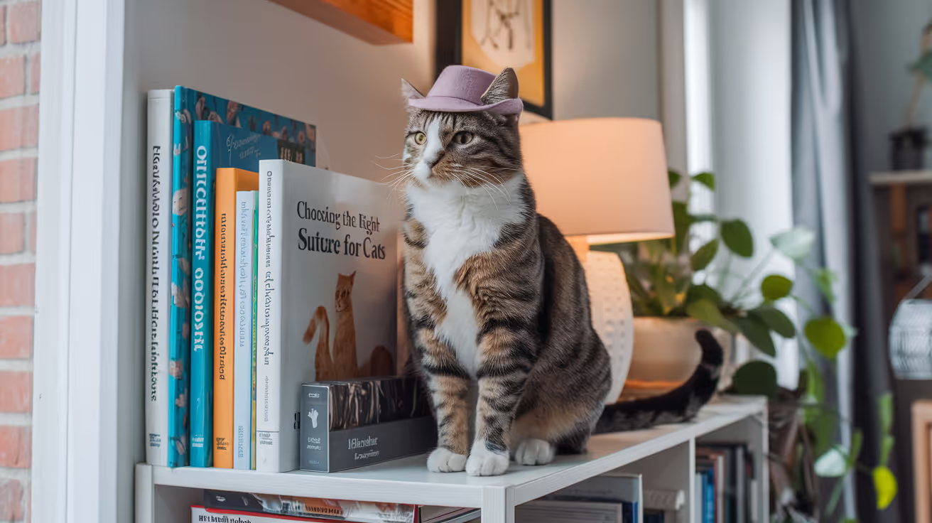
Cats are not small dogs. Their tissue is more delicate, their healing is faster, and their skin is thinner and more reactive to foreign material than most canine breeds.
Suture selection in cats follows the same basic logic as in dogs match the material's properties to the tissue's needs but with specific adjustments for feline physiology. The materials that work best in cats tend toward lower tissue reaction, faster absorption, and finer gauge than their dog equivalents.
Quick answer: In cats, PDS (polydioxanone) is preferred for deep structural layers like the linea alba; Monocryl (poliglecaprone 25) is preferred for subcutaneous and subcuticular closure due to lower tissue reaction than PDS in feline tissue; nylon or absorbable monofilament (3-0 or 4-0) for skin. Suture sizes in cats run 3-0 to 4-0 for most layers. Clinician's Brief notes Monocryl "may induce less tissue reaction than PDS, especially in cats."
Key takeaways
- Monocryl is the preferred subcutaneous suture in cats due to lower tissue reaction than PDS in feline tissue.
- PDS remains the standard for linea alba because subcutaneous absorption speed of Monocryl is too fast for fascial support.
- 3-0 and 4-0 are the standard sizes for most feline surgical layers.
- Vicryl (braided multifilament) causes more tissue reaction than monofilament alternatives in cats.
- Tissue adhesive is a practical and effective skin closure option for many feline procedures.
- Suture removal is needed only for non-absorbable skin sutures most feline surgery uses absorbable materials throughout.
Why cat suture selection differs from dogs
Three feline-specific factors drive material selection:
1. Thinner, more reactive skin. Cat dermis is more sensitive to suture-related inflammation. Materials that cause more tissue reaction (braided multifilament, natural gut) produce more visible swelling, licking behavior, and risk of suture track infection.
2. Faster tissue healing. Cats' subcutaneous and skin layers heal in 7 to 10 days rather than 10 to 14 days. This allows faster-absorbing materials to be appropriate choices at layers where dogs might need longer retention.
3. Smaller tissue volumes. Cats' smaller bodies require finer gauges. A 2-0 suture that is appropriate in a 20 kg dog would cause unnecessary tissue damage in a 4 kg cat.
Suture material guide by tissue layer
Abdominal wall / linea alba
Best choice: PDS (polydioxanone) 2-0 to 3-0
The linea alba is the structural layer of abdominal closure. It heals slowly. Monocryl loses tensile strength too quickly for fascial support: Clinician's Brief states Monocryl "is a poor choice for tissue that is slow to regain tensile strength, such as fascia (linea alba) or tendons."
PDS retains approximately 70% of its tensile strength at 2 weeks and 50% at 4 to 6 weeks appropriate for feline fascial healing timelines.
Published feline cadaver research (Frontiers in Veterinary Science, 2019) supports using 3-0 PDS with a 5 mm suture bite-to-stitch interval for feline linea alba closure.
For how abdominal wall closure works in the context of spay surgery, see absorbable vs non-absorbable decision.
Subcutaneous tissue
Best choice: Monocryl (poliglecaprone 25) 3-0 to 4-0
For subcutaneous closure in cats, Monocryl outperforms Vicryl and PDS on the tissue reaction criterion. Clinician's Brief (Suture Choice for Today's Veterinarian): "Poliglecaprone 25 may induce less tissue reaction than polydioxanone especially in cats."
Monocryl is absorbed by 90 to 119 days, which exceeds the healing timeline by a comfortable margin.
WSAVA 2015 conference notes Biosyn (glycomer 631) as comparable to Monocryl: "Biosyn is relatively rapidly absorbed with approximately 50% loss of tensile strength at 2 to 3 weeks and is completely absorbed at 90 to 110 days. When compared with Vicryl, Biosyn is stronger and has greater knot security." Biosyn is an appropriate alternative where Monocryl is unavailable.
For how monofilament vs. multifilament affects tissue reaction in this layer, see monofilament vs multifilament decision. For suture material selection in dogs as a comparison, see suture material selection in dogs.
Subcuticular / intradermal skin
Best choice: Monocryl 4-0
For intradermal closure in cats, Monocryl provides:
- Low tissue drag (smooth passage through delicate feline dermis)
- Adequate tensile strength through the 7 to 10 day feline skin healing period
- Dissolution timeline that exceeds healing without requiring removal
A PMC study comparing absorbable and non-absorbable sutures for intradermal skin closure in cats found that monofilament materials cause "less tissue drag and induce less inflammation than multifilament sutures."
For intradermal closure technique in cats specifically, see needle selection alongside material.
Skin (external interrupted sutures)
Options:
Tissue adhesive (n-butyl cyanoacrylate) is widely used in feline skin closure, particularly after scrotal and prescrotal neuter incisions. It provides waterproof closure, requires no needle passage through delicate skin, and eliminates removal visits.
Suture size selection in cats
Veterinary Practice News guidance: "Suture size should be the smallest size that will adequately hold the tissue." In cats this is almost always 3-0 or 4-0 for standard soft tissue layers.
For how suture size decisions are made alongside material selection, see suture size alongside material.
Materials to avoid in cats
Surgical gut (catgut): natural material with unpredictable absorption rate and high inflammatory response in cats. Largely replaced by synthetic materials; not recommended.
Silk: multifilament non-absorbable; highest tissue reaction of any suture material; associated with sinus tract formation; not appropriate for feline surgery.
Chromic gut: improved over plain gut but still variable absorption and more inflammatory than synthetic options.
Vicryl for subcutaneous closure: higher tissue reaction than Monocryl in cats; suitable but not the preferred choice where Monocryl is available.
What suture choice means for your cat's recovery
If your cat had absorbable sutures throughout:
- No removal visit is needed
- You may feel a slight ridge under the incision for 2 to 4 weeks (suture material dissolving)
- No suture material should emerge through the skin in normal healing
If your cat had non-absorbable skin sutures:
- A scheduled removal visit at 10 to 14 days is required
- Do not attempt to remove sutures at home incorrect removal can open the wound
- Use an E-collar to prevent licking, which can loosen sutures or introduce bacteria
Signs of suture reaction (contact your vet):
- Swelling that increases rather than decreases after day 5
- A small firm lump forming along the incision line
- Discharge at a suture site
- Persistent licking at a specific point on the incision
Frequently asked questions
Why did my vet use a different suture for my cat than they used on my dog?
Cats have more reactive skin and a faster healing timeline than most dogs. The materials optimized for feline tissue (particularly Monocryl for subcutaneous closure) differ from the broader range used in dogs. This is an intentional species-specific selection.
My cat keeps licking one point on her incision but it looks fine. Should I be worried?
A cat persistently targeting one spot often indicates localized irritation possibly a suture knot that is slightly prominent, early suture reaction, or mild discomfort at that site. Keep the E-collar on and contact your vet. They may want to assess the site to rule out early dehiscence or reaction.
Do tissue adhesives work as well as sutures in cats?
For thin-skinned areas, low-tension incisions, and cats prone to licking and suture removal, tissue adhesive performs comparably to sutures and eliminates the need for needle passage through delicate feline skin. It is not appropriate for high-tension wounds or areas requiring layered closure.
Suture selection in cats is not a trivial detail. The thin, reactive feline dermis, faster healing timeline, and smaller tissue volumes all argue for the same general direction: the finest gauge that provides adequate strength, in the material with the lowest tissue reaction profile, using absorbable materials wherever removal creates compliance challenges. Monocryl and PDS together cover the vast majority of feline surgical closure needs.
Resources
- Clinician's Brief. How to Fine-Tune Suture Choices for Today's Veterinarian. cliniciansbrief.com
- WSAVA 2015 (VIN). So Many Pieces of String: How to Choose Appropriate Suture Material. vin.com
- Frontiers in Veterinary Science. Optimal Suture Bite Size for Closure of Feline Linea Alba. PMC, 2019. ncbi.nlm.nih.gov
- Veterinary Practice News. What Suture Size Should I Use? veterinarypracticenews.com

Get a Free Poster
Enhance your workspace with a high-quality radiographs reference poster, designed for veterinary professionals. This free physical poster will be shipped directly to you—just fill out the form to request your copy.





Taking Great TPLO Radiographs
Click Below to Watch Live Video Demos


We'll send you a Free Wall Poster with all the steps
Step #1
Getting Ready
Ensuring a clean surgical field starts with proper skin preparation. This video demonstrates the best practices for:
- Shaving the patient – Achieving a close, even shave while minimizing skin irritation
- The Dirty Scrub – The initial skin prep step to remove surface debris and reduce bacterial load before the sterile scrub.
Following these techniques helps reduce infection risk and improve surgical outcomes. Watch the video to see how it’s done effectively!


Step #2
Reduce Your Risks
Many surgeons are shocked to find out that their patients are not protected from biofilms and resistant bacteria when they use saline and post-op antibiotics.
That’s Where Simini Comes In.
Why leave these risks and unmanaged? Just apply Simini Protect Lavage for one minute. Biofilms and resistant bacteria can be removed, and you can reduce two significant sources of infection.
Step #3
Take the Course
Preventing surgical infections is critical for patient safety and successful outcomes. This course covers:
- Aseptic techniques – Best practices to maintain a sterile field.
- Skin prep & draping – Proper methods to minimize contamination.
- Antibiotic stewardship – When and how to use perioperative antibiotics effectively.
Stay up to date with the latest evidence-based protocols. Click the link to start learning and earn CE credits!

Things to know

Common Closure Errors in Small Animal Surgery
Explore common closure errors in small animal surgery, their causes, prevention, and how to ensure optimal healing for your pet’s recovery.
Most surgical complications that appear in the first two weeks of recovery are not random. They trace back to specific, identifiable mistakes in wound closure mistakes that are preventable, detectable, and in many cases correctable if caught early.
The errors are consistent enough across procedures that they can be named, explained, and recognized.
Quick answer: The five most common closure errors in small animal surgery are: sutures tied too tight (causing ischemia and tissue necrosis), sutures tied too loose (leaving the wound inadequately apposed), wrong suture material for the tissue (mismatched strength or absorption rate), failure to close dead space (leaving fluid-filling pockets that become seromas or abscesses), and wrong closure pattern for wound tension (using simple interrupted across a high-tension wound without tension-relieving technique). Each error has a predictable consequence and recognizable early signs.
Key takeaways
- Sutures tied too tight restrict blood flow, causing tissue ischemia and necrosis that creates infected dead tissue.
- Sutures tied too loose fail to appose wound edges, leading to dehiscence and infection.
- Wrong suture material means either premature loss of strength (wound opens) or excess foreign material reaction.
- Unclosed dead space fills with serum, creating an ideal bacterial growth medium.
- Wrong pattern for wound tension allows sutures to cut through tissue or fail catastrophically.
- Most closure errors become visible within 3 to 7 days owner monitoring in this window enables early intervention.
Error 1: Sutures tied too tight
What happens: when the knot is overtightened, the loop of suture compresses tissue between the wound edge and the entry/exit points. That compression restricts blood flow.
Consequences:
- Tissue between the suture and the wound edge becomes ischemic (inadequately perfused)
- Ischemic tissue cannot mount an immune response bacteria colonize it readily
- Dead tissue becomes a substrate for infection
- The strangulated tissue eventually sloughs, opening the wound from within
How to recognize it:
- Within 24 to 48 hours: the skin at each suture entry point looks pale or whitened rather than pink
- Within 3 to 5 days: necrotic (black, brown, or grey) tissue appears at the wound margins
- The tissue may start to smell before it visibly changes
Why it happens: the temptation to tie tight for security. Wound edges should appose, not compress. The suture should draw tissue into contact, not hold it under pressure.
For how suture tension decisions affect the wound closure protocol overall, see wound closure principles relating to tension.
Error 2: Sutures tied too loose
What happens: loose sutures allow the wound edges to remain separated or to move relative to each other. The wound never achieves apposition the edges must touch for healing to proceed.
Consequences:
- Dehiscence: the wound opens, either gradually (edges drift apart) or suddenly (with one jump or activity)
- Exposed subcutaneous tissue is at high infection risk
- Re-closure is required under sedation or anesthesia
How to recognize it:
- Immediately post-surgery: small visible gaps between sutures
- Within 2 to 4 days: the wound edges separate at one or multiple points
- The wound may drain and appear wider than it should be
Why it happens: attempting to minimize suture marks by under-tensioning, or placing sutures too far apart.
Error 3: Wrong suture material
What happens: material mismatch between the suture's properties and the tissue's needs.
Material too weak for the load
Using Monocryl (fast-absorbing) to close the linea alba in a large breed dog. Monocryl loses most of its tensile strength by 21 days. The linea alba takes 4 to 6 weeks to regain adequate strength. The suture fails before the tissue is ready to hold alone.
Consequence: incisional hernia, wound opening, or catastrophic dehiscence.
Material too reactive for the tissue
Using silk (high tissue reactivity) in a buried internal layer. Silk provokes a significant chronic inflammatory response, creating sinus tracts that drain permanently.
Consequence: persistent drainage from a wound that appears to have healed; often misidentified as infection.
Braided material in a contaminated wound
Using Vicryl (braided polyglactin) in a bite wound or heavily contaminated case. Bacteria colonize the interstices between braided strands, shielded from the immune response.
Consequence: wound infection despite antibiotic therapy, because the suture itself harbors the organism.
For how suture material selection avoids these errors, see suture material selection to prevent closure errors.
Error 4: Failure to close dead space
What happens: dead space is any gap remaining between tissue planes after closure. These gaps fill with serum (tissue fluid). Serum is protein-rich and warm the ideal bacterial growth medium.
Consequences:
- Seroma: a fluid pocket under the skin that creates a painless swelling, often discovered 3 to 5 days post-surgery
- Infected seroma: the serum becomes colonized with bacteria, producing an abscess
- Delayed healing: the tissue planes cannot adhere if fluid separates them
How to recognize it:
- Soft, fluctuant swelling at or near the surgical site, appearing days after surgery
- The wound surface may appear normal while a large pocket forms beneath
Prevention: subcutaneous closure to eliminate the fat layer gap, walking sutures to tack skin to fascia over larger defects, drain placement when dead space cannot be sutured closed.
For how dead space management is addressed as part of the closure sequence, see dead space management as a closure step.
Error 5: Wrong pattern for wound tension
What happens: simple interrupted sutures are placed across a wound under significant tension without any tension-relieving technique. Each suture bears the full tensile load of the wound at that point.
Consequences:
- Sutures cut through skin: the suture entry point becomes a linear tear perpendicular to the wound
- Wound dehiscence: one or more sutures fail, creating a gap
- The wound may appear to be holding for several days, then fail suddenly when activity increases or a seroma develops
Prevention: recognize high-tension wounds pre-closure and apply tension-relieving technique horizontal mattress, walking sutures, or undermining of skin edges before placing the primary closure.
Veterinary Surgery Online: "If tissues are fragile and suture pulls out easily, use cruciate or horizontal mattress pattern instead of simple interrupted suture pattern for less stress on each bite."
For how specific tension-relieving patterns prevent this error, see tension-relieving patterns that prevent closure failure.
Error 6: Incorrect layer closure sequence
What happens: layers are closed out of sequence, or a layer is skipped entirely. Most commonly: the subcutaneous layer is not closed, leaving dead space; or the fascial layer is closed with the same material and tension as the subcutaneous layer, leaving the structural closure inadequately supported.
Consequences depend on which layer is affected:
- Missed subcutaneous layer: dead space, seroma, delayed healing
- Inadequate fascial closure: incisional hernia weeks to months later
- Skin closed over unclosed deep layers: surface looks fine; structural failure develops silently
For how the layered closure sequence prevents these errors, see layered closure sequence in small animal surgery.
What owners can monitor at home
The first 5 to 7 days are the highest-risk window. Most closure errors become externally visible during this period.
Check twice daily for:
- Any visible gap between suture points
- Skin at suture entry points: should be pink and pliable, not white or dark
- Swelling, especially fluctuant swelling (feels like a fluid-filled balloon under the skin)
- Discharge: small amounts of serous (clear, slightly yellow) fluid is normal; green, brown, or foul-smelling discharge is not
- Wound pulling apart or gaping
When to call the vet:
- Any visible gap or suture failure
- Discharge that is purulent or foul-smelling
- Dark or pale tissue at wound margins
- Fluctuant swelling appearing 3 or more days post-surgery
- Dog is chewing at or licking the wound despite E-collar
For the full post-operative monitoring protocol after closure, see post-operative monitoring after closure.
Frequently asked questions
One of my dog's sutures has a small bump around it. Is that a problem?
A small firm bump at a suture site within the first 3 to 5 days is usually normal a mild inflammatory reaction to the suture material. A soft, fluctuant (fluid-filled) bump, especially one that appears at the wound itself or between suture sites, is more likely to be a seroma and should be assessed by your vet.
My dog's wound opened two days after surgery. What caused it?
Common causes: the dog licked or chewed the wound (removing sutures), activity that placed excessive force on the closure, sutures that were inadequately tensioned, or excessive wound tension that exceeded the repair strength. Contact your vet the same day open wounds generally need re-closure promptly to prevent infection and further dehiscence.
Can I prevent closure errors at home?
You cannot change what happened in the operating room, but you can prevent the most common post-operative causes of closure failure: keep the E-collar on at all times, restrict activity as directed, keep the wound dry, and attend all scheduled rechecks. Most secondary failures (wound opening after initial surgery) are caused by licking, jumping, or premature bathing.
Closure errors are not random they are the predictable consequences of specific technical decisions made during wound closure. Recognizing which error you are looking at early, based on the pattern of what is happening to the wound, is the first step in getting it corrected before a small problem becomes a large one.
Resources
- Veterinary Surgery Online. Wound Closure Continued. vetsurgeryonline.com
- VCA Animal Hospitals. Care of Surgical Incisions in Dogs. vcahospitals.com
- Veterian Key. Selection of Suture Materials, Suture Patterns, and Drains for Wound Closure. veteriankey.com
- AAHA. Oh, Sew Easy: A Guide to Sutures. aaha.org

Closure Considerations in Geriatric Dogs and Cats
Learn essential closure considerations for geriatric dogs and cats to ensure safe, effective surgical outcomes and recovery.
Senior pets undergo surgery more frequently than younger ones age brings a higher burden of tumors, orthopedic disease, and organ conditions requiring surgical management. And yet age-related tissue changes make wound closure more technically demanding in precisely the patients who also tolerate complications least well.
Understanding what changes with age in the tissue helps explain why closure technique must be adjusted for geriatric patients.
Quick answer: Geriatric dogs and cats present four specific closure challenges: thinner, less elastic skin that tears at suture entry points; delayed healing from reduced perfusion and immune function; comorbidities (diabetes, hyperadrenocorticism, CKD) that impair healing independently; and reduced collagen synthesis that weakens tissue intrinsic strength. Closure modifications include: PDS preferred over faster-absorbing materials to match extended healing timelines, smaller needle sizes, cruciate or horizontal mattress patterns instead of simple interrupted in fragile skin, intradermal closure to eliminate licking targets, and extended suture removal timing (full 14 days or beyond).
Key takeaways
- Feline and canine skin becomes thinner and less elastic with age, increasing cut-through risk at suture entry points.
- PDS is preferred in geriatric patients because delayed healing requires longer-duration tensile strength.
- Intradermal absorbable closure eliminates the removal visit stress and licking target for older pets.
- Cruciate or horizontal mattress patterns distribute bite force more broadly in fragile skin.
- Comorbidities (diabetes, Cushing's, CKD) significantly impair healing independently of the closure technique.
- Suture removal extends to 14 days or beyond in geriatric patients with slow wound healing.
Four age-related tissue changes that affect closure
1. Thin, inelastic skin
Older dogs and cats undergo dermal thinning with age. Collagen cross-linking changes make the dermis less elastic and more prone to tearing. A suture placed 4 to 5 mm from the wound edge in young tissue holds reliably; in thin geriatric skin, it may cut through at the entry point under tension.
This is the same mechanism that explains suture cut-through in fragile or inflamed tissue and the same modifications apply: cruciate or horizontal mattress patterns over simple interrupted, smaller needle sizes, and wider spacing between sutures to reduce focal stress concentration.
Practical modification: consider cruciate sutures instead of simple interrupted for skin closure in geriatric patients, particularly in cats over 12 years and large breed dogs over 8 to 9 years.
2. Delayed wound healing
Wound healing depends on adequate perfusion (to deliver immune cells, oxygen, and nutrients) and immune function (to control contamination and drive the repair phase). Both decline with age.
The implications for closure timing:
- The repair phase takes longer to build tensile strength
- Sutures may be needed for the full 14-day window rather than the shorter end (10 to 12 days feasible in young, healthy patients)
- The risk of premature suture removal is higher
Practical modification: schedule recheck at day 14 rather than day 10 to 12 for geriatric patients. If wound healing appears delayed at the recheck, defer suture removal by 3 to 5 additional days.
For suture removal timing modified for geriatric cats, see suture removal timing in geriatric cats. For dogs, see suture removal timing in geriatric dogs.
3. Comorbidities
Diabetes mellitus, hyperadrenocorticism (Cushing's disease), chronic kidney disease, and hypothyroidism each impair wound healing through distinct mechanisms:
Geriatric patients should have pre-operative bloodwork and a cardiovascular assessment before elective surgery. Uncontrolled diabetes or Cushing's disease substantially increases wound complication risk and should ideally be managed before elective procedures.
4. Reduced collagen synthesis
Collagen is the primary structural protein in healed wounds. Its synthesis rate and quality decline with age. The result: the healed wound is intrinsically weaker at the same timepoint compared to a wound in a younger patient.
Practical modification: longer-duration absorbable sutures (PDS rather than Monocryl for deep layers in geriatric patients with any delay in expected healing) provide extended structural support through the slower healing process.
Suture material modifications
Deep layers: PDS preferred over faster-absorbing alternatives
PDS (polydioxanone) retains significant tensile strength for 4 to 6 weeks and absorbs over 180 to 210 days. In a healthy young dog, this is more duration than needed for fascial healing. In a geriatric patient with delayed healing, it is appropriate.
Monocryl (poliglecaprone 25) loses most of its strength by 21 days. In a geriatric patient whose linea alba is healing slowly, the suture may lose functional strength before the tissue has adequate intrinsic strength to compensate.
Skin: intradermal absorbable over external non-absorbable
Two specific advantages in geriatric patients:
No removal visit: older patients particularly cats are stressed by veterinary visits. An intradermal closure eliminates the removal visit entirely.
No external licking target: geriatric dogs and cats may be less consistent E-collar users, and their owners may be less strict about compliance. Removing the external suture material eliminates the most common source of self-trauma.
For how intradermal closure works in this context, see intradermal closure in older dogs.
Pattern modifications
Cruciate or horizontal mattress over simple interrupted
When tissue is fragile and sutures are at risk of cutting through, the cruciate pattern distributes the bite force differently and is significantly faster to place (JAVMA 2016 data). Horizontal mattress sutures spread tension across 8 to 10 mm from each wound edge, further reducing focal stress.
Specific indication in geriatric patients: cats over 12 years with hyperadrenocorticism, Cushing's-treated dogs with thinned skin, any patient where simple interrupted sutures are pulling through the tissue at placement.
Wider spacing
Reducing suture spacing (more sutures per wound length) might seem protective, but it increases the total number of suture-skin interface points each one a potential cut-through site. Wider spacing with an appositional pattern that distributes tension is often more appropriate.
For how these pattern modifications compare in the context of high-tension and fragile-tissue closure, see pattern modifications for fragile skin.
Obese geriatric patients: compounded challenges
Obesity and aging frequently coincide in middle-to-senior-aged dogs and cats. The combined effect:
- Reduced perfusion (fat is poorly vascularized)
- Greater dead space (more fat tissue to approximate)
- Higher skin tension (weight on the wound)
- Both thin skin (age) and thick subcutaneous fat (obesity)
For how obesity specifically affects closure technique, see closure considerations in obese dogs.
Post-operative monitoring: more frequent and more important
What to monitor
The monitoring frequency and detail appropriate for geriatric patients is greater than for young, healthy patients:
- Twice-daily wound checks
- Document changes over time (photograph the wound at each check)
- Watch for systemic signs geriatric patients developing wound infections may show systemic signs (lethargy, inappetence, fever) before local signs become obvious
- Check suture integrity specifically suture cut-through, loosening, or loss is more likely in geriatric patients
Nutrition during recovery
Collagen synthesis requires adequate dietary protein. Geriatric patients with reduced appetite or pre-existing protein restriction (for CKD management) may have compromised wound healing from nutritional factors. Discuss any feeding changes with your vet before and during recovery.
For the post-operative monitoring protocol applicable to geriatric patients, see post-operative monitoring in geriatric patients.
Frequently asked questions
My 14-year-old cat is having a mass removed. Are older cats at higher risk for wound complications?
Yes, but manageable risk. The key factors are: the cat's overall health status, whether comorbidities are controlled, the size and location of the mass, and the closure technique chosen. An experienced vet will adjust the technique for a geriatric patient, choosing materials and patterns that match the tissue's actual properties. Careful pre-operative assessment, appropriate anesthesia monitoring, and attentive post-operative care reduce this risk substantially.
My older dog's wound opened two days after surgery. Is age the cause?
Possibly, but other factors are equally likely: E-collar non-compliance (allowing licking), premature activity, excessive wound tension at closure, or concurrent illness. Age-related healing delay typically manifests as very slow progress over 7 to 14 days, not sudden acute failure in the first 48 hours. A wound opening in the first 2 days most often reflects one of the other factors.
Should my senior dog get intradermal sutures specifically?
It is worth discussing with your vet. The main advantages for senior dogs are: no removal visit (less stress), no external material to lick (reduces E-collar dependence), and finer healed scar. The main requirement is that the wound must be low-to-moderate tension and clean for intradermal closure to be appropriate. Many routine procedures in geriatric dogs meet these criteria.
Geriatric closure requires adjusting every assumption that applies to a young, healthy patient. The healing timeline is longer. The tissue tolerates suture entry points less well. The comorbidities stack against normal wound healing. Each modification PDS instead of Monocryl for deep layers, cruciate instead of interrupted for fragile skin, intradermal instead of external for skin is a response to a specific age-related change in what the tissue can support.
Resources
- Clinician's Brief. How to Fine-Tune Suture Choices for Today's Veterinarian. cliniciansbrief.com
- VCA Animal Hospitals. Care of Surgical Incisions in Cats. vcahospitals.com
- JAVMA 2016 (Kieves et al.). Comparison of Tensile Strength Among Simple Interrupted, Cruciate, Intradermal, and Subdermal Suture Patterns in Ex Vivo Canine Skin. pubmed.ncbi.nlm.nih.gov
Absorbable vs Non-Absorbable Sutures in Veterinary Surgery
Explore the differences between absorbable and non-absorbable sutures in veterinary surgery, including uses, benefits, and care tips.
When your dog or cat has surgery, sutures are doing different jobs in different layers of the body. Some need to stay permanently. Others should dissolve quietly on their own. The decision between absorbable and non-absorbable isn't a preference: it follows a logical set of rules based on what each layer of tissue needs.
Understanding the difference helps you know what your vet placed, whether a recheck visit is needed, and what to watch for during healing.
Quick answer: Absorbable sutures dissolve through hydrolysis over weeks to months and are used for internal tissue layers (muscle, fascia, subcutaneous tissue). Non-absorbable sutures resist breakdown and are used for skin closure (requiring removal) or long-term structural repairs like ligament reconstruction. The material, pattern, and placement location determine which type is correct for each situation.
Key takeaways
- Absorbable sutures dissolve on their own and are used for internal layers in nearly all soft tissue surgery.
- Non-absorbable sutures do not break down and either need removal or remain permanently in place.
- PDS (polydioxanone) and Monocryl (poliglecaprone) are the most common absorbable materials in small animal surgery.
- Nylon and polypropylene are the most common non-absorbable materials for skin closure.
- No removal is needed for absorbable skin sutures, reducing recheck visits for many patients.
- Multifilament sutures carry higher infection risk than monofilament in contaminated wound environments.
How absorbable sutures work
Absorbable sutures break down through hydrolysis. Water penetrates the suture filaments and causes the polymer chains to dissolve. This happens over a predictable timeline that varies by material.
The WSAVA 2016 Suture Materials review describes the ideal absorbable suture: "pliable, sterile, with uniform tensile strength, non-inflammatory, degrading at just the right time, strong enough to support the repair."
The key word is timing. The suture must maintain strength long enough for the tissue to heal, then disappear without causing a foreign body reaction.
Common absorbable suture materials in veterinary surgery
Key distinction: Monocryl and PDS are monofilament (single strand). Vicryl is multifilament (braided). Monofilament materials cause less tissue drag and lower infection risk than braided materials in contaminated environments.
Chromic catgut is rarely used today in small animal surgery due to its unpredictable absorption and inflammatory response. Synthetic materials have replaced it in most veterinary contexts.
Common non-absorbable suture materials
Veterian Key (Surgery of the Abdominal Cavity) specifically cautions: "Monofilament, nonabsorbable suture material (polybutester, polypropylene, nylon) has been associated with suture sinus formation and should be avoided" in continuous abdominal wall patterns. Non-absorbable materials belong at the skin surface, not in deep tissue layers.
For how material choice affects infection risk at the wound site, see infection risk differences between absorbable and non-absorbable.
Where each type is used by tissue layer
The University of Saskatchewan WCVM Lab notes: "For skin sutures (simple interrupted or cruciate), a monofilament nonabsorbable material (polypropylene, polybutester, or nylon) is a good choice."
For material selection in dog-specific procedures, see material selection for dog surgery. For cat-specific decisions, see material selection for cat surgery.
Monofilament vs multifilament within each category
Both absorbable and non-absorbable sutures come in monofilament and multifilament forms. This distinction matters as much as absorbable vs. non-absorbable in infection-prone environments.
- Monofilament: single strand, smooth surface, less bacterial wicking, lower tissue drag
- Multifilament (braided): multiple strands twisted or braided together, stronger handling properties, higher infection risk in contaminated fields
In clean elective surgery, either may be appropriate. In contaminated wounds, bite wounds, or infection-prone cases, monofilament is preferred.
For a full comparison of monofilament vs. multifilament properties, see monofilament vs multifilament alongside absorbable vs non-absorbable.
Non-absorbable sutures: when removal is and isn't needed
Requires removal (10 to 14 days):
- External skin sutures placed using nylon, Prolene, or other non-absorbable materials
- Your vet will clip and pull each suture individually using suture scissors or a seam ripper
Does not require removal:
- Non-absorbable sutures used internally for permanent structural repairs (ligament reconstruction, vascular procedures) remain in place indefinitely
Absorbable skin sutures:
- Used when the surgeon places an intradermal or buried closure
- No removal needed; the material dissolves on its own
For timing of non-absorbable skin suture removal in dogs, see when non-absorbable sutures need removal in dogs. For cats, see when non-absorbable sutures need removal in cats.
What this means for your pet's recovery
Understanding suture type helps you know what to expect at home:
- No visible sutures = intradermal absorbable closure. No removal needed. Monitor for swelling or separation.
- Visible sutures = external closure. A recheck is scheduled for removal at 10 to 14 days. Do not try to remove them yourself.
- All internal sutures dissolve. You will never see or feel them surface through the skin in normal healing.
- A small firm lump along the incision can be a suture knot reacting as it absorbs. This is common with Vicryl in cats and usually resolves within 2 to 4 weeks.
Frequently asked questions
Why doesn't my vet use the same suture for every layer?
Each tissue layer has a different healing timeline and functional requirement. The subcutaneous layer heals in 3 to 4 weeks. The linea alba takes months to reach full strength. Matching suture absorption rate to healing timeline is what prevents premature closure failure.
Are absorbable sutures weaker than non-absorbable?
Not at placement. Most absorbable sutures are placed with equivalent or superior initial tensile strength compared to non-absorbable materials of the same size. The difference is what happens over time: absorbable sutures lose strength as they dissolve, which is intentional.
I can feel a hard lump under my dog's incision. Is a suture failing?
A firm knot under the skin is most likely a buried suture knot in the subcutaneous or intradermal layer. This is normal, especially in the first 2 to 4 weeks. It should gradually soften. A soft, fluid-filled swelling is more likely a seroma. A painful, warm, firm lump may indicate infection. Contact your vet if you're unsure.
Choosing between absorbable and non-absorbable sutures is not arbitrary. Every placement decision reflects what the tissue needs at that depth, on that timeline, under that amount of tension. When the right suture is matched to the right layer, the result is a wound that heals cleanly without the complications that bring patients back for second procedures.
Resources
- WSAVA 2016 Congress (VIN). Suture Materials. vin.com
- Veterian Key. Selection of Suture Materials, Suture Patterns, and Drains for Wound Closure. veteriankey.com
- Veterian Key. Surgery of the Abdominal Cavity. veteriankey.com
- WCVM University of Saskatchewan. Lab 6 Part 4: Incision Closure. wcvm.usask.ca
Closure Protocol Checklist for Veterinary Surgeons
Comprehensive closure protocol checklist for veterinary surgeons to ensure safe, effective surgical outcomes and patient care.
A protocol checklist exists for the same reason a preflight checklist does: the steps are well known, the consequences of skipping them are serious, and cognitive load under pressure increases the probability of omission.
This checklist is the practical translation of closure principles into sequential, verifiable steps from the moment the primary procedure is complete to the moment discharge instructions are given.
Quick answer: A complete veterinary surgical closure checklist covers five phases: (1) pre-close assessment (hemorrhage control, irrigation, tissue viability, dead space identification), (2) deep layer closure (appropriate material, correct bite dimensions, knot security), (3) subcutaneous closure (dead space elimination confirmed), (4) skin closure (method appropriate to wound and patient), and (5) discharge instructions (E-collar, activity restriction, twice-daily monitoring, recheck timing). Verification at each phase prevents the errors that produce post-operative complications.
Key takeaways
- Pre-close hemorrhage control must be confirmed before any sutures are placed active bleeding under a closed wound produces hematoma.
- Irrigation before closure reduces bacterial count at the wound margin.
- Bite dimensions and knot security should be verified at the deep layer before moving to subcutaneous.
- Dead space confirmation at subcutaneous closure prevents seroma formation.
- Skin closure method selection should be matched to wound tension, patient compliance risk, and removal feasibility.
- Discharge instructions are part of the closure protocol incomplete instructions produce avoidable post-operative complications.
Phase 1: Pre-close assessment
Before the first closure suture is placed, the following must be confirmed:
Hemorrhage control
- No active bleeding points remain
- All ligatures on ovarian and uterine stumps (spay) or vascular pedicles are intact and secure
- Lap sponge or pad count matches none retained in the abdomen
Active bleeding under a closed wound does not stop it produces a hematoma that can become infected, place pressure on the closure, and obscure the wound assessment during monitoring.
Irrigation
- Abdominal or wound lavage with warm sterile saline has been performed
- If peritoneal contamination occurred: copious irrigation (multiple warm saline flushes)
- No lavage fluid pools remaining before closure
DVM360 (Basic Principles of Wound Management): "Primary closure should eliminate dead space and provide good anatomical apposition of tissue."
Tissue viability
- All tissue edges are pink and bleeding when cut (viable)
- No devitalized (grey, brown, or non-bleeding) tissue remains at the margins
- If contamination was significant: wound suitable for primary closure or decision made for delayed primary
Foreign body / instrument check
- Instrument count complete
- No suture material loops, clamp caps, or other items retained in the wound
For the principles that underpin each pre-close verification step, see closure principles that pre-close assessment implements.
Phase 2: Deep layer closure
Linea alba / fascia
- [ ] Material selected: PDS or Biosyn for most patients; size matched to patient weight (0 to 2-0 for medium-large dogs; 2-0 to 3-0 for cats and small dogs)
- [ ] Pattern: simple continuous or interrupted based on tissue quality and contamination status
- [ ] Bite dimensions: 5 to 10 mm from incision edge; 5 to 10 mm between bites
- [ ] Bites perpendicular to wound (not oblique)
- [ ] Fascia engaged in every bite not just muscle belly
- [ ] No fat incorporated in bites (prevents healing)
- [ ] Start knot secured with adequate throws (minimum 4 for PDS; 5 for feline linea alba per published guidance)
- [ ] End knot secured with additional throws (continuous pattern end knots require extra throws)
- [ ] No gaps visible when forceps are run along the closed linea alba
Joint capsule (orthopedic cases)
- [ ] Full-thickness bites through capsule wall
- [ ] Inverting or appositional pattern as appropriate for the joint
- [ ] No suture material crosses the joint space
For how the deep layer closure relates to overall wound closure principles, see layered closure in the deep layer context.
Phase 3: Subcutaneous closure
- [ ] Material: Monocryl 2-0 to 3-0 or Vicryl 2-0 to 3-0
- [ ] Pattern: simple continuous
- [ ] Dead space confirmed eliminated: wound edges at subcutaneous level are in contact
- [ ] No fluid pocket remains between the deep closure and the skin
- [ ] Drain placement assessed: if dead space cannot be eliminated by suturing, drain has been placed and exits through a separate stab incision (not the primary wound)
- [ ] Knots buried
For common subcutaneous closure errors that produce dead space, see subcutaneous errors and dead space.
Phase 4: Skin closure
Method selection (complete one)
External interrupted (nylon or Prolene):
- [ ] 3-0 to 4-0 for most dogs; 4-0 for cats
- [ ] Sutures 4 to 6 mm apart, placed 4 to 5 mm from wound edge
- [ ] No blanching at wound margins after tying
- [ ] Removal planned for day 10 to 14
Intradermal (Monocryl 4-0):
- [ ] Start and end knots buried
- [ ] No suture material crosses the epidermis
- [ ] Wound edges fully apposed without puckering
- [ ] No removal visit required communicate this to owner
Staples:
- [ ] Staple remover available at discharge or removal visit
- [ ] Not used in cats or dogs under 15 kg unless specifically indicated
- [ ] Not used over high-tension incisions
Post-skin closure inspection
- [ ] All wound margins apposed no gaps
- [ ] No suture marks from overtightened knots
- [ ] No inversion of wound edges
- [ ] Skin color normal no blanching or dark discoloration at wound margins
For how skin closure method selection is made, see skin closure method selection for this patient.
Phase 5: Pre-discharge verification
Wound dressing (if applicable)
- [ ] Clean, non-adherent primary dressing applied if needed
- [ ] Drain exit covered with sterile absorbent bandage if drain is in place
Discharge instructions (verbal and written)
E-collar:
- [ ] E-collar fitted and in place before the patient goes home
- [ ] Owner instructed: worn at all times, including overnight
- [ ] Alternative explained if owner expects compliance issues (inflatable collar, surgical suit)
Activity restriction:
- [ ] Leash-only walks; no running, jumping, or rough play for 10 to 14 days
- [ ] Confined when unsupervised (crate, single room)
Wound monitoring:
- [ ] Owner instructed to check the wound twice daily
- [ ] Normal findings explained: mild swelling and redness for 2 to 3 days, thin serous crust at wound margins
- [ ] Abnormal findings explained with clear same-day action: purulent discharge, increasing swelling after day 3, wound opening, odor, pale gums
Suture removal:
- [ ] If external sutures: recheck scheduled at day 10 to 14
- [ ] If intradermal: recheck still scheduled (wound assessment) but no suture removal needed communicate clearly
Medications:
- [ ] Pain management dispensed and instructions given
- [ ] Antibiotics dispensed (if indicated) with full course completion emphasized
For the post-operative monitoring protocol owners follow from discharge, see post-operative monitoring from discharge.
Common errors the checklist catches
Frequently asked questions
Does every surgery have a formal closure checklist?
Many clinics use informal mental checklists; fewer use formal written protocols. Published evidence from human surgery (and increasingly from veterinary practice) shows that formal written checklists reduce surgical complications the same principle applies to closure. The value of a formal checklist is that it does not depend on the surgeon remembering every step under the cognitive load of a busy operating list.
Can owners access this kind of checklist information?
Not in surgical detail the linea alba bite dimensions and knot throw counts are for surgical teams. What owners can access is the discharge instruction section (Phase 5) and understanding what those instructions are designed to prevent makes owners more likely to follow them precisely.
What should I do if I notice a step was missed at home?
If you discover that an E-collar was not provided, that the wound is showing early signs of a problem, or that activity restriction was not maintained, contact your vet the same day. Earlier intervention for any closure problem produces better outcomes than waiting.
A checklist is only as useful as the discipline to use it. The value is not in the list itself but in the consistent verification it provides ensuring that none of the steps that determine post-operative outcomes are left to chance or tired recall.
Resources
- WCVM University of Saskatchewan. Lab 6 Part 4: Incision Closure. wcvm.usask.ca
- DVM360. Basic Principles of Wound Management. dvm360.com
- Veterinary Evidence (2017). Choice of Suture Pattern for Linea Alba Closure. veterinaryevidence.org
- Metropolitan Veterinary Associates. Abdominal Surgery: Post-Operative and Incisional Care. metro-vet.com

Closure Protocol for Orthopedic Incisions in Dogs
Learn the best closure protocol for orthopedic incisions in dogs to ensure optimal healing and reduce complications.
Orthopedic incisions differ from soft tissue incisions in several important ways. They are placed over joints or bone, are subject to movement-related tension from the moment the dog wakes from anesthesia, and the underlying surgical work bone cuts, implant placement, or joint reconstruction depends on the structural integrity of the closure above it to remain protected during healing.
A closure failure over an orthopedic site is not just a wound problem. It is a potential pathway to implant infection, joint sepsis, or loss of the surgical repair itself.
Quick answer: Orthopedic closure in dogs proceeds in layers: joint capsule (if opened), deep fascial layer, subcutaneous tissue, and skin. Joint capsule closure uses strong absorbable monofilament (PDS 0 to 2-0) in interrupted or continuous pattern this layer restores joint integrity. Fascia and deep tissue use PDS or Biosyn. Subcutaneous layer uses Monocryl or Vicryl. Skin uses interrupted nylon, Prolene, or staples. All external sutures are removed at 10 to 14 days. Activity restriction through this period is mandatory.
Key takeaways
- Joint capsule closure is the most critical layer in joint surgeries it restores synovial seal and joint stability.
- PDS (polydioxanone) is the standard for joint capsule and deep fascial closure due to long strength retention.
- Movement-related tension makes orthopedic incisions higher risk for dehiscence than abdominal incisions.
- Staples are commonly used for skin closure in orthopedic cases because they are fast and tolerate some movement.
- Activity restriction through 10 to 14 days is more critical in orthopedic than soft tissue cases.
- Infection at an orthopedic site can reach implants or joint space, making it far harder to treat.
How orthopedic incisions differ from soft tissue
Location over bone or joint: the incision must be closed with enough tension resistance to withstand the dog's movement and weight-bearing. Each time the dog moves or bears weight, the closure is under dynamic load.
Underlying implants: many orthopedic procedures involve plates, screws, pins, or prosthetic components. These implants cannot mount an immune response if bacteria reach them through a closure failure, infection becomes extremely difficult to resolve without implant removal.
Joint space exposure: procedures that open the joint capsule (articular fracture repair, joint replacement, arthroscopy conversion to open) create direct communication between the skin surface and the joint space during surgery. Closure must restore this barrier completely.
For how layered closure principles apply in this context, see layered closure in orthopedic incisions.
Layer 1: Joint capsule (where opened)
Why this is the most critical closure
The joint capsule contains synovial fluid and forms the sealed environment in which the joint functions. When opened for surgery, it must be closed in a way that:
- Restores the synovial seal (prevents joint fluid leakage)
- Maintains mechanical stability of the joint
- Does not constrict joint movement
Pattern: simple interrupted or simple continuous, depending on capsule length and surgeon preference. Interrupted sutures allow individual adjustment and do not create a single point of failure if one suture is compromised.
Material: PDS 0 to 2-0 (monofilament absorbable, long-duration strength). Biosyn is an alternative with a similar absorption profile.
Bite depth: full-thickness bites through the capsule wall to ensure structural engagement. The capsule is relatively thin but tough partial-thickness bites do not hold reliably under joint motion.
Layer 2: Deep fascia and muscle fascia
After joint capsule closure (if applicable), the deep fascial layers overlying the surgical site are closed.
Pattern: simple continuous or interrupted, depending on the length and complexity of the fascial incision.
Material: PDS 0 to 2-0, matched to patient size and tissue thickness. The same considerations as abdominal fascial closure apply the material must retain strength through 4 to 6 weeks while the fascia heals.
Key principle: suture bites must engage the fascial layer, not just the muscle belly above it. Muscle tissue is not load-bearing in this context it tears through under the repetitive load of a walking dog.
For the fascial closure technique and why it matters for strength, see fascial layer closure in orthopedic context.
Layer 3: Subcutaneous tissue
Subcutaneous closure in orthopedic cases serves the same function as in other surgeries: eliminate dead space below the skin to prevent seroma formation and reduce infection risk.
Pattern: simple continuous absorbable.
Material: Monocryl 2-0 to 3-0, or Vicryl of equivalent size. Monocryl is preferred in high-infection-risk cases (such as revision surgeries or patients with prior infections).
Key principle: close subcutaneous tissue in a separate step from deep fascia. In orthopedic cases with significant soft tissue dissection, there may be substantial dead space that requires careful attention.
Layer 4: Skin
Skin closure in orthopedic surgery has two specific considerations not shared with all soft tissue procedures:
Movement pressure: the limb moves constantly during recovery. Even with strict activity restriction, the dog will shift weight, turn, and reposition. Skin closure must tolerate this without loosening.
E-collar compliance: dogs recovering from orthopedic surgery may not tolerate an E-collar if it interferes with how they position the limb. Assess this before selecting a skin closure method.
Common choices for orthopedic skin closure:
For how suture removal timing applies to orthopedic skin sutures, see suture removal timing after orthopedic closure. For the full TPLO closure protocol specifically, see TPLO-specific closure protocol.
Infection risk in orthopedic closure
Orthopedic surgical site infection (SSI) carries consequences beyond a soft tissue wound infection. Bacteria that reach the implant surface form a biofilm that resists both host immune response and antibiotic penetration.
Risk factors for orthopedic SSI:
- Implant presence (plates, screws, prosthetics)
- Extended surgery time
- Contaminated environment or revision surgery
- Poor tissue handling during closure
- Inadequate dead space elimination
Closure practices that reduce infection risk:
- Monofilament materials in all buried layers
- Thorough subcutaneous dead space closure
- Minimal suture material consistent with adequate strength
- Intradermal or interrupted skin closure that minimizes external licking targets
For infection risk and closure technique in the broader context, see infection risk factors in closure.
Post-operative monitoring for orthopedic incisions
The first 72 hours: the highest risk period for acute complications. Watch for:
- Excessive swelling around the incision (beyond normal post-operative swelling)
- Wound discharge
- Behavioral changes suggesting pain (not eating, not moving, excessive vocalization)
Days 3 to 10: wound is in the active healing phase. Watch for:
- Sutures remaining intact and wound edges apposed
- Gradual reduction in swelling
- No signs of fever or systemic illness
Day 10 to 14: recheck and suture removal visit. The vet will assess wound healing before removing sutures. In high-tension or movement-affected incisions, some sutures may be left until day 14 even if the wound looks healed at day 10.
For the post-operative monitoring checklist that applies to orthopedic incisions, see post-operative monitoring after orthopedic closure.
Frequently asked questions
My dog had TPLO surgery and the vet used staples on the skin. Is that standard?
Yes. Staples are commonly used for orthopedic skin closure because they are fast to place and tolerate some movement better than fine interrupted sutures. Published research confirms equivalent healing outcomes to sutures for straight incisions. The staple remover visit at 10 to 14 days is a brief, low-stress procedure.
Why is activity restriction so important after orthopedic closure?
Every time the dog runs, jumps, or moves abruptly, the closure over the joint or bone takes dynamic load. Before the tissues have healed, this force can pull sutures through tissue, open the wound, or disrupt the deeper joint capsule repair. Strict leash-only activity for the first 10 to 14 days protects the closure while it heals.
The wound looks healed at day 10 but the vet wants to wait until day 14. Why?
Orthopedic incisions are under more mechanical stress than abdominal incisions. Even when the surface looks healed, the deep fascial and joint capsule layers are still in the early repair phase. The vet is being appropriately cautious about premature suture removal in a high-stress closure location.
Orthopedic closure is the most consequence-laden closure in small animal surgery. A failure does not just mean a wound opens it means the surgical repair underneath is exposed, potentially contaminated, and at risk. The layer-by-layer protocol exists to build redundancy into the closure so that no single layer bears all the risk.
Resources
- Clinician's Brief. How to Fine-Tune Suture Choices for Today's Veterinarian. cliniciansbrief.com
- VCA Animal Hospitals. Care of Surgical Incisions in Dogs. vcahospitals.com
- Veterian Key. Selection of Suture Materials, Suture Patterns, and Drains for Wound Closure. veteriankey.com

Choosing Closure Technique Based on Tissue Type
Learn how to choose the best closure technique based on different tissue types for optimal healing and minimal complications.
No single closure technique works for every tissue. Skin needs edge apposition with minimal tension. Fascia needs mechanical strength. Hollow organs need watertight inverting patterns that prevent luminal contamination. Subcutaneous tissue needs dead space elimination.
Each tissue layer requires a different approach and matching the technique to the tissue is one of the most fundamental principles in wound closure.
Quick answer: Closure technique is matched to tissue type based on three factors: mechanical load (how much tension the tissue bears), healing timeline (how long the tissue needs support), and contamination risk (whether luminal contents or bacteria can compromise the closure). Fascia uses continuous or interrupted patterns with large-gauge long-retaining material. Hollow organs use inverting patterns (Cushing, Lembert, Connell) to prevent luminal contact. Subcutaneous tissue uses continuous or interrupted absorbable patterns to eliminate dead space. Skin uses interrupted, continuous, intradermal, or staple methods based on tension, cosmetics, and patient factors.
Key takeaways
- Fascia (linea alba) requires continuous or interrupted patterns with PDS or Biosyn the most mechanically critical closure layer.
- Hollow organs use inverting patterns (Cushing, Lembert) to prevent suture ends from contacting luminal contents.
- Muscle belly is generally not sutured fascia closure provides the structural support.
- Subcutaneous tissue uses simple continuous patterns to eliminate dead space and support skin.
- Skin closure technique is chosen based on wound tension, cosmetic needs, and removal feasibility.
- Needle type matches tissue: cutting needles for skin, taper-point for viscera and muscle.
Layer 1: Fascia and linea alba
Why this layer is the most mechanically critical
The linea alba is the aponeurotic junction where the abdominal wall muscles insert. It is the primary load-bearing structure in abdominal wall closure not the muscle itself.
If the fascial closure fails, the muscle bellies on either side retract and the entire abdominal wall opens. This creates incisional hernia at minimum, and evisceration at worst.
Technique
Pattern: simple continuous or simple interrupted.
Continuous is faster and equally strong when the suture material is intact. Interrupted is safer if suture integrity is uncertain failure of one suture does not open the entire closure.
Bite depth and spacing: bites must engage the full fascial thickness. Clinician's Brief (spay closure guide): "Take bites 5 to 10 mm from the incision edge and 5 to 10 mm apart to ensure adequate tissue purchase and tension distribution."
Material: PDS (polydioxanone) or Biosyn (glycomer 631) both monofilament absorbable materials that retain strength for 4 to 6 weeks while the linea alba heals. Size 0 to 2-0 depending on patient size.
For the full fascial closure technique, see fascial layer closure technique.
Layer 2: Muscle belly
In most small animal surgeries, the muscle belly itself is not sutured separately. The fascia that sheathes the muscle provides the structural closure muscle fibers do not hold suture well under load and can tear through.
Exception: deep epaxial muscle closure in dorsal spinal approaches, where muscle belly approximation is performed to reduce dead space. In these cases, a simple continuous pattern with absorbable suture is used, with tension kept minimal.
For how muscle layer closure relates to the layered sequence, see muscle layer as part of layered closure.
Layer 3: Hollow organs (stomach, intestine, bladder, uterus)
Why inverting patterns are used
Hollow organ closures face a unique problem: the luminal surface is in contact with bacteria (intestine), acidic contents (stomach), or urine (bladder). If the suture line protrudes into the lumen, these contents contaminate the closure from the inside. Inverting patterns bury the suture line beneath the seromuscular layer.
Common inverting patterns:
- Cushing: a continuous pattern that takes seromuscular bites parallel to the wound; the bites invert the tissue as the suture is tightened
- Lembert: an interrupted inverting pattern; similar seromuscular bites, tied individually
- Connell: a full-thickness inverting continuous pattern used when complete wall penetration is needed (e.g., some uterine closures)
Material: 3-0 to 4-0 monofilament absorbable on a taper-point needle. Vicryl (braided) should be avoided in the urinary bladder it can lose tensile strength prematurely in an alkaline urine environment.
Two-layer vs. one-layer closure: most enterotomy and gastrotomy closures use two layers a full-thickness first layer followed by an inverting seromuscular second layer for maximum leakage protection.
Layer 4: Subcutaneous tissue
Purpose of this layer
Subcutaneous closure eliminates dead space the gaps between tissue planes that fill with serum and create the ideal environment for seroma formation and bacterial growth.
Pattern: simple continuous absorbable. Bites are placed in the subcutaneous fat perpendicular to the wound, with each loop drawing the fat planes together.
Material: Monocryl (poliglecaprone 25) 2-0 to 3-0, or Vicryl of equivalent size. Monocryl is preferred in contaminated or high-infection-risk cases because its smooth monofilament surface resists bacterial adhesion.
For the full subcutaneous closure technique in dogs, see subcutaneous closure technique in dogs. For cats, see subcutaneous closure technique in cats.
Layer 5: Skin
Skin closure technique is the most variable of all layers influenced by wound tension, cosmetic requirements, patient size, and whether a removal visit is feasible.
For the complete skin closure method comparison, see skin closure methods compared by indication.
Needle selection by tissue type
The needle type must match the tissue. The wrong needle creates unnecessary trauma.
For full needle selection guidance, see needle selection alongside tissue type.
Frequently asked questions
Why did the vet use a different suture for each layer of my dog's surgery?
Because each layer has different mechanical needs and healing timelines. The linea alba needs long-lasting structural support. The subcutaneous layer needs dead space elimination and moderate short-term support. The skin needs surface apposition with or without cosmetic consideration. Using the same material and technique everywhere would mean either overengineering some layers or underprotecting others.
What happens if the wrong pattern is used for a hollow organ?
Using an appositional (non-inverting) pattern on a hollow organ for example, simple interrupted on an enterotomy leaves the full-thickness suture line exposed to the intestinal contents on the luminal side. This increases leakage risk and bacterial contamination of the suture material itself. Inverting patterns are specifically designed to prevent this by burying the closure.
Does tissue type change for cats versus dogs?
The tissue types are the same. The differences are in size cats use finer sutures (typically one size smaller than equivalent-weight dogs), and their thin skin tolerates less tension. Intradermal closure is particularly common in cats because their skin does not tolerate the suture-mark scarring that larger dogs accept.
Closure technique selection is not a preference it is a requirement that each tissue layer imposes on the surgeon. Fascia demands structural strength. Viscera demands contamination prevention. Subcutaneous tissue demands dead space closure. Skin demands surface apposition under the appropriate tension. Meeting each demand with the appropriate technique is what separates reliable healing from unpredictable complications.
Resources
- Clinician's Brief. How to Fine-Tune Suture Choices for Today's Veterinarian. cliniciansbrief.com
- Veterian Key. Selection of Suture Materials, Suture Patterns, and Drains for Wound Closure. veteriankey.com
- Veterinary Practice News. The Must-Read Guide to Selecting Sutures (Dr. Kendra Freeman, DACVS). veterinarypracticenews.com
- AAHA. Oh, Sew Easy: A Guide to Sutures. aaha.org

Secondary Intention Healing in Dogs and Cats
Learn about secondary intention healing in dogs and cats, including its process, benefits, risks, and care tips for pet owners.
Not every wound can be closed with sutures. Not every wound should be.
When a wound is too large, too infected, or has lost too much tissue to close edge to edge, the body uses a different route: secondary intention healing, where the wound fills in from the bottom up, without surgical closure.
For owners managing a dog or cat through this process, understanding what is supposed to happen at each stage makes the difference between appropriate monitoring and unnecessary alarm.
Quick answer: Secondary intention healing is the process by which open wounds heal without surgical closure. The wound fills with granulation tissue from the base, contracts inward from the edges, and is covered by new epithelium from the margins inward. It is slower than primary (sutured) healing, requires more intensive home care, and takes longer in cats than in dogs. Most wounds appropriate for secondary intention healing do close, though the timeline varies significantly by wound size, location, and the animal's health.
Key takeaways
- Secondary intention healing applies when wounds cannot or should not be sutured: Too infected, too large, too much tissue loss, or dehisced wounds that cannot be re-closed.
- The wound heals from the base up: Granulation tissue fills the wound bed before the surface closes.
- Wound contraction is the primary closure mechanism: In most cases, the wound shrinks significantly through contraction before epithelium covers the remaining area.
- Cats heal more slowly than dogs by secondary intention: They produce less granulation tissue and contract wounds more slowly.
- Active open wound management is required: The wound cannot simply be left alone. Regular cleaning, dressing changes, and monitoring are essential throughout.
- Infection monitoring is critical: An open wound has ongoing exposure to bacteria. Recognizing infection in a wound being managed by secondary intention is a core owner skill.
Primary vs. secondary intention healing: the key distinction
Primary intention healing occurs when wound edges are brought together by sutures, staples, or tissue glue. A spay incision is the classic example. The two edges bond, and the wound heals across the margin rather than filling in.
Secondary intention healing occurs when the wound is left open. The wound heals by:
- Filling from the base with granulation tissue
- Contracting inward as myofibroblasts (specialized cells) pull the wound edges toward each other
- Epithelializing as new skin cells migrate from the wound margin inward to cover the contracted wound surface
Primary healing takes days to two weeks for a clean surgical incision. Secondary intention healing takes weeks to months, depending on wound size and individual factors.
When secondary intention is chosen or necessary:
- The wound is too infected to close safely: suturing an infected wound traps bacteria and nearly always leads to dehiscence
- Too much tissue has been lost for primary closure without excessive tension
- The wound has already dehisced and the tissue is not in a suitable state for re-suturing
- The wound location or anatomy makes primary closure technically difficult
- The wound is a bite wound with deep contamination that requires ongoing drainage
The stages of secondary intention healing
Stage 1: Inflammation and debridement
The same initial response as any wound: blood vessels dilate, white blood cells flood the area, and the immune system begins clearing bacteria and dead tissue.
In an open wound undergoing secondary intention healing, this stage is extended compared to a sutured incision because there is no skin surface to protect the wound from the environment. Bacteria continue to contact the wound throughout healing, which is why open wound management with appropriate dressings is essential from day one.
What you see: The wound bed looks red, moist, and may have discharge. Some tissue may appear necrotic (dark, brown, or black) early on and will be cleared through debridement.
Stage 2: Granulation tissue formation
This is the most visible and distinctive stage of secondary intention healing.
Granulation tissue is a specialized tissue composed of new blood vessels, fibroblasts, and collagen. It fills the wound bed from the base upward, gradually reducing the depth of the wound.
What healthy granulation tissue looks like:
- Bright red or deep pink color
- Slightly bumpy or granular surface texture (which gives the tissue its name)
- Moist appearance
- Bleeds easily when touched, which is normal
What unhealthy granulation tissue looks like:
- Pale, grey, or brown: suggests insufficient blood supply or ongoing infection
- Excessive height above wound margin (proud flesh or exuberant granulation): overgrown tissue that blocks epithelialization, more common in horses but can occur in dogs and cats, particularly on legs
- Sloughing or detaching tissue: suggests active infection
For how infection affects the healing timeline in wounds undergoing secondary intention healing, including how to recognize when a granulating wound has become secondarily infected, that guide covers the stage-by-stage signs.
Stage 3: Wound contraction
As granulation tissue fills the wound bed, specialized cells called myofibroblasts begin contracting the wound from the edges inward.
Wound contraction is the most powerful closure mechanism in secondary intention healing. In trunk and body wounds, contraction can close a large proportion of the wound area before epithelialization is needed.
Limb wounds contract less efficiently than trunk wounds. The skin on legs has less mobility and elasticity than trunk skin, which limits how much the wound can contract. Wounds approaching or exceeding 50% of the limb circumference may require reconstructive surgery (skin graft or flap) if secondary intention healing stalls.
What you see: The wound visibly shrinks in size from day to day and week to week. The margins move inward. This is the most encouraging phase to observe as an owner.
Stage 4: Epithelialization and maturation
As the wound contracts and the granulation bed reaches near-surface level, new epithelial cells migrate from the wound margins inward, covering the remaining wound surface.
Epithelialization produces new skin that initially appears thin, pale, and fragile compared to normal skin. Over weeks to months, the new skin thickens and gains pigmentation.
What you see: A thin, pale or pinkish skin surface covering the wound, initially fragile and easily disrupted. The wound area will be hairless initially. Some permanent hair loss may remain over scarred tissue.
Cats vs. dogs in secondary intention healing
Cats and dogs differ measurably in how they heal open wounds, and these differences affect management decisions and owner expectations.
Research on open cutaneous wound healing found that cats produced significantly less granulation tissue than dogs, with a peripheral rather than central distribution pattern. Epithelialization and total wound closure were slower in cats across the 21-day measurement period.
In practical terms: if your cat is healing by secondary intention, the process takes longer than it would for a dog with an equivalent wound. Extended bandage change schedules, longer monitoring periods, and more patience are required.
For secondary healing in cat surgical wounds specifically, including how secondary intention becomes the management pathway after dehiscence, that guide covers the transition from sutured wound to open wound management in the feline context.
Home care during secondary intention healing
Managing a wound healing by secondary intention at home requires consistent daily attention.
Bandage and dressing changes
Your veterinarian will prescribe a dressing protocol appropriate to the wound stage. This typically involves:
- Wet-to-dry dressings in early stages: Saline-moistened gauze that, when removed, mechanically debrides the wound surface
- Non-adherent moist dressings in the granulation phase: Protect the forming granulation tissue without disrupting it on removal
- Transition to protective dressings in the epithelialization phase: Protect fragile new epithelium from trauma and contamination
Dressing changes are typically daily during active wound stages, reducing in frequency as healing progresses. Follow your veterinarian's specific schedule.
Cleaning
Clean the wound with saline or a vet-approved wound wash at each dressing change. Do not use hydrogen peroxide, alcohol, or undiluted iodine, which damage the fragile cells forming in the wound bed.
Gentle irrigation with saline via syringe under mild pressure is more effective than dabbing or swabbing.
Infection monitoring
An open wound has ongoing bacterial exposure. Infection monitoring during secondary intention healing is a consistent, ongoing responsibility.
For identifying infection in wounds healing by secondary intention, including how the signs of infection in an already-open wound differ from signs of infection in a sutured wound, that guide covers the identification markers clearly.
Signs that an open wound has become infected or that existing infection has worsened:
- Wound bed tissue changing from bright red to pale, grey, or dark
- Discharge changing from clear or serosanguineous to purulent (yellow, green, thick)
- Foul or unusual odor from the wound
- Surrounding skin becoming increasingly red or warm
- Your dog or cat becoming lethargic, reduced appetite, or feverish
Licking prevention
Open wounds require continuous licking prevention, just as sutured wounds do. An open wound is, if anything, more accessible and more tempting to the animal.
E-collar or recovery suit use throughout the full secondary intention healing period is essential. The healing window for secondary intention is weeks to months, not days.
For context on wounds that progress to secondary healing from the perspective of what dehiscence means for the wound management plan, that guide explains the pathway from surgical wound to open wound management.
When secondary intention healing needs veterinary reassessment
Contact your vet if:
- The wound is not visibly progressing (shrinking, filling) over two to three weeks
- The granulation tissue is pale, sloughing, or growing above the wound margin level
- Discharge is increasing in volume or changing from clear to purulent
- You see signs of systemic illness alongside the wound changes
- The wound reopens or enlarges rather than contracting
Some wounds, particularly on limbs, stall during secondary intention healing and require surgical intervention to close. Even wounds that stall can usually be surgically closed once the wound bed is clean, because secondary intention healing will have reduced the wound area considerably, making reconstruction simpler.
Frequently asked questions
How long does secondary intention healing take in dogs?
Timeline depends entirely on wound size, location, depth, the animal's health, and whether infection is present. Small wounds may close in two to four weeks. Large wounds, particularly on limbs, can take months. Your veterinarian will give you expectations specific to your dog's wound.
Is secondary intention healing painful?
Open wounds involve ongoing tissue exposure and regular dressing changes, both of which can be uncomfortable. Pain management is typically addressed by your veterinarian with appropriate medications during active healing phases. Dressing changes should be done gently, and your vet can advise on whether sedation for wound care is appropriate in particularly painful cases.
Can a wound be closed surgically after starting secondary intention healing?
Yes, often. Secondary intention healing can reduce a large wound to a smaller, surgically manageable size. Once granulation tissue is healthy and the wound is free of infection, reconstruction surgery, such as a skin flap or graft, can close what remains. This is a common approach for large wounds.
Do secondary intention wounds leave more scarring than sutured wounds?
Yes. Open wound healing produces more scar tissue than sutured primary closure, and the scar covers a larger area. Hair may not regrow over the scar tissue. However, most secondary intention healed wounds in dogs and cats are functionally excellent even if cosmetically less ideal.
Can secondary intention healing become infected?
Yes, and this is one of the primary concerns during the healing period. An open wound has ongoing bacterial exposure at every dressing change and during any gap in barrier protection. Consistent cleaning protocol, appropriate dressings, and licking prevention minimize infection risk, but monitoring throughout healing is essential.
Secondary intention healing is slower and demands more from owners than sutured wound management. But it is also a genuinely effective biological process that closes wounds that could not be closed any other way. Understanding what each stage looks like, and what deviations from normal look like, transforms home wound management from anxiety-inducing to systematic.
Resources
The following sources were used as reference and background for this article:
- MSPCA-Angell. 2nd Intention Healing in Full-Thickness Skin Wound Management, Revisited. mspca.org
- Veterinary Partner (VIN). Wound Healing in Dogs and Cats. veterinarypartner.vin.com
- Bohling, M.W. et al. Cutaneous wound healing in the cat: a macroscopic description and comparison with cutaneous wound healing in the dog. Veterinary Surgery. pubmed.ncbi.nlm.nih.gov
- Today's Veterinary Practice. Moist Wound Healing: The New Standard of Care. todaysveterinarypractice.com
- DVM360. Basic Principles of Wound Management. dvm360.com
- Vetrix. 7 Steps of Effective Veterinary Wound Management. rethinkhealing.com
Choosing Suture Material for Dog Surgery
Learn how to choose the best suture material for dog surgery with expert tips on types, uses, and care for optimal healing.
Suture material selection is one of the first decisions made in surgical planning, and it is more nuanced than "this one dissolves, this one doesn't."
The right material for each layer of tissue depends on how long that layer needs support, whether the wound is clean or contaminated, how thick the tissue is, and what the patient's body will tolerate without excessive reaction.
Quick answer: For dogs: PDS (polydioxanone) is the preferred absorbable monofilament for deep structural layers (linea alba, fascia) because of its long strength retention; Monocryl (poliglecaprone 25) for subcutaneous closure because of minimal tissue reaction; Vicryl (polyglactin 910) where braided handling properties are preferred over monofilament; nylon or Prolene for external skin sutures. Monofilament materials are preferred in contaminated wounds or high-infection-risk cases.
Key takeaways
- PDS retains strength for 4 to 6 weeks, making it the standard for slow-healing structural layers.
- Monocryl absorbs in 90 to 119 days with minimal tissue reaction, ideal for subcutaneous closure.
- Vicryl is braided, offering better handling and knot security but higher infection risk in contaminated fields.
- Monofilament sutures carry lower infection risk because their smooth surface resists bacterial adhesion.
- Nylon loses only 30% tensile strength in 2 years, making it reliable for external skin sutures needing removal.
- Silk and catgut are largely obsolete in modern small animal surgery due to high tissue reaction.
The decision framework: what each layer needs
Every tissue layer in canine surgery has different mechanical requirements and healing timelines. The suture material must match both.
Absorbable materials: which to use and when
PDS (polydioxanone)
Absorbable monofilament. The most commonly used material for structural layers in canine surgery.
Properties (Clinician's Brief, 2012): "Polydioxanone and polyglyconate are slow to absorb." PDS retains approximately 70% of its tensile strength at 2 weeks and 50% at 4 to 6 weeks. Full absorption at 180 to 210 days.
Best for: linea alba, fascia, deep muscle closure, joint capsule in orthopedic surgery, any layer that requires holding strength through 4 to 6 weeks of healing.
Drawback: high "memory" (returns to original shape if kinked), which makes handling slightly less smooth than braided alternatives.
For how PDS compares in fascial closure specifically, see absorbable sutures used in muscle closure.
Monocryl (poliglecaprone 25)
Absorbable monofilament. The preferred subcutaneous and intradermal suture in most canine procedures.
Clinician's Brief: "Poliglecaprone 25 is a rapidly absorbable suture. In the first week, it loses 40 to 50% of its tensile strength and 100% at 21 days postimplantation. This rapid absorption makes it a good choice for rapidly healing tissue, such as subcutaneous and urinary bladder tissue."
Best for: subcutaneous closure, intradermal skin closure, urinary bladder closure.
Important limitation: Clinician's Brief explicitly states Monocryl "is a poor choice for tissue that is slow to regain tensile strength, such as fascia (linea alba) or tendons." Do not use it for structural layers.
Vicryl (polyglactin 910)
Absorbable multifilament (braided). Maintains strength for approximately 2 to 3 weeks and absorbs by 56 to 70 days.
Best for: subcutaneous tissue closure, vessel ligation, subcutaneous fat layer in medium-duration support applications.
Trade-off: braided structure provides excellent knot security and pliability (easier handling than monofilaments), but the interstices between strands can harbor bacteria. In clean elective surgery, this is acceptable. In contaminated wounds, monofilament is preferred.
Clinician's Brief: "Polyglactin 910 and polyglycolic acid are soft, pliable, and easy to handle. These sutures are appropriate for vessel ligation and subcutaneous tissue closure."
Not recommended for: urinary bladder closure (may lose strength prematurely in alkaline urine environment) or contaminated wounds.
For how Vicryl compares within the monofilament vs. multifilament decision, see monofilament vs multifilament decision. For the equivalent material guide for cats, see suture material selection in cats for comparison.
Non-absorbable materials: which to use and when
Nylon (polyamide)
Monofilament non-absorbable. The most common skin closure suture in veterinary surgery.
Properties: "Monofilament nylon degrades slowly; it loses only 30% of its tensile strength in 2 years. The monofilament suture is noncapillary and inexpensive and causes minimal tissue reaction." (Clinician's Brief)
Drawbacks: poor knot security relative to braided materials (requires 4 to 5 throws for a secure knot) and high memory.
Best for: external skin sutures requiring removal at 10 to 14 days, where low tissue reaction and cost efficiency matter.
Prolene (polypropylene)
Monofilament non-absorbable. Very similar to nylon in application but maintains strength indefinitely without any degradation.
Best for: cardiovascular tissue, permanent repairs where long-term structural support is required, and intradermal skin closure in wounds under tension where prolonged support is desired.
Silk
Multifilament non-absorbable (technically loses strength over about a year, but is functionally non-absorbable). Offers excellent knot security and the easiest handling of any suture material.
Important limitation: silk has the highest tissue reaction of any suture material. Modern veterinary surgery has largely replaced it with synthetic materials. Appropriate for securing drains or retracting tissues, not for wound closure.
For the full absorbable vs. non-absorbable framework, see absorbable vs non-absorbable suture decision.
Monofilament vs. braided: the infection risk trade-off
The practical difference between monofilament and braided materials becomes significant in two situations: contaminated wounds, and wounds in high-risk patients.
Monofilament advantage: smooth surface resists bacterial adhesion. Bacteria cannot colonize the interstices between strands because there are none.
Multifilament disadvantage: the "wicking" effect braided interstices can draw fluid and bacteria from the wound surface through the suture strand by capillary action.
In clean elective surgery, either type is acceptable with appropriate technique. In contaminated wounds, bite wounds, or patients with immune compromise, monofilament is the safer choice for every layer.
For how material type affects infection risk within the closure decision, see how tissue type influences material choice.
Suture size alongside material: a reminder
Size and material are separate decisions. The right material in the wrong size creates problems. For a dog's linea alba:
- Right material (PDS), wrong size (3-0 in a 30 kg dog) = inadequate tensile strength, risk of hernia
- Right material (PDS), right size (0 or 2-0) = appropriate support through healing
For the complete suture size selection guide, see suture size alongside material selection. For needle selection alongside suture material, see needle selection alongside suture material.
Materials to avoid in dogs
Chromic catgut: natural, unpredictable absorption, higher inflammatory response than synthetic alternatives. Replaced by synthetic absorbable sutures in modern practice.
Silk for wound closure: high tissue reactivity; may cause chronic sinus tracts if buried. Acceptable only for drain fixation or similar temporary use.
Non-absorbable sutures in internal layers: sutures that cannot be retrieved if a problem develops create a permanent foreign body. Use absorbable materials for all buried layers.
Frequently asked questions
My dog had surgery with Vicryl for internal layers and nylon for skin. Is that a standard combination?
Yes, entirely standard. Vicryl for subcutaneous closure and nylon for external skin is one of the most common material combinations in routine canine soft tissue surgery. Vicryl's knot security and handling properties make it well-suited to the subcutaneous layer, and nylon for skin allows easy, visible monitoring and clean removal at 10 to 14 days.
Why did the vet use PDS for some layers and Monocryl for others in the same surgery?
Because those layers have different healing timelines and different structural requirements. The linea alba takes 4 to 6 weeks to regain strength and needs PDS. The subcutaneous tissue heals in 2 to 3 weeks and needs only Monocryl. Using PDS everywhere would be more material than the subcutaneous layer requires; using Monocryl on the linea alba would leave the structural closure without support before healing is complete.
Does suture material matter for my dog's recovery experience?
Yes, indirectly. Larger or more reactive materials in internal layers produce more post-operative inflammation, which causes more discomfort. Monofilament materials in contaminated sites reduce infection risk. Absorbable intradermal closure eliminates the licking target and removal visit. All of these affect how comfortable and smooth your dog's recovery is.
Suture material selection in dogs is a matching exercise: match the material's properties to the tissue's needs at each layer. PDS for long-healing structural layers. Monocryl for fast-healing supportive layers. Monofilament for contaminated environments. The right answer at every layer is the smallest, most appropriate material that holds through the tissue's healing timeline without unnecessary reaction.
Resources
- Clinician's Brief. How to Fine-Tune Suture Choices for Today's Veterinarian. cliniciansbrief.com
- WSAVA 2016 (VIN). Suture Materials. vin.com
- Veterinary Practice News. What Suture Size Should I Use? veterinarypracticenews.com
- Veterian Key. Selection of Suture Materials, Suture Patterns, and Drains. veteriankey.com

Preventing Seroma Formation Through Proper Closure
Learn how proper surgical closure techniques prevent seroma formation and promote faster healing in pets.
After surgery, a soft, fluid-filled lump near the incision can appear within days. That lump is a seroma, and it forms when tissue fluid collects in the space left behind after surgery.
Most seromas are not dangerous. But they cause discomfort, slow healing, and can get infected if not managed. The good news is that proper surgical closure and attentive home care dramatically reduce the risk.
Quick answer: A seroma forms when fluid accumulates in dead space left under the skin after surgery. Preventing it requires layered closure to eliminate that space, sometimes combined with surgical drains. After surgery, strict activity restriction is the single most controllable owner factor. Most seromas resolve on their own; large or infected ones need veterinary attention.
Key takeaways
- Dead space is the main cause of seroma formation after veterinary surgery.
- Layered closure eliminates dead space by securing each tissue layer separately during wound closure.
- Drains are placed when dead space cannot be fully eliminated through suturing alone.
- Activity restriction is critical because movement keeps fluid from re-accumulating.
- Soft, fluctuant swelling near the incision is the classic owner-visible sign of seroma.
- Most seromas resolve without treatment if activity is restricted and the area is monitored.
What is a seroma and why does it form?
A seroma is a pocket of clear, straw-colored fluid that collects under the skin after surgery. It is not pus and is not typically infected.
When tissue is cut and dissected during surgery, the layers separate. If they are not brought back into contact, a gap remains. That gap fills with serum leaking from surrounding tissue and blood vessels.
Seromas are most common after spay surgery and large mass removal, where tissue manipulation creates the most dead space.
Kingsdale Animal Hospital notes: "With a large lump removal, there is excessive dead space following the removal. Your veterinarian should either place a drain or a special closure technique where the dead space is minimized."
How surgical closure prevents seroma
Layered closure technique
The most effective prevention is closing each tissue layer separately before closing the skin. This physically eliminates the gaps where fluid would otherwise pool.
Veterinary Surgery Online describes the standard approach: each layer from deep to superficial is closed in sequence using absorbable sutures. Common suture choices include:
Sizes shift smaller in cats and dogs under 15 kg, and larger in bigger breeds.
For more on how layered closure works step by step, see layered closure technique in small animal surgery.
Subcutaneous closure
A dedicated subcutaneous closure layer is one of the most practical dead space elimination steps. Sutures placed in the subcutaneous tissue bring the two sides of the dissected tissue back together.
This reduces the volume of fluid that can accumulate before it reaches a problematic level.
For technique details, see subcutaneous closure techniques in dogs.
Walking sutures
In large breed dogs or after wide tissue dissection, walking sutures are placed to anchor the skin or subcutaneous tissue to the underlying muscle fascia. This eliminates the pocket without relying on fluid resorption.
Walking sutures are particularly useful after extensive tumor removals or limb surgeries in large or obese dogs.
For details on how and when walking sutures are used, see drain placement and closure strategy in dogs.
When drains are used
Sometimes dead space cannot be fully closed with sutures alone. The tissue is too fragile, too mobile, or the dissected area is too large. In these cases, surgical drains are placed.
A drain allows fluid to exit the body rather than accumulate. Common options include:
- Passive drains (Penrose): soft silicone tubes that allow gravity-assisted fluid drainage
- Active drains (Jackson-Pratt): suction-based systems that actively draw fluid out
Drains are not indefinite. Most are removed within 3 to 5 days once daily output drops below a threshold set by the veterinarian.
For how drains are closed around and monitored, see closure around surgical drains in dogs and cats.
Owner role: the most overlooked prevention step
Great surgical technique can still result in seroma if the dog is too active after surgery. Motion prevents the tissue planes from adhering back together.
Great Pet Care (Dr. Rhiannon Koehler) states: "Preventing surgical seromas is a joint effort. Your veterinarian works to prevent seromas by tacking down dead space during surgery, and you work to prevent seromas by following activity restriction recommendations."
Activity restriction guidelines:
- No running, jumping, or rough play for the full recovery period
- Leash walks only for bathroom breaks during the first 10 to 14 days
- Crate rest is strongly recommended, especially for excitable or young dogs
- No stairs without supervision if the incision is in the hindquarters or abdomen
Recognizing a seroma at home
Seromas typically appear 2 to 5 days after surgery. They feel soft, fluctuant (moveable, like a water balloon), and painless on gentle palpation.
Signs consistent with a seroma (monitor, report to vet):
- Soft, rounded swelling near the incision
- Clear or slightly yellow fluid if drain is present
- Swelling that appears after the initial post-op swelling resolves
Signs that require same-day vet contact:
- Warmth, redness, or hardness at the swelling site
- Foul odor or cloudy discharge
- Dog in obvious pain when area is touched
- Fever alongside swelling
For monitoring guidance specific to surgical closures, see post-operative monitoring of surgical closures.
Treatment when a seroma develops
Most small seromas resorb on their own within 2 to 4 weeks if activity restriction is maintained. The body naturally reabsorbs the fluid as the tissue layers adhere.
When treatment is needed:
- Large seromas: aspiration (draining with a needle) at the vet clinic
- Recurrent seromas: drain placement or pressure bandaging
- Infected seromas: antibiotics, drainage, and wound management
Aspiration is typically not performed in the first 10 days after surgery unless the swelling is causing significant discomfort, because early aspiration can introduce bacteria.
For closure mistakes that commonly contribute to seroma formation, see common closure errors in small animal surgery.
Frequently asked questions
How do I know if my dog has a seroma or an infection?
A seroma feels soft and fluid-filled, is generally painless, and does not have a foul odor. An infection causes warmth, firmness, redness, tenderness, and often discharge with an odor. When in doubt, contact your vet. Cytology of aspirated fluid can definitively distinguish the two.
Is it safe to drain a seroma at home?
No. Home aspiration risks introducing bacteria and causing an infection. Seromas that need draining should be evaluated and drained at the veterinary clinic using sterile technique.
My dog's spay incision has a small lump. Is it definitely a seroma?
Not necessarily. Small lumps near incisions can be seromas, hematomas (blood accumulation), or reactions to suture material. Your vet can assess it by feel and, if needed, aspirate a small sample to identify the fluid type.
Seroma prevention is built into every well-planned surgical closure. When dead space is eliminated through layered technique, walking sutures, or drains, fluid has nowhere to collect. The owner's job after surgery is equally important: strict rest gives those tissue layers the chance to stick together.
Resources
- Great Pet Care. Seroma in Dogs: Causes, Symptoms, and Treatment (Dr. Rhiannon Koehler). greatpetcare.com
- Kingsdale Animal Hospital. What Is A Seroma In Dogs And How To Prevent It. kingsdale.com
- Veterinary Surgery Online. Wound Closure: Continued. vetsurgeryonline.com
- VCA Animal Hospitals. Bite Wounds in Dogs. vcahospitals.com
Closure Protocol for Laparotomy in Cats
Learn the detailed closure protocol for laparotomy in cats, including step-by-step surgical techniques and post-op care.
Feline laparotomy closure follows the same layered sequence as dogs linea alba, subcutaneous tissue, skin but with meaningful differences in material selection, suture sizing, and the specific risks that matter most in cats.
The feline linea alba is narrower than in dogs. The skin is thinner. And the risk of suture sinus formation from non-absorbable fascial sutures is higher in cats than the equivalent canine procedure.
Quick answer: Feline laparotomy closure proceeds in three layers: (1) linea alba PDS or Vicryl in simple continuous pattern, 2-0 or 3-0 depending on cat size; (2) subcutaneous tissue Monocryl or Vicryl 3-0 to 4-0 in simple continuous; (3) skin intradermal Monocryl 4-0 (preferred) or interrupted nylon. Non-absorbable monofilament sutures for fascial closure are discouraged in cats published evidence links them to increased suture sinus formation and infection.
Key takeaways
- PDS or absorbable monofilament is preferred for feline linea alba non-absorbable sutures carry increased sinus formation risk.
- Simple continuous is as reliable as interrupted for feline linea alba with correct knot security.
- Intradermal Monocryl 4-0 is the preferred skin closure in cats because feline skin tolerates external sutures poorly.
- Suture bite size matters: 3 to 5 mm from the edge, 3 to 5 mm between bites.
- The postumbilical region achieves lower loads to failure an anatomical consideration for feline linea alba repair.
- Tissue glue can supplement intradermal skin closure for small, clean cat incisions.
Why feline laparotomy closure differs from dogs
Narrower linea alba: the feline linea alba is anatomically smaller than in a similar-weight dog. Bites must be precise too far from the incision edge risks missing the linea entirely; too close risks inadequate tissue purchase.
Published evidence against non-absorbable fascial sutures in cats: Feline Gastrointestinal Surgery (Williams, 2014): "Non-absorbable monofilament suture material (nylon or polypropylene) has been used for fascial closure in cats, but it reportedly leads to an increased risk of suture sinus formation and infection, and thus its use cannot be recommended."
Thinner, more reactive skin: feline skin tolerates external suture material less well than canine skin. Suture marks, irritation, and self-trauma are more common and more pronounced in cats.
Postumbilical anatomy: published cadaveric biomechanical data (PMC5813137) found: "The postumbilical region achieved lower loads to failure" in feline linea alba testing. This suggests the caudal linea requires at least equivalent, if not greater, care than the cranial region.
For how laparotomy closure in dogs compares on the same layered structure, see laparotomy closure in dogs.
Layer 1: Linea alba
Pattern
Simple continuous or simple interrupted. Published evidence (Veterinary Evidence, systematic review): "Incidence of complications using either a simple continuous or simple interrupted suture pattern in the linea alba is low if appropriate suture material is chosen and appropriate surgical technique is used."
Simple continuous is faster and distributes tension evenly. The security of a continuous closure depends entirely on the end knots both must be tied with the correct number of throws.
Feline Gastrointestinal Surgery: "Polydioxanone requires five throws for the start knot and seven for the end knot."
Material and size
Preferred: PDS (polydioxanone) 2-0 to 3-0
Alternative: Vicryl (polyglactin 910) 2-0 to 3-0. Vicryl provides adequate holding strength and is acceptable for feline fascial closure in most clean procedures.
V-Loc 180 (barbed, extended absorption): Feline Gastrointestinal Surgery notes this can also be considered for feline fascial closure. The knotless design eliminates the knot-security concern of continuous patterns.
Not recommended: nylon or polypropylene for buried fascial closure in cats (suture sinus formation risk).
Bite dimensions
Veterinary Evidence (systematic review, 550 dogs and cats): "Suture bites were placed 3 to 5 mm from the incised edges."
This is narrower than human abdominal closure guidelines reflecting the smaller fascial width available in cats.
Layer 2: Subcutaneous tissue
Pattern: simple continuous absorbable
Material: Monocryl 3-0 to 4-0 (preferred for low tissue reaction) or Vicryl 3-0 to 4-0 (acceptable in clean cases)
Purpose: eliminate dead space below the skin, reduce skin closure tension, reduce seroma risk
Cats have relatively little subcutaneous fat compared to most dogs, which means the subcutaneous layer is thin and the closure bites are shallow. The surgeon takes care not to incorporate dermis in this layer, which would distort skin apposition at the next layer.
For muscle layer context within the feline laparotomy closure, see muscle layer in feline laparotomy.
Layer 3: Skin
Preferred method: intradermal Monocryl 4-0
Intradermal closure eliminates all external suture material no external knots, no suture tracks, no removal visit. In cats, this is the preferred method for routine laparotomy closure because:
- Cats lick and chew external sutures aggressively
- Feline skin reacts more visibly to suture-track scarring
- The E-collar compliance challenge is greater in cats
Tissue adhesive supplementation: for small, clean feline laparotomy incisions, cyanoacrylate tissue adhesive can be applied over the intradermal closure to seal the wound ends. This supplements, does not replace, the intradermal layer.
Alternative: interrupted nylon or Prolene 4-0
Used when:
- The wound has any tension that exceeds what intradermal closure can hold
- The surgeon prefers external sutures for post-operative monitoring
- The skin is irregular or intradermal closure is technically difficult
External sutures require a removal visit at 10 to 14 days. In cats, this visit may require sedation or very calm restraint.
For suture removal timing in cats, see suture removal timing for feline laparotomy closure.
Post-operative care: feline-specific considerations
E-collar: mandatory regardless of skin closure method. Cats can reach abdominal incisions with their hind legs even with an intradermal closure. A self-inflicted wound through licking disrupts the intradermal layer before it has healed.
Activity restriction: cats jump. A cat returning to jumping after laparotomy closure subjects the linea alba to significant dynamic loading before it has healed. Strict activity restriction (keep in small room or large crate) for 10 to 14 days minimum.
Monitoring: check twice daily for redness, discharge, swelling, or any separation at the wound line. A small amount of dried serous crust at the intradermal wound ends is normal; purulent or growing discharge is not.
For the closure protocol checklist applicable to feline laparotomy, see closure checklist for feline laparotomy.
Frequently asked questions
The vet used Vicryl for my cat's abdominal layer. Is that appropriate?
Yes. Vicryl (polyglactin 910) is an absorbable material that provides adequate holding strength for feline fascial closure in clean, elective procedures. It is a braided suture, which means slightly higher infection risk than monofilament in contaminated cases, but for routine laparotomy in a healthy cat, it is within the standard of care.
Why is my cat's incision just a thin line with no visible stitches?
Your vet used intradermal closure the suture runs inside the dermis and is not visible externally. There is nothing to remove. The incision line should narrow and flatten over the first 10 to 14 days as the dermis heals.
My cat keeps trying to lick the incision despite the E-collar. What should I do?
Try an inflatable collar (donut collar) if a standard E-collar is causing distress some cats tolerate these better. Surgical recovery suits (body wraps) are another option, though they are harder to keep clean. The goal is preventing licking, not choosing a specific collar type. Contact your vet if the cat is distressed enough to injure itself trying to remove the collar.
Feline laparotomy closure is not simply a smaller version of canine closure. The published evidence against non-absorbable fascial sutures in cats, the anatomical differences in linea alba width and regional strength, and the behavioral challenges of managing a cat during recovery all require protocol adjustments specific to the species.
Resources
- Feline Gastrointestinal Surgery (Williams, 2014). Journal of Feline Medicine and Surgery. journals.sagepub.com
- PMC5813137. Biomechanical Properties of Feline Ventral Abdominal Wall and Celiotomy Closure Techniques. ncbi.nlm.nih.gov
- Veterinary Evidence (2017). Choice of Suture Pattern for Linea Alba Closure Knowledge Summary. veterinaryevidence.org
- VCA Animal Hospitals. Care of Surgical Incisions in Cats. vcahospitals.com

Layered Closure Technique in Small Animal Surgery
Learn about the layered closure technique in small animal surgery, its benefits, steps, and tips for optimal healing in pets.
Closing a surgical wound is not a single action. Every incision goes through multiple tissue layers, and each layer needs its own closure before the next one can be addressed.
The layered closure technique is the standard approach in small animal surgery because it mirrors the anatomy of the wound. Each layer is repaired separately, from deep to superficial, restoring both structural integrity and the biological conditions needed for healing.
Quick answer: The layered closure technique closes a surgical wound in sequence from the deepest layer outward, typically: muscle and fascia, subcutaneous tissue, and skin. Each layer uses its own suture material and pattern matched to the tissue's mechanical needs and healing timeline. This method distributes wound tension across all layers, eliminates dead space at each level, and produces stronger, faster-healing wounds than single-layer closure.
Key takeaways
- Each tissue layer is closed separately, from muscle and fascia through to skin.
- Absorbable sutures are used for all internal layers they dissolve as the tissue heals.
- Dead space is eliminated at each layer, not only at the skin level.
- Tension is distributed across all layers, preventing the skin closure from bearing the full load.
- Used in spays, tumor removals, wound repairs, and most soft tissue surgeries in dogs and cats.
- Owner activity restriction is what allows the layered closure to actually adhere and heal.
Why layered closure is the standard
A single-layer closure places all wound tension on one suture line and leaves dead space in every layer below the skin. The result is a wound that has to fight the combined effects of tension, fluid accumulation, and tissue separation all at once.
Layered closure distributes those forces:
- Tension is shared across multiple suture lines
- Dead space is eliminated at each layer rather than accumulating beneath a single closure
- Tissue planes are restored to anatomical contact at every depth
The technique mimics how the body is built. Each layer had its own structure before the incision was made, and it needs its own closure to restore that structure.
The layered sequence: deep to superficial
Layer 1: Muscle and fascia
The deepest functional layer and the one that bears the most structural load after abdominal surgery. The linea alba or muscle fascia must be incorporated in suture bites the muscle belly itself does not hold sutures reliably.
Material: PDS (polydioxanone) or Biosyn, absorbable monofilament, size 0 to 3-0 depending on patient sizePattern: Simple continuous (standard) or interrupted (contaminated or poor-quality tissue)
For muscle layer closure technique in full detail, see muscle layer closure within layered technique.
Layer 2: Fascia (where distinct from muscle)
In some procedures, a separate fascial layer (such as the external rectus sheath) is closed after the muscle layer and before the subcutaneous fat.
Material: PDS, absorbable monofilament, same size range as muscle layerPattern: Simple continuous
For fascial layer closure details, see fascial layer closure within layered technique.
Layer 3: Subcutaneous tissue
The fat and connective tissue layer just beneath the skin. Closing this layer eliminates the dead space where seromas form and reduces tension on the skin edges above.
Material: Monocryl (poliglecaprone 25) or Vicryl (polyglactin 910), absorbable, size 2-0 to 4-0Pattern: Simple continuous
Cats: A 1987 JAVMA study found seroma formation in 9 of 12 cats when subcutaneous tissue was not sutured.
For dog-specific subcutaneous technique, see subcutaneous layer closure in dogs. For cats, see subcutaneous layer closure in cats.
Layer 4: Skin
The final layer. Multiple options are appropriate depending on wound type and patient factors.
For skin layer options and when each is used, see skin layer as the final closure layer.
Suture material by layer: quick reference
Sizes shift down in cats and small dogs, and up in large breed dogs.
Benefits over single-layer closure
The layered approach is not more complex for the sake of complexity. Each step solves a problem that the previous layer alone cannot.
For the principles that guide the decision to use layered closure, see principles that guide layered closure.
Dead space management within layered closure
Dead space forms at every layer where tissue was dissected. Layered closure addresses it at each level rather than leaving it to accumulate beneath the skin.
At the subcutaneous layer specifically, fluid accumulation is prevented by bringing the fat tissue back into contact using continuous absorbable sutures. When extensive dead space remains despite suturing (after large tumor removal or mastectomy), walking sutures or drains may supplement layered closure.
For dead space management strategies when layered closure alone is not enough, see dead space management during layered closure.
When layered closure is used
Layered closure is the standard for nearly all soft tissue surgery in small animals:
- Spay surgery (ovariohysterectomy): linea alba, subcutaneous, skin
- Neuter surgery: subcutaneous and skin at minimum for prescrotal approach
- Tumor excision: adds skin mobilization and walking sutures as needed
- Wound repair: applied after debridement of traumatic wounds
- Laparotomy: linea alba, subcutaneous, skin; same principle regardless of what was done inside
What owners should understand about layered closure
You will see only the skin closure the last 5 to 10 minutes of a procedure that may have taken one to two hours. The layers underneath are doing the structural work that allows the skin to heal correctly.
Your role:
- Activity restriction: the layers only adhere if the dog is rested. Motion disrupts the tissue planes trying to stick together
- E-collar compliance: licking the skin layer does not directly affect the deep layers, but it disrupts the epidermis and introduces bacteria
- No bathing: moisture weakens the skin suture line and creates infection risk before the dermal seal is complete
- Attend rechecks: your vet confirms deep healing at the scheduled follow-up, not just surface healing
Frequently asked questions
How do I know if the deep layers failed even though the skin looks healed?
Signs of deep layer failure include a soft bulge near the incision (seroma or hernia), discharge tracking from a point below the skin, return of lameness after orthopedic surgery, or abdominal discomfort. The skin can appear healed while a deeper problem develops. Report any new lumps or behavioral changes to your vet even after the incision looks fine.
Do cats and dogs get the same layered closure technique?
The sequence is the same, but the materials and sizes differ. Cats have thinner tissue and heal faster, so finer sutures are used at every layer. The subcutaneous closure is particularly important in cats because of their prominent fat layer and high seroma risk when it is omitted.
Why does my dog still need to rest when the skin sutures have been removed?
Skin suture removal at 10 to 14 days reflects external wound healing. The muscle and fascial layers take 4 to 6 weeks to approach functional strength. Full tissue remodeling continues for months. Resuming full activity at suture removal risks the deep layers before they are ready.
Layered closure works because it respects the anatomy of the wound. Each layer that was opened is closed back to its original position. When every layer is repaired with the right material and pattern, the wound heals as the body intended from the inside out, without the complications that arise when shortcuts are taken.
Resources
- Veterian Key. Surgery of the Abdominal Cavity. veteriankey.com
- PubMed. Tissue reaction to suture material in the feline linea alba. JAVMA, 1987. pubmed.ncbi.nlm.nih.gov
- WCVM University of Saskatchewan. Lab 6 Part 4: Incision Closure. wcvm.usask.ca
- Veterinary Surgery Online. Wound Closure: Continued. vetsurgeryonline.com
Cosmetic Skin Closure in Veterinary Surgery
Learn about cosmetic skin closure techniques in veterinary surgery to improve healing and appearance in pets.
Not every skin closure aims for the same outcome. Routine interrupted sutures close the wound reliably, but they leave external knots and suture tracks that create some degree of visible scarring.
Cosmetic skin closure prioritizes minimizing that scar. The techniques used primarily intradermal and subcuticular closure hide the suture line within the dermis, reduce surface inflammation, and produce a finer, less visible healed incision.
Quick answer: Cosmetic skin closure in veterinary surgery primarily uses intradermal (subcuticular) closure a continuous suture pattern placed within the dermis just below the skin surface. This eliminates external suture material, reduces suture-track scarring, and avoids the need for removal visits. Published veterinary research confirms equivalent wound strength and improved cosmetic outcomes compared to external suture methods. It is most appropriate for clean, low-to-moderate-tension incisions in cosmetically sensitive areas.
Key takeaways
- Intradermal (subcuticular) closure is the primary cosmetic skin closure technique in veterinary surgery.
- No external suture material is visible, eliminating suture marks and reducing self-trauma risk.
- Published research confirms decreased scar formation and equivalent healing strength.
- Monocryl (4-0 poliglecaprone 25) is the most commonly used material for cosmetic closure in dogs and cats.
- No removal visit is needed when absorbable materials are used, reducing stress for both the pet and owner.
- Appropriate only for low-to-moderate tension wounds high-tension wounds require tension-relieving patterns first.
What makes closure "cosmetic"
Cosmetic closure is defined less by a specific technique than by a set of goals: minimizing the visible scar, reducing post-healing skin irregularity, and producing a healed incision that is as inconspicuous as possible.
These goals are achieved by:
- Hiding suture material within the skin layers (no external knots or loops)
- Avoiding percutaneous suture tracts (channels where epithelium migrates down the suture track and creates visible marks)
- Using fine-gauge monofilament materials that cause minimal inflammatory reaction
- Ensuring precise wound edge apposition with no inversion or puckering
PMC (Veterinary Sciences, 2023, PMC9960444) confirms: "Advantages of the buried continuous intradermal closure pattern include decreased scar formation because of the promotion of epithelialization due to adequate skin apposition and minimal skin tension, elimination of the need for suture removal, reduction of tissue inflammation and risk of infection by avoiding the formation of percutaneous suture tracts, and reduction of self-induced trauma."
Primary technique: intradermal (subcuticular) closure
The intradermal pattern places a continuous suture horizontally within the dermis. Each needle pass is 2 to 3 mm below the skin surface, alternating sides of the wound, advancing 4 to 6 mm with each pass.
What makes it cosmetically superior:
For complete intradermal technique detail in dogs, see intradermal closure for cosmetic outcomes in dogs. For cats, see intradermal closure for cosmetic outcomes in cats.
When cosmetic closure is used
Common cosmetic closure scenarios
- Elective spay surgery: the most common use intradermal closure for the skin layer is standard practice in many clinics
- Neuter surgery prescrotal approach: clean, low-tension incision well suited to intradermal closure
- Mass removal in visible body areas: face, lateral body, or areas where owners will see the scar
- Biopsy sites: small, clean wounds where healing without a visible scar is straightforward
- Post-surgical cosmetic concerns: breeds with high-show-dog status where scar appearance matters to owners
When cosmetic closure is not the right choice
- High-tension wounds: intradermal sutures cannot hold against significant wound tension without additional support from tension-relieving patterns placed first
- Contaminated or infected wounds: buried suture material in a contaminated wound risks trapping bacteria
- Irregular or non-linear wounds: the continuous horizontal pattern requires a straight wound for even apposition
- Cases where wound monitoring is critical: internal sutures give no visible access for individual suture assessment
For how cosmetic closure fits within the full range of skin closure options, see cosmetic closure within skin closure options.
Material selection for cosmetic closure
Best choice: 4-0 Monocryl (poliglecaprone 25)
Multiple published studies compare intradermal suture materials in dogs and cats. Monocryl consistently achieves the best cosmetic scores:
- PMC9960444 (2023): Monocryl outperformed polypropylene on cosmetic scoring; both "found sufficient for intradermal suturing in dogs"
- PMC8614295 (2021): Monocryl outperformed Caprosyn (polyglytone 6211) in cosmetic, clinical, and histological evaluation
Why Monocryl over alternatives:
- Smooth monofilament surface: least tissue drag of common absorbable materials
- Adequate tensile strength through the 10 to 14 day skin healing period
- Loses most strength at 3 weeks after healing is complete then absorbs fully by 90 to 119 days
- Lower tissue reaction than PDS in cats (Clinician's Brief)
For how Monocryl compares to alternatives in the context of full suture material selection, see suture material for cosmetic skin closure.
Tissue adhesive as cosmetic closure adjunct
Tissue adhesive (n-butyl cyanoacrylate) can supplement intradermal closure or serve as the sole skin closure for very small, low-tension incisions.
Properties:
- Waterproof
- No needle penetration of skin required
- Sloughs off naturally as the wound heals no removal needed
- Appropriate for small neuter incisions in cats, minor biopsy sites, and pediatric wounds
Not appropriate for:
- Large wounds under tension
- Wounds with any contamination
- Any wound where the adhesive is applied over poorly apposed edges
What owners should expect with cosmetic closure
Immediately post-surgery:
- No visible sutures or knots at the skin surface
- The incision appears as a thin, slightly raised line
- A small amount of dried blood or serous discharge at the wound ends (where knots are buried) is normal
During healing (days 1 to 14):
- Gradual flattening and narrowing of the incision line
- A slight ridge or firmness may be palpable this is the suture material within the dermis and is normal
- Redness should fade, not intensify, over the first 5 days
At healed state:
- A fine, linear scar noticeably less visible than what forms after external interrupted closure
- No suture marks or perpendicular lines from external stitches
For suture removal timing context (applicable when external sutures are used instead), see timing of cosmetic closure removal.
Frequently asked questions
Is cosmetic closure more expensive than standard closure?
Sometimes, because intradermal closure requires more technical precision and slightly more time than placing simple interrupted sutures. Many clinics include it as a standard component of spay and neuter procedures at no additional cost. Ask your vet whether intradermal closure is included in the quoted procedure price.
My dog had cosmetic closure but still has a visible scar. Is that normal?
All surgery creates some scarring cosmetic closure minimizes it but does not eliminate it. The scar from intradermal closure is typically a thin, flat line without the perpendicular marks left by external suture entry points. How prominent the healed scar is depends on the wound size, the dog's breed and skin type, and how well activity restriction was maintained during healing.
Can cosmetic closure be used for wounds that were not surgically created?
It depends on the wound. Clean, fresh traumatic lacerations that have been properly debrided may be amenable to intradermal closure if they are straight, low-tension, and uncontaminated. Bite wounds, heavily contaminated wounds, and wounds more than a few hours old are generally not appropriate for primary cosmetic closure.
Cosmetic skin closure reflects a specific set of priorities: minimize the visible mark the surgery leaves behind. When the wound type and tension allow it, intradermal closure reliably delivers that result a fine, suture-mark-free scar that heals without removal visits, self-trauma, or external irritation.
Resources
- PMC (Veterinary Sciences, 2023). Comparison of Absorbable and Nonabsorbable Sutures for Intradermal Skin Closure in Dogs. ncbi.nlm.nih.gov
- PMC (Veterinary Sciences, 2021). A Controlled Trial of Polyglytone 6211 versus Poliglecaprone 25 for Intradermal Suturing in Dogs. ncbi.nlm.nih.gov
- AAHA. Oh, Sew Easy: A Guide to Sutures. aaha.org
- Veterinary Surgery Online. Intradermal Skin Closure. vetsurgeryonline.com

