

Closure in Contaminated or Dirty Wounds in Dogs
Learn how to safely manage closure in contaminated or dirty wounds in dogs to prevent infection and promote healing.

This article is for informational purposes only and is not a substitute for professional veterinary advice. Every case is unique, so always consult your veterinarian for guidance specific to your pet.
This content is intended for veterinary professionals for educational purposes. It does not replace clinical judgment or tailored advice. Always rely on your training, expertise, and the specific context of your patients.
A contaminated wound is not a wound that looks bad it is a wound where the bacterial count exceeds what the body can control if the wound is closed. Trapping bacteria under skin produces a far worse outcome than leaving the wound open: an abscess, tissue necrosis, or systemic infection.
Recognizing contamination level is what drives the closure decision. It is not about how the wound looks. It is about what the wound contains.
Quick answer: Contaminated and dirty wounds in dogs are managed through staged closure: open wound management (daily debridement, lavage, and bandage changes) until bacterial counts fall and tissue is viable, then surgical closure at day 3 to 5 (delayed primary) or after 5 days over granulation tissue (secondary closure). Immediate primary closure of heavily contaminated or dirty wounds produces consistently worse outcomes than staged management. MSD Veterinary Manual: antibiotics are indicated for all dirty, infected, or puncture wounds.
Key takeaways
- Primary closure of contaminated wounds is generally contraindicated it traps bacteria.
- Delayed primary closure (day 3 to 5) is appropriate for mildly to moderately contaminated wounds.
- Secondary closure (after day 5, over granulation tissue) is used for heavily infected wounds.
- Daily open wound care (lavage, debridement, bandage changes) is mandatory during the open phase.
- Wound classification (clean-contaminated, contaminated, dirty) determines the closure approach.
- Antibiotics are indicated for all dirty, infected, or puncture wounds while culture results are pending.
The core principle: match closure timing to contamination level
The decision to close a contaminated wound is driven by one question: has the bacterial count been reduced to a level the body can manage once the wound is sealed?
DVM360 (Wound Management Proceedings): "Primary wound closure should be performed when the animal is otherwise in good condition. Additional requirements are a short time lapse (under 6 hours) since injury and a minimal degree of contamination and tissue trauma."
If those conditions are not met, immediate closure traps the contamination below the skin surface in an anaerobic environment precisely the conditions under which most wound pathogens thrive.
Wound classification and what it means for closure
Vetrix: "Wounds are classified differently according to how long the wound has been present and the degree of contamination."
Initial wound management: what happens first
Before any closure decision is made, the wound must be stabilized.
Steps at first presentation:
- Protect the wound from further contamination: cover with a sterile bandage while the patient is assessed
- Pain management: analgesia before wound manipulation
- Lavage: copious irrigation with sterile saline under pressure removes visible and microscopic debris. Pressure lavage (via syringe and 18-gauge needle at 8 to 13 psi) is significantly more effective than gravity irrigation
- Debridement: remove all devitalized (non-bleeding, grey, or brown) tissue. Surgical debridement is not performed within the first 24 hours if intense vasoconstriction is present viable tissue may be misidentified as dead and removed
DVM360: "Surgical debridement consists of removing non-bleeding tissue. Surgical debridement is not performed immediately after the injury. During the first 24 hours after an injury, the blood vessels are vasoconstricted. If surgical debridement is performed while intense vasoconstriction is present, viable skin might be removed."
- Culture: if infection is suspected or established, a deep wound swab is submitted for culture and sensitivity
- Antibiotics: MSD Veterinary Manual: "Antimicrobial treatment should be instituted in all cases of dirty, infected, or puncture wounds. A broad-spectrum bactericidal antimicrobial (e.g., a first-generation cephalosporin) is generally recommended pending culture results."
For how infection risk management shapes this initial decision, see infection risk in contaminated wound closure.
Open wound management phase (days 1 through closure)
Once the wound is debrided and bandaged, the open management phase begins.
Bandage types used
Wet-to-dry dressings: gauze moistened with saline is applied to the wound surface, allowed to dry, then removed. The removal mechanically debrides loose necrotic tissue and slough. Used in the early inflammatory phase when debridement is the priority.
DVM360: "An adherent bandage material used as the contact bandage layer has wide mesh openings without cotton filler. The wide mesh entraps loose necrotic tissue and foreign bodies, which are then removed when the dressing is changed."
Honey or sugar dressings: osmotic agents that draw fluid from the wound, inhibit bacterial growth, and facilitate autolytic debridement. Used as an alternative or adjunct to wet-to-dry in the debridement phase.
Non-adherent dressings: once the wound is clean and granulation tissue is forming, non-adherent contact layers protect the granulation bed without disrupting it.
Frequency
Daily bandage changes, or more frequently if the bandage becomes saturated. Each change includes lavage and wound assessment.
Delayed primary closure: the 3-to-5-day window
Delayed primary closure is performed when open management has reduced the bacterial load and confirmed tissue viability, but before granulation tissue has formed.
Signs the wound is ready for delayed primary closure:
- No purulent discharge
- Wound margins viable and pink
- No odor
- Dog systemically well (no fever, normal appetite)
- Wound bed moist and clean
Veterinary Surgery Online: "Delayed primary closure is surgical closure of the wound 3 to 5 days following injury and before granulation tissue has formed."
At closure: wound edges are debrided to fresh margins, the wound is lavaged one final time, dead space is assessed and managed (drain placement if needed), and closure proceeds with standard layered technique.
For how delayed primary closure relates to the broader contaminated wound context, see delayed primary closure in the contaminated wound context.
Secondary closure: over granulation tissue
When the wound was too contaminated or infected for closure within 5 days, granulation tissue forms in the wound bed. Secondary closure is closure over this granulation tissue.
DVM360: "When the wound is ready to be closed, the edges of the wound are debrided and closed over the granulation tissue. The granulation tissue is left in place and the wound edges pulled over it. Secondary closure is usually associated with tension on the skin edges. Undermining of the skin has to be performed. Walking sutures, releasing incisions, and mattress sutures can be used to reduce tension."
Granulation tissue in the wound bed is a positive sign it indicates that necrotic tissue has been eliminated, the inflammatory phase is resolving, and the wound is systemically contained. Closing over granulation tissue is not ideal cosmetically but is clinically effective.
For how bite wounds specifically reach the closure decision point, see bite wound contamination and closure.
What owners need to manage at home during open wound care
Open wound management at home between bandage changes requires attention and cooperation:
E-collar at all times: the dog must not lick the wound. Saliva introduces oral bacteria directly into the open wound.
Bandage integrity: a wet, soiled, or loose bandage becomes a contamination source rather than a protection. If the bandage is compromised before the next scheduled change, contact the vet.
Watch for systemic signs: fever (warm ears, lethargy, inappetence) indicates infection is progressing systemically and warrants same-day vet contact, not waiting for the scheduled change.
Activity restriction: the wound must not bear mechanical stress during the open management phase. The granulation tissue that is forming is fragile direct trauma disrupts it and delays closure readiness.
For how the strategy shifts once closure is attempted, see emergency wound closure strategy.
Frequently asked questions
My dog was bitten by another dog and the wound was left open. Is that normal?
Yes, in most cases. Bite wounds are classified as contaminated regardless of their appearance because cat and dog oral flora are heavily populated with bacteria that cause soft tissue infections. Most bite wounds are managed with open wound care for 3 to 5 days before delayed primary closure. Immediate closure would trap the bacteria from the bite.
The wound looks clean now. Why can't it just be closed?
Visual appearance alone is not enough to judge closure readiness in contaminated wounds. The decision also depends on how long the wound has been open, what bacteria are present, whether the tissue margins are viable, and what the bacterial count trend is. Your vet is looking at all of these, not just the surface.
My dog's wound is being left open and it smells. Should I be worried?
Some odor is expected from any open wound with necrotic tissue or bacterial activity. Moderate odor that is improving with each bandage change is part of the normal debridement process. A strong, worsening odor especially combined with increasing redness, swelling, or systemic signs is different, and that warrants same-day contact with your vet.
Contaminated wound closure is never about whether the wound can be sutured. Any wound can be sutured. It is about whether the conditions under the suture line will allow healing rather than catastrophic infection. Staged management creates those conditions deliberately before asking the skin to seal.
Resources
- MSD Veterinary Manual. Initial Wound Management in Small Animals. msdvetmanual.com
- DVM360. Wound Management: Proceedings. dvm360.com
- DVM360. Basic Principles of Wound Management. dvm360.com
- Veterinary Surgery Online. Wound Closure. vetsurgeryonline.com

Get a Free Poster
Enhance your workspace with a high-quality radiographs reference poster, designed for veterinary professionals. This free physical poster will be shipped directly to you—just fill out the form to request your copy.






Taking Great TPLO Radiographs
Click Below to Watch Live Video Demos


We'll send you a Free Wall Poster with all the steps
Step #1
Getting Ready
Ensuring a clean surgical field starts with proper skin preparation. This video demonstrates the best practices for:
- Shaving the patient – Achieving a close, even shave while minimizing skin irritation
- The Dirty Scrub – The initial skin prep step to remove surface debris and reduce bacterial load before the sterile scrub.
Following these techniques helps reduce infection risk and improve surgical outcomes. Watch the video to see how it’s done effectively!


Step #2
Reduce Your Risks
Many surgeons are shocked to find out that their patients are not protected from biofilms and resistant bacteria when they use saline and post-op antibiotics.
That’s Where Simini Comes In.
Why leave these risks and unmanaged? Just apply Simini Protect Lavage for one minute. Biofilms and resistant bacteria can be removed, and you can reduce two significant sources of infection.
Step #3
Take the Course
Preventing surgical infections is critical for patient safety and successful outcomes. This course covers:
- Aseptic techniques – Best practices to maintain a sterile field.
- Skin prep & draping – Proper methods to minimize contamination.
- Antibiotic stewardship – When and how to use perioperative antibiotics effectively.
Stay up to date with the latest evidence-based protocols. Click the link to start learning and earn CE credits!

Things to know

Interrupted vs Continuous Suturing in Cats
Learn the differences between interrupted and continuous suturing in cats, including benefits, risks, and best practices for healing wounds.
Cats are not small dogs. This matters in surgery including in how their wounds are closed. Feline skin is thinner, more mobile, and more prone to self-inflicted wound disruption than canine skin.
These properties influence which suture patterns work best and which materials are best tolerated.
This guide explains the main suture patterns used in feline surgery, what research shows about outcomes, and what owners should expect from each type of closure.
Quick answer: Feline surgery most commonly uses buried continuous intradermal (BCID) closure: sutures placed beneath the skin with no external stitches. BCID has significantly lower dehiscence rates than simple interrupted sutures in cats. No suture removal is needed with absorbable intradermal sutures.
Key takeaways
- BCID closure is preferred in most feline surgeries: significantly lower dehiscence rate than simple interrupted external sutures
- ARSHI Veterinary Letters (2024): BCID had significantly fewer complications including dehiscence than simple interrupted in feline OVH
- Intradermal closure eliminates suture removal stress: absorbable sutures dissolve over 60 to 90 days
- Cats are prone to suture self-trauma: external sutures are accessible to grooming; intradermal sutures are not exposed
- Suture size matters in cats: smaller gauges (4-0 or 5-0) minimize tissue drag and inflammatory reaction in feline skin
- Monofilament sutures are preferred over braided for feline skin to reduce bacterial adherence and tissue drag
Why feline suturing differs from canine
Cats have several anatomical and behavioral characteristics that influence closure technique selection:
Thin, mobile skin: feline dermis is thinner than canine dermis. External sutures can cut through the skin edge more easily, particularly if tension is placed across the closure.
Self-grooming behavior: cats groom surgical incisions relentlessly. An E-collar reduces but does not eliminate this behavior. External sutures that protrude through the skin are at higher risk of being chewed or pulled out. Intradermal sutures that sit below the surface are inherently less accessible.
Smaller incisions: many feline procedures (neuters, spays, lumpectomies) involve relatively short incisions where intradermal placement is technically feasible for most surgeons.
Higher inflammatory response to some materials: cats can show more pronounced tissue reaction to certain suture materials than dogs, particularly braided multifilament absorbable sutures.
Suture patterns used in cats
Buried continuous intradermal (BCID)
The standard approach for feline elective surgery skin closure.
NCBi (PMC study in cats): BCID eliminates suture removal, decreases scar formation, reduces tissue inflammation and SSI risk by avoiding percutaneous suture tracts, and reduces self-induced trauma.
ARSHI Veterinary Letters (2024): "This study evaluated two suture techniques, BCID and simple interrupted (SI), in 42 female cats using nonabsorbable nylon sutures.
The results indicated a significantly higher incidence of complications, notably dehiscence, in the SI group. Consequently, BCID is recommended because of its lower complication rate and reduced risk of infection from self-trauma."
How it works: absorbable monofilament suture (poliglecaprone 25 or polydioxanone) is placed within the dermal layer in a continuous running pattern. Knots are buried within the tissue. No suture material protrudes through the skin surface. The incision appears as a clean line with no external stitches visible.
Suture material: NCBi (PMC study): "Monofilament or multifilament suture materials have been used intradermally for closure of skin incisions in dogs, cats, and humans, but there are no studies comparing absorbable and nonabsorbable monofilament suture material for a continuous intradermal closure in small animals."
The comparison of absorbable vs. nonabsorbable for intradermal closure in cats showed both are usable, but absorbable materials have the advantage of not requiring removal.
Simple interrupted
Each suture is a separate unit placed through full skin thickness and tied externally. Standard for skin closure in many species.
SustainableVet: "Simple interrupted sutures: Provide strong closure and allow precise wound edge alignment, commonly used in feline neuters."
NCBi (PMC study on intradermal closure): "Common interrupted external suture patterns in the skin include the simple interrupted and cruciate patterns.
These patterns allow more precise approximation of the skin edges along with more security than continuous buried suture patterns."
Disadvantages in cats: ARSHI (2024) found significantly higher dehiscence rates in cats compared to BCID. External sutures are accessible to grooming. They require a suture removal appointment at 10 to 14 days stressful for cats and owners.
When appropriate: wounds that cannot be closed intradermally (highly contaminated wounds, wounds under significant tension, locations where intradermal technique is not technically feasible), or where the surgeon prefers external closure for monitoring.
Cruciate sutures
A variation of interrupted sutures where each suture crosses in an X pattern. Distributes tension over a wider area; reduces skin edge inversion. Used in higher-tension skin closures in cats.
Ford interlocking (locking continuous)
A continuous pattern where each bite is locked. Used primarily for body wall and fascia closure rather than skin. Provides the speed advantage of a continuous pattern with improved security.
Suture material selection in cats
Gauge
SustainableVet: "Suture size selection: Smaller sizes (4-0 or 5-0) are preferred for delicate feline skin to minimize trauma and promote fine healing."
Smaller gauge sutures cause less tissue drag on passage through feline dermis and produce smaller suture tracts if external sutures are used.
Monofilament vs. braided
SustainableVet: "Monofilament sutures: These have smooth surfaces that reduce tissue drag and lower infection risk compared to braided sutures."
Braided multifilament sutures have a higher surface area for bacterial colonization and more tissue drag. In thin feline skin, monofilament materials are strongly preferred for both skin and subcutaneous layers.
Absorbable materials for internal layers
SustainableVet: "Absorbable sutures: Materials like polydioxanone (PDS) or poliglecaprone (Monocryl) dissolve over time, ideal for internal tissue layers to avoid suture removal stress."
Typical feline surgery layered closure:
- Body wall/linea alba (if abdominal): absorbable, monofilament, continuous
- Subcutaneous layer: absorbable, monofilament, continuous (closes dead space)
- Skin: intradermal, absorbable monofilament 4-0 or 5-0 (no external sutures)
What owners should know
No external sutures visible: with intradermal closure, the incision should appear as a smooth line. This is correct and not a sign that closure was incomplete.
E-collar is still required: even without external sutures, cats can traumatize a healing wound through licking. The E-collar protects the intradermal closure from grooming during the initial healing phase.
No suture removal appointment: absorbable intradermal sutures dissolve without requiring a procedure. If nonabsorbable intradermal sutures were used, your vet will advise on removal.
What to monitor: a small amount of bruising or mild swelling in the first 2 to 3 days is normal. Increasing redness after day 3, any discharge, or opening of the incision line requires veterinary assessment.
For suturing in dogs for comparison, see interrupted vs. continuous suturing in dogs. For tissue adhesives as an alternative, see tissue adhesives in dog and cat surgery.
Frequently asked questions
Why does my cat have no visible stitches after surgery?
Intradermal closure places sutures beneath the skin surface. The incision is closed; the sutures are simply not visible from outside. This is intentional and provides better outcomes in cats than external sutures.
Does my cat still need an E-collar if there are no external stitches?
Yes. Cats can traumatize a healing wound through licking even without external sutures to grab. The E-collar is required throughout the initial healing phase regardless of closure technique.
When will the stitches dissolve?
Absorbable intradermal sutures typically dissolve over 60 to 90 days depending on the material. The dissolution is internal you will not see suture material emerge or fall out.
My cat's incision opened slightly at one end. What should I do?
Contact the vet same day. Minor dehiscence at a closure end is the most common feline complication. Small gaps may close conservatively; larger openings may need reclosure.
Can my cat's incision be closed with tissue glue instead of sutures?
Tissue glue suits very short, low-tension wounds in cats but not longer incisions like spays. Ask your vet whether glue or sutures are more appropriate for the specific wound.
Can staples be used to close cat skin instead of sutures?
Staples are occasionally used in cats but require removal at 10 to 14 days and cause more tissue reaction than fine monofilament sutures. For most elective feline surgery, intradermal sutures are preferred.
Resources
- ARSHI Veterinary Letters. Comparison of Buried Continuous Intradermal and Simple Interrupted Suture Patterns for Skin Closure in Feline Ovariohysterectomy. journal.ipb.ac.id
- NCBi PMC. Comparison of Absorbable and Nonabsorbable Sutures for Intradermal Skin Closure in Cats. ncbi.nlm.nih.gov
- ScienceDirect. Comparison of Continuous Intradermal with Simple Interrupted Suture Pattern in Dogs. sciencedirect.com
- Academia. Comparison of Absorbable and Nonabsorbable Sutures for Intradermal Skin Closure in Cats. academia.edu

Skin Closure Methods in Small Animal Surgery
Explore detailed skin closure methods in small animal surgery, including sutures, staples, and adhesives for effective wound healing.
The skin is the last layer your vet closes and the only one you can see. But that single visible layer has five or six viable options, each with different trade-offs in speed, cosmetics, infection risk, and whether a removal visit is needed.
Understanding each method helps you know what your pet received and what to expect during healing.
Quick answer: The main skin closure methods in small animal surgery are: simple interrupted sutures (most flexible, any wound), continuous sutures (fast, uniform tension), intradermal (subcuticular) sutures (best cosmetics, no removal), skin staples (fastest, equivalent healing to sutures), and tissue adhesive (no needle, for small low-tension wounds). Published research confirms equivalent healing between intradermal sutures and staples; tissue adhesive has the least favorable cosmetic outcome of the three.
Key takeaways
- Simple interrupted sutures are the most versatile skin closure, usable in any wound shape.
- Intradermal (subcuticular) closure produces the best cosmetic result and eliminates the removal visit.
- Skin staples are equivalent to sutures in healing outcomes and faster to place.
- Tissue adhesive is appropriate only for small, clean, low-tension wounds.
- Continuous sutures close wounds faster than interrupted but depend entirely on end-knot integrity.
- Removal is required at 10 to 14 days for all external non-absorbable methods (interrupted, continuous, staples).
Overview: the five main skin closure methods
Simple interrupted sutures
The standard, most widely used skin closure in small animal surgery. Each stitch is placed and tied independently.
How they work: the needle enters one side of the wound, crosses to the other, and the two ends are tied in a square knot. Each stitch is independent failure of one does not open the entire wound.
Advantages:
- Maximum flexibility: works on curved, irregular, or variable-tension wounds
- If one suture fails or becomes infected, only that point opens
- Precise tension control at each stitch
- Easy to assess individual stitch sites during monitoring
Disadvantages:
- Time-consuming for long incisions
- Multiple knots create multiple potential bacterial adhesion points
- Requires removal visit at 10 to 14 days
For how simple interrupted fits within appositional closure patterns, see cruciate pattern for skin.
Simple continuous suture
A running stitch placed from one end of the wound to the other without cutting and re-tying between each bite.
Variations:
- Simple continuous: the most common running pattern; faster than interrupted
- Ford interlocking (blanket stitch): each loop locks on the previous one; more secure than simple continuous
- Subcuticular (intradermal): placed within the dermis, not on the surface a distinct technique (see below)
Advantages: fast; fewer knots; even tension distribution
Key risk: if the suture breaks at any point, or the end knots fail, the entire wound line is potentially compromised. For this reason, continuous patterns are best suited to clean, well-tensioned wounds in cooperative patients under reliable activity restriction.
Intradermal (subcuticular) closure
The only skin closure method that leaves no external material. The suture runs horizontally within the dermis, buried completely beneath the skin surface.
Published research (PMC9913468, University of Thessaly): "Intradermal suture was the best, however not significantly better than staples, which are applied easier and in significantly less time."
What makes it preferred for cosmetic cases:
- No percutaneous suture tracts (the main source of suture marks)
- No external knots or loops to lick, chew, or remove
- Equivalent wound strength at day 10 to 14 compared to other methods
- No removal visit required when absorbable Monocryl is used
Best choice: 4-0 Monocryl (poliglecaprone 25). This is the most-studied material for intradermal closure in dogs and cats.
For the full intradermal technique and material guide in dogs, see intradermal closure as a skin closure method. For cats, see intradermal closure in cats.
Skin staples
Small stainless steel or titanium clips applied with a staple gun in 2 to 3 seconds per staple.
PMC9913468 confirms: "Staples are applied easier and in significantly less time" than intradermal sutures. "Clinical healing was similar in all cases" at day 10 to 14 evaluation.
Advantages:
- Fastest skin closure method
- Reduces anesthesia time (direct patient safety benefit)
- Equivalent cosmetic outcomes to external sutures in most patients
- Removal is less stressful than suture removal in published clinical assessment
Limitations:
- Require specific staple-removal clamp for extraction
- More prone to rotating and falling out in cats and dogs under 15 kg
- Not appropriate for curved or irregular wounds where precise edge positioning is needed
For a detailed staples vs. sutures comparison, see staples vs sutures for skin closure.
Tissue adhesive
Tissue adhesive (n-butyl cyanoacrylate, the same basic compound as surgical superglue) bonds skin edges without needle penetration.
PMC9913468: "Glue had a less favorable outcome" compared to both intradermal sutures and staples. However, this reflects its use as a primary closure on longer wounds for its intended application (small, low-tension incisions), it performs well.
Where tissue adhesive works:
- Feline scrotal neuter incisions (very small, minimal tension)
- Minor biopsy sites
- Small superficial lacerations on cooperative patients
- As an adjunct over intradermal closure to seal the wound ends
Where it does not work:
- Wounds over 2 to 3 cm in length
- Any wound under significant tension
- Contaminated wounds
- Areas with significant movement (joints, axilla, groin)
Choosing between methods: decision framework
Subcuticular pattern: clarifying the terminology
"Subcuticular" and "intradermal" are often used interchangeably. Both describe a continuous horizontal pattern placed within the dermis. The key is that both are buried no external material. The distinction from "subcutaneous closure" is depth: subcutaneous sutures close the fat layer; subcuticular/intradermal sutures close the dermal layer just below the epidermis.
For the cosmetic closure as a method within this range, see cosmetic skin closure as a method. For a full explanation of when subcuticular closure is the right choice, see subcuticular closure as a skin closure option.
Post-operative monitoring regardless of method
All skin closure methods require the same basic post-operative monitoring:
- Check twice daily for redness extending beyond the wound edge, discharge, odor, or separation
- E-collar for all methods that leave any external material (interrupted, continuous, staples)
- E-collar even for intradermal closure licking disrupts epidermal healing
- Keep wound dry until vet clears bathing
For how suture removal timing applies to these methods, see timing of skin closure removal in dogs.
Frequently asked questions
My dog has no visible sutures after surgery. How was the skin closed?
Your vet used an intradermal (subcuticular) absorbable closure. The suture runs inside the dermis and dissolves on its own. The incision will appear as a clean line without any external crossing stitches. No removal visit is needed.
Is one skin closure method safer than another?
All methods used appropriately in the right wound type have similar safety profiles. The risk comes from using the wrong method for the wrong wound such as tissue adhesive in a high-tension wound, or continuous sutures in a contaminated wound where individual suture failure needs to be manageable.
Can I tell from looking at the wound which method was used?
Yes, usually. Visible crossing stitches = interrupted sutures. Looped stitch along the wound = continuous. No visible suture material = intradermal. Small metal clips = staples. A thin shiny line without stitches = tissue adhesive or intradermal.
Every skin closure method closes the wound. What separates them is the trade-off between speed, cosmetics, infection risk, patient compliance, and owner convenience for the removal visit. The right method is the one that serves this specific patient's wound, body type, and post-operative situation best.
Resources
- PMC (Veterinary Sciences, 2023). Evaluation of Incisional Wound Healing in Dogs after Closure with Staples or Tissue Glue vs. Intradermal Suture. ncbi.nlm.nih.gov
- WCVM University of Saskatchewan. Lab 6 Part 4: Incision Closure. wcvm.usask.ca
- Clinician's Brief. How to Fine-Tune Suture Choices for Today's Veterinarian. cliniciansbrief.com
- DVM360. How to Apply Practical Suturing, Stapling, and Wound Drainage Techniques. dvm360.com
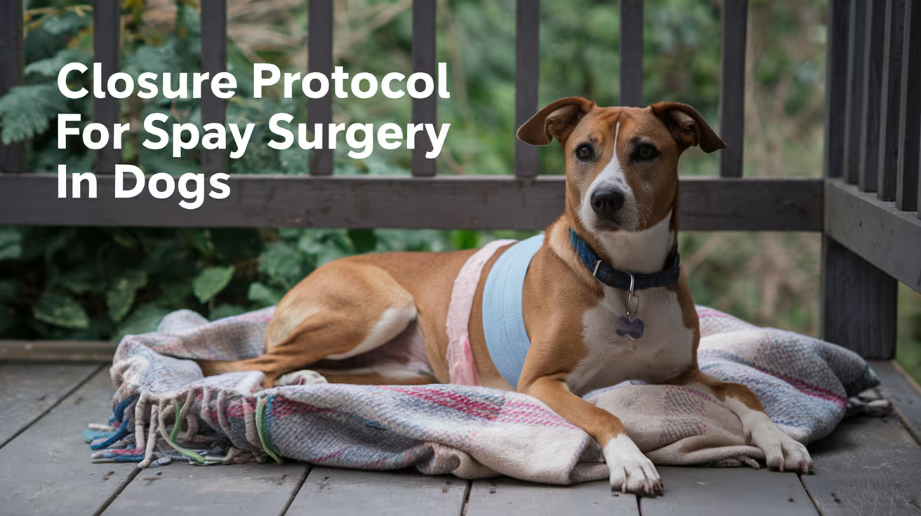
Closure Protocol for Spay Surgery in Dogs
Learn the detailed closure protocol for spay surgery in dogs, including step-by-step suturing techniques and post-op care tips.
The spay incision is one of the most commonly performed surgical wounds in veterinary practice. The closure is routine, but "routine" does not mean it can be done carelessly inadequate linea alba closure is the leading cause of post-spay incisional hernia, and insufficient skin closure is the most common source of early post-operative complications owners observe at home.
Understanding the three-layer protocol helps you know what your dog received and what to expect during recovery.
Quick answer: Canine spay closure proceeds in three layers: (1) linea alba PDS or polyglyconate 0 to 2-0, simple continuous, bites 5 to 10 mm from the incision edge and 5 to 10 mm apart; (2) subcutaneous tissue Monocryl or Vicryl 2-0 to 3-0, simple continuous; (3) skin interrupted nylon 3-0 to 4-0 or intradermal Monocryl 4-0. Secure knots are critical for continuous patterns: minimum 4 throws at the start of the continuous, additional throws at the end knot.
Key takeaways
- Linea alba is the structural layer inadequate closure here causes hernia, not just a surface wound problem.
- Bite dimensions matter: 5 to 10 mm from the incision edge; 5 to 10 mm between bites.
- Knot security is essential in continuous patterns published evidence links terminal knot failure to body wall dehiscence.
- Subcutaneous closure eliminates dead space and reduces skin tension for better wound healing.
- Intradermal skin closure is increasingly standard for routine spays, with no removal visit needed.
- Suture size varies with patient size: small dogs use 2-0 to 3-0; large dogs use 0 to 2-0.
Why the spay closure protocol matters
An ovariohysterectomy creates a ventral midline laparotomy. The linea alba is incised, the abdominal cavity is entered, the ovaries and uterus are removed, and the abdomen is closed in layers.
The closure is not just sealing the skin it is restoring the mechanical integrity of the abdominal wall. A dog that hernias through a spay incision has not had a skin problem; she has had a linea alba failure.
Veterinary Evidence (systematic review, linea alba closure): knot security is significantly affected by suture type, number of throws per knot, and surgeon experience and these factors should all be considered when performing surgery.
Layer 1: Linea alba closure
What the linea alba is
The linea alba is the midline aponeurosis a band of fibrous tissue created where the left and right abdominal wall muscles join. It is the primary mechanical structure of the ventral abdominal wall. The spay incision passes through this tissue to access the abdomen.
Technique
Pattern: simple continuous (preferred for speed and even tension distribution) or simple interrupted (preferred when tissue quality is questionable or infection is a concern).
WVS Academy (canine OVH surgical procedure guide): "Suture bites should be placed in the fascia and muscle 5 to 10 mm from the incision, and 5 to 10 mm apart. Place your first knot in the intact muscle directly adjacent to the incision. This ensures that the knot does not sit within the linea alba creating a gap. Tie a surgeon's knot and then 4 to 5 single-throw knots."
WCVM (University of Saskatchewan, Lab 6): "Take adequate bite (5 to 10 mm) of external rectus fascia or linea alba on either side of abdominal incision. Avoid large amounts of muscle as adds minimal strength and decreases apposition. Avoid fat as prevents healing."
Key principle: bites must be perpendicular to the wound not oblique. Oblique bites create longer bites with a wider gap between the incorporated tissue and the wound edge, reducing the mechanical strength of the closure.
Material
Preferred: PDS (polydioxanone) or polyglyconate (Maxon)
- Both are monofilament absorbable materials that retain strength for 4 to 6 weeks while the linea alba heals
- Monofilament surface minimizes bacterial adhesion compared to braided alternatives
- Size: 0 to 2-0 in most dogs; 2-0 to 3-0 in dogs under 10 kg
Alternative: Vicryl (polyglactin 910), size 0 to 2-0 acceptable for clean spay in a healthy dog; braided structure means slightly higher bacterial adhesion risk but clinically acceptable in routine clean surgery.
Knot security reminder
For continuous patterns, the end knot must be tied with additional throws beyond the start knot. WCVM: "Place appropriate number of throws for material and pattern (e.g., one extra throw at beginning and 2 to 3 throws extra at end of a simple continuous pattern) to ensure knot security."
For how the linea alba closure relates to the layered closure principle, see linea alba closure in the layered closure context.
Layer 2: Subcutaneous closure
Subcutaneous closure serves two functions: eliminating dead space beneath the skin, and reducing the tension on the skin closure.
Pattern: simple continuous absorbable
Material:
- Monocryl (poliglecaprone 25) 2-0 to 3-0: preferred for low tissue reaction
- Vicryl (polyglactin 910) 2-0 to 3-0: acceptable alternative
Technique: bites engage the subcutaneous fat perpendicular to the wound. Each loop draws the fat layers together, eliminating the space below the skin where serum would otherwise accumulate.
For how subcutaneous closure prevents seroma formation, see subcutaneous closure and seroma prevention.
Layer 3: Skin closure
Option 1: Interrupted external sutures (nylon or Prolene 3-0 to 4-0)
Best when:
- Post-operative wound monitoring is a priority
- The dog cannot reliably wear an E-collar (external sutures allow the vet to assess the wound directly at the removal visit)
- Any wound tension exists that exceeds what intradermal can hold
Spacing: 4 to 6 mm between sutures, placed 4 to 5 mm from the wound edge.
Removal: day 10 to 14, at the post-operative recheck.
Option 2: Intradermal (subcuticular) Monocryl 4-0
Increasingly the standard for routine canine spay closure in many practices.
WCVM: "For the intradermal, an absorbable suture material is indicated. A monofilament is typically preferred because of decreased tissue drag. The skin heals rapidly so a rapidly absorbable suture, like poliglecaprone 25, can be used."
Advantages:
- No external material for the dog to lick or chew
- No removal visit required
- Fine, less visible scar
- Knots are buried WCVM notes that "poorly buried knots are frequently associated with excess licking, irritation and increased infection rates"
For the full intradermal technique in the context of canine spay closure, see intradermal closure for spay incisions. For the cat spay closure comparison, see cat spay closure compared to dog spay.
Size-adjusted material guide
Post-operative care: what owners do
Activity restriction: leash walks only for 10 to 14 days. No running, jumping, or rough play. Metropolitan Veterinary Associates: "Dogs should be kept from jumping up/down on/from high surfaces, running up steps or any other activity that puts tension on the incision. Excess tension can lead to dehiscence."
E-collar compliance: non-negotiable, regardless of whether external or intradermal sutures were placed. A dog can disrupt an intradermal closure through licking before the dermal healing is complete.
Wound monitoring: twice daily. Normal: mild swelling and redness for 2 to 3 days, then progressive improvement. Abnormal: increasing redness, swelling after day 3, discharge, wound opening, or odor.
For the full post-operative monitoring protocol applicable to spay recovery, see monitoring the spay closure at home. For suture removal timing, see suture removal at the post-spay recheck.
Frequently asked questions
My dog has no visible stitches after her spay. Does that mean no stitches were used?
No it means intradermal closure was used. The suture runs inside the dermis, is completely buried, and dissolves on its own. There is nothing to remove and no external material to lick. A thin incision line is all that is visible.
The vet said my dog's spay used a "continuous pattern." Is that different from separate stitches?
Yes. Continuous (running) suture means a single thread runs the length of the closure in a sequence of loops. It is faster to place and distributes tension evenly along the entire closure. Separate (interrupted) sutures are placed and tied individually. Both are standard the choice reflects surgeon preference and patient factors.
How do I know if the linea alba closure is holding?
You cannot assess the linea alba directly it is the internal fascial layer below the skin. The external sign that the linea alba has failed is a soft, doughy bulge near the incision that appears weeks to months after surgery this is an incisional hernia. More immediate signs of early closure failure: the incision opening (dehiscence), abdominal contents visible, or your dog showing signs of significant pain or abdominal discomfort. Any of these require immediate veterinary assessment.
The spay closure protocol is a specific instance of the general layered closure principle applied to a clean ventral midline laparotomy. Linea alba for structure, subcutaneous for dead space elimination and skin tension reduction, skin for external protection. Getting each layer right material, bite dimensions, pattern, and knot security is what makes a routine spay a reliably routine recovery.
Resources
- WVS Academy. Canine OVH: Surgical Procedure. wvs.academy
- WCVM University of Saskatchewan. Lab 6 Part 4: Incision Closure. wcvm.usask.ca
- Veterinary Evidence (2017). Choice of Suture Pattern for Linea Alba Closure. veterinaryevidence.org
- Metropolitan Veterinary Associates. Abdominal Surgery: Post-Operative and Incisional Care. metro-vet.com

Tension-Relieving Sutures in Veterinary Surgery
Learn about tension-relieving sutures in veterinary surgery, their types, uses, benefits, and care for better healing in pets.
When wound edges cannot be brought together without significant pulling force, the problem is tension. Standard interrupted sutures placed across a high-tension wound concentrate that force at each suture entry point creating ischemia at the tissue-suture interface and dramatically increasing the risk of sutures cutting through and the wound opening.
Tension-relieving sutures work differently. They distribute that force across a wider area, or recruit more tissue to share the load, so no single point bears enough force to fail.
Quick answer: Tension-relieving sutures are patterns specifically designed to distribute wound tension across more tissue and a wider surface area than standard interrupted sutures. The main types used in veterinary surgery are: horizontal mattress (broad tension distribution, parallel to wound), vertical mattress (deep plus superficial bite for edge eversion), near-far-far-near (appositional and tension-relieving, stays in until healed), and walking sutures (advances skin subcutaneously before surface closure). Each addresses tension at a different structural level.
Key takeaways
- Horizontal mattress sutures spread tension across 8 to 10 mm from the wound edge, reducing cut-through risk.
- Vertical mattress sutures provide deep tissue purchase plus edge eversion ideal in high-tension zones.
- Near-far-far-near (NFFN) is both cosmetic and tension-relieving, staying in through full healing.
- Temporary mattress sutures can be placed first, then removed after 3 to 4 days once appositional sutures are secured.
- Stent sutures use tubing or pads under the knot to prevent large sutures from cutting through fragile skin.
- Walking sutures address tension at the subcutaneous level before skin closure begins.
Why wound tension causes problems
Tension at the wound edge reduces blood supply to the tissue between the suture and the wound margin. Without adequate perfusion, that tissue cannot heal, mount an immune response, or resist bacteria.
The consequences of uncorrected wound tension:
- Sutures cut through skin (when tension exceeds the tissue's tolerance at the suture entry point)
- Wound dehiscence (when sutures fail or the tissue gives out)
- Tissue necrosis at the wound margins
- Delayed healing
- Infection in poorly perfused tissue
VCA (Care of Surgical Incisions in Dogs): "If the surgical procedure involved tissue loss, the incision may be under a lot of tension. Excessive tension across an incision line may cause the wound to gape open and delay healing. To minimize tension on the incision line, your veterinarian may use a special tension-relieving suture pattern, or a type of skin suture called a stent suture."
For how tension directly contributes to high-tension wound closure failure, see high-tension wound closure using tension-relieving sutures.
Horizontal mattress sutures
The horizontal mattress pattern is placed parallel to the wound edge. Each stitch enters the skin, crosses the wound, re-enters on the opposite side, returns parallel to the wound, and exits the skin. The result is a U-shaped loop lying flat across the wound surface.
Veterian Key: "This type of suture can be used in areas of tension as the pressure exerted by the horizontal sutures is spread evenly over a broad area, which reduces the likelihood of tearing through the tissue edges. The action is: holding the needle with needle holders, insert the needle approximately 8 to 10 mm away from the edge of the incision on the far side."
Properties:
- Spreads tension over 8 to 10 mm from the wound edge on each side
- Can be used as a temporary stay stitch to approximate wound edges while interrupted or intradermal sutures are placed
- Can remain in place for several days after the primary closure if tension persists
- Risk of suture marks if left beyond 7 days
University of Minnesota (Large Animal Surgery): "If tension is greater than can be managed with a NFFN suture, vertical and horizontal mattress sutures may be placed temporarily to relieve tension. After 3 to 4 days, the mattress sutures can be removed, leaving just the appositional pattern."
For how mattress sutures are used as the primary tension pattern in small animal surgery, see mattress sutures in small animal surgery.
Vertical mattress sutures
The vertical mattress pattern takes a deep bite far from the wound edge (the "far" component), crosses to the opposite side, returns with a shallower bite close to the wound edge (the "near" component). The loop is vertical crossing the wound at two depths.
Properties:
- Provides strong tissue purchase deep to the skin holds against tension at the fascial level
- Everts wound edges (turns edges outward) counters the tendency of high-tension wounds to invert
- Reduces dead space just below the skin edge by drawing deep tissue into the closure
- More likely to cause suture marks than horizontal mattress if left beyond 10 days
Best for:
- High-tension wounds where deep tissue purchase is needed
- Areas where wound inversion is a problem (e.g., over joints, areas with thick overlying muscle)
- Skin over orthopedic surgical sites
Near-far-far-near (NFFN) sutures
The NFFN pattern is a tension-relieving suture that is also appositional meaning it holds wound edges together while distributing tension, rather than merely providing mechanical support from the outside.
University of Minnesota (Large Animal Surgery, Suturing Skin chapter): "A near-far-far-near type pattern is cosmetic as well as tension relieving. As it is an appositional pattern and will stay in until the tissue is healed, it should not be bigger than 2-0 or 0 in non-bovine patients."
Pattern sequence:
- Near entry close to the wound edge on one side
- Far exit exits the skin far from the wound edge on the opposite side
- Re-enters far re-enters far from the wound on the same side it just exited
- Near exit exits close to the wound edge on the original side
The alternating near-far bites distribute tension while the pattern itself holds wound edge apposition. This makes it more cosmetically acceptable than a horizontal mattress and appropriate for wounds where the suture will be the primary closure rather than a temporary adjunct.
For the clinical comparison of NFFN and walking suture approaches to tension, see NFFN pattern in high-tension wound context.
Stent sutures
Stent sutures are not a pattern they are a modification applied to any external suture in high-tension situations. A piece of rubber tubing, surgical sponge, or button is threaded under the knot of a mattress or interrupted suture.
Why stents are used:
- In thin-skinned or fragile patients, a large-gauge suture alone can cut through skin at the knot
- The stent distributes the contact area of the knot across a wider skin surface
- Allows use of stronger suture material (size 0 to 1) without the cut-through risk
VCA: "Your veterinarian may use... a type of skin suture called a stent suture, which involves the addition of some tubing or a button to the skin layer."
University of Minnesota (Large Animal Surgery): "Larger suture (1-2) can be used for the mattress sutures. Stents (tubing or pads) are often used to prevent the larger suture from cutting through the skin."
Walking sutures: the subcutaneous approach
While the above patterns address tension at the skin surface, walking sutures address it before skin closure begins by advancing the dermis toward the defect at the subcutaneous level.
For full detail on the walking suture technique, see walking sutures for large defects in large breed dogs.
Choosing between tension-relieving options
Suture material for tension-relieving patterns
University of Minnesota: In large animals, mattress sutures use "larger suture (1 to 2)" while the appositional pattern uses "2-0 or 0."
For dogs: horizontal and vertical mattress sutures typically use the same material as other skin sutures but at a size one step larger than the primary closure so if the wound would normally close with 3-0, tension sutures might use 2-0. Monofilament non-absorbable (nylon, Prolene) is preferred to minimize bacterial adhesion. PDS is used when an absorbable material is needed in a tension-relieving position.
For how tissue type and patient factors such as obesity affect closure technique selection, see tissue type and closure technique selection. In obese patients where tissue fragility increases cut-through risk, stent sutures are especially relevant; see tension management in obese dogs.
Frequently asked questions
My dog had large mattress sutures placed around a smaller standard closure. Why two types?
Your vet used the mattress sutures as tension relief to protect the primary appositional closure. The mattress sutures bear the bulk of the wound tension, reducing the load on the finer interrupted or intradermal sutures that produce the actual skin edge contact. In some cases, the mattress sutures are removed after 3 to 4 days, leaving only the appositional closure; in others, they stay through the full healing period.
Are tension-relieving sutures removed at the same time as regular sutures?
Usually around the same time (10 to 14 days), but staggered removal is common for walking sutures and some mattress patterns. In cases with significant tension, the vet may leave some tension-relieving sutures beyond 14 days if the wound is not yet stable. Ask your vet specifically about the removal plan for each suture type placed.
Do tension-relieving sutures hurt more than regular sutures?
Not typically, though mattress sutures do involve deeper tissue bites that may cause more post-operative soreness than fine interrupted skin sutures. Pain management after surgery covers this. The tenderness is usually at its peak for 24 to 48 hours, then diminishes as the wound stabilizes.
The purpose of every tension-relieving technique is the same: ensure that no single point in the closure bears enough force to fail. Whether that means spreading force across the surface with a horizontal mattress, anchoring it deep with a vertical mattress, distributing it along the appositional line with NFFN, or advancing the skin before surface closure with walking sutures, the outcome goal is uniform: a closure that holds while the tissue heals.
Resources
- Veterian Key. Suturing Techniques and Common Surgical Procedures. veteriankey.com
- University of Minnesota. Suturing Skin (Large Animal Surgery Supplemental Notes). open.lib.umn.edu
- VCA Animal Hospitals. Care of Surgical Incisions in Dogs. vcahospitals.com
- WSAVA 2007 (VIN). Reconstructive Surgery. vin.com
Suture Size Selection in Small Animal Surgery
Learn how to choose the right suture size for small animal surgery to ensure optimal healing and minimize complications.
Every surgical suture comes in multiple sizes, and size matters as much as material. A suture that is too large for the tissue causes unnecessary inflammation and delays healing. One that is too small fails to hold under the mechanical forces the wound faces.
Selecting the right size requires understanding how sutures are sized, what each tissue needs, and how patient weight influences the decision.
Quick answer: Suture sizes follow the USP (United States Pharmacopeia) system, where more zeroes mean smaller diameter so 4-0 is smaller than 2-0, and 2-0 is smaller than 0. The guiding principle is to use the smallest size that provides adequate tensile strength for the tissue. For small animals: linea alba in medium dogs takes 0 to 2-0; subcutaneous layers take 2-0 to 3-0; skin takes 3-0 to 4-0. Cats and small dogs use sizes one unit finer throughout.
Key takeaways
- The USP size system runs from 11-0 (smallest) to 7 (largest) more zeroes means smaller diameter.
- Use the smallest size that provides adequate tensile strength larger sizes cause more inflammation.
- Linea alba in medium dogs: 0 to 2-0 PDS or Biosyn.
- Subcutaneous tissue in dogs: 2-0 to 3-0 Monocryl or Vicryl.
- Skin in dogs and cats: 3-0 to 4-0 for external sutures; 4-0 for intradermal.
- Cats and small dogs use sizes one unit finer than dogs of average size throughout.
Understanding the USP suture sizing system
The United States Pharmacopeia (USP) system is the standard for suture sizing in veterinary and human surgery.
How it works:
- Sutures are assigned a numerical designation based on diameter
- Size 1 is larger than size 0
- Below size 0, additional zeroes are added: 0, 2-0, 3-0, 4-0, 5-0, etc.
- Each additional zero indicates a smaller diameter
- 11-0 is the smallest (microsurgery); 7 is the largest (orthopedic/heavy tissue)
Veterinary Surgery Online explains: "The more zeros, the smaller the material, so 6-0 is actually size 000000, and is pronounced 'six ought' or 'six zero'."
AAHA (Oh, Sew Easy, 2022): "Optimal suture size is determined as the smallest size necessary to achieve a tension-free wound closure."
The core principle: smallest effective size
Veterinary Practice News (Dr. Kendra Freeman, DACVS): "The general principle is to use the smallest suture strong enough for the tissue. This allows for less suture material to be present, which may potentially contribute to inflammation and tissue reaction."
What happens when suture is too large:
- More foreign material in the tissue
- Greater inflammatory response
- Higher infection risk at the suture site
- Possible tissue strangulation if pulled too tight
What happens when suture is too small:
- Suture cuts through the tissue under mechanical load
- Wound dehiscence
- Need for re-closure
Published research (Frontiers in Veterinary Science, 2023): A study comparing USP 2-0, 3-0, and 4-0 PGA sutures in canine abdominal incisions found 4-0 suture had lower inflammatory response markers while maintaining adequate mechanical strength. The authors concluded "USP 4-0 PGA suture has more advantages to suturing canine abdominal surgical incisions."
Size guide by tissue type and patient
Linea alba / fascial closure
Veterinary Practice News: "2-0 or 0 suture is appropriate for the linea alba in medium- to large-size animals."
For why the fascial layer specifically requires larger suture than adjacent muscle, see tissue type as a guide for suture size.
Subcutaneous tissue
Gastrointestinal and urinary tract
Veterinary Practice News: "3-0 or 4-0 suture is generally appropriate for the gastrointestinal tract and urinary bladder."
These organs require fine suture because oversized suture causes excessive lumenal reaction and stenosis risk. Taper-point needles are paired with these sizes to minimize tissue injury.
Skin (external interrupted)
Skin (intradermal)
4-0 is the standard for intradermal (subcuticular) closure across all patient sizes. The dermis is a uniform layer that does not vary as much with patient size as deeper structural layers.
Needle size relationship to suture size
Suture size and needle size are linked the needle is sized to match the suture. Common veterinary needle types:
For how needle selection alongside suture size affects closure quality, see needle size alongside suture size.
Common sizing errors
Too large for the tissue:Most common in the skin layer, where surgeons sometimes default to a larger size "for security." The result is more visible suture marks, more inflammation, and longer healing time.
Too small for the load:More common in the linea alba, where a 3-0 suture is used in a large breed dog that needs 0 or 2-0. Tension exceeds the suture's strength before healing occurs.
Mismatched sizing across layers:Each layer needs its own appropriate size. Using the same suture size throughout all layers (a practice shortcut) results in either oversized subcutaneous sutures or undersized fascial sutures.
For how suture size selection connects to material selection across procedures, see size alongside material selection in dogs. For cats, see size alongside material selection in cats.
Frequently asked questions
My vet used 3-0 suture for my large dog's spay. Is that too small?
It depends on the layer. 3-0 for the subcutaneous and skin layers of a medium-large dog is appropriate. 3-0 for the linea alba in a dog over 25 kg would likely be undersized 0 or 2-0 is standard for that layer. If you are concerned, ask your vet specifically which sizes were used at each layer.
Do smaller sutures dissolve faster?
No. Absorption timeline is determined by the suture material, not by its size. A 3-0 Monocryl and a 2-0 Monocryl absorb over the same timeline approximately 90 to 119 days. The difference is tensile strength, not absorption rate.
Can suture sizes vary between different materials of the same USP designation?
Yes, slightly. Veterinary Surgery Online notes: "It is important to note that the USP dimensions for catgut are different to those of other sutures. For example, 2-0 Catgut is larger than 2-0 PDS." When comparing materials, the USP designation is a starting reference, not an exact interchangeable standard across all material types.
Suture size selection follows one rule applied at every layer: the smallest size that adequately holds the tissue under its expected mechanical load. Getting it right means less inflammation, less reaction, and faster healing. Getting it wrong in either direction creates predictable complications that show up in the first two weeks of recovery.
Resources
- Veterinary Practice News. The Must-Read Guide to Selecting Sutures (Dr. Kendra Freeman, DACVS). veterinarypracticenews.com
- Veterinary Surgery Online. Suture Size. vetsurgeryonline.com
- AAHA. Oh, Sew Easy: A Guide to Sutures. aaha.org
- Frontiers in Veterinary Science (2023). Biomechanical and tissue reaction: the effects of varying suture size on canine abdominal wall stitching. frontiersin.org

Common Closure Errors in Small Animal Surgery
Explore common closure errors in small animal surgery, their causes, prevention, and how to ensure optimal healing for your pet’s recovery.
Most surgical complications that appear in the first two weeks of recovery are not random. They trace back to specific, identifiable mistakes in wound closure mistakes that are preventable, detectable, and in many cases correctable if caught early.
The errors are consistent enough across procedures that they can be named, explained, and recognized.
Quick answer: The five most common closure errors in small animal surgery are: sutures tied too tight (causing ischemia and tissue necrosis), sutures tied too loose (leaving the wound inadequately apposed), wrong suture material for the tissue (mismatched strength or absorption rate), failure to close dead space (leaving fluid-filling pockets that become seromas or abscesses), and wrong closure pattern for wound tension (using simple interrupted across a high-tension wound without tension-relieving technique). Each error has a predictable consequence and recognizable early signs.
Key takeaways
- Sutures tied too tight restrict blood flow, causing tissue ischemia and necrosis that creates infected dead tissue.
- Sutures tied too loose fail to appose wound edges, leading to dehiscence and infection.
- Wrong suture material means either premature loss of strength (wound opens) or excess foreign material reaction.
- Unclosed dead space fills with serum, creating an ideal bacterial growth medium.
- Wrong pattern for wound tension allows sutures to cut through tissue or fail catastrophically.
- Most closure errors become visible within 3 to 7 days owner monitoring in this window enables early intervention.
Error 1: Sutures tied too tight
What happens: when the knot is overtightened, the loop of suture compresses tissue between the wound edge and the entry/exit points. That compression restricts blood flow.
Consequences:
- Tissue between the suture and the wound edge becomes ischemic (inadequately perfused)
- Ischemic tissue cannot mount an immune response bacteria colonize it readily
- Dead tissue becomes a substrate for infection
- The strangulated tissue eventually sloughs, opening the wound from within
How to recognize it:
- Within 24 to 48 hours: the skin at each suture entry point looks pale or whitened rather than pink
- Within 3 to 5 days: necrotic (black, brown, or grey) tissue appears at the wound margins
- The tissue may start to smell before it visibly changes
Why it happens: the temptation to tie tight for security. Wound edges should appose, not compress. The suture should draw tissue into contact, not hold it under pressure.
For how suture tension decisions affect the wound closure protocol overall, see wound closure principles relating to tension.
Error 2: Sutures tied too loose
What happens: loose sutures allow the wound edges to remain separated or to move relative to each other. The wound never achieves apposition the edges must touch for healing to proceed.
Consequences:
- Dehiscence: the wound opens, either gradually (edges drift apart) or suddenly (with one jump or activity)
- Exposed subcutaneous tissue is at high infection risk
- Re-closure is required under sedation or anesthesia
How to recognize it:
- Immediately post-surgery: small visible gaps between sutures
- Within 2 to 4 days: the wound edges separate at one or multiple points
- The wound may drain and appear wider than it should be
Why it happens: attempting to minimize suture marks by under-tensioning, or placing sutures too far apart.
Error 3: Wrong suture material
What happens: material mismatch between the suture's properties and the tissue's needs.
Material too weak for the load
Using Monocryl (fast-absorbing) to close the linea alba in a large breed dog. Monocryl loses most of its tensile strength by 21 days. The linea alba takes 4 to 6 weeks to regain adequate strength. The suture fails before the tissue is ready to hold alone.
Consequence: incisional hernia, wound opening, or catastrophic dehiscence.
Material too reactive for the tissue
Using silk (high tissue reactivity) in a buried internal layer. Silk provokes a significant chronic inflammatory response, creating sinus tracts that drain permanently.
Consequence: persistent drainage from a wound that appears to have healed; often misidentified as infection.
Braided material in a contaminated wound
Using Vicryl (braided polyglactin) in a bite wound or heavily contaminated case. Bacteria colonize the interstices between braided strands, shielded from the immune response.
Consequence: wound infection despite antibiotic therapy, because the suture itself harbors the organism.
For how suture material selection avoids these errors, see suture material selection to prevent closure errors.
Error 4: Failure to close dead space
What happens: dead space is any gap remaining between tissue planes after closure. These gaps fill with serum (tissue fluid). Serum is protein-rich and warm the ideal bacterial growth medium.
Consequences:
- Seroma: a fluid pocket under the skin that creates a painless swelling, often discovered 3 to 5 days post-surgery
- Infected seroma: the serum becomes colonized with bacteria, producing an abscess
- Delayed healing: the tissue planes cannot adhere if fluid separates them
How to recognize it:
- Soft, fluctuant swelling at or near the surgical site, appearing days after surgery
- The wound surface may appear normal while a large pocket forms beneath
Prevention: subcutaneous closure to eliminate the fat layer gap, walking sutures to tack skin to fascia over larger defects, drain placement when dead space cannot be sutured closed.
For how dead space management is addressed as part of the closure sequence, see dead space management as a closure step.
Error 5: Wrong pattern for wound tension
What happens: simple interrupted sutures are placed across a wound under significant tension without any tension-relieving technique. Each suture bears the full tensile load of the wound at that point.
Consequences:
- Sutures cut through skin: the suture entry point becomes a linear tear perpendicular to the wound
- Wound dehiscence: one or more sutures fail, creating a gap
- The wound may appear to be holding for several days, then fail suddenly when activity increases or a seroma develops
Prevention: recognize high-tension wounds pre-closure and apply tension-relieving technique horizontal mattress, walking sutures, or undermining of skin edges before placing the primary closure.
Veterinary Surgery Online: "If tissues are fragile and suture pulls out easily, use cruciate or horizontal mattress pattern instead of simple interrupted suture pattern for less stress on each bite."
For how specific tension-relieving patterns prevent this error, see tension-relieving patterns that prevent closure failure.
Error 6: Incorrect layer closure sequence
What happens: layers are closed out of sequence, or a layer is skipped entirely. Most commonly: the subcutaneous layer is not closed, leaving dead space; or the fascial layer is closed with the same material and tension as the subcutaneous layer, leaving the structural closure inadequately supported.
Consequences depend on which layer is affected:
- Missed subcutaneous layer: dead space, seroma, delayed healing
- Inadequate fascial closure: incisional hernia weeks to months later
- Skin closed over unclosed deep layers: surface looks fine; structural failure develops silently
For how the layered closure sequence prevents these errors, see layered closure sequence in small animal surgery.
What owners can monitor at home
The first 5 to 7 days are the highest-risk window. Most closure errors become externally visible during this period.
Check twice daily for:
- Any visible gap between suture points
- Skin at suture entry points: should be pink and pliable, not white or dark
- Swelling, especially fluctuant swelling (feels like a fluid-filled balloon under the skin)
- Discharge: small amounts of serous (clear, slightly yellow) fluid is normal; green, brown, or foul-smelling discharge is not
- Wound pulling apart or gaping
When to call the vet:
- Any visible gap or suture failure
- Discharge that is purulent or foul-smelling
- Dark or pale tissue at wound margins
- Fluctuant swelling appearing 3 or more days post-surgery
- Dog is chewing at or licking the wound despite E-collar
For the full post-operative monitoring protocol after closure, see post-operative monitoring after closure.
Frequently asked questions
One of my dog's sutures has a small bump around it. Is that a problem?
A small firm bump at a suture site within the first 3 to 5 days is usually normal a mild inflammatory reaction to the suture material. A soft, fluctuant (fluid-filled) bump, especially one that appears at the wound itself or between suture sites, is more likely to be a seroma and should be assessed by your vet.
My dog's wound opened two days after surgery. What caused it?
Common causes: the dog licked or chewed the wound (removing sutures), activity that placed excessive force on the closure, sutures that were inadequately tensioned, or excessive wound tension that exceeded the repair strength. Contact your vet the same day open wounds generally need re-closure promptly to prevent infection and further dehiscence.
Can I prevent closure errors at home?
You cannot change what happened in the operating room, but you can prevent the most common post-operative causes of closure failure: keep the E-collar on at all times, restrict activity as directed, keep the wound dry, and attend all scheduled rechecks. Most secondary failures (wound opening after initial surgery) are caused by licking, jumping, or premature bathing.
Closure errors are not random they are the predictable consequences of specific technical decisions made during wound closure. Recognizing which error you are looking at early, based on the pattern of what is happening to the wound, is the first step in getting it corrected before a small problem becomes a large one.
Resources
- Veterinary Surgery Online. Wound Closure Continued. vetsurgeryonline.com
- VCA Animal Hospitals. Care of Surgical Incisions in Dogs. vcahospitals.com
- Veterian Key. Selection of Suture Materials, Suture Patterns, and Drains for Wound Closure. veteriankey.com
- AAHA. Oh, Sew Easy: A Guide to Sutures. aaha.org
Closure Protocol for Tumor Excision in Dogs
Learn the detailed closure protocol for tumor excision in dogs to ensure proper healing and reduce complications after surgery.
Tumor excision surgery is not one procedure it is as many procedures as there are tumors and locations. A small, benign lipoma on the flank closes in minutes. A mast cell tumor with 2 to 3 cm margins on the distal limb may require a flap, a graft, or staged reconstruction.
The closure protocol that follows the excision is entirely shaped by the margin required, the location of the mass, and how much skin remains after the specimen is removed.
Quick answer: Tumor excision closure in dogs follows layered technique: deep tissue (if fascia or muscle was included in the excision), subcutaneous tissue, and skin. Benign masses with marginal excision close primarily with standard techniques. Malignant masses requiring wide margins often leave defects that require tension management (undermining, walking sutures) or reconstruction (flaps, grafts). PMC5771180 (12 dogs, mammary tumor excision) confirms: undermining, walking sutures, and tension-relieving vertical mattress sutures are the primary adjuncts for large ventral skin defect closure.
Key takeaways
- Margin width determines closure complexity marginal excision closes easily; wide margins often require reconstruction.
- En bloc deep margin (including fascia below the tumor) creates a deeper defect requiring fascial and possibly muscle closure.
- Undermining releases skin for tension-free primary closure without recruiting distant tissue.
- Walking sutures and vertical mattress sutures are the primary techniques for large skin defects after tumor excision.
- Flaps and grafts are used when primary closure would produce ischemia-inducing tension.
- Drain placement is indicated when significant dead space cannot be eliminated by suturing alone.
How margin requirements shape closure
Surgical margins define how much normal tissue is removed around the tumor to reduce local recurrence risk. The margin required depends on tumor type and behavior:
Today's Veterinary Practice: "Wide excision of skin or subcutaneous masses frequently leaves large skin defects that can be difficult to close. When primary closure cannot be obtained due to excessive skin tension, consider either immediate or staged flap or graft reconstruction."
Deep layer closure
When the excision includes the fascia below the tumor (the "deep margin"), the fascial defect must be closed before subcutaneous closure.
Pattern: simple continuous or interrupted absorbableMaterial: PDS 0 to 2-0, depending on the size of the fascial defect and patient sizeKey principle: the fascial closure must engage the fascial tissue, not just the muscle belly. Muscle does not hold suture under load.
If the deep margin included a full muscle belly cross-section, the defect is assessed for primary closure feasibility. Very large muscle defects may require a mesh or fascial graft if primary tension-free closure is not achievable.
Subcutaneous closure and dead space management
Tumor excision frequently creates significant dead space a three-dimensional cavity where the mass occupied the tissue. This space fills with serum if not closed, producing a seroma that can become infected or delay healing.
Dead space management options:
- Subcutaneous sutures: simple continuous Monocryl or Vicryl 2-0 to 3-0, placed to obliterate the cavity
- Walking sutures: tacking the dermis to the underlying fascia to eliminate the gap above the fascial closure
- Drain placement: when the cavity cannot be fully obliterated by suturing Penrose drains allow serosanguinous fluid to escape
Veterian Key (Skin Reconstruction Options): "The use of various subcutaneous suture patterns will eliminate dead space, resist tensile forces trying to disrupt the incision, and help maintain wound apposition for better wound closure."
For how delayed closure applies when tumor margins intersect contaminated tissue, see delayed closure for contaminated tumor excision wounds.
Skin closure for primary-closeable defects
When sufficient skin remains after excision for primary closure, the skin edges are brought together using standard tension assessment and technique.
Pre-closure tension check: before placing any skin sutures, assess whether the edges can be approximated without blanching. If they can meet with light finger pressure and the skin at each edge remains pink, primary closure is feasible.
Techniques:
- Simple interrupted or cruciate (nylon or Prolene 3-0 to 4-0) for standard-tension wounds
- Horizontal mattress for wounds with moderate tension
- Vertical mattress and/or walking sutures when tension is significant
PMC5771180 (12 dogs, mammary tumor excision with wide margins): "Undermining, walking sutures, and tension-relieving techniques were followed to close the large skin defect without much tension. Tension-relieving vertical mattress sutures were placed to relieve tension at the suture site."
For the complete tension management technique guide, see high-tension closure after tumor excision.
When primary closure is not achievable
Some excision defects are too large or too poorly positioned (e.g., distal limb, face) for standard primary closure. Options:
Cosmetic skin closure approach: for tumor sites where scarring is a concern, see cosmetic closure after tumor excision.
Skin advancement flaps
Adjacent skin is incised, undermined, and advanced to cover the defect. Flaps maintain their blood supply through an intact pedicle. Fail if the pedicle is kinked or the flap is placed under excessive tension.
Staged reconstruction
For large or complex defects, the wound is managed open (with daily bandage changes) for 3 to 5 days while the patient stabilizes and the defect is assessed. Secondary closure or flap reconstruction is planned after this period.
Second intention healing
For small defects in non-critical locations (trunk, dorsum), second intention healing (contraction and epithelialization without surgical closure) produces acceptable results. No closure surgery is required; the wound is managed with daily dressings.
Post-excision margin assessment and re-excision
If histopathology returns with "incomplete margins" (tumor cells at the surgical margin), re-excision may be indicated. The closure from the first surgery is still healing when this decision is made typically at 7 to 14 days.
Re-excision timing considerations:
- Primary closure must have adequate wound strength before re-excision
- The previous scar track is included in the new excision specimen
- Reconstruction for the second excision is planned before the procedure
For the closure protocol checklist applicable to tumor excision, see closure checklist for tumor excision procedures.
Post-operative monitoring
Days 1 to 5: highest-risk period for acute closure failure and seroma formation.
- Check the wound twice daily
- Drain output (if placed): monitor volume and color decreasing output and clearing color indicate the seroma risk is resolving
- Swelling should peak around day 2 to 3, then gradually decrease
- Any sudden increase in swelling after the initial post-operative period suggests seroma or hematoma
Days 5 to 14: wound consolidation phase.
- Continued twice-daily checks
- Suture removal at 10 to 14 days for external sutures
- If a drain was placed, it is removed once output is minimal (typically 3 to 7 days post-operatively)
For tumor excision closure in cats, see tumor excision closure protocol in cats.
Frequently asked questions
The vet said my dog's tumor margins were wide and the closure was complex. Does that mean more complications?
Not necessarily. Wide margin excision is the appropriate surgery for malignant tumors it gives the best chance of complete removal. A complex closure performed well (with appropriate tension management) heals as reliably as a simple closure. The complexity is in the operating room; the recovery timeline is similar.
My dog has a Penrose drain coming out of the wound. When will it be removed?
Drains are typically removed when output has dropped to a small amount (under 1 to 2 mL per day) and the color has become clear serous rather than bloody. This usually happens 3 to 7 days after surgery, depending on how much dead space was present. Your vet will assess drain output at each recheck.
Can tumors grow back after surgery?
Local recurrence depends on whether complete margins were achieved. If histopathology confirms clean margins (no tumor cells at the edges), local recurrence is unlikely. Incomplete margins substantially increase local recurrence risk. Systemic spread (metastasis) depends on tumor type, grade, and staging your oncologist or surgeon can discuss the specific risk for your dog's tumor.
Tumor excision closure is the final step in a procedure where the outcome is determined largely by what came before it: the margin achieved, the tissue preserved, and the dead space created. Getting those decisions right before closure begins is what makes the closure itself straightforward.
Resources
- PMC5771180. Studies on Reconstruction of Large Skin Defects Following Mammary Tumor Excision in Dogs. ncbi.nlm.nih.gov
- Today's Veterinary Practice. Fundamentals of Surgical Oncology in Small Animals. todaysveterinarypractice.com
- Veterian Key. Summary of Skin Reconstruction Options. veteriankey.com
- Veterinary Key Points (Dr. Stephen Birchard). Complete Surgical Excision of Mast Cell Tumor in Dogs and Cats. drstephenbirchard.blogspot.com

Preventing Dehiscence in Dog Surgical Wounds
Learn how to prevent dehiscence in dog surgical wounds with expert tips on care, suturing, and monitoring to ensure safe healing.
A sutured surgical wound has two phases of risk. The first is the operating room. The second is the ten to fourteen days your dog spends at home while that wound heals.
Most dehiscence, the reopening of a surgically closed wound, happens because of something that occurs after the dog goes home. That puts a meaningful amount of prevention directly in the owner's hands.
Quick answer: Dehiscence is the breakdown and separation of a surgical wound's sutured edges. In dogs, the most common causes are licking or chewing the wound, excessive activity that stresses healing tissue, and infection. Wounds are most vulnerable in the first seven to fourteen days, when tensile strength is still building. Prevention centers on consistent E-collar use, strict activity restriction, daily monitoring, and prompt veterinary contact at the first sign of infection.
Key takeaways
- Dehiscence most commonly occurs in the first seven to fourteen days: This is when the wound has the least tensile strength and is most vulnerable to disruption.
- Licking is the leading preventable cause: Even brief access to a wound can undo days of healing.
- Dogs are more physically disruptive than cats: Larger size and greater activity drive higher mechanical stress on abdominal incisions.
- Infection significantly raises dehiscence risk: Bacterial enzymes actively degrade suture material and surrounding tissue.
- Abdominal and limb wounds carry higher risk: Movement at these sites creates ongoing tension against healing tissue.
- Abdominal evisceration is a surgical emergency: Any significant opening of an abdominal incision requires immediate veterinary care.
What dehiscence is and when it happens
Dehiscence is defined as the separation of a surgically closed wound after closure. It can be partial, where only part of the wound edge separates, or complete, where the entire incision comes apart.
In dogs, dehiscence most often occurs within the first seven to fourteen days post-surgery. This timing reflects a biological reality: tensile strength in healing tissue builds slowly. At day seven, a well-healing wound has regained only a fraction of the strength of normal skin. At day fourteen, it is stronger but still vulnerable.
The most common areas where dehiscence occurs in dogs are:
- Abdominal incisions (spay, tumor removal, exploratory surgery) where the combination of movement, tension, and internal organ pressure creates ongoing mechanical stress
- Limb wounds over joints, where flexion and extension pull at the sutured edges with every movement
- Areas under direct pressure, such as the trunk in overweight dogs
Dehiscence is distinct from minor surface irregularities. A small seroma, slight scabbing, or minor discharge near an otherwise closed incision is not dehiscence. Dehiscence is when wound edges visibly separate.
The main causes of dehiscence in dogs
Licking and chewing
Licking is the most common preventable cause of wound dehiscence in dogs.
A dog that can access the wound will mechanically disrupt forming tissue with each lick, introduce oral bacteria that increase infection risk, and may physically pull sutures loose. Large dogs, in particular, can access abdominal incisions with ease unless the collar is correctly fitted and consistently used.
E-collar requirements for dogs:
- Must extend at least two inches past the tip of the nose
- Must be kept on at all times, including overnight and when unsupervised
- Soft recovery collars and recovery suits are acceptable alternatives if they reliably prevent wound access
- Check periodically that the collar has not been removed or repositioned by the dog
Excessive physical activity
Dogs that return to normal activity too soon impose mechanical forces that sutures and healing tissue cannot yet withstand.
For abdominal incisions, the specific risks are: jumping, which creates impact stress on landing; running, which requires core muscle engagement; and stair climbing, which flexes abdominal structures. These movements can pop sutures or create tension across the healing tissue that exceeds what the early-stage wound can sustain.
Typical activity restriction for routine dog surgeries is ten to fourteen days of leash-only walks for bathroom purposes, no running or jumping, and no stairs where avoidable. Complex or larger surgeries may require longer restriction.
Confinement to a single room or a large crate for the restriction period is appropriate. Dogs that appear recovered often feel well before the wound is structurally ready.
Infection
Infection causes dehiscence through a specific mechanism. Many bacteria produce collagenase, an enzyme that degrades the collagen being deposited to rebuild the wound and that can weaken suture material through the same enzymatic process.
An infected incision is not just inflamed. It is being actively weakened by the bacterial activity within it.
For this reason, signs of incision infection, including spreading redness, warmth, discharge, and odor, require same-day veterinary contact. Treated early, most incision infections can be managed without dehiscence. Left untreated, they significantly raise the probability of wound breakdown.
For understanding infection as a cause of wound breakdown, including how infection progresses through the healing stages and when it begins to threaten wound integrity, that guide covers the relationship between infection and healing in detail.
Tension on the closure
Tension on the wound edges is the most common cause of dehiscence from the surgical side. When sutures are closed under excessive tension, the tissue between the suture and the wound edge can be cut through (suture cutout) or the tissue can lose blood supply and die (ischemic necrosis), both leading to wound breakdown.
Owners cannot prevent this directly, but factors that increase tension post-operatively include:
- Weight gain or significant swelling
- Excessive movement at the incision site
- Trauma to the closed wound area
Maintaining normal weight during recovery and avoiding rough handling of the incision area reduces post-operative tension.
Dog's individual health status
Several patient factors increase the baseline risk of dehiscence:
- Obesity: increased tension on closures, reduced tissue perfusion
- Diabetes: impaired immune function and slower cellular healing response
- Long-term steroid use: delays wound healing and reduces tissue strength
- Advanced age: slower healing and reduced skin elasticity
- Hypoproteinemia (low blood protein): impairs collagen synthesis
If your dog has any of these conditions, your veterinarian will account for them in post-operative care instructions. Follow those instructions precisely.
Dog vs. cat: key differences for owners
Dogs and cats share most of the same causes of dehiscence but differ in the practical prevention challenges.
For dehiscence prevention in cats for comparison, that guide covers the feline-specific biology and prevention approach, which differs meaningfully from dogs.
Recognizing dehiscence early
Signs that warrant immediate veterinary contact:
- Visible gap between wound edges, even a small one
- One or more sutures appear missing, cut through, or pulled out
- Discharge emerging from a specific point along an otherwise closed incision
- Visible tissue beneath the skin surface at the wound site
- Swelling that appears to be bulging outward through the wound
Emergency signs: go immediately
- Visible internal organs or tissue protruding through the wound
- Any abdominal incision separation beyond minimal gaping
- Your dog is distressed, collapsed, or in extreme pain
If you see any wound separation, cover the area loosely with a clean, damp cloth. Do not push anything back in. Do not clean or attempt to close the wound yourself. Go to your vet or an emergency clinic immediately.
Prevention checklist for owners
From day one through the recheck appointment:
- E-collar or recovery suit on at all times, confirmed fitting
- Activity restricted to leash-only bathroom walks
- No running, jumping, rough play, stairs, or off-leash access
- Wound checked morning and evening under good lighting
- Incision area kept dry: no bathing until veterinary clearance
- Medications given on schedule, full antibiotic course completed if prescribed
- Recheck appointment attended as scheduled
For understanding the specific intersection of incision infection that leads to dehiscence and how to recognize when an incision infection is progressing toward wound breakdown, that guide covers the warning signs at each point.
If dehiscence occurs
The treatment approach depends on the extent of the dehiscence and the state of the wound.
Partial dehiscence, clean tissue: The vet may re-suture the wound, allow secondary intention healing, or manage with open wound care depending on wound size and location.
Partial dehiscence, infected or necrotic tissue: Debridement is required before re-closure. Open wound management with bandage changes will be needed until the tissue bed is clean enough to re-close.
For understanding secondary healing when dehiscence occurs and what the open wound management pathway looks like from the owner's perspective, that guide covers the full secondary intention process in dogs and cats.
Abdominal dehiscence with evisceration: This is a surgical emergency. Any visible internal tissue or organ through the wound opening means go immediately to the nearest emergency clinic. Cover the exposed tissue loosely with a clean, moistened cloth during transport. Do not push anything back, do not apply pressure, and do not attempt to clean the area.
Abdominal wounds carry a uniquely serious risk because the abdominal wall holds internal organs in place. Even partial separation of an abdominal incision warrants same-day veterinary evaluation, not a wait-and-see approach.
For guidance on abdominal incision complications including dehiscence, particularly the specific anatomy and risk profile of abdominal wounds in dogs, that guide covers post-operative abdominal incision management in full detail.
Frequently asked questions
How do I know if my dog's incision is just healing normally or starting to dehisce?
Normal healing produces a consistent line with edges touching, minor redness that fades each day, and no discharge after the first day or two. Dehiscence is when edges visibly separate, a gap appears, or discharge re-emerges from a previously dry incision. Any gap between wound edges, even a few millimeters, warrants a veterinary call.
My dog is acting totally normal. Can I let them off the leash?
No. Dogs frequently feel well before their wounds are structurally ready to handle normal activity. Feeling normal and being healed are not the same thing at one week post-surgery. Activity restriction continues until your veterinarian confirms at the recheck that healing is sufficient.
Do large dogs have higher dehiscence risk than small dogs?
Large dogs create more mechanical force on abdominal incisions through their body weight and movement. However, dehiscence risk is driven more by individual factors, including obesity, health status, and compliance with restrictions, than by size alone.
What if my dog's E-collar breaks or gets wet?
Contact your veterinarian for a replacement as soon as possible. In the interim, use a recovery suit or improvise a barrier that prevents wound access. Do not leave the wound unprotected overnight.
Can I clean a dehisced wound at home before going to the vet?
No. A reopened surgical wound requires veterinary assessment before any home care. Attempting to clean a dehisced surgical incision at home risks introducing bacteria to an already compromised wound. Go to your vet.
Preventing dehiscence in a dog's surgical wound is largely within the owner's control in the post-operative period. The E-collar stays on. Activity is restricted. The wound is checked daily. Infection signs are acted on immediately. These four consistent behaviors prevent the majority of dehiscence events that occur after dogs go home from surgery.
Resources
The following sources were used as reference and background for this article:
- MSPCA-Angell. Wound Dehiscence: Causes, Prevention, Management. mspca.org
- The Veterinary Nurse. An Overview of Postoperative Wound Care: Surgical Wound Dehiscence. theveterinarynurse.com
- Pavletic, M.M. Preventing Wound Dehiscence: Tension-Relieving Techniques. Standards of Care.
- Zarasyl. Understanding a Dog's Wound Healing Phases. zarasyl.com
- Veterinary Partner (VIN). Wound Healing in Dogs and Cats. veterinarypartner.vin.com
Closure Protocol for Laparotomy in Cats
Learn the detailed closure protocol for laparotomy in cats, including step-by-step surgical techniques and post-op care.
Feline laparotomy closure follows the same layered sequence as dogs linea alba, subcutaneous tissue, skin but with meaningful differences in material selection, suture sizing, and the specific risks that matter most in cats.
The feline linea alba is narrower than in dogs. The skin is thinner. And the risk of suture sinus formation from non-absorbable fascial sutures is higher in cats than the equivalent canine procedure.
Quick answer: Feline laparotomy closure proceeds in three layers: (1) linea alba PDS or Vicryl in simple continuous pattern, 2-0 or 3-0 depending on cat size; (2) subcutaneous tissue Monocryl or Vicryl 3-0 to 4-0 in simple continuous; (3) skin intradermal Monocryl 4-0 (preferred) or interrupted nylon. Non-absorbable monofilament sutures for fascial closure are discouraged in cats published evidence links them to increased suture sinus formation and infection.
Key takeaways
- PDS or absorbable monofilament is preferred for feline linea alba non-absorbable sutures carry increased sinus formation risk.
- Simple continuous is as reliable as interrupted for feline linea alba with correct knot security.
- Intradermal Monocryl 4-0 is the preferred skin closure in cats because feline skin tolerates external sutures poorly.
- Suture bite size matters: 3 to 5 mm from the edge, 3 to 5 mm between bites.
- The postumbilical region achieves lower loads to failure an anatomical consideration for feline linea alba repair.
- Tissue glue can supplement intradermal skin closure for small, clean cat incisions.
Why feline laparotomy closure differs from dogs
Narrower linea alba: the feline linea alba is anatomically smaller than in a similar-weight dog. Bites must be precise too far from the incision edge risks missing the linea entirely; too close risks inadequate tissue purchase.
Published evidence against non-absorbable fascial sutures in cats: Feline Gastrointestinal Surgery (Williams, 2014): "Non-absorbable monofilament suture material (nylon or polypropylene) has been used for fascial closure in cats, but it reportedly leads to an increased risk of suture sinus formation and infection, and thus its use cannot be recommended."
Thinner, more reactive skin: feline skin tolerates external suture material less well than canine skin. Suture marks, irritation, and self-trauma are more common and more pronounced in cats.
Postumbilical anatomy: published cadaveric biomechanical data (PMC5813137) found: "The postumbilical region achieved lower loads to failure" in feline linea alba testing. This suggests the caudal linea requires at least equivalent, if not greater, care than the cranial region.
For how laparotomy closure in dogs compares on the same layered structure, see laparotomy closure in dogs.
Layer 1: Linea alba
Pattern
Simple continuous or simple interrupted. Published evidence (Veterinary Evidence, systematic review): "Incidence of complications using either a simple continuous or simple interrupted suture pattern in the linea alba is low if appropriate suture material is chosen and appropriate surgical technique is used."
Simple continuous is faster and distributes tension evenly. The security of a continuous closure depends entirely on the end knots both must be tied with the correct number of throws.
Feline Gastrointestinal Surgery: "Polydioxanone requires five throws for the start knot and seven for the end knot."
Material and size
Preferred: PDS (polydioxanone) 2-0 to 3-0
Alternative: Vicryl (polyglactin 910) 2-0 to 3-0. Vicryl provides adequate holding strength and is acceptable for feline fascial closure in most clean procedures.
V-Loc 180 (barbed, extended absorption): Feline Gastrointestinal Surgery notes this can also be considered for feline fascial closure. The knotless design eliminates the knot-security concern of continuous patterns.
Not recommended: nylon or polypropylene for buried fascial closure in cats (suture sinus formation risk).
Bite dimensions
Veterinary Evidence (systematic review, 550 dogs and cats): "Suture bites were placed 3 to 5 mm from the incised edges."
This is narrower than human abdominal closure guidelines reflecting the smaller fascial width available in cats.
Layer 2: Subcutaneous tissue
Pattern: simple continuous absorbable
Material: Monocryl 3-0 to 4-0 (preferred for low tissue reaction) or Vicryl 3-0 to 4-0 (acceptable in clean cases)
Purpose: eliminate dead space below the skin, reduce skin closure tension, reduce seroma risk
Cats have relatively little subcutaneous fat compared to most dogs, which means the subcutaneous layer is thin and the closure bites are shallow. The surgeon takes care not to incorporate dermis in this layer, which would distort skin apposition at the next layer.
For muscle layer context within the feline laparotomy closure, see muscle layer in feline laparotomy.
Layer 3: Skin
Preferred method: intradermal Monocryl 4-0
Intradermal closure eliminates all external suture material no external knots, no suture tracks, no removal visit. In cats, this is the preferred method for routine laparotomy closure because:
- Cats lick and chew external sutures aggressively
- Feline skin reacts more visibly to suture-track scarring
- The E-collar compliance challenge is greater in cats
Tissue adhesive supplementation: for small, clean feline laparotomy incisions, cyanoacrylate tissue adhesive can be applied over the intradermal closure to seal the wound ends. This supplements, does not replace, the intradermal layer.
Alternative: interrupted nylon or Prolene 4-0
Used when:
- The wound has any tension that exceeds what intradermal closure can hold
- The surgeon prefers external sutures for post-operative monitoring
- The skin is irregular or intradermal closure is technically difficult
External sutures require a removal visit at 10 to 14 days. In cats, this visit may require sedation or very calm restraint.
For suture removal timing in cats, see suture removal timing for feline laparotomy closure.
Post-operative care: feline-specific considerations
E-collar: mandatory regardless of skin closure method. Cats can reach abdominal incisions with their hind legs even with an intradermal closure. A self-inflicted wound through licking disrupts the intradermal layer before it has healed.
Activity restriction: cats jump. A cat returning to jumping after laparotomy closure subjects the linea alba to significant dynamic loading before it has healed. Strict activity restriction (keep in small room or large crate) for 10 to 14 days minimum.
Monitoring: check twice daily for redness, discharge, swelling, or any separation at the wound line. A small amount of dried serous crust at the intradermal wound ends is normal; purulent or growing discharge is not.
For the closure protocol checklist applicable to feline laparotomy, see closure checklist for feline laparotomy.
Frequently asked questions
The vet used Vicryl for my cat's abdominal layer. Is that appropriate?
Yes. Vicryl (polyglactin 910) is an absorbable material that provides adequate holding strength for feline fascial closure in clean, elective procedures. It is a braided suture, which means slightly higher infection risk than monofilament in contaminated cases, but for routine laparotomy in a healthy cat, it is within the standard of care.
Why is my cat's incision just a thin line with no visible stitches?
Your vet used intradermal closure the suture runs inside the dermis and is not visible externally. There is nothing to remove. The incision line should narrow and flatten over the first 10 to 14 days as the dermis heals.
My cat keeps trying to lick the incision despite the E-collar. What should I do?
Try an inflatable collar (donut collar) if a standard E-collar is causing distress some cats tolerate these better. Surgical recovery suits (body wraps) are another option, though they are harder to keep clean. The goal is preventing licking, not choosing a specific collar type. Contact your vet if the cat is distressed enough to injure itself trying to remove the collar.
Feline laparotomy closure is not simply a smaller version of canine closure. The published evidence against non-absorbable fascial sutures in cats, the anatomical differences in linea alba width and regional strength, and the behavioral challenges of managing a cat during recovery all require protocol adjustments specific to the species.
Resources
- Feline Gastrointestinal Surgery (Williams, 2014). Journal of Feline Medicine and Surgery. journals.sagepub.com
- PMC5813137. Biomechanical Properties of Feline Ventral Abdominal Wall and Celiotomy Closure Techniques. ncbi.nlm.nih.gov
- Veterinary Evidence (2017). Choice of Suture Pattern for Linea Alba Closure Knowledge Summary. veterinaryevidence.org
- VCA Animal Hospitals. Care of Surgical Incisions in Cats. vcahospitals.com
Tension-Relieving Sutures in Veterinary Surgery
Learn about tension-relieving sutures in veterinary surgery, their types, uses, benefits, and care for better healing in pets.
When wound edges cannot be brought together without significant pulling force, the problem is tension. Standard interrupted sutures placed across a high-tension wound concentrate that force at each suture entry point creating ischemia at the tissue-suture interface and dramatically increasing the risk of sutures cutting through and the wound opening.
Tension-relieving sutures work differently. They distribute that force across a wider area, or recruit more tissue to share the load, so no single point bears enough force to fail.
Quick answer: Tension-relieving sutures are patterns specifically designed to distribute wound tension across more tissue and a wider surface area than standard interrupted sutures. The main types used in veterinary surgery are: horizontal mattress (broad tension distribution, parallel to wound), vertical mattress (deep plus superficial bite for edge eversion), near-far-far-near (appositional and tension-relieving, stays in until healed), and walking sutures (advances skin subcutaneously before surface closure). Each addresses tension at a different structural level.
Key takeaways
- Horizontal mattress sutures spread tension across 8 to 10 mm from the wound edge, reducing cut-through risk.
- Vertical mattress sutures provide deep tissue purchase plus edge eversion ideal in high-tension zones.
- Near-far-far-near (NFFN) is both cosmetic and tension-relieving, staying in through full healing.
- Temporary mattress sutures can be placed first, then removed after 3 to 4 days once appositional sutures are secured.
- Stent sutures use tubing or pads under the knot to prevent large sutures from cutting through fragile skin.
- Walking sutures address tension at the subcutaneous level before skin closure begins.
Why wound tension causes problems
Tension at the wound edge reduces blood supply to the tissue between the suture and the wound margin. Without adequate perfusion, that tissue cannot heal, mount an immune response, or resist bacteria.
The consequences of uncorrected wound tension:
- Sutures cut through skin (when tension exceeds the tissue's tolerance at the suture entry point)
- Wound dehiscence (when sutures fail or the tissue gives out)
- Tissue necrosis at the wound margins
- Delayed healing
- Infection in poorly perfused tissue
VCA (Care of Surgical Incisions in Dogs): "If the surgical procedure involved tissue loss, the incision may be under a lot of tension. Excessive tension across an incision line may cause the wound to gape open and delay healing. To minimize tension on the incision line, your veterinarian may use a special tension-relieving suture pattern, or a type of skin suture called a stent suture."
For how tension directly contributes to high-tension wound closure failure, see high-tension wound closure using tension-relieving sutures.
Horizontal mattress sutures
The horizontal mattress pattern is placed parallel to the wound edge. Each stitch enters the skin, crosses the wound, re-enters on the opposite side, returns parallel to the wound, and exits the skin. The result is a U-shaped loop lying flat across the wound surface.
Veterian Key: "This type of suture can be used in areas of tension as the pressure exerted by the horizontal sutures is spread evenly over a broad area, which reduces the likelihood of tearing through the tissue edges. The action is: holding the needle with needle holders, insert the needle approximately 8 to 10 mm away from the edge of the incision on the far side."
Properties:
- Spreads tension over 8 to 10 mm from the wound edge on each side
- Can be used as a temporary stay stitch to approximate wound edges while interrupted or intradermal sutures are placed
- Can remain in place for several days after the primary closure if tension persists
- Risk of suture marks if left beyond 7 days
University of Minnesota (Large Animal Surgery): "If tension is greater than can be managed with a NFFN suture, vertical and horizontal mattress sutures may be placed temporarily to relieve tension. After 3 to 4 days, the mattress sutures can be removed, leaving just the appositional pattern."
For how mattress sutures are used as the primary tension pattern in small animal surgery, see mattress sutures in small animal surgery.
Vertical mattress sutures
The vertical mattress pattern takes a deep bite far from the wound edge (the "far" component), crosses to the opposite side, returns with a shallower bite close to the wound edge (the "near" component). The loop is vertical crossing the wound at two depths.
Properties:
- Provides strong tissue purchase deep to the skin holds against tension at the fascial level
- Everts wound edges (turns edges outward) counters the tendency of high-tension wounds to invert
- Reduces dead space just below the skin edge by drawing deep tissue into the closure
- More likely to cause suture marks than horizontal mattress if left beyond 10 days
Best for:
- High-tension wounds where deep tissue purchase is needed
- Areas where wound inversion is a problem (e.g., over joints, areas with thick overlying muscle)
- Skin over orthopedic surgical sites
Near-far-far-near (NFFN) sutures
The NFFN pattern is a tension-relieving suture that is also appositional meaning it holds wound edges together while distributing tension, rather than merely providing mechanical support from the outside.
University of Minnesota (Large Animal Surgery, Suturing Skin chapter): "A near-far-far-near type pattern is cosmetic as well as tension relieving. As it is an appositional pattern and will stay in until the tissue is healed, it should not be bigger than 2-0 or 0 in non-bovine patients."
Pattern sequence:
- Near entry close to the wound edge on one side
- Far exit exits the skin far from the wound edge on the opposite side
- Re-enters far re-enters far from the wound on the same side it just exited
- Near exit exits close to the wound edge on the original side
The alternating near-far bites distribute tension while the pattern itself holds wound edge apposition. This makes it more cosmetically acceptable than a horizontal mattress and appropriate for wounds where the suture will be the primary closure rather than a temporary adjunct.
For the clinical comparison of NFFN and walking suture approaches to tension, see NFFN pattern in high-tension wound context.
Stent sutures
Stent sutures are not a pattern they are a modification applied to any external suture in high-tension situations. A piece of rubber tubing, surgical sponge, or button is threaded under the knot of a mattress or interrupted suture.
Why stents are used:
- In thin-skinned or fragile patients, a large-gauge suture alone can cut through skin at the knot
- The stent distributes the contact area of the knot across a wider skin surface
- Allows use of stronger suture material (size 0 to 1) without the cut-through risk
VCA: "Your veterinarian may use... a type of skin suture called a stent suture, which involves the addition of some tubing or a button to the skin layer."
University of Minnesota (Large Animal Surgery): "Larger suture (1-2) can be used for the mattress sutures. Stents (tubing or pads) are often used to prevent the larger suture from cutting through the skin."
Walking sutures: the subcutaneous approach
While the above patterns address tension at the skin surface, walking sutures address it before skin closure begins by advancing the dermis toward the defect at the subcutaneous level.
For full detail on the walking suture technique, see walking sutures for large defects in large breed dogs.
Choosing between tension-relieving options
Suture material for tension-relieving patterns
University of Minnesota: In large animals, mattress sutures use "larger suture (1 to 2)" while the appositional pattern uses "2-0 or 0."
For dogs: horizontal and vertical mattress sutures typically use the same material as other skin sutures but at a size one step larger than the primary closure so if the wound would normally close with 3-0, tension sutures might use 2-0. Monofilament non-absorbable (nylon, Prolene) is preferred to minimize bacterial adhesion. PDS is used when an absorbable material is needed in a tension-relieving position.
For how tissue type and patient factors such as obesity affect closure technique selection, see tissue type and closure technique selection. In obese patients where tissue fragility increases cut-through risk, stent sutures are especially relevant; see tension management in obese dogs.
Frequently asked questions
My dog had large mattress sutures placed around a smaller standard closure. Why two types?
Your vet used the mattress sutures as tension relief to protect the primary appositional closure. The mattress sutures bear the bulk of the wound tension, reducing the load on the finer interrupted or intradermal sutures that produce the actual skin edge contact. In some cases, the mattress sutures are removed after 3 to 4 days, leaving only the appositional closure; in others, they stay through the full healing period.
Are tension-relieving sutures removed at the same time as regular sutures?
Usually around the same time (10 to 14 days), but staggered removal is common for walking sutures and some mattress patterns. In cases with significant tension, the vet may leave some tension-relieving sutures beyond 14 days if the wound is not yet stable. Ask your vet specifically about the removal plan for each suture type placed.
Do tension-relieving sutures hurt more than regular sutures?
Not typically, though mattress sutures do involve deeper tissue bites that may cause more post-operative soreness than fine interrupted skin sutures. Pain management after surgery covers this. The tenderness is usually at its peak for 24 to 48 hours, then diminishes as the wound stabilizes.
The purpose of every tension-relieving technique is the same: ensure that no single point in the closure bears enough force to fail. Whether that means spreading force across the surface with a horizontal mattress, anchoring it deep with a vertical mattress, distributing it along the appositional line with NFFN, or advancing the skin before surface closure with walking sutures, the outcome goal is uniform: a closure that holds while the tissue heals.
Resources
- Veterian Key. Suturing Techniques and Common Surgical Procedures. veteriankey.com
- University of Minnesota. Suturing Skin (Large Animal Surgery Supplemental Notes). open.lib.umn.edu
- VCA Animal Hospitals. Care of Surgical Incisions in Dogs. vcahospitals.com
- WSAVA 2007 (VIN). Reconstructive Surgery. vin.com
Closing Fascial Layers in Veterinary Surgery
Learn the best practices for closing fascial layers in veterinary surgery to ensure strong healing and reduce complications.
Fascia is the connective tissue that wraps, separates, and supports muscles and organs. It does not get much attention as a tissue type, but it is the primary holding layer in most veterinary wound closures.
When the linea alba is sutured after abdominal surgery, what is actually being closed is fascia. When a joint capsule is repaired after orthopedic surgery, fascia is doing the structural work. Getting fascial closure wrong leads to hernia, dehiscence, or implant failure complications that require return surgery.
Quick answer: Fascial closure uses absorbable monofilament sutures (PDS or Vicryl, size 0 to 3-0 depending on patient size) placed in a simple continuous or interrupted pattern that incorporates adequate tissue bites (4 to 10 mm). The key principle is that sutures must engage the fascia itself, not just adjacent muscle or fat. Fascia is the holding layer it has the tensile strength muscle belly and fat tissue lack.
Key takeaways
- Fascia is the primary tensile-holding layer in most abdominal and orthopedic wound closures.
- Sutures must engage the fascia itself, not surrounding muscle belly or fat tissue.
- PDS is the preferred material for fascial closure because of its long strength retention profile.
- Simple continuous pattern is the standard for most fascial closures in small animals.
- Interrupted pattern is used when contamination or poor tissue quality raises continuous failure risk.
- Incisional hernia is the most serious complication of inadequate fascial closure.
What fascia is and why it matters
Fascia is a dense, fibrous connective tissue made primarily of collagen. Unlike muscle, fat, or loose connective tissue, fascia resists tension. When sutures are placed in it, they hold.
In veterinary surgery, the most commonly encountered fascial structures requiring closure:
Understanding which fascial structure is being closed helps explain why suture selection and bite size are procedure-specific.
The critical rule: sutures must engage fascia
Veterian Key (Surgery of the Abdominal Cavity) is explicit: "Because the holding layer of abdominal incisions consists of fascia rather than muscle, dehiscence is common if the rectus fascia is not incorporated in sutures."
This is the most common technical error in fascial closure: placing sutures in the muscle belly adjacent to the fascia rather than through the fascia itself. The consequence is predictable the sutures pull through the non-holding tissue and the wound fails.
Technical guidance:
- Incorporate 4 to 10 mm of fascia in each bite
- Ensure the needle actually passes through the white fibrous tissue, not the adjacent muscle
- In the linea alba, the European Hernia Society recommends a 5 mm suture bite-to-stitch interval as optimal for small animals (supported by published feline cadaver research)
For how fascial closure fits within muscle layer closure, see muscle layers closed alongside fascia.
Suture materials for fascial closure
Why PDS is preferred:
Fascial healing is slow. The linea alba regains meaningful tensile strength over 4 to 6 weeks. PDS maintains strength through that entire period before losing tensile capacity. Vicryl's faster absorption may leave the closure unsupported before full healing occurs in larger patients.
For how suture material selection affects fascial closure outcomes, see suture selection for fascial closure.
Suture patterns for fascial closure
Simple continuous (standard)
The preferred pattern for most fascial closures in small animals. Published data on canine linea alba closure (Rosin and Crowe, 530 coeliotomies) found a complication rate of just 0.19% with simple continuous technique.
Properties:
- Distributes tension evenly along the entire closure length
- Faster than interrupted
- Relies on two secure end knots for integrity
Simple interrupted
Used when the risk of continuous pattern failure is elevated:
- Contaminated or infected wounds
- Poor tissue quality from chronic steroid use, malnutrition, or disease
- Cases where post-operative activity compliance is uncertain (some surgeons use 2-0 suture with interrupted pattern in these cases, per WCVM guidance)
Mattress patterns (tension-relieving)
Horizontal or vertical mattress sutures distribute tension over a wider tissue area and are used when standard bite size is insufficient to achieve tension-free apposition.
For a full discussion of tension-relieving applications, see tension relief during fascial closure.
Fascial closure in specific contexts
Abdominal surgery
The linea alba is the only fascial structure requiring closure for a standard ventral midline celiotomy. The muscle bellies of the rectus abdominis are not closed only the fibrous sheath (the fascia) is incorporated.
Bite size: 4 to 10 mm of fascia per bite. Bites placed every 4 to 8 mm apart. Suture should be taut but not strangulating.
Orthopedic surgery
Joint capsule closure is the functional equivalent of fascial closure for orthopedic procedures. The joint capsule is dense fibrous tissue that must be closed securely to restore joint stability and prevent fluid leakage.
PDS or Vicryl in interrupted or continuous patterns are used. The suture must engage the capsule tissue, not the surrounding soft tissue.
High-tension wounds
When the fascial edges are difficult to appose without excessive tension, a far-near-near-far (tension-relieving) pattern or pre-placed sutures may be used to bring edges into contact before final closure.
For how high-tension closures are managed at the fascial level, see high-tension closure techniques for fascia.
Complications of inadequate fascial closure
Incisional hernia: the most serious consequence. Abdominal contents push through the fascial gap. Visible as a soft, reducible bulge at the incision line. Requires surgical repair.
Wound dehiscence: fascial failure at depth causes the wound to separate, potentially opening the abdominal cavity to contamination.
Joint instability: inadequate joint capsule closure allows abnormal joint movement, effusion, and impaired healing after orthopedic procedures.
Seroma at fascial level: when fascial edges do not appose, fluid accumulates in the potential space between them, even if subcutaneous closure above has been performed.
For how fascial closure integrates into the full layered technique, see fascial layer within layered closure.
Frequently asked questions
Can my dog develop a hernia even though the skin closure looks fine?
Yes. The skin heals independently of the fascial layer. A dog can have a healed skin incision with a fascial hernia forming beneath it. New soft swelling at an incision site weeks after skin suture removal should be evaluated by your vet, as it may represent an early incisional hernia.
What does "incorporating fascia" mean in practical terms for a surgeon?
The surgeon can see the difference between white fibrous fascial tissue and the adjacent reddish muscle belly or yellow fat. The needle must visibly pass through the white tissue on both sides of the incision. When it does, the suture has the holding power it needs. When it does not, the suture will eventually fail under tension.
Is fascial closure more important in large dogs than small ones?
It is equally important in all patients, but the consequences of failure are proportionally more significant in large dogs. A large dog's body weight applies more tension to the fascial repair. Large breeds are also more active during recovery, further stressing the closure. This is why suture size scales up with patient size.
Fascial closure is where wound integrity is actually built. The skin may heal on its own given enough time, but the fascial layer does not. When fascia is properly engaged with the right material and an adequate tissue bite, the wound has a structural foundation that holds through the entire healing period. When it is not, the foundation is absent and the consequences appear weeks later.
Resources
- Veterian Key. Surgery of the Abdominal Cavity. veteriankey.com
- WCVM University of Saskatchewan. Lab 6 Part 4: Incision Closure. wcvm.usask.ca
- Veterinary Evidence. Choice of Suture Pattern for Linea Alba Closure. veterinaryevidence.org
- Veterian Key. Suturing Techniques and Common Surgical Procedures. veteriankey.com
Mattress Sutures in Small Animal Surgery
Learn about mattress sutures in small animal surgery, their types, uses, and benefits for effective wound closure in pets.
Some wounds cannot be closed with a simple interrupted suture. The tension would pull the suture straight through the skin edge, the tissue would strangulate, or the wound edges would invert rather than appose.
Mattress sutures exist for these situations. They take a larger, deeper bite of tissue than simple interrupted sutures, distributing tension over a greater area and allowing closure that would otherwise fail.
Quick answer: Mattress sutures are everting suture patterns used in high-tension wounds, fragile skin, or areas where simple interrupted sutures risk tissue cut-through or strangulation. Horizontal mattress sutures distribute tension parallel to the wound over 2 to 3 cm. Vertical mattress sutures take deep and superficial bites to evert wound edges and close dead space simultaneously. Both types are removed at 10 to 14 days if non-absorbable, or earlier if used as temporary tension-relieving sutures before final closure.
Key takeaways
- Horizontal mattress sutures distribute tension parallel to the wound over a wider tissue bite.
- Vertical mattress sutures take a far-near-near-far path to evert edges and relieve deep tension.
- Both are everting patterns they turn wound edges slightly outward, which is preferable to inversion for skin healing.
- Mattress sutures are indicated for high-tension wounds, fragile skin, or areas where simple interrupted patterns would fail.
- Temporary use is common: used to achieve initial apposition, then removed after final sutures are in place.
- Over-tightening causes tissue necrosis the most important technical error to avoid.
What mattress sutures are
Mattress sutures are a category of interrupted suture patterns distinguished by a wider and deeper tissue bite than simple interrupted sutures. Both the horizontal and vertical variants belong to the everting pattern classification they turn wound edges slightly outward rather than aligning them flush.
This slight eversion is beneficial for skin wound healing. University of Melbourne Virtual Vet Surgery explains: "Slight eversion of the skin edges is preferable to inversion in respect to wound healing." Inverted edges trap epithelium beneath the wound, while everted edges allow direct surface apposition and proper epithelial migration.
For how mattress sutures fit within the broader classification of appositional and everting closure patterns, see mattress sutures as everting patterns.
Horizontal mattress suture
How it is placed
- Needle enters skin on the far side of the wound, 5 to 8 mm from the edge
- Needle passes across the wound and exits on the near side
- Needle re-enters the near side at the same depth, 5 to 10 mm along the wound from the first bite
- Needle crosses back across the wound and exits on the far side
- The two ends are tied, creating a "U" shape that bridges the wound with a suture running parallel to it on each side
Properties
- Distributes tension over 2 to 3 cm of tissue rather than 4 to 8 mm
- Provides moderate wound edge eversion
- Lower risk of tissue strangulation than vertical mattress when properly placed
- Does not close deep dead space (takes bites in the superficial dermis only)
Pronorth Medical notes: "The advantages of horizontal mattress sutures include robust closure and reduced risk of tissue strangulation. This technique is particularly preferred in scenarios where wounds are subjected to high tension, such as in the areas around joints."
When horizontal mattress is the right choice:
- Joint-adjacent wounds under significant tension
- Wounds in areas with thick skin (trunk, neck, lateral thigh)
- Pre-suturing before mass excision to stretch skin before removal
- Temporary tension-relieving before apposition-pattern final closure
Stent use
In high-tension wounds, horizontal mattress sutures can cut into skin if the suture presses directly on the surface. MSPCA-Angell notes that stents (tubing or pads placed under the suture) prevent this. The stent distributes the suture's pressure over a broader skin surface area.
Vertical mattress suture
How it is placed
- Needle enters far from the wound edge (5 to 8 mm), passes deep through the dermis, exits on the far side
- Needle returns from the far side, taking a superficial bite very close to the wound edge (1 to 2 mm)
- Needle crosses back close to the wound edge on the near side and exits
- Ends are tied, creating a deep and superficial anchor on each side of the wound
Properties
- Distributes tension over both deep and superficial tissue in the same suture
- Stronger wound edge eversion than horizontal mattress
- Simultaneously closes some dead space through the deep bite component
- Higher risk of tissue strangulation if over-tightened
MSPCA-Angell (Atlas of Small Animal Wound Management, Pavletic, 4th ed.): "In the central zone of tension, vertical mattress sutures are used to further protect the incision from wound dehiscence." 2-0 suture is typically used for vertical mattress placements due to the mechanical forces involved.
When vertical mattress is the right choice:
- High-tension wounds in areas with thin skin (sighthound breeds, geriatric dogs on steroids)
- Wounds where dead space closure and skin tension relief are both needed
- Central "tension zone" within a longer wound where the tension is greatest
- Reconstructive procedures after large mass excision
For high-tension wound management using mattress sutures in specific clinical scenarios, see mattress sutures in high-tension wounds.
Horizontal vs vertical: a direct comparison
Temporary mattress sutures
University of Minnesota (Large Animal Surgery notes) describes a common technique: mattress sutures can be placed temporarily to achieve wound apposition, then simple interrupted sutures placed between them in the now-apposed wound, then the mattress sutures removed after 3 to 4 days once the wound edges are stable.
This technique allows the final wound closure to be appositional (better cosmetic outcome) while using mattress sutures to manage the initial tension before the tissue settles.
For how mattress sutures relate to other tension-relieving options used in high-tension closures, see tension relief techniques for high-tension wounds.
Suture material and removal
Material choices:
- Non-absorbable monofilament (nylon, Prolene): most common for skin mattress sutures; size 2-0 to 3-0
- Must be removed at 10 to 14 days for most wounds
- Horizontal mattress sutures left in place longer than 7 days risk producing suture marks
When to use absorbable material for mattress sutures:
- When suture removal may be difficult (uncooperative patient, remote location)
- Buried vertical mattress patterns (more technical)
For how suture size is selected for mattress patterns alongside other considerations, see suture size for mattress patterns.
Aftercare for wounds with mattress sutures
What to watch for:
- Suture marks (linear skin indentations parallel to the suture): appear if sutures are too tight or left in too long
- Tissue necrosis under the suture (pale or gray skin): indicates over-tightening; contact vet same day
- Wound opening between sutures: normal healing space or early sign of tension overcoming the closure
What to avoid:
- Any activity that increases wound tension (running, jumping, stretching)
- Moisture to the incision before suture removal
- Allowing the dog or cat to lick even brief licking can dislodge or contaminate a tight mattress suture
For where mattress sutures fit within the complete range of skin closure methods, see mattress sutures within skin closure options.
Frequently asked questions
My dog has what looks like two parallel rows of sutures along the incision. Are those mattress sutures?
Possibly. Horizontal mattress sutures create a visible "U" shape with both legs of the suture parallel to the incision on either side of the wound. If the sutures appear as pairs running parallel to the wound rather than individual crossing stitches, they are likely horizontal mattress sutures.
Are mattress sutures more painful than simple interrupted sutures?
Mattress sutures take a larger bite of tissue, which means there is more suture material in the wound. They do not necessarily cause more pain during healing, but they can cause more discomfort during removal if swelling has occurred around the suture loops. Ask your vet about appropriate pain management for the post-operative period.
What happens if mattress sutures are left in too long?
Suture marks (permanent indentations in the healed skin) form when non-absorbable sutures are left in beyond 7 to 10 days. The skin epithelializes down the suture tract, leaving a visible line. Remove on schedule or sooner if wound tension has resolved.
Mattress sutures exist for the wounds that simple interrupted sutures cannot close without failure. Horizontal patterns for wide tension distribution, vertical patterns for simultaneous dead space and skin closure each has a specific indication. Used correctly and removed on schedule, they allow wounds to heal that would otherwise dehisce under the forces working against them.
Resources
- MSPCA-Angell. Incisional Tension Relief: Simple Intraoperative Options (Pavletic, Atlas of Small Animal Wound Management, 4th ed.). mspca.org
- University of Minnesota. Suturing: Skin Closure (Large Animal Surgery). open.lib.umn.edu
- Veterian Key. Suturing Techniques and Common Surgical Procedures. veteriankey.com
- Pronorth Medical. Choosing Horizontal vs Vertical Mattress Sutures. pronorthmed.ca

