

Protecting
Pets, People & Planet
Join a group of veterinarians leveraging the latest technologies to deliver excellent care to their patients while being a responsible and positive force for their local and global communities.
100% secure. We do not share your information


Recent Articles
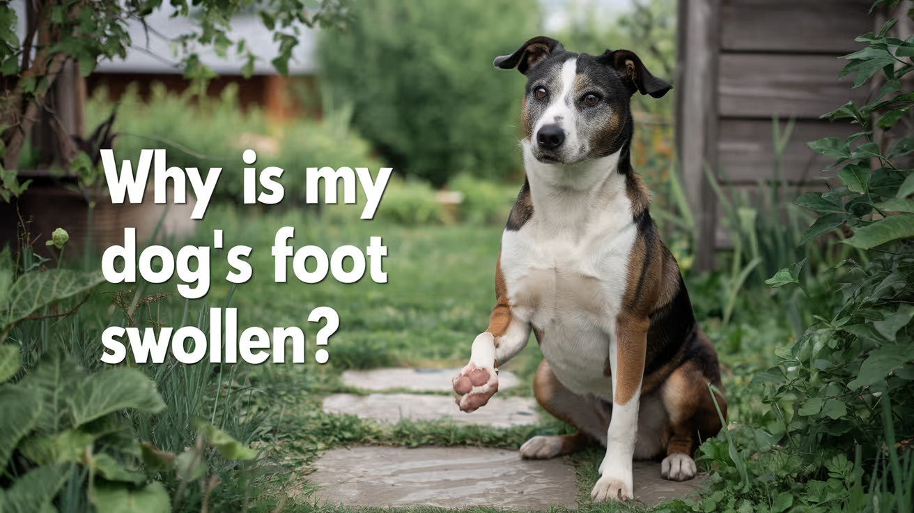
Why Is My Dog's Foot Swollen?
Discover why your dog's foot is swollen, common causes, treatments, and when to see a vet for proper care.
A swollen dog foot is one of the most common reasons owners call their vet. It can be as simple as a bee sting or as serious as a deep infection or bone tumor. The location, speed of onset, and other symptoms all help narrow down the cause.
This guide covers every common cause, how to assess severity at home, and the exact signs that mean your dog needs same-day care.
Quick answer: A swollen dog foot usually has a local cause: injury, insect sting, foreign object, or infection. Warm, increasing, or discharging swelling needs same-day vet care. Multiple swollen paws suggest allergies.
Key takeaways
- Single-paw swelling almost always has a local cause: injury, sting, foreign body, or infection
- Interdigital furunculosis (infected hair follicle cysts between toes) is among the most misidentified causes of recurring paw swelling
- Foreign objects like foxtails and glass splinters can migrate deep into tissue if left untreated
- Pododermatitis is the clinical term for inflamed paw skin; allergies are the most common underlying driver
- Swelling spreading up the leg or paired with face or neck swelling is an emergency requiring immediate care
- Most mild swelling from a sting or minor sprain improves within 24 to 48 hours with rest and monitoring
How to assess your dog's swollen foot before calling the vet
Before looking up causes, do a quick home assessment. This takes two minutes and tells you how urgent the situation is.
Step 1: Which paw?One paw = likely local cause. All four paws = likely allergic or systemic.
Step 2: Feel for heat.Warm or hot tissue means active inflammation or infection.
Step 3: Look between every toe.Redness, swelling between the digits, or visible nodules between the toes points to interdigital furunculosis or a foreign body.
Step 4: Check the pads.Burns appear as red, blistered, or peeling pad tissue. Cuts and punctures are often visible on the pad surface.
Step 5: Look at each nail.A broken nail or swollen nail bed causes localized pain and swelling at the toe tip.
VCA Animal Hospitals: "A deeper infection, like an abscess, will appear as a warm, soft to mildly firm swelling under the skin."
8 common causes of a swollen foot in dogs
1. Injury or trauma
Sprains, cuts, fractured toes, torn nails, and pad burns are the most frequent causes of sudden single-paw swelling. Dogs running on rough, hot, or icy terrain are especially prone.
PetMD notes that pad burns are particularly common in summer: check the pads after any walk on hot pavement. If your palm cannot hold against the pavement for five seconds, it is too hot for your dog's feet.
Signs: sudden lameness after activity, localized swelling, visible wound or bruising.
2. Foreign object
Thorns, glass, foxtails, and grass awns lodge between the toes or penetrate the pad. The body mounts an inflammatory response around the object, causing swelling that worsens over days.
SpectrumCare: "Foxtails and grass awns can lodge between the toes or in the pad and trigger pain, swelling, and infection."
Foxtails are particularly dangerous because they are barbed and can migrate deeper into tissue over days, eventually requiring surgical removal. If you cannot see and safely remove the object, do not probe. See a vet.
3. Insect sting or bite
Bees, wasps, fire ants, and spiders cause rapid single-paw swelling that appears within minutes of the sting. The paw may look puffy and your dog may lick or hold it up.
Dyer Animal Clinic advises watching for anaphylaxis signs: hives, difficulty breathing, excessive swelling spreading beyond the paw. These require emergency care immediately.
Localized sting swelling that stays in the paw and is not worsening can be monitored at home for 24 to 48 hours.
4. Infection: bacterial and fungal
Bacterial or fungal infections enter through cuts, puncture wounds, or damaged skin. Infected paws are typically warm, red, swollen, and often have an odor or discharge. VCA lists the visual signs: "Skin infections on the feet may result in red, moist lesions between the toes."
Pododermatitis (inflammation of the paw skin) is the umbrella clinical term. Common causes include:
- Secondary bacterial infection from chronic licking
- Yeast overgrowth in skin folds between toes
- Fungal infections (ringworm can affect paws)
- Demodectic mange (mite overgrowth)
Pododermatitis does not resolve without treatment. Antibiotics, antifungal medication, or both are typically required.
5. Interdigital furunculosis
This is one of the most commonly missed causes of recurring paw swelling. Interdigital furunculosis occurs when hair follicles between the toes become infected, forming painful reddish-purple nodules that may rupture and drain.
AKC notes that the condition is especially prevalent in short-coated, heavyset breeds: Bulldogs, Labrador Retrievers, and Chinese Shar-Pei are among the most commonly affected. Chronic licking due to allergies is a major driver.
Signs: swollen, painful nodules between the toes, draining tracts, recurrent swelling in the same location.
For how furuncles between the toes connect to abscess formation, see abscess as a cause of limb swelling.
6. Allergic reaction and pododermatitis
Environmental allergens (pollen, grass, lawn chemicals, road salt) and food allergies frequently cause paw inflammation. Dogs with allergies lick their feet chronically, creating secondary infection on top of the allergic reaction.
Wakefield Pet Vet: "Allergic reactions typically cause itching, redness, and sometimes blistering between the toes or on paw pads."
Allergic paw swelling usually affects multiple paws. It tends to recur seasonally or after contact with the trigger substance. Dogs with seasonal allergies often have their worst paw symptoms in spring and fall.
7. Bursitis
Bursae are small fluid-filled sacs that cushion joints. Repeated pressure on bony prominences, especially the elbow and hock, can cause bursitis. Affected joints may appear as soft, fluctuant swellings near a joint.
For how bursitis specifically produces foot and joint swelling, see bursitis as a common cause of foot swelling.
8. Cysts, tumors, and nail bed disease
Cysts, mast cell tumors, and subungual (under-nail) tumors can all present as localized swelling on or near the foot. Petcube notes that toenail tumors are more prevalent in large black-coated breeds such as Standard Poodles, Gordon Setters, and Schnauzers.
A lump that is growing, firm, or pigmented should always be evaluated by a vet rather than monitored at home.
For an overview of lumps that cause swelling on the legs, see lumps that can cause swelling.
Severity triage: what to do right now
Home care for mild cases
For minor swelling without infection signs:
- Keep the dog calm and limit walking
- Soak the paw in warm (not hot) water with Epsom salts for 10 minutes. PetMD recommends this as an excellent short-term measure regardless of cause.
- Gently clean any visible wound with mild soap and warm water
- Apply a cool damp cloth for 10 to 15 minutes to reduce swelling from sprains or stings
- Do not apply antibiotic ointments without vet guidance some formulations are toxic to dogs if licked
For pressure-related paw and leg swelling in dogs that rest in one position for extended periods, see pressure-related swelling in dogs.
Frequently asked questions
How do I treat my dog's swollen paw at home?
For mild swelling with no wound or discharge, rest the dog and soak the paw in warm Epsom salt water for 10 minutes. Check carefully for a foreign object or insect stinger. Do not use human antibiotic creams without vet guidance. If swelling has not reduced within 24 to 48 hours or is getting worse, contact your vet.
When should I be worried about my dog's swollen paw?
Be concerned immediately if the swelling is warm, increasing, or has discharge or odor. Call the vet the same day if your dog refuses to bear weight, if the swelling is spreading up the leg, or if there is any sign of an allergic reaction such as facial swelling or breathing difficulty.
What can I give my dog for a swollen paw?
Do not give human pain medications (ibuprofen, acetaminophen, aspirin) to dogs; many are toxic. For minor swelling, warm Epsom salt soaks are safe and effective for short-term relief. Your vet can prescribe appropriate anti-inflammatory medication if needed after examining the paw.
My dog's foot was normal this morning and is swollen now. What happened?
Sudden single-paw swelling most often means an insect sting, a foreign object picked up during activity, or a minor sprain. Check each toe carefully for a stinger, thorn, or visible wound. If swelling is mild and localized, monitor for 24 hours. If increasing or painful, call the vet the same day.
Can swelling in a dog's foot go away on its own?
Minor swelling from a bee sting or small sprain typically resolves within 24 to 48 hours with rest. Infections, foreign objects, interdigital furunculosis, and bursitis do not resolve without treatment. If swelling has not improved within 48 hours, veterinary assessment is needed.
My dog's foot smells bad and is swollen. Is that serious?
Yes. Odor from a swollen paw strongly indicates infection. A foul smell means bacteria are actively present. Common sources include interdigital furunculosis, a nail bed infection, or an abscess. This requires prompt veterinary treatment and should not be left to resolve on its own.
Resources
- PetMD. Dogs Swollen Paws: Causes and Treatments. petmd.com
- VCA Animal Hospitals. First Aid for Limping Dogs. vcahospitals.com
- AKC. Pododermatitis on Dog Paw: Causes, Symptoms and Treatment. akc.org
- SpectrumCare. Paw Swelling in Dogs. spectrumcare.pet
- Dyer Animal Clinic. Reasons Your Dog's Paw is Swollen. dyeranimalclinic.com
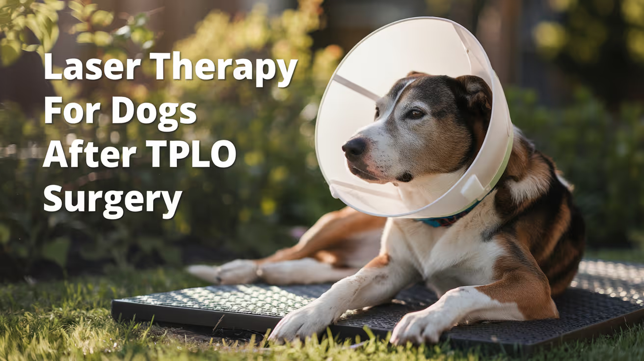
Laser Therapy for Dogs After TPLO Surgery
Learn how laser therapy helps dogs recover faster and with less pain after TPLO surgery for cruciate ligament repair.
Laser therapy also called photobiomodulation (PBMT) or low-level laser therapy (LLLT) is one of the most commonly offered adjunct treatments during TPLO recovery. Many specialist and rehabilitation centres include it routinely.
But the evidence for its benefits is more nuanced than the marketing suggests, and owners deserve an honest picture of what it does and does not reliably achieve.
Quick answer: Laser therapy after TPLO uses specific light wavelengths to reduce inflammation and support tissue healing. Evidence for early pain reduction is moderately supported; evidence for improved radiographic bone healing is weak. It is a safe adjunct but should not replace rehabilitation exercises, pain medication, or activity restriction.
Key takeaways
- Laser therapy reduces postoperative inflammation and may improve gait scores: a TPLO study found better hindlimb function at 8 weeks in treated dogs
- Evidence for improving radiographic bone healing is weak: three controlled studies found no statistically significant difference in healing time
- The 2024 AVMA randomized trial found no significant difference in CRP, weight bearing, pain scores, or SSI rates between PBMT and sham groups
- Sessions typically begin within the first few days of surgery and continue through the rehabilitation phase
- Laser therapy is safe with few contraindications: avoid eyes and active tumour sites; safe over the TPLO incision once closed
- It works best as part of a multimodal plan: exercise therapy and pain medication carry stronger evidence than laser alone
What laser therapy does
Laser therapy for dogs, also known as photobiomodulation, involves using specific wavelengths of light to penetrate tissues and promote cellular regeneration and healing. The laser light stimulates the production of ATP (adenosine triphosphate), enhancing cell repair and growth, reducing inflammation, and increasing blood circulation.
Photobiomodulation therapy has been shown to decrease inflammation, and increase analgesia, vascularization, and tissue healing after musculoskeletal injury or surgery.
The mechanism is photochemical: light energy at specific wavelengths (typically 630 to 980 nm) is absorbed by mitochondria.
This increases ATP production, modulates reactive oxygen species, and influences gene expression related to inflammation and healing.
The effects are local confined to the tissue depth the light reaches.
For post-TPLO use, the targets are: the surgical incision, the osteotomy site in the proximal tibia, and the surrounding periarticular soft tissues.
What the clinical evidence shows
Pain and function
Research following TPLO surgery showed that dogs receiving LLLT had better hindlimb function and gait scores at 8 weeks compared to controls. This is especially valuable in orthopedic recovery, where early weight-bearing can prevent muscle atrophy and joint stiffness.
In a controlled veterinary study, dogs with surgical incisions treated with laser therapy exhibited significantly less inflammatory cell infiltration and tissue necrosis within the first week post-op compared to untreated controls.
The 2024 randomized trial
54 client-owned dogs with CCL rupture undergoing unilateral TPLO surgery were enrolled. The study population was randomly assigned to either a treatment group receiving PBMT (24 dogs) or a control group (30 dogs). PBMT was performed immediately after induction, and at 6 hours, 24 hours, 48 hours, and 8 weeks postoperatively. Evaluation of CRP, pain scores, evidence of SSI, and percentage weight bearing were assessed at all time points.
The trial found the therapy showed promise but no statistically significant difference between groups on any primary outcome measure.
Bone healing
Three studies compared LLLT to a control and concluded that LLLT treatment did not make a significant difference in improving radiographic bone healing. The studies collectively provide weak evidence for this outcome.
This is an important distinction: laser therapy may support soft tissue healing, pain, and early function but it does not appear to accelerate the osteotomy healing visible on radiographs.
When to start and how often
Laser therapy uses focused light energy on the surgical site to support healing.
Most rehabilitation programmes begin laser therapy within the first 1 to 3 days after TPLO surgery, often at the surgical centre before discharge or at the first rehabilitation visit.
Typical post-TPLO laser protocol:
- Frequency: 3 to 5 sessions per week in the first 2 to 3 weeks
- Frequency: 1 to 2 sessions per week from weeks 3 to 8
- Session duration: 5 to 15 minutes depending on the laser system and dosing protocol
- Total sessions: typically 6 to 12 in the first 8-week recovery phase
The protocol varies by laser system, power output, and the individual patient's response.
Realistic expectations
Laser therapy is a useful adjunct in TPLO recovery. It is not a substitute for the treatments with stronger evidence: pain medication, activity restriction, and structured rehabilitation exercises.
Laser therapy could be particularly helpful for dogs with weight-bearing and gait issues while recovering from TPLO surgery after a cruciate injury.
Dogs with significant early swelling, wound sensitivity, or slow initial weight-bearing progress may benefit most. Dogs recovering well with standard multimodal analgesia and rehabilitation may show less measurable difference.
For the bone healing timeline that laser therapy supports during recovery, see TPLO bone healing time in dogs explained.
For the full recovery plan that laser therapy fits into, see 10 essential TPLO recovery tips for pet owners.
For the physical therapy that is the primary evidence-based adjunct, see when to start physical therapy after TPLO surgery.
For swelling management in the recovery period, see how long does swelling last after TPLO surgery.
Frequently asked questions
Is laser therapy safe over the TPLO incision?
Yes, once the incision is closed. Laser therapy is safe over sutured incisions and can be applied at the surgical site from the first post-operative day in most protocols.
Avoid direct application over open wounds or actively infected tissue.
How many laser sessions does a dog need after TPLO?
Typically 6 to 12 sessions across the first 8 weeks.
Start at 3 to 5 sessions per week for the first 2 to 3 weeks, tapering to 1 to 2 per week through the rehabilitation phase.
The exact protocol depends on the laser system and rehabilitation plan.
Does laser therapy replace pain medication after TPLO?
No. Laser therapy is an adjunct to pharmaceutical pain management, not a replacement. Post-TPLO pain management requires NSAIDs, and often gabapentin or other analgesics.
Laser therapy may reduce the pain burden and support earlier mobility but does not provide sufficient analgesia on its own.
Can I do laser therapy at home with a consumer device?
Consumer-grade red light therapy devices exist but operate at lower power densities than veterinary therapeutic lasers. The clinical evidence discussed in this article relates to veterinary-grade PBMT devices.
Home devices may offer some benefit but cannot replicate the dosing of professional equipment. Discuss with your rehabilitation veterinarian before purchasing.
Will laser therapy prevent my dog from needing more medication?
Possibly. If laser therapy reduces post-operative inflammation and pain, some dogs may need lower doses of pain medication or taper off sooner.
This should be guided by your veterinarian based on your dog's individual recovery trajectory.
Resources
- AVMA Journal. Photobiomodulation Therapy in Dogs Undergoing TPLO After Cranial Cruciate Ligament Rupture. avmajournals.avma.org
- Veterinary Evidence. Does LLLT Improve Radiographic Healing for Dogs with CCL Rupture Undergoing TPLO Surgery? veterinaryevidence.org
- AKC. Laser Therapy For Dogs: Uses, Side Effects, and Alternatives. akc.org
- Erchonia. Laser Therapy for Post-Surgical Recovery in Pets. erchonia.com
All Articles
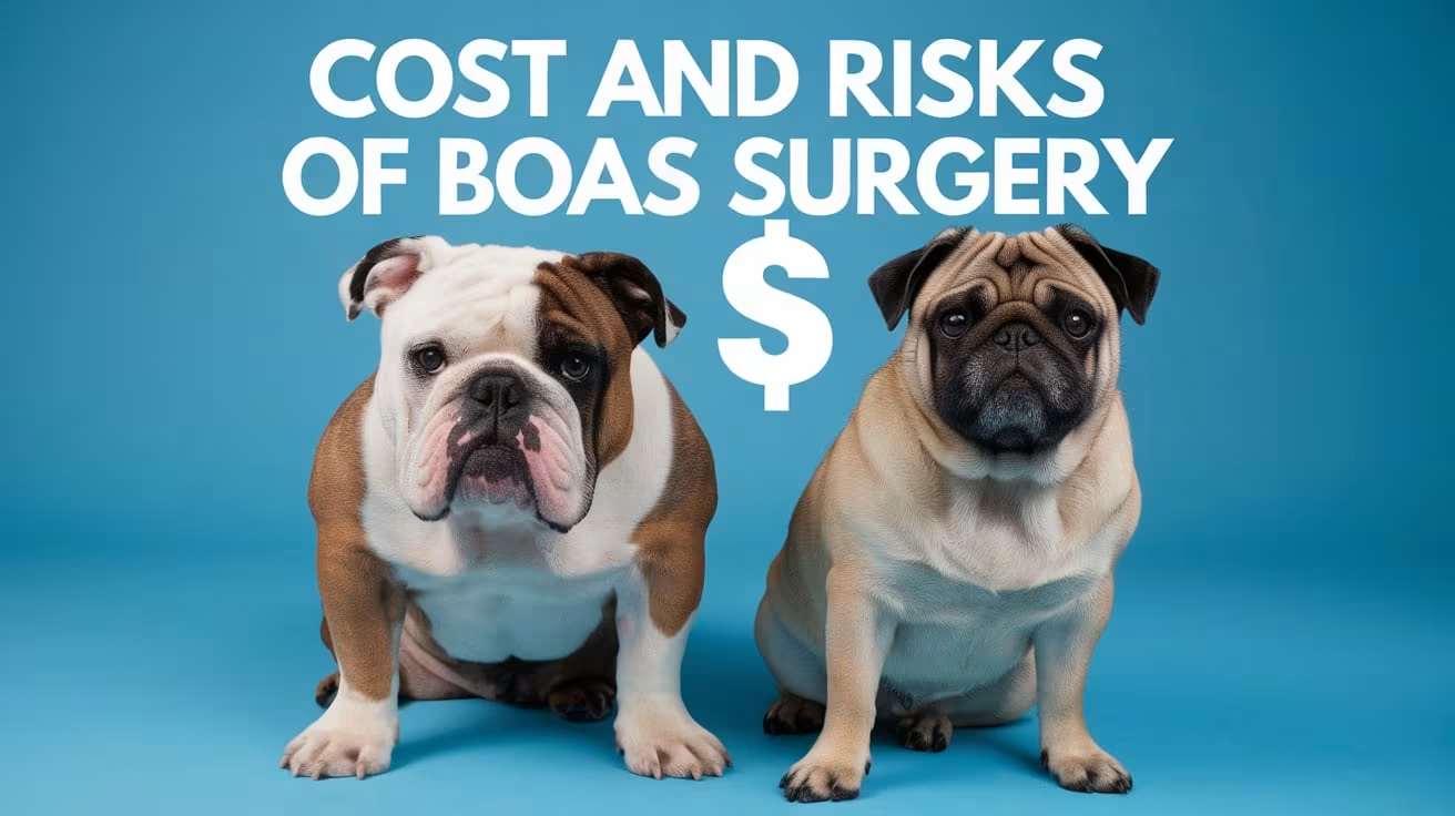
Cost and Risks of BOAS Surgery in Bulldogs and Pugs
Learn the cost and risks of BOAS surgery in Bulldogs and Pugs. Understand pricing, complications, and how early surgery can improve safety and outcomes
BOAS surgery is not a minor decision, financially or medically. For Bulldog and Pug owners especially, the cost can run into thousands of dollars, and the anesthetic risks unique to these breeds require careful planning.
Understanding what drives the cost and what the real risk profile looks like helps you approach the decision with clear expectations.
Quick answer: Full corrective BOAS surgery for Bulldogs and Pugs typically costs between $2,000 and $5,000 in the US, depending on the number of corrections needed, the surgeon's experience, and geographic location. Dogs needing only nostril correction may pay $500 to $2,000. Anesthetic risk is elevated in brachycephalic breeds and must be managed by an experienced team. Most pet insurance policies cover BOAS surgery if the condition was not noted before enrollment.
Key takeaways
- Full BOAS surgery typically costs $2,000 to $5,000: The range reflects procedure complexity, surgeon experience, and location.
- Nostril correction alone starts around $500 to $2,000: The less complex the case, the lower the cost.
- Bulldogs typically cost more than Pugs: Greater body mass and more complex anatomy require more anesthesia and surgical time.
- Anesthetic risk is real but manageable: Brachycephalic breeds are higher risk under anesthesia; experienced teams plan for this.
- Pet insurance can help significantly: Early enrollment is essential because BOAS is often classified as a hereditary condition.
- Delaying surgery increases both cost and risk: Secondary complications require more complex procedures.
Why Bulldogs and Pugs are among the most costly BOAS patients
Not all brachycephalic breeds have the same surgical complexity. Bulldogs and Pugs are consistently among the most expensive BOAS patients because of their anatomy.
English Bulldogs frequently have multiple concurrent defects: severely stenotic nares, a dramatically elongated soft palate, and often a hypoplastic trachea. They are also heavier dogs, requiring more anesthesia and more intensive monitoring. Post-operative swelling risk is high, and overnight hospitalization is more commonly recommended.
Pugs typically have severely stenotic nares and an elongated soft palate. They are smaller than Bulldogs, which reduces some costs, but their airway anatomy is still significantly compressed. Pugs also have a high rate of aberrant nasal turbinates, which may require additional turbinate reduction procedures in specialist settings.
What BOAS surgery costs in the US
Cost varies based on several factors. The ranges below reflect current US pricing across different settings.
Procedure-by-procedure cost breakdown
Total cost ranges by case complexity
One French Bulldog owner in California reported a total BOAS surgery bill over $4,800, of which MetLife Pet covered nearly $4,400, illustrating both the real cost ceiling and the value of pet insurance for these breeds.
What drives cost up or down
Number of procedures required
A dog needing only nostril widening costs considerably less than one needing nostril correction, palate shortening, saccule removal, and possibly turbinate reduction in the same surgical event.
The more defects present, the longer the surgery, the more anesthetic time, and the more intensive the post-operative monitoring required.
Surgeon and facility type
General practitioners experienced in BOAS surgery charge less than board-certified veterinary surgeons at specialty centers. The tradeoff is expertise and equipment.
For straightforward cases in young, otherwise healthy dogs, a skilled general practitioner may be entirely appropriate. For older dogs, dogs with suspected laryngeal collapse, or dogs with concurrent health issues, specialist care is worth the additional cost.
Geographic location
Veterinary costs vary significantly by location. Urban specialty hospitals in major cities carry higher overhead and charge accordingly. Rural and suburban general practices are typically less expensive.
Hospitalization and monitoring
Post-operative monitoring is essential for brachycephalic dogs because post-surgical airway swelling peaks in the first 24 to 48 hours. Most surgeons recommend at minimum several hours of post-operative monitoring, and many recommend overnight stays.
Overnight hospitalization adds $200 to $600 or more to the total bill depending on the facility.
Additional diagnostics
Pre-surgical imaging (radiographs, CT scan) to assess tracheal size and detect concurrent conditions adds $200 to $800 to the pre-surgical cost. This is often recommended for Bulldogs specifically, given the prevalence of hypoplastic trachea in this breed.
The real surgical risks
BOAS surgery carries specific risks that are higher in brachycephalic breeds than in other dogs. Understanding these helps you ask the right questions before proceeding.
Anesthetic complications
Brachycephalic dogs are at elevated risk under anesthesia for several reasons:
- The compromised airway makes intubation more difficult
- Maintaining an open airway during induction before the endotracheal tube is placed is more challenging
- Recovery from anesthesia carries a higher risk of airway obstruction as the dog wakes and the muscles relax
- Some dogs cannot be safely extubated without emergency intervention
An experienced anesthetic team will:
- Preoxygenate the dog before induction
- Have the endotracheal tube ready for immediate placement
- Keep a tracheostomy kit available as a contingency
- Monitor the dog closely through extubation and into recovery
This is why choosing a surgical team experienced with brachycephalic breeds matters significantly.
Post-operative airway swelling
Tissue swelling after soft palate resection is the most common post-operative complication.
The operated area is a confined space in the airway. Swelling that would be minor elsewhere can be significant here, potentially narrowing the airway further in the critical hours after surgery.
Signs of concerning post-operative swelling include: increased breathing noise after initial improvement, visible distress, and labored breathing. These require immediate veterinary attention.
Most surgeons keep brachycephalic surgical patients in hospital for monitoring precisely because of this risk.
Bleeding
Soft tissue surgery in the throat carries bleeding risk. The soft palate has a good blood supply. Laser palatoplasty reduces but does not eliminate this risk compared to cold scalpel techniques.
Significant post-operative bleeding is uncommon but requires immediate intervention.
Aspiration
BOAS dogs frequently have concurrent gastrointestinal dysfunction. Post-operatively, the risk of aspiration of stomach contents is elevated. Strict fasting protocols before surgery and careful recovery positioning reduce this risk.
Incomplete resolution
Not all dogs achieve complete resolution of breathing difficulty after surgery. Dogs with hypoplastic tracheas retain the tracheal limitation regardless of what is corrected surgically. Dogs with advanced laryngeal collapse may show improvement but often not full resolution.
Setting realistic expectations before surgery is part of the pre-surgical consultation.
The cost of not having surgery
Untreated BOAS has its own financial consequences that owners sometimes underweight when evaluating the surgery decision.
Progressive secondary complications:
Without surgical correction, everted laryngeal saccules worsen, and laryngeal collapse eventually develops. Laryngeal collapse is far more difficult to treat and carries a much higher complication rate when surgical intervention is attempted at that stage.
Emergency presentations:
Dogs with untreated moderate to severe BOAS are significantly more likely to present as respiratory emergencies, particularly in warm weather. Emergency veterinary care for acute respiratory distress is expensive and carries higher procedural risk than planned surgery.
Reduced quality of life and lifespan:
Chronic oxygen restriction, poor sleep, and persistent gastrointestinal distress from BOAS affect a dog's overall health trajectory. This is difficult to quantify financially but is a real consideration in the decision.
Pet insurance and BOAS surgery
Most comprehensive pet insurance policies that cover illnesses will cover BOAS surgery, provided the condition was not noted before the policy was in force.
Critical points for Bulldog and Pug owners:
- Enroll your dog in pet insurance as early as possible, ideally before any BOAS symptoms are documented
- Many insurers classify BOAS as a hereditary or congenital condition, which may affect coverage depending on the specific policy language
- Accident-only policies will not cover BOAS, as it is classified as a medical condition
- Review your policy's waiting periods, as some have extended waiting periods for hereditary conditions
Some insurers, including Spot Pet Insurance, market coverage that includes prescription diets and follow-up visits alongside the surgical claim. A real-world example: a French Bulldog owner received nearly $4,400 in reimbursement from MetLife Pet on a total bill of over $4,800.
Contacting your insurer before booking surgery to confirm coverage is always advisable.
Financing options
For owners without pet insurance who face a significant surgery bill, several options exist:
- CareCredit: A healthcare credit card commonly accepted at veterinary practices, offering deferred interest plans
- Scratchpay: A veterinary-specific financing platform
- Payment plans: Some veterinary practices offer in-house payment plans, particularly for regular clients
- Breed-specific rescue organizations: Some brachycephalic breed rescues maintain funds for medical assistance
Understanding when surgery is actually necessary helps you prioritize surgical spending on the dogs who most need it rather than treating every brachycephalic dog prophylactically.
Questions to ask your veterinarian before BOAS surgery
- What specific procedures does my dog need and why?
- What is the itemized estimate including hospitalization and medications?
- What are the surgeon's qualifications and BOAS surgical volume?
- What is the post-operative monitoring protocol?
- What are the specific risks given my dog's age, weight, and concurrent health status?
- What are realistic expectations for improvement?
- Does my dog's trachea appear normal on imaging?
For a full understanding of what BOAS is and the anatomy being corrected, that background helps you understand what the surgeon describes during the consultation. For recovery expectations after surgery and what changes to make at home, planning ahead reduces surprises during the recovery period. For Pug owners specifically, the guide on pug-specific nose surgery costs addresses the breed-specific cost and recovery picture in more detail.
Frequently asked questions
Is BOAS surgery worth the cost for a Bulldog or Pug?
For dogs with moderate to severe BOAS, yes. The alternative is progressive airway obstruction, increasing risk of respiratory emergencies, and a significant reduction in quality of life. Owner-reported satisfaction after surgery is approximately 94%, and many describe the improvement as dramatic.
How much does soft palate surgery cost on its own?
Soft palate resection alone typically costs $800 to $2,500 depending on the surgeon and facility. It is most commonly combined with stenotic nares correction in the same anesthetic event.
Can I reduce the cost of BOAS surgery?
Cost can vary significantly between providers. Choosing a general practitioner experienced in BOAS surgery rather than a specialist center is less expensive, though appropriate for only less complex cases. Having pet insurance before symptoms are documented is the most effective cost-reduction strategy.
How long is the hospital stay after BOAS surgery?
Most brachycephalic surgical patients stay in hospital for several hours to overnight after surgery. The exact duration depends on how quickly your dog wakes from anesthesia, how the post-operative airway looks, and your surgeon's protocol. Bulldogs often require longer observation than smaller breeds.
Will my dog need repeat surgery?
For most dogs, a single surgical intervention addresses the correctable defects adequately. Dogs may experience some gradual return of soft palate elongation over years. Repeat surgery is uncommon but is occasionally needed, particularly in dogs that were very young at first surgery.
The cost of BOAS surgery for Bulldogs and Pugs is significant but well-defined. The risk is real but manageable with the right team. What is less well-defined, and what often surprises owners, is the cost of waiting: progressive secondary damage that makes surgery harder, more expensive, and less effective the longer it is delayed. Planning early, insuring early, and choosing a surgeon with genuine brachycephalic experience are the three most important financial and medical decisions in this process.
Resources
The following sources were used as reference and background for this article:
- ScienceInsights. What Is BOAS Surgery in Dogs? Risks, Cost and Recovery. scienceinsights.org
- Vety. How Much Does BOAS Surgery Cost? vety.com
- Dogster. How Much Does Stenotic Nares Surgery Cost? dogster.com
- MetLife Pet Insurance. Stenotic Nares Surgery Cost for Dogs. metlifepetinsurance.com
- Insurify. Does Pet Insurance Cover BOAS Surgery? insurify.com
- Spot Pet Insurance. Does Pet Insurance Cover BOAS Surgery? spotpet.com
- Today's Veterinary Practice. A Guide to Brachycephalic Obstructive Airway Syndrome Surgery. todaysveterinarypractice.com
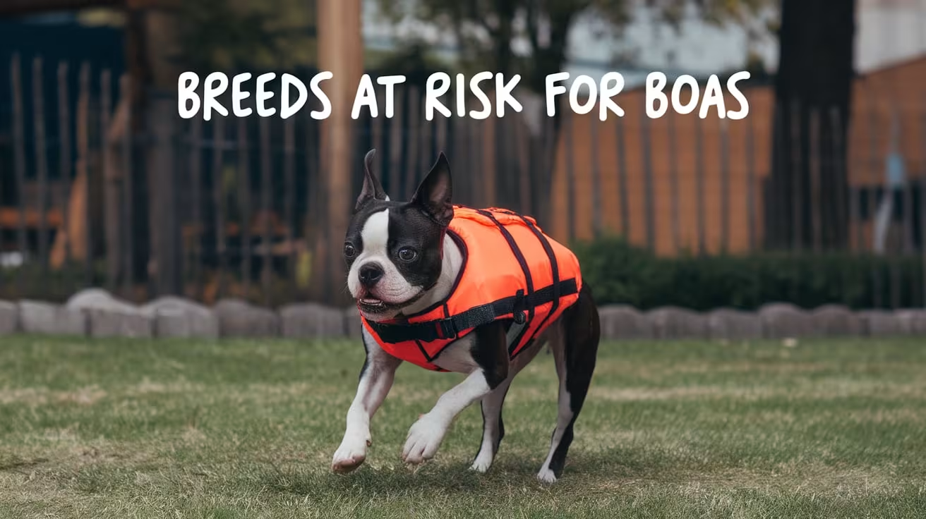
Breeds at Risk for BOAS and Early Warning Signs
Discover which dog breeds are at risk for BOAS and the early warning signs to watch for, from noisy breathing to subtle changes in sleep and activity
Flat-faced dogs are among the most popular breeds in the US. They are also among the most medically vulnerable.
Not every brachycephalic dog develops clinically significant BOAS. But some breeds are at dramatically higher risk than others, and within those breeds, individual variation means some dogs are much more severely affected than others.
Knowing where your dog sits in this picture is the starting point for responsible care.
Quick answer: The highest-risk breeds for BOAS are English Bulldogs, French Bulldogs, Pugs, Boston Terriers, and Pekingese. Research suggests up to 50% of dogs in the three most severely affected breeds have clinically significant breathing obstruction. Early warning signs include noisy breathing at rest, exercise intolerance, overheating, and disrupted sleep. These signs are often normalized but should be evaluated by a veterinarian.
Key takeaways
- Some breeds have BOAS prevalence rates of 50% or higher: This is not a rare condition in the most severely affected breeds.
- 60% of owners do not recognize BOAS signs as medical symptoms: Normalization delays diagnosis and worsens outcomes.
- Early signs can be subtle: Reluctance to exercise, restless sleep, and occasional gagging are often the first indicators.
- Not all brachycephalic breeds carry the same risk: Risk varies significantly by skull shape and muzzle length.
- BOAS is progressive: Signs that seem manageable at age one may become serious by age three or four without intervention.
- Puppies should be evaluated early: Veterinary assessment between 6 and 12 months allows intervention before secondary damage develops.
Why breed matters in BOAS
BOAS results from the shortened skull structure of brachycephalic breeds. The degree of risk is closely tied to how extreme the brachycephaly is.
Dogs with very flat faces, extremely short muzzles, and wide skulls have the most compressed nasal passages, the least room for the soft palate, and the highest risk of significant airway obstruction. Dogs with a slightly longer muzzle, even within a traditionally flat-faced breed, tend to have less severe BOAS.
This is why individual dogs within the same breed vary. Two French Bulldogs from different breeding lines can have very different airway anatomy.
Highest-risk breeds
These breeds consistently show the highest rates of clinically significant BOAS in research studies.
English Bulldog
The English Bulldog is widely considered one of the most severely affected breeds.
Bulldogs frequently have stenotic nares, elongated soft palates, and a hypoplastic (narrowed) trachea, which cannot be surgically corrected. The combination of multiple defects makes BOAS management in Bulldogs particularly complex.
Research published in the journal PLOS ONE found that approximately 50% of English Bulldogs, French Bulldogs, and Pugs assessed in studies showed clinically significant BOAS by objective respiratory function testing.
French Bulldog
The French Bulldog has become the most popular dog breed in the US for multiple consecutive years. That popularity has made BOAS in French Bulldogs an increasingly common presentation in veterinary clinics.
French Bulldogs typically have severely stenotic nares and elongated soft palates. They are also prone to aberrant nasal turbinates that further restrict airflow. Exercise intolerance and sleep disruption are particularly common in this breed.
Pug
Pugs are among the most recognizably flat-faced breeds and among the highest-risk for BOAS.
UK research found that around 20% of pugs see a veterinarian specifically for respiratory disease in any two-year period. When objective testing is used, the true proportion with some degree of airway restriction is considerably higher.
Pugs are also common candidates for stenotic nares correction, and pug-specific airway surgery costs and recovery differ somewhat from larger breeds. For the pug-specific picture, the guide on pug-specific surgery cost and recovery covers the detail relevant to pug owners.
Boston Terrier
Boston Terriers occupy a middle position in BOAS severity. Their muzzle, while short, is typically not as extreme as Bulldogs or Pugs, and some individuals have relatively functional airways.
However, within the breed, more extreme conformation is associated with higher BOAS risk. Boston Terriers with more pronounced facial foreshortening and nostril stenosis are more likely to have significant airway obstruction.
Pekingese
The Pekingese is one of the most severely affected breeds in terms of BOAS prevalence. A cross-breed study found that among 14 brachycephalic breeds assessed in the UK, only 10.9% of Pekingese scored Grade 0 (no significant breathing abnormality). This means nearly 90% showed some degree of respiratory dysfunction.
Other affected breeds
A significant number of other breeds carry elevated BOAS risk:
Breeds with longer muzzles, such as the Pomeranian and Maltese, show considerably lower rates of significant BOAS despite being classified as brachycephalic.
Early warning signs of BOAS
The signs of BOAS can be divided into those that are obvious and those that are easy to overlook. Both matter.
Obvious signs
Noisy breathing at rest:
Stertor is a low, snoring sound produced in the throat. Stridor is a higher-pitched wheeze that originates from the larynx or trachea. Both indicate airway obstruction. A dog breathing normally at rest should breathe quietly.
Exercise intolerance:
A brachycephalic dog that stops, refuses to continue, or collapses after only a short walk is showing a significant sign. This is not laziness. It is the inability to take in enough oxygen to sustain exertion.
Open-mouth breathing at rest:
Dogs breathe through the nose at rest unless they are hot or just exercised. A dog that routinely breathes through the mouth at rest is unable to move adequate air through the nasal passages.
Coughing or gagging:
Particularly after eating or drinking, or after exertion. Often caused by the elongated soft palate interfering with swallowing.
Subtle signs that are often missed
Reluctance to exercise:
A dog that consistently chooses to stop playing, sit down on walks, or avoid stairs may be doing so because exertion becomes quickly uncomfortable.
Restless or disturbed sleep:
Dogs with BOAS often cannot enter deep sleep because their airway partially collapses when they relax. They wake frequently, reposition, or sleep with their head propped against furniture or the wall to keep the airway more open.
Frequent swallowing and lip-licking:
Often associated with regurgitation and esophageal dysfunction that accompanies BOAS.
Sleeping with the head elevated:
A dog that consistently props its head on the edge of the bed, couch, or a pillow may be doing so to keep the airway aligned and reduce obstruction.
Gastrointestinal symptoms:
Vomiting, regurgitation, and reflux occur in a high proportion of BOAS dogs due to the interaction between respiratory effort and gastrointestinal function.
Signs most commonly normalized by owners
Research has consistently found that approximately 60% of owners of brachycephalic dogs do not recognize signs of BOAS as medical problems.
The following are the most frequently normalized signs:
- Loud snoring: Often described as "cute" or "just how they sleep"
- Snorting or snuffling when excited: Attributed to personality rather than anatomy
- Rapid overheating: Seen as a breed trait rather than a ventilation problem
- Refusal to walk far: Interpreted as laziness rather than oxygen limitation
A dog that cannot breathe freely is not a healthy dog. The flat-faced appearance that owners find endearing comes at a real biological cost, and recognizing that cost is an act of responsible ownership.
When to see a veterinarian
Any brachycephalic dog should receive a veterinary airway assessment, regardless of how minor the owner thinks the symptoms are.
Schedule a veterinary assessment promptly if your dog shows:
- Noisy breathing at rest or during sleep
- Exercise intolerance disproportionate to their fitness level
- Frequent gagging, regurgitation, or vomiting
- Episodes of distress, panic, or labored breathing
- Blue or purple gums at any point (emergency: go immediately)
Routine evaluation is recommended:
- For all flat-faced breed puppies between 6 and 12 months of age
- For adult brachycephalic dogs that have not been assessed
For a clear understanding of what BOAS is and how it develops in the airway, that guide provides the anatomical foundation for understanding why these signs occur.
The importance of early diagnosis
BOAS is progressive. Early-stage stenotic nares and a mildly elongated soft palate become moderate obstruction over time. Moderate obstruction leads to everted laryngeal saccules. Everted laryngeal saccules contribute to laryngeal collapse, which is the most severe and least surgically correctable stage of BOAS.
Dogs treated surgically before age two have significantly better outcomes than those treated later.
Understanding when affected breeds need surgery and managing mild BOAS in at-risk breeds are the two most important next steps for owners of high-risk dogs whose signs are currently mild.
Frequently asked questions
My Frenchie snores but seems happy. Does that mean BOAS is not serious?
Happy behavior does not mean comfortable breathing. Dogs adapt to chronic difficulty and do not communicate discomfort the way humans do. A dog that snores at rest has measurable airway obstruction. Whether intervention is warranted depends on the degree of obstruction, which a veterinary assessment can determine.
Are some individual dogs within high-risk breeds unaffected?
Yes. Not every Bulldog or Pug develops clinically significant BOAS. Dogs with slightly less extreme conformation within their breed may have functional airways throughout their lives. Objective respiratory function testing is the most accurate way to assess individual dogs.
Can BOAS develop later in life, or is it present from birth?
The anatomical abnormalities are present from birth, but the clinical signs often become more apparent as the dog ages. Secondary changes, such as everted laryngeal saccules, develop over time as a result of the chronic breathing strain. This is why a dog that seemed manageable at age one may be more significantly affected by age three or four.
Is there a test to find out how severe my dog's BOAS is?
Yes. Veterinarians use a grading system based on clinical examination, sometimes with mild sedation to assess the soft palate and laryngeal saccules. Some specialist centers use objective respiratory function testing. Most general practitioners can provide an initial assessment and refer to a specialist if surgery is being considered.
Should I avoid buying a flat-faced breed because of BOAS?
This is a personal decision, but it deserves serious consideration. The BOAS welfare issue is significant and well-documented. If you do own or plan to own a brachycephalic dog, committing to early veterinary assessment, appropriate lifestyle management, and surgical intervention if needed is the responsible path.
The popularity of flat-faced breeds has grown dramatically over the past decade. The medical burden that comes with that popularity is real, significant, and mostly preventable through early diagnosis and appropriate care. Knowing which breeds are at risk and what the early signs look like is the most important tool an owner has.
Resources
The following sources were used as reference and background for this article:
- Liu, N.C. et al. (2017). Conformational risk factors of brachycephalic obstructive airway syndrome. PLOS ONE. ncbi.nlm.nih.gov
- PetMD. Brachycephalic Airway Syndrome in Dogs. petmd.com
- Cornell University College of Veterinary Medicine. Brachycephalic Obstructive Airway Syndrome (BOAS). vet.cornell.edu
- Joii Pet Care. Brachycephalic Obstructive Airway Syndrome (BOAS) in Dogs. joiipetcare.com
- Cross-breed BOAS study. A cross-sectional study into the prevalence and conformational risk factors of BOAS across fourteen brachycephalic dog breeds. ncbi.nlm.nih.gov
- Today's Veterinary Practice. An Overview of Brachycephalic Obstructive Airway Syndrome. todaysveterinarypractice.com
Recovery and Lifestyle Changes After BOAS Surgery
Learn how to care for your dog after BOAS surgery. Recovery tips, diet, exercise, and lifestyle changes to improve comfort and long-term health
BOAS surgery corrects the structural defects causing airway obstruction. What happens in the weeks after surgery determines how well those corrections hold.
Recovery from BOAS surgery requires more than rest. It involves specific feeding protocols, activity restrictions, monitoring for complications, and a set of permanent lifestyle changes that protect the improved airway for the rest of your dog's life.
Quick answer: Most dogs recover from BOAS surgery within two to three weeks, with full tissue healing taking up to six weeks. The first 48 hours are the most critical, with post-operative swelling the primary risk. At home, your dog will need soft food for two weeks, strict activity restriction, harness use, and regular rechecks. Long-term lifestyle changes including weight management, heat avoidance, and controlled exercise are permanent requirements for all brachycephalic dogs.
Key takeaways
- The first 48 hours are the highest-risk period: Post-operative airway swelling peaks in this window and must be monitored closely.
- Soft food is required for the first two weeks: Hard food, bones, and chews can disrupt healing oral tissue.
- No running or jumping for two weeks: Activity restriction protects the surgical sites and reduces swelling.
- Recheck appointments are not optional: A 3 to 5 day recheck and a 10 to 14 day recheck are standard.
- Surgery improves but does not cure BOAS: Lifelong lifestyle management remains essential.
- Most dogs show dramatic improvement: Approximately 95 to 98% of owners report significant improvement in breathing and quality of life.
The first 48 hours: what to expect
The period immediately after surgery is the most medically significant part of recovery.
Your dog will come home groggy, quiet, and possibly with mild nasal bleeding from the nostril correction. This is expected and typically resolves within the first day.
Normal signs in the first 48 hours:
- Mild bloody discharge from the nostrils for the first 24 hours
- Noisy or labored-sounding breathing due to post-operative tissue swelling
- Excessive swallowing or retching in the first five to seven days as the throat heals
- Grogginess and low energy from anesthesia
- Reduced appetite
Paradoxically, breathing may sound worse immediately after surgery than before. This is because tissue swelling in the operated area temporarily reduces airflow before the healing process begins to improve it. This is expected and generally resolves within a few days as swelling subsides.
Signs requiring immediate veterinary contact:
- Breathing that is severely labored or distressed, not just noisy
- Gum color change to blue, purple, or white
- Significant active bleeding from the nose or mouth
- Complete collapse or loss of consciousness
- Inability to swallow or extreme distress when attempting to eat or drink
If you are unsure whether what you are seeing is within the range of normal, call your veterinarian. It is always better to call and be reassured than to wait on a genuine complication.
Recovery timeline
Feeding during recovery
Feeding protocol during recovery is one of the most specific and important aspects of aftercare.
Days 1 to 4: soft food only
Feed soft food rolled into small meatballs or very soft wet food. Supervise every meal and watch the 20 to 30 minutes after eating closely in the first week.
Feed four to five small meals per day rather than one or two large ones. This reduces the volume in the stomach at any time and lowers the risk of regurgitation, which is particularly dangerous in a dog with a freshly operated throat.
Days 5 to 14: transition to soaked kibble
If your dog normally eats dry kibble, transition by soaking it thoroughly until it is soft throughout before serving.
No hard food, hard treats, chews, or bones for the full two weeks post-surgery. No raw food for two weeks while the oral incisions heal.
Beyond two weeks:
With veterinary clearance, a gradual return to normal diet is usually appropriate. Continue feeding multiple smaller meals rather than one large meal permanently, as this reduces GI strain in brachycephalic dogs.
Activity restrictions
Strict activity restriction for the first two weeks is essential.
The surgical sites in the nose and throat are healing tissue. Exertion causes increased breathing effort, which in turn stresses the healing areas. Excessive activity also raises body temperature, which drives additional respiratory demand.
Two-week activity protocol:
- No running, jumping, or rough play
- No stairs if avoidable
- Short, leash-only bathroom trips on a Y-front harness
- Keep the dog calm during greetings and interactions
If your dog is the type that becomes frantic with excitement, consider limiting visitor activity in the home for the first two weeks. A calm environment is a medical requirement during this period, not just a preference.
After two weeks, with veterinary clearance:
- Begin short, gentle leash walks
- Increase duration gradually over the following weeks
- Watch for any signs of respiratory distress during exercise
- Full return to normal activity typically takes four to six weeks
Medications during recovery
Your vet will send your dog home with a medication protocol. Follow it precisely.
Common post-operative medications include:
- Pain relief: Anti-inflammatory pain medications for the first several days. Do not skip doses even if your dog seems comfortable.
- Antibiotics: If prescribed, complete the full course.
- Anti-nausea or reflux medication: Many BOAS dogs benefit from reflux management both during and beyond the immediate recovery window.
Do not give any over-the-counter human pain medications. Many, including ibuprofen and acetaminophen, are toxic to dogs.
The E-collar
Most dogs come home with an Elizabethan collar to prevent them from pawing at the nose or face.
Keep it on until the recheck appointment confirms the surgical sites are healing appropriately. Many dogs are initially distressed by the collar, but they adapt within a day or two. A soft recovery collar is often better tolerated than a rigid plastic one.
Recheck appointments
Two recheck appointments are standard after BOAS surgery.
First recheck: 3 to 5 days post-surgery
This appointment allows the surgeon to assess early healing, confirm there are no post-operative complications, and address any concerns you have about the recovery so far.
Second recheck: 10 to 14 days post-surgery
This is the primary assessment point for healing. The surgeon will evaluate the nasal and palatal tissue, confirm that healing is progressing normally, and typically give you guidance on when to begin gradual return to activity.
Do not skip these appointments even if your dog appears to be doing well. Some complications are not visible from the outside.
Long-term lifestyle changes after BOAS surgery
Surgery is the most effective intervention for correcting BOAS anatomy. But it does not change the underlying brachycephalic conformation. Your dog will always have a flat face, and the permanent lifestyle management appropriate for that anatomy continues for life.
Understanding what BOAS is and why lifestyle changes are needed even after surgery helps frame these permanent changes not as restrictions but as protection for the improved airway.
Weight management
Maintaining a lean body condition is the single most important long-term lifestyle factor.
Weight gain after surgery narrows the airway with fat tissue, compounding any structural obstruction that remains. Set a healthy body condition target with your vet and monitor it at every wellness visit.
Permanent harness use
Collars should never be used on brachycephalic dogs for leash walking. A Y-front harness is the permanent standard.
Heat and humidity avoidance
Even after surgery substantially improves airflow, brachycephalic dogs remain less efficient at thermoregulation than other breeds.
Walk in early morning or evening. Keep the dog in air conditioning during hot months. Never leave a brachycephalic dog in a warm car. These precautions do not expire after surgery.
Controlled exercise
Regular moderate exercise is important for maintaining healthy weight and cardiovascular fitness. High-intensity activity in warm conditions remains a risk even post-surgery.
Calibrate exercise to what your dog can comfortably manage. Watch for signs of distress during walks, particularly in warm weather.
Ongoing veterinary monitoring
Annual airway assessment remains appropriate for post-surgical BOAS dogs. Surgery corrects the defects addressed at the time of the procedure, but brachycephalic dogs can develop progressive changes over time, and some dogs require revision surgery years after the initial procedure.
What to expect from long-term outcomes
The long-term outlook after BOAS surgery is genuinely positive for most dogs.
Research shows 95 to 98% of dogs experience improvement in airway symptoms after surgery. Many owners describe the change as dramatic: quieter breathing, better sleep, more willingness to exercise, and less overheating.
Some residual breathing noise is common and normal. The goal of surgery is significant improvement in airflow and quality of life, not complete elimination of all breathing sound. A dog that breathes more quietly, exercises more freely, and sleeps without repeated waking is a success, even if some snoring remains.
Understanding the risks that may affect recovery and the surgical decision that preceded this recovery helps put the recovery window in its full medical context.
Frequently asked questions
How long does it take for a dog to recover from BOAS surgery?
Surface healing of the nasal and palatal tissue takes two to three weeks. Full internal tissue healing takes up to six weeks. Most dogs are noticeably more comfortable within a few days of surgery, but the activity and diet restrictions apply for the full two-week window regardless of how well the dog appears to be doing.
My dog sounds worse after surgery than before. Is that normal?
Yes, in the immediate post-operative period. Tissue swelling after the procedure temporarily reduces the airflow improvement. As swelling resolves over the first few days, breathing typically becomes progressively quieter. If breathing sounds severely distressed rather than just noisy, contact your veterinarian.
When can my dog eat normally again?
Soft food for the first four to five days, soaked kibble for days five to fourteen, then gradual return to normal diet with veterinary clearance. No hard food, hard treats, or bones for the full two weeks.
Will my dog still snore after BOAS surgery?
Some residual snoring is common and expected. Surgery improves airflow, but it does not change the fundamental brachycephalic anatomy. Most owners report that snoring is significantly reduced and less disruptive after surgery, even if it does not disappear entirely.
Does my dog need lifestyle changes permanently, even after surgery?
Yes. Weight management, harness use, heat avoidance, and controlled exercise are lifelong requirements for all brachycephalic dogs, regardless of surgical history. Surgery improves the airway within the structural constraints of the breed. The lifestyle management protects that improved airway.
Recovery from BOAS surgery is manageable with clear expectations and consistent follow-through. The first two weeks are the most demanding in terms of monitoring and restriction. What follows those two weeks is a dog that breathes more comfortably, sleeps more soundly, and handles the world more easily. The lifestyle changes that come after surgery are not burdens. They are how you protect what the surgery achieved.
Resources
The following sources were used as reference and background for this article:
- Centenary Vets. BOAS Surgery and Desexing Aftercare. centenaryvets.com.au
- My Pets Vets. Post Operative Care Following BOAS Surgery. mypetsvetsltd.co.uk
- Animal Clinic of Billings. Brachycephalic Obstructive Airway Care and Surgery. animalclinicofbillings.com
- Veterinary Healthcare Associates. BOAS Surgery: Everything You Need to Know. vhavets.com
- Berwick Vet. BOAS Surgery in Dogs: Procedure, Benefits and Recovery. berwickvet.com.au
- VCA Animal Hospitals. Brachycephalic Airway Syndrome in Dogs. vcahospitals.com
BOAS Surgery: When Should You Consider It for Your Dog?
Find out when BOAS surgery is needed for flat-faced dogs. Learn key signs, benefits, risks, recovery, and how it improves your dog’s quality of life
Surgery is not the first conversation most owners want to have about their flat-faced dog. But for dogs with moderate to severe BOAS, it is often the most important one.
The question is not whether BOAS surgery is scary. It is whether the alternative, leaving the airway obstruction untreated, is worse. In most moderate to severe cases, it clearly is.
This guide helps you understand when surgery becomes necessary, what it actually involves, and what you can expect from outcomes.
Quick answer: BOAS surgery is indicated for dogs with moderate to severe airway obstruction causing noticeable breathing difficulty, exercise intolerance, or disrupted sleep. It is also recommended as a preventive measure in younger dogs with significant anatomical defects, before secondary damage like everted laryngeal saccules develops. Dogs treated before age two have significantly better outcomes. Surgery is not appropriate for every brachycephalic dog, and mild cases are often managed with lifestyle changes first.
Key takeaways
- Surgery is the most effective treatment for moderate to severe BOAS: Lifestyle management helps mild cases but cannot correct structural defects.
- Earlier is better: Dogs treated surgically before age two consistently show better long-term outcomes than those treated later.
- Not every brachycephalic dog needs surgery: Severity assessment by a veterinarian is the first step.
- BOAS surgery typically addresses multiple defects in one procedure: Stenotic nares, elongated soft palate, and everted laryngeal saccules are corrected as needed.
- Owner-reported satisfaction is high: Studies show approximately 94% of owners report quality-of-life improvement after surgery.
- Anesthetic risk is a real consideration: Brachycephalic breeds carry higher anesthetic risk, which must be planned for carefully.
Who needs BOAS surgery?
Not every flat-faced dog requires surgery. The decision depends on the severity of airway obstruction and the degree to which it is affecting the dog's quality of life.
Surgery is typically recommended when a dog shows:
- Noisy breathing at rest (stertor or stridor) that is moderate to loud
- Exercise intolerance disproportionate to their fitness level
- Frequent episodes of respiratory distress or coughing
- Disrupted sleep from airway obstruction
- Repeated overheating with minimal exertion
- Frequent regurgitation or vomiting linked to the breathing effort
- Objective assessment placing the dog at Grade 2 or Grade 3 on the BOAS grading scale
Surgery is also considered preventively for:
- Young dogs (under two years) with significant anatomical defects, even if symptoms are currently mild
- Dogs with stenotic nares severe enough to restrict airflow substantially, before secondary changes develop
Surgery is generally not indicated for:
- Dogs with Grade 0 or Grade 1 BOAS that are well-managed with lifestyle adjustments
- Dogs with severe laryngeal collapse, where the prognosis for surgical improvement is significantly worse
For dogs on the borderline of needing surgery, a specialist consultation and objective airway testing can provide a clearer picture.
Why timing matters so much
BOAS is a progressive condition. The structural defects present at birth create an increasing cycle of damage over time.
Here is the progression without treatment:
- Stenotic nares and elongated soft palate restrict airflow from the start
- The dog works harder to breathe, creating increased negative pressure in the airway
- Laryngeal saccules evert (turn outward) into the airway, adding obstruction
- Ongoing strain causes laryngeal cartilage to weaken
- Laryngeal collapse develops, a severe secondary complication that is difficult to treat and carries a guarded prognosis
Dogs that are treated surgically in the early stages, before laryngeal collapse develops, have the best chance at significantly improved breathing. Dogs treated after severe laryngeal collapse has occurred have a far less favorable prognosis even with surgery.
This is why veterinarians now frequently recommend surgery for dogs under two years of age with significant airway defects, even when the owner feels the dog is currently managing.
Waiting until the dog is clearly struggling often means waiting until the most serious secondary damage has already occurred.
What BOAS surgery involves
BOAS surgery is not a single procedure. It is a combination of corrections tailored to the specific defects present in each individual dog.
The surgeon assesses the airway at the time of anesthesia induction, which is why the pre-surgical consultation and the surgical event are typically combined into a single anesthesia event. For understanding BOAS before surgery, knowing what each structural defect is and how it contributes to airway obstruction helps you understand what the surgeon is correcting and why each component matters.
Stenotic nares correction (rhinoplasty)
Narrowed nostrils are widened by removing a small wedge of tissue from each nostril.
This is often the first step and can be done as a standalone procedure in mild cases. It improves nasal airflow immediately. Dogs typically breathe more quietly through the nose within days of this correction.
Stenotic nares correction alone costs approximately $500 to $2,000 when performed without other procedures.
Soft palate resection (staphylectomy or palatoplasty)
The elongated soft palate is shortened and sometimes thinned to prevent it from blocking the entrance to the larynx.
This is the most technically demanding component of BOAS surgery because the soft palate must be shortened by exactly the right amount. Too little does not resolve the obstruction. Too much can create problems swallowing.
Surgeons use scalpel, laser, or bipolar device depending on their training and equipment. Laser palatoplasty has advantages in reducing bleeding and post-operative swelling.
Everted laryngeal saccule removal (sacculectomy)
If the laryngeal saccules have everted into the airway, they are removed.
This is typically addressed at the same surgical session as the nares and palate correction, when present.
Turbinectomy (nasal turbinate reduction)
In some dogs, particularly those with significant aberrant nasal turbinates, this additional procedure removes or reduces turbinate tissue to improve nasal airflow.
This is more commonly performed at specialist centers and is not part of all BOAS surgery protocols.
What to expect from the procedure
BOAS surgery typically takes one to two hours depending on the complexity of the case.
Before surgery:
- A thorough pre-surgical examination including assessment of trachea and lung health
- Imaging (radiographs or CT) may be used to evaluate tracheal size and detect concurrent conditions
- Discussion of anesthetic risk management for brachycephalic dogs
During surgery:
- General anesthesia with careful induction, as brachycephalic dogs are at elevated anesthetic risk
- Continuous monitoring of vitals throughout
- Airway examined and corrections performed as appropriate to the individual dog
After surgery:
- Your dog will typically remain at the clinic for several hours to overnight for monitoring
- Post-operative swelling in the airway is the primary risk in the first 24 to 48 hours
- Pain management medications will be provided
For a detailed picture of what recovery and lifestyle look like after surgery, including the first days at home and what long-term changes to expect, that guide covers the full recovery window.
What outcomes to expect
The results of BOAS surgery are generally very positive when surgery is timed appropriately.
Typical outcomes:
- Significant reduction in noisy breathing, often within days of surgery
- Improved exercise tolerance over the following weeks to months
- Better sleep quality
- Reduction in gastrointestinal symptoms in many dogs
- Reduced overheating risk
Research and owner-reported outcomes are consistently positive. Studies show that approximately 94% of owners report improved quality of life in their dogs after BOAS surgery. The improvement is often described as dramatic by owners who had normalized their dog's breathing as "just how they are."
What surgery cannot fix:
- A hypoplastic (narrowed) trachea: this is not surgically correctable
- Advanced laryngeal collapse: surgery can be attempted but outcomes are less predictable
- The underlying brachycephalic conformation: the dog will always have a flat face; surgery improves the airway within that structure
The anesthetic risk conversation
Brachycephalic dogs carry higher anesthetic risk than other breeds. This is worth understanding, not as a reason to avoid surgery, but as a reason to choose an experienced surgical team.
Risks specific to brachycephalic dogs under anesthesia include:
- Airway obstruction during induction before the endotracheal tube is placed
- Difficult intubation due to compressed anatomy
- Post-extubation obstruction as the dog wakes and the airway loses tone
- Higher rate of post-operative respiratory complications
These risks are manageable with proper preparation. A surgeon experienced in brachycephalic airway management will plan for these contingencies, have a temporary tracheostomy kit available if needed, and monitor the dog through extubation and into recovery.
Questions to ask before surgery:
- How many BOAS surgeries has this surgeon performed?
- Is a board-certified surgeon performing or supervising the procedure?
- What monitoring equipment is available during and after surgery?
- What is the plan if the dog cannot be safely extubated?
Choosing the right surgeon
For straightforward BOAS cases in otherwise healthy young dogs, an experienced general practitioner with BOAS surgical training may be appropriate.
For more complex cases, including older dogs, dogs with suspected laryngeal collapse, dogs with concurrent tracheal issues, or dogs with significant anesthetic risk factors, referral to a board-certified veterinary surgeon is strongly recommended.
Board-certified veterinary surgeons (Diplomates of the American College of Veterinary Surgeons) have completed three to five years of post-graduate surgical residency training. Be cautious of practitioners who market themselves as specialists without formal board certification.
For understanding which breeds are most likely to need surgery and how breed-specific anatomy affects surgical planning, that context is useful when discussing your dog's case with a surgeon.
When surgery is not the right answer
Surgery does not benefit every dog with BOAS.
Situations where surgery may not be indicated:
- Dogs with very mild BOAS (Grade 0 to 1) whose symptoms are well-managed with lifestyle changes
- Dogs with severe laryngeal collapse, where surgical correction has limited benefit
- Dogs with significant concurrent health conditions that increase anesthetic risk beyond a reasonable threshold
For dogs in the mild category, the guide on non-surgical alternatives to consider first covers what conservative management involves and how to know when it is no longer enough.
For a full financial picture before making a decision, the guide on costs and risks to weigh before deciding covers what to expect in terms of pricing, insurance, and the risk profile specific to Bulldogs and Pugs.
Frequently asked questions
At what age should BOAS surgery be done?
Most veterinary guidelines recommend surgery between 1 and 2 years of age for dogs with significant BOAS, before secondary damage develops. Some dogs are treated younger if symptoms are severe. Older dogs can also benefit from surgery, though outcomes are generally better when intervention happens earlier.
Will my dog need to stay overnight after BOAS surgery?
Most dogs stay at the clinic for at least several hours post-surgery for monitoring as anesthesia wears off. Many vets recommend overnight observation because post-operative airway swelling is most significant in the first 24 hours. Your veterinarian will advise based on your dog's individual situation.
Can BOAS surgery be done at the same time as spaying or neutering?
In some cases, yes. When a brachycephalic dog is already going under anesthesia for another procedure, combining BOAS correction can reduce overall anesthetic exposure. However, the decision depends on the dog's overall health, the complexity of the BOAS correction needed, and the surgical team's capabilities. Discuss this option with your veterinarian.
How do I know if my dog's BOAS is severe enough to need surgery?
A veterinary assessment using the BOAS grading scale is the definitive answer. Grade 2 and Grade 3 dogs generally benefit from surgical correction. Grade 1 dogs may benefit from preventive surgery or from monitoring with lifestyle management. A specialist consultation can provide objective assessment if you are uncertain after an initial vet evaluation.
What is the success rate of BOAS surgery?
Owner-reported success rates are high, with approximately 94% of owners in follow-up studies reporting improved quality of life. The most significant factor in outcome is the timing of surgery relative to disease progression. Dogs treated before laryngeal collapse develops have the best prognosis.
Deciding whether your dog needs BOAS surgery is not about fear of surgery. It is about understanding what untreated airway obstruction does to a dog over time, and whether the surgical benefit outweighs the risk. For the majority of dogs with moderate to severe BOAS, the evidence consistently shows that surgery improves quality of life meaningfully. The earlier it happens, the better.
Resources
The following sources were used as reference and background for this article:
- Today's Veterinary Practice. A Guide to Brachycephalic Obstructive Airway Syndrome Surgery. todaysveterinarypractice.com
- Vet Help Direct. How Will I Know If My Dog Needs BOAS Surgery? vethelpdirect.com
- Veterinary Surgical Center of Long Island. What Is BOAS and How Can Surgery Help? vscli.com
- VSC Sarasota. BOAS Surgery for Dogs. vscsarasota.com
- Improve International. 10 Signs Your Patient Needs BOAS Surgery. improveinternational.com
- ScienceInsights. What Is BOAS Surgery in Dogs? Risks, Cost and Recovery. scienceinsights.org
What Is BOAS (Brachycephalic Obstructive Airway Syndrome)?
Learn what BOAS in dogs is, why flat-faced breeds like pugs and bulldogs struggle with it, symptoms to watch for, and treatment options.
If you own a Bulldog, Pug, French Bulldog, or Boston Terrier, you have probably heard your dog snore, snort, or struggle to catch their breath after a short walk.
Most owners assume it is just the breed. It is not.
That noisy breathing is a medical symptom. In many flat-faced dogs, it signals brachycephalic obstructive airway syndrome, a condition that affects the structure of the airway itself and worsens over time without treatment.
Quick answer: BOAS is a breathing disorder caused by the shortened skull structure of flat-faced dog breeds. It results in narrowed nostrils, an elongated soft palate, and other airway abnormalities that restrict airflow. BOAS is progressive, meaning it gets worse with age, and can lead to serious complications including heatstroke, collapse, and heart strain if left untreated.
Key takeaways
- BOAS stands for brachycephalic obstructive airway syndrome: It is a set of structural airway abnormalities, not a single defect.
- It is caused by selective breeding for flat faces: The skull shortens, but the soft tissue does not, creating crowding and obstruction.
- BOAS is progressive: Signs that seem mild today will worsen over months and years without intervention.
- Snoring and noisy breathing are symptoms, not quirks: Many owners normalize these signs and delay diagnosis.
- Both surgery and lifestyle management are treatment options: Which is appropriate depends on the severity of the dog's airway obstruction.
- Early diagnosis leads to significantly better outcomes: Dogs treated before age two tend to recover better from surgery.
What does brachycephalic mean?
The word brachycephalic comes from Greek: brachys (short) and kephale (head).
It describes dogs selectively bred for a flattened, wide skull. The facial bones shorten dramatically, but the soft tissue, including the soft palate, throat, and nasal passages, does not shrink proportionally.
The result is too much tissue crammed into too little space. Airflow is restricted at multiple points. The dog has to work much harder than a normal dog just to breathe.
What causes BOAS?
BOAS is a structural condition, not an infection or disease in the traditional sense.
It results directly from the anatomical changes that come with a shortened skull. These changes affect three main areas of the airway, each creating its own form of obstruction.
Stenotic nares
Stenotic nares are abnormally narrow nostrils.
A dog's primary airway for breathing is through the nose. When the nostrils are pinched or nearly closed, the dog cannot take in enough air through normal nasal breathing.
This forces them to breathe through the mouth, increases respiratory effort, and contributes to overheating because dogs rely on airflow through the nasal passages to regulate temperature.
Elongated soft palate
The soft palate is the flap of tissue at the back of the mouth that separates the nasal passages from the throat.
In brachycephalic dogs, the soft palate is often longer than the shortened skull can accommodate. It droops into the airway, partially blocking airflow with every breath.
This is one of the most common and significant components of BOAS. Dogs with a severely elongated soft palate often produce a loud snoring or rattling noise when breathing, even at rest.
Everted laryngeal saccules
Inside the larynx are small pouches called laryngeal saccules.
When a dog works harder to breathe, as BOAS dogs chronically do, increased negative pressure in the airway can pull these saccules outward into the airway. They then contribute directly to the obstruction.
Everted laryngeal saccules are considered a secondary change. They develop over time as a consequence of the breathing strain from the primary defects above.
Hypoplastic trachea
Some brachycephalic breeds, particularly English Bulldogs, also have a narrowed trachea (windpipe).
A hypoplastic trachea limits airflow further and is one of the more serious components of BOAS because it cannot be corrected surgically. It is also a significant risk factor for anesthetic complications.
Aberrant nasal turbinates
The turbinates are bony structures inside the nasal passages that normally direct airflow and warm incoming air.
In BOAS dogs, the turbinates sometimes grow abnormally and extend into regions where they obstruct airflow rather than helping it. This is increasingly recognized as an important contributor to nasal resistance in affected dogs.
How BOAS affects the body beyond breathing
BOAS does not only affect the airway. The constant effort of breathing against resistance puts strain on multiple body systems.
Secondary effects of BOAS include:
- Gastrointestinal problems: Repeated swallowing of air, increased abdominal pressure, and esophageal dysfunction cause regurgitation, vomiting, and reflux in many BOAS dogs. Studies suggest a majority of severely affected dogs have concurrent GI issues.
- Heart strain: Increased effort to breathe over time raises pressure in the chest. Research has documented right-sided cardiac changes in BOAS dogs, similar to patterns seen in humans with obstructive sleep apnea.
- Poor sleep quality: Many BOAS dogs cannot enter deep sleep because their airway partially collapses when they relax. They wake repeatedly, leading to chronic sleep deprivation.
- Overheating risk: Dogs cool themselves primarily through panting. When airflow is restricted, panting is less effective. BOAS dogs are at significantly higher risk of heatstroke in warm weather or after modest exercise.
- Skin and eye problems: Excessive facial skin folds and prominent eyes in some brachycephalic breeds create additional health concerns alongside the airway issues.
Grading BOAS severity
Veterinarians use grading systems to assess how severely a dog's airway is affected. A common approach uses three or four grades:
Grading is typically done by a veterinarian after a physical examination and often a mild sedation assessment to evaluate the soft palate and laryngeal saccules directly.
Signs of BOAS to watch for
Many of the most significant signs of BOAS are normalized by owners of flat-faced breeds. Recognizing them as symptoms is the first step toward getting appropriate care.
Respiratory signs:
- Noisy breathing at rest (snoring, snorting, rattling)
- Labored breathing after minimal exertion
- Open-mouth breathing when resting
- Visible effort to inhale (neck extended, elbows out)
- Coughing or gagging
Heat and exercise intolerance:
- Rapid overheating during walks or play
- Reluctance to exercise, even short distances
- Excessive panting that does not resolve quickly
Gastrointestinal signs:
- Frequent regurgitation or vomiting
- Gulping and swallowing after eating
Sleep disturbances:
- Restless sleep, frequent waking
- Sleeping with the head propped up to keep the airway open
Emergency signs requiring immediate veterinary attention:
- Blue or purple gums (cyanosis): indicates oxygen deprivation
- Collapse or loss of consciousness
- Severe open-mouth breathing with extreme distress
Is BOAS the same as normal breed breathing?
No. This is one of the most important misconceptions to address.
The snoring, snorting, and grunting sounds that many flat-faced breed owners accept as normal are actually signs of airway obstruction. Research suggests that approximately 60% of owners do not recognize these sounds as medical symptoms.
A dog breathing normally does not snore loudly at rest, overheat after a short walk, or struggle to sleep through the night. When these things happen routinely, they reflect structural obstruction that warrants veterinary assessment.
"Normal for the breed" does not mean healthy. BOAS is a welfare issue, and early recognition significantly changes the outcome.
Diagnosis
Diagnosis of BOAS involves several steps.
Physical examination: Your vet will observe your dog's breathing pattern, assess nostril size visually, and listen for airway sounds.
Sedated assessment: A definitive evaluation of the soft palate and laryngeal saccules often requires mild sedation, because dogs breathe differently when examined awake.
Imaging: Radiographs or CT scanning may be used to evaluate the trachea, lungs, and in some specialist settings, the nasal turbinates.
Respiratory function testing: Some specialist centers use whole-body barometric plethysmography, an objective tool that measures airflow restriction during normal breathing.
Many veterinarians recommend screening brachycephalic breeds as early as 6 to 12 months of age, before secondary changes like everted laryngeal saccules and laryngeal collapse develop.
Treatment overview
Treatment depends on the severity of the dog's airway obstruction and which components are present.
For mild cases: Lifestyle management including weight control, harness use, limiting exercise in heat, and avoiding stress can meaningfully reduce symptoms. For a detailed look at non-surgical management options for mild cases, including what these approaches involve and their limits, that guide covers conservative care in full.
For moderate to severe cases: Surgery to correct the primary defects, typically narrowed nostrils and elongated soft palate, is the most effective intervention. Understanding when BOAS requires surgical intervention and what the procedure involves helps you make an informed decision with your veterinarian.
Financial planning: For owners navigating the decision, a clear picture of the cost and risks of BOAS surgery is an important part of the planning process.
Prognosis
The outlook for dogs with BOAS depends heavily on when it is diagnosed and treated.
- Dogs with mild to moderate BOAS treated early, typically before age two, tend to have significantly better outcomes.
- Dogs with severe cases, particularly those that have developed laryngeal collapse (a tertiary complication of long-standing untreated BOAS), face a more guarded prognosis.
- BOAS is a genetic condition. Dogs with significant airway problems should not be bred.
For understanding which dogs are at highest risk and what early signs to look for, the guide on breeds most commonly affected by BOAS covers breed-specific risk profiles in detail.
Frequently asked questions
Is BOAS painful for dogs?
Not in the way a wound is painful, but BOAS causes chronic discomfort and strain. Dogs with untreated BOAS experience constant respiratory effort, poor sleep, overheating, and sometimes gastrointestinal distress. The chronic oxygen restriction of severe BOAS significantly reduces quality of life.
Can BOAS be cured?
BOAS cannot be cured in the sense that the underlying brachycephalic conformation cannot be changed. However, surgery can significantly improve airflow by correcting the specific anatomical defects. Many dogs experience dramatic improvement in breathing quality and exercise tolerance after surgery.
Is it normal for my Pug or Bulldog to breathe noisily?
Noisy breathing in flat-faced breeds is extremely common, but it is not normal in the sense of being healthy. It indicates restricted airflow. How significant the restriction is varies by individual dog, which is why a veterinary assessment is the right next step when these sounds are present.
At what age should a brachycephalic dog be evaluated for BOAS?
Most veterinary guidelines recommend evaluation between 6 and 12 months of age. Early intervention, before secondary changes develop, leads to better surgical outcomes and a better long-term quality of life.
Does every flat-faced dog have BOAS?
Not every brachycephalic dog has clinically significant BOAS, but most have some degree of anatomical variation that creates breathing resistance. Some dogs have very mild cases that are manageable with lifestyle adjustments. Others require surgical correction to breathe comfortably.
BOAS is not a personality trait of flat-faced breeds. It is a structural medical condition that causes real discomfort and carries real health risks. Recognizing the signs, getting a veterinary assessment early, and understanding the treatment options are the most important things an owner of a brachycephalic dog can do. The earlier the diagnosis, the better the outcome.
Resources
The following sources were used as reference and background for this article:
- PetMD. Brachycephalic Airway Syndrome in Dogs. petmd.com
- VCA Animal Hospitals. Brachycephalic Airway Syndrome in Dogs. vcahospitals.com
- Cornell University College of Veterinary Medicine. Brachycephalic Obstructive Airway Syndrome (BOAS). vet.cornell.edu
- University of Florida College of Veterinary Medicine. Brachycephalic Obstructive Airway Syndrome (BOAS). smallanimal.vethospital.ufl.edu
- Today's Veterinary Practice. An Overview of Brachycephalic Obstructive Airway Syndrome. todaysveterinarypractice.com
- Liu, N.C. et al. (2017). Conformational risk factors of brachycephalic obstructive airway syndrome. PLOS ONE. ncbi.nlm.nih.gov
Alternatives to Hip Replacement Surgery in Large Dogs
Explore effective alternatives to hip replacement surgery in large dogs, including surgical options, non-surgical care, regenerative therapies, and long-term outcomes
Total hip replacement is the gold standard for severe hip disease in large dogs. It's also the most expensive, most complex, and most demanding surgical option available.
For owners who can't access THR or whose dogs aren't suitable candidates, meaningful alternatives exist. None produce outcomes identical to THR in large dogs, but several provide real benefit when chosen and managed appropriately.
Quick answer: The main alternatives to THR in large dogs are FHO (removes the femoral head, provides pain relief with variable function), conservative management (NSAIDs, weight loss, physio, supplements), and regenerative therapies such as PRP or stem cell injections (temporary pain relief, may need repeat treatments). Hip braces provide partial support for mild to moderate cases. The right choice depends on the dog's condition severity, age, health status, and what the owner can sustain long-term.
Key takeaways
- FHO is the most accessible surgical alternative to THR, offering pain relief at lower cost.
- Large dogs have more variable FHO outcomes than small dogs; functional expectations should be realistic.
- Conservative management works for mild to moderate disease and as a bridge before surgery.
- Weight loss is the highest-impact non-surgical intervention for any large dog with hip pain.
- Regenerative therapies (PRP, stem cell) offer temporary relief and typically require repeat treatments.
- No alternative matches THR outcomes in large dogs for complete joint function restoration.
Why alternatives matter for large dogs specifically
Large dogs face a unique challenge: FHO is less reliable in them than in small dogs because the false joint must support more body weight, and THR is the preferred surgical option when feasible.
But THR isn't always feasible. Cost ($4,000 to $8,000+ per hip), health contraindications, lack of local specialist access, and owner circumstances all create situations where alternatives are genuinely needed rather than simply preferred.
For the full picture of what THR involves and why it's preferred in large dogs, see what THR involves before considering alternatives.
Surgical alternative: FHO
What it is
FHO (femoral head ostectomy) removes the femoral head and neck, eliminating bone-on-bone contact and allowing the body to form a fibrous "false joint" of scar tissue.
How it performs in large dogs
In small dogs, FHO produces excellent outcomes. In large dogs, results are more variable. Global Vet Specialists' published literature review found that in their large multi-study analysis, 42% of dogs (across all sizes) had poor outcomes on objective assessment. Large dogs bear more load on the false joint, which may not provide adequate mechanical support.
That said, FHO in large dogs consistently delivers pain relief, which is often the primary goal. Many large dogs achieve comfortable daily function even if their gait isn't identical to pre-disease movement.
FHO for large dogs is most appropriate when:
- THR is not feasible (cost, health, access)
- Pain relief is the primary goal rather than athletic restoration
- The dog is a lower-activity family pet rather than a working or competition dog
Cost: $2,000 to $4,500 per hip. Significantly lower than THR.
For the main surgical alternative comparison at the procedure level, see FHO as the main surgical alternative.
Conservative management
Conservative management doesn't fix the underlying anatomy. What it does is manage symptoms within the limits of the damaged joint, often effectively enough that quality of life is maintained for months or years.
Weight management
The single most impactful intervention for any overweight large dog with hip pain. Every pound above ideal body weight adds mechanical load to a compromised joint. Even 10 to 15% weight reduction produces measurable improvement in mobility and pain scores in dogs with hip arthritis.
In large dogs, where body weight is already substantial, this impact is amplified. A 90 lb dog at ideal weight places far less stress on a dysplastic hip than the same dog at 105 lbs.
NSAIDs (anti-inflammatory medication)
Carprofen, meloxicam, and grapiprant are the most commonly used NSAIDs for canine hip pain. They reduce joint inflammation and pain, enabling better activity and quality of life.
Long-term NSAID use requires:
- Bloodwork monitoring every 6 months (kidney and liver function)
- Administration with food to reduce GI effects
- Dose adjustment to the minimum effective dose
Long-term NSAID use has its own risks. When dose escalation is needed to maintain effect or side effects develop, this path's limitations have been reached.
Physical therapy and targeted exercise
Physical therapy builds the muscle mass that partially compensates for joint instability. For large dogs, stronger hindquarter muscles provide dynamic stabilization that can meaningfully reduce the functional impact of hip disease.
Key exercises for large dogs with hip pain:
- Controlled leash walking on soft ground (builds muscle without excessive impact)
- Hydrotherapy: swimming or underwater treadmill removes joint loading while building muscle; particularly valuable for large dogs whose weight makes land exercise painful
- Sit-to-stands: targets quadriceps and hip extensors
- Cavaletti poles and balance work: proprioceptive training
For conservative management in the context of hip dysplasia specifically, see conservative management for large dogs.
Joint supplements
Omega-3 fatty acids (fish oil: EPA and DHA), glucosamine, chondroitin, and green-lipped mussel extract support joint health and reduce inflammation. Evidence quality varies, but omega-3s in particular have the strongest published evidence in canine hip arthritis.
Adequan injections
Polysulfated glycosaminoglycan (Adequan) injectable joint modifier is one of the few non-surgical joint interventions with consistent published evidence in dogs. Administered as a loading course then maintained less frequently. Supports cartilage health and has anti-inflammatory properties.
Regenerative therapies
PRP (platelet-rich plasma)
PRP uses a concentration of the dog's own blood platelets, which contain growth factors that may support tissue repair and reduce inflammation. Injected directly into the joint.
What it delivers: Temporary pain relief and potentially some tissue support. Evidence in canine hip disease is emerging rather than established.
Limitations: Effects are temporary (weeks to several months); repeat treatments are required. Cost ranges from $2,000 to $5,000 for a treatment course. This is an adjunct therapy, not a cure.
Stem cell therapy
Adipose-derived or bone marrow-derived mesenchymal stem cells are injected into the affected joint to support tissue repair and modulate inflammation.
What it delivers: Variable; some dogs show meaningful improvement in comfort and function for months. Evidence is actively developing.
Limitations: Expensive ($2,000 to $5,000+), effects temporary, requires repeat treatments. Not a replacement for structural management of severe disease.
Both regenerative therapies are most appropriate as adjuncts to conservative management or as alternatives when surgery isn't possible, not as primary treatments for end-stage hip disease.
Hip braces
Hip braces provide external mechanical support to an unstable hip joint and may reduce pain by limiting painful range of motion.
Cost: $200 to $800, making them the most affordable support option.
What they deliver: Partial pain reduction and some mobility support, particularly for mild to moderate hip instability. They don't address the underlying joint damage.
Limitations: Braces don't prevent disease progression. They must be properly fitted and monitored for skin irritation. Not appropriate as a substitute for surgery in dogs with severe disease.
For the non-surgical FHO alternatives in the specific context of small-to-medium dogs, see non-surgical FHO alternatives.
Setting realistic expectations for large-dog alternatives
Global Vet Specialists makes the position clear: THR should be the primary recommendation over FHO for large dogs with rare exceptions. Conservative management has a 50 to 60% success rate for managing canine hip dysplasia without surgery (EnduraPet, citing published research), meaning it works for about half of dogs who try it.
What this means practically:
- Conservative management is worth trying first for early to moderate disease
- If conservative management works, it may delay or avoid surgery for meaningful periods
- As disease progresses, alternatives become less effective and surgery becomes more appropriate
- For dogs who aren't surgical candidates, a well-designed conservative program can maintain quality of life meaningfully, even if it can't match THR outcomes
For candidacy factors that determine who is a THR candidate, see candidacy factors that make alternatives relevant.
Comparison of alternatives
Frequently asked questions
My large dog had FHO and is still limping significantly. What now?
Persistent significant lameness after FHO in a large dog should be evaluated. Causes include insufficient rehabilitation, residual bone contact, or inadequate false joint development. Radiographs can assess whether bone contact is occurring. Intensified rehabilitation may help. If the limping is severe and ongoing, THR conversion is technically possible, though more complex than primary THR. Consult a specialist who performs THR for assessment.
Is conservative management ever the long-term plan for a large dog?
Yes, for appropriate patients. Large dogs with mild to moderate hip disease that responds adequately to conservative care, or dogs whose age or health status makes surgery inappropriate, can be managed long-term with conservative care. The key is consistent management and realistic expectations about the limitations of this path as disease progresses.
Can PRP or stem cells cure hip dysplasia?
No. These therapies do not reverse the anatomic abnormality or the established arthritis. They may reduce inflammation and pain temporarily and support tissue maintenance. They are adjunct tools in a broader management plan, not curative treatments.
For large dogs who can't have THR, the alternatives don't offer the same outcome. They offer a different kind of outcome: meaningful pain reduction, maintained function within the limits of the damaged joint, and extended quality of life. For many dogs and owners, that's a real and worthwhile goal. Managing expectations clearly is what makes the alternative path sustainable.
Resources
- Global Vet Specialists. Total Hip Replacement vs Femoral Head Ostectomy. globalvetspecialists.org
- EnduraPet. Treatment Options for Dogs with Hip Dysplasia. endurapet.com
- VCA Animal Hospitals. Total Hip Replacement in Dogs. vcahospitals.com
What to Expect During Recovery from Total Hip Replacement in Dogs
Learn what to expect during recovery from total hip replacement (THR) in dogs, including activity restrictions, rehab milestones, follow-ups, and long-term outcomes
THR recovery is fundamentally different from FHO recovery. Where FHO encourages early movement to form the false joint, THR requires strict rest in the early phase to allow the implant to osseointegrate: to bond with the surrounding bone.
Getting the recovery wrong in THR carries higher stakes than in FHO, because the consequence of premature activity isn't just a slower false joint. It's potential implant loosening or luxation that may require surgical revision.
Quick answer: THR recovery involves three phases. Weeks 1 to 4: strict crate rest with leash-only bathroom trips; no running, jumping, or stairs. Weeks 4 to 12: progressive leash walking, increasing from 5 to 20 minutes. Week 12 to 16: return to normal activity with vet clearance. Most dogs walk normally by week 14. Full activity including running and free play is typically permitted at 14 to 16 weeks post-surgery.
Key takeaways
- Weeks 1 to 4 are strict rest: no running, jumping, stairs, or off-leash activity.
- Most dogs bear weight within the first week on the new implant.
- Texas A&M protocol: 5-minute leash walks from week 4, increasing 5 minutes per week.
- Implant luxation risk is highest in the first 6 to 8 weeks: activity restriction is not optional.
- 14 weeks is the typical milestone for lameness resolution.
- Annual radiographic monitoring is required for the rest of the dog's life.
The first 24 to 72 hours: in the hospital
Most dogs undergoing THR are hospitalized for 1 to 3 days after surgery, sometimes up to 5 days. This is longer than FHO, reflecting the greater complexity of THR and the need for more intensive early monitoring.
During hospital recovery:
- Continuous pain management through injectable and then oral medications
- Monitoring of the surgical site for bleeding, swelling, or discharge
- Assessment of the dog's neurological status (sciatic nerve runs near the hip)
- Radiographs taken immediately post-operatively to confirm implant position
- Initial weight-bearing assessment
Most dogs begin tentative weight-bearing on the new hip within the first 1 to 3 days, which reflects the immediate stability of the implant. Arizona Canine Orthopedics notes: "Your dog's walking should improve daily."
Coming home: immediate priorities
When your dog is discharged, you'll receive detailed written instructions specific to your surgeon's protocol. The most important immediate priorities:
Pain management: Give all medications on schedule. Pain control in the early weeks directly influences how comfortable the dog is during the critical rest period. Never skip doses.
E-collar: Must be worn at all times until the incision is fully healed (10 to 14 days). Licking introduces bacteria to the surgical site and risks implant infection, which is catastrophic in THR.
Incision monitoring: Check daily for redness, swelling, or discharge. The incision must stay dry. No bathing until cleared.
Home setup before surgery day:
- Non-slip rugs or mats throughout the dog's movement area
- Ramp for car entry/exit (no jumping on or off anything)
- Baby gates blocking all stairs for 6 to 8 weeks
- Crate or exercise pen for supervised confinement
- Ground-level sleeping area; no furniture access
Weeks 1 to 4: strict activity restriction
This is the most important phase of THR recovery. The implant is integrating with the bone during this period, and premature loading or impact can prevent osseointegration or cause the implant to shift position.
Global Vet Specialists is explicit: "INADEQUATE RESTRICTION OF ACTIVITY IS A COMMON CAUSE OF POSTOPERATIVE COMPLICATIONS AND INCREASED VETERINARY COSTS."
What is permitted:
- Crate rest or small room confinement when unsupervised
- Short, slow leash walks on a 6-foot leash for bathroom trips only
- Non-slip surfaces at all times
What is strictly prohibited:
- Running or trotting
- Jumping on or off anything (including furniture, car, steps)
- Stairs (carry the dog or use a ramp for unavoidable situations)
- Off-leash activity
- Rough play or interaction with other pets
Managing a restless dog: Many dogs feel significantly better within 1 to 2 weeks of THR because their chronic pain has been removed. They want to run and play. This is the most dangerous time for the implant. Mental enrichment through puzzle feeders, scent games, and training helps substitute for physical activity. Sedatives prescribed by your vet are appropriate if the dog is consistently difficult to keep calm.
Weeks 4 to 8: beginning controlled exercise
After the 6-week recheck confirms early healing, the walking program begins.
Texas A&M protocol: "After the first four weeks of crate confinement, slow five-minute leash walks are allowed two to three times a day. These leash walks are increased by five minutes each week until they are 20 minutes long."
Arizona Canine Orthopedics protocol: "Home care must be limited to slow and brief, leash-controlled walks for 4 weeks. One month after surgery, leash walking may be gradually increased to up to 20 minutes over the next 4-week period."
Walk guidelines during this phase:
- Flat, even ground only (avoid rough terrain or steep gradients)
- Slow pace to encourage weight-bearing on the operated leg
- 6-foot leash maximum length
- Stop if the dog is significantly more lame after the walk than before
Still prohibited through week 8:
- Off-leash activity
- Running or trotting
- Jumping
- Stairs (supervised only if unavoidable, using a ramp where possible)
Weeks 8 to 12: progressive rehabilitation
The 8-week recheck with radiographs confirms the progress of osseointegration. If the recheck is clear, activity increases further.
Walk duration: 20-minute leash walks, 2 to 3 times daily, maintained through week 12.
Physical therapy: This phase is when formal rehabilitation exercises are most beneficial:
- Passive range-of-motion exercises: gentle hip flexion and extension, maintaining joint mobility
- Sit-to-stands: targeted quadriceps and hip extensor strengthening
- Controlled uphill walking: engages the hip extensors more effectively than flat walking
- Hydrotherapy (if available): underwater treadmill allows full muscle engagement without joint impact; excellent for muscle rebuilding in this phase
VCA Animal Hospitals: "Physical rehabilitation with a qualified canine rehabilitation practitioner should begin shortly after surgery."
For arthroscopy recovery comparisons if your dog has had multiple joint procedures, see what recovery from joint surgery looks like.
For a full understanding of the procedure you're recovering from, see what the surgery involves.
Week 12 to 16: return to activity
The 12-week recheck is the major milestone. Radiographs assess full osseointegration and implant stability. If results are favorable:
- Off-leash activity in a secure area may begin
- Free play and running are gradually permitted
- Stairs are typically cleared
- Swimming is often permitted or encouraged
Arizona Canine Orthopedics: "Dogs are allowed to resume normal activities at approximately 14 to 16 weeks following surgery."
Full return to vigorous activity (for working dogs, competition, extended athletic activity) should be discussed with your surgeon specifically. Global Vet Specialists notes some strenuous activities should be avoided for life: those where the dog may jump and land with all weight supported by the hind legs.
For how recovery compares with the FHO timeline, see FHO recovery for comparison.
Signs of complications: when to call
Luxation (the prosthetic ball coming out of the socket) is the most serious early complication. Signs include sudden severe lameness, the leg appearing shorter or in an abnormal position, and obvious pain. This is a surgical emergency: contact your surgeon or emergency vet immediately.
Dr. Buzby ToeGrips: "If your dog is limping after a total hip replacement, it is important to speak with your veterinary surgeon right away."
Long-term monitoring: annual rechecks
THR recovery doesn't end at 16 weeks. Annual radiographic monitoring throughout the dog's life allows early detection of:
- Implant loosening (particularly with cemented systems)
- Bone resorption around the implant
- Wear changes in the polyethylene liner
- Any compensatory changes in the opposite hip, stifles, or spine
Global Vet Specialists protocol: "Checkups are completed at 10 to 14 days after surgery, at 6 weeks, and annually thereafter. Both the six-week and annual checkups will consist of an examination and radiographs."
For the long-term outcomes that this ongoing monitoring protects, see long-term outcomes that recovery leads to.
For alternatives with easier recovery if THR seems too demanding, see alternatives with easier recovery.
Frequently asked questions
Why does THR require such strict rest when FHO encourages early movement?
The mechanisms are opposite. FHO creates a scar tissue false joint that forms in response to movement. THR places a precision-engineered implant that must form a stable bond with the bone (osseointegration) before it can be loaded. Premature activity prevents osseointegration and risks the implant shifting, which can require revision surgery.
My dog is walking fine two weeks after THR. Can I let them off the leash?
No. Looking fine is not the same as being healed. Osseointegration takes 6 to 8 weeks to establish adequately. A dog who appears comfortable at two weeks is walking on an implant that is not yet fully bonded to the bone. Off-leash activity at this stage risks luxation or loosening.
Can my dog use stairs during recovery?
Not during the first 6 to 8 weeks. After the 6-week recheck, your surgeon will advise when stairs are appropriate. A ramp is the preferred solution during the restriction period. If stairs are unavoidable, carry the dog for the first several weeks.
What if my dog refuses to rest?
Talk to your vet about sedative support. Many practices prescribe mild sedatives during the strict rest phase specifically for this scenario. Puzzle feeders, scent games, and calm indoor training are the most effective behavioral strategies. A dog that cannot be confined safely is a genuine complication risk.
THR recovery asks a lot of both the dog and the owner. The activity restrictions are demanding, the timeline is long, and the stakes for getting it wrong are real. But the outcome data for dogs who follow the protocol correctly is among the most positive in veterinary orthopedics. The investment of 16 weeks of careful recovery is what produces the years of pain-free function that the surgery promises.
Resources
- Texas A&M Veterinary Medical Teaching Hospital. Total Hip Replacement. vethospital.tamu.edu
- Global Vet Specialists. Post-Operative Care Instructions. globalvetspecialists.org
- Arizona Canine Orthopedics & Sports Medicine. Total Hip Replacement. arizonacanineorthopedics.com
- Dr. Buzby's ToeGrips. Total Hip Replacement in Dogs: What to Expect. toegrips.com
Long-Term Outcomes After Canine Hip Replacement Surgery
Explore the long-term outcomes after canine hip replacement surgery, including success rates, implant durability, complications, and quality of life improvements
The question that matters most about total hip replacement isn't how the surgery goes. It's what happens years afterward.
For most dogs, the answer is consistently good: pain-free function, restored activity, and implants that last the remainder of the dog's life. The published data on THR long-term outcomes is among the most positive in veterinary orthopedic surgery.
Quick answer: Long-term studies report success rates of 91 to 100% for canine THR. The BFX cementless stem study found over 97% of dogs returned to normal activity. Texas A&M reports 90 to 95% good-to-excellent function in their long-term outcomes. Over 90% of owners report satisfaction with THR results even years post-surgery. In most cases, implants last the dog's entire lifetime.
Key takeaways
- 91 to 100% success rates across long-term THR studies in dogs.
- Over 97% return to normal activity including walking, running, and swimming, per BFX stem studies.
- Implants are designed to outlast the dog: modern cementless systems form durable bone integration.
- Lameness typically resolves by 14 weeks post-surgery, with full function by 6 months.
- Owner satisfaction exceeds 90% in long-term outcome assessments.
- Complications requiring revision occur in 5 to 10% of cases; most are manageable.
What the research shows: published long-term data
Texas A&M Veterinary Medical Teaching Hospital
One of the most widely cited THR outcome sources in veterinary surgery: "Studies have shown that 90 to 95% of dogs have good-to-excellent function following this procedure. Hip replacement provides years of pain-free activity that would otherwise not have been possible."
BFX cementless stem system
The BFX system (BioMedtrix) is one of the most common cementless THR systems in veterinary use. Published studies on this system have reported that over 97% of dogs regain normal function, representing the highest performance data in the published THR literature.
Long-term studies overall
Long-term follow-up studies report success rates ranging from 91 to 100% across different centers, implant systems, and patient populations. This range reflects the high baseline performance of well-executed THR rather than significant variability in outcomes.
Owner satisfaction
Over 90% of owners report satisfaction with THR outcomes, even years after surgery, according to long-term outcome assessments. This is one of the highest owner satisfaction rates in veterinary orthopedic surgery.
Economic perspective
Multiple long-term analyses note that while THR has a high upfront cost, it often reduces long-term expense by eliminating ongoing pain medication and reducing the frequency of chronic management visits. For dogs who would otherwise require years of escalating medical management, THR frequently compares favorably in total lifetime cost.
How function improves over time: the recovery trajectory
Long-term outcomes are built on a recovery trajectory that unfolds over 6 months.
Week 1: Most dogs begin tentative weight-bearing within the first week. The implant is immediately stable in most cases.
Weeks 6 to 8: Lameness significantly improves. Dogs begin walking more normally and using the leg with increasing confidence. The 6-week recheck radiographs assess implant position and early osseointegration.
Week 14: Lameness is typically fully resolved in successful cases. Most dogs walk normally with minimal discomfort at this milestone.
Months 3 to 6: Activity levels expand from controlled leash walks to free play, swimming, and running as the surgeon approves. The hip musculature strengthens further.
6 months and beyond: The majority of dogs have resumed their normal pre-disease lifestyle. Full function is maintained long-term through the stable prosthetic joint.
Texas A&M notes: "The main benefit of total hip replacement is the total relief of a painful hip joint. Dogs are very good at concealing pain, but once the affected hip joint is replaced, the dog's activity and attitude should improve dramatically."
Implant longevity: how long does the replacement last
Modern THR implants are designed to last the dog's entire lifetime. For dogs who typically live 10 to 15 years, a well-placed implant from middle age often functions for the rest of the dog's life without requiring revision.
Cementless systems: Bone grows into the titanium-coated or porous surface of cementless implants, forming a biological bond. This osseointegration eliminates the cement-bone interface that is the primary failure point in cemented systems. Cementless systems are associated with better long-term durability for most dogs.
Cemented systems: Bone cement provides immediate rigid fixation. Aseptic loosening (where the cement interface fails over time without infection) occurs in 5 to 15% of cemented replacements. This can require revision surgery years after the original procedure.
The choice between systems is made by the surgeon based on individual anatomy, bone quality, and other patient factors.
For the procedure details that produce these outcomes, see what THR involves.
For the week-by-week recovery process that leads to these long-term outcomes, see recovery that leads to these outcomes.
What long-term function actually looks like
Dogs who have successful THR can typically:
- Walk and trot normally with no visible limp
- Run, play, and climb stairs
- Swim and participate in low-impact athletic activities
- For working dogs: return to service roles (some limitations apply for high-impact activities)
- Live without daily pain medication
Global Vet Specialists notes that most endurance activities, including long walks, swimming, and running, are acceptable when rehabilitation is complete. However, some high-impact activities should be avoided for life: those where the dog may jump and land with all weight supported by the hind legs (such as Frisbee competition). Competition and strenuous exercise after THR should be discussed with the surgeon before surgery.
How THR long-term outcomes compare to FHO
Both procedures aim to eliminate hip pain. Long-term, they produce meaningfully different functional levels:
For how these outcomes compare in the context of the FHO decision, see comparing THR outcomes with FHO. For FHO long-term outcomes specifically, see FHO long-term outcomes for comparison.
Factors that affect long-term outcomes most
Surgeon expertise and case selection
The surgeon's experience with THR is one of the most significant factors. Implant placement precision directly determines whether the joint is stable, well-aligned, and biomechanically correct. Cases at centers with high THR volume consistently produce better outcomes than those at lower-volume practices.
Post-surgical rehabilitation
Recovery from THR requires strict activity restriction for 6 to 8 weeks, followed by structured progressive rehabilitation. Dogs who receive consistent rehabilitation rebuild hip musculature faster, reducing the load on the implant and producing better long-term gait quality.
VCA Animal Hospitals: "Physical rehabilitation with a qualified canine rehabilitation practitioner should begin shortly after surgery."
Long-term weight management
Excess body weight places additional mechanical stress on the prosthetic joint. Dogs who remain lean throughout their post-surgical life produce less wear on the polyethylene liner and reduce the risk of implant loosening. Maintaining ideal body condition is the most important long-term care variable after recovery is complete.
Annual monitoring
Annual radiographic assessment of the implant allows early detection of any loosening, bone resorption, or other changes before they become symptomatic or require urgent intervention. Global Vet Specialists recommends annual checkups with radiographs throughout the dog's life.
For candidacy factors that influence who achieves the best long-term outcomes, see candidacy factors that affect outcomes.
What happens if the implant fails long-term
Implant failure is uncommon but does occur. The main failure modes and their management:
- Aseptic loosening: the cement or bone-implant interface fails without infection; revision THR or conversion to FHO are options
- Implant luxation (dislocation): the prosthetic ball comes out of the socket; often managed with closed reduction, sometimes requires revision
- Late implant infection: requires implant removal; significant complication
The Dr. Buzby ToeGrips review notes: "Serious post-op problems happen in only 5 to 10% of canine patients." Most complications are manageable when caught early, which is why annual monitoring is recommended.
Frequently asked questions
Will my dog need a second surgery eventually?
Most dogs don't. Modern cementless implants, when well-placed in appropriately screened candidates, are designed to outlast the dog. Revision surgery is more common with cemented systems (5 to 15% aseptic loosening rate long-term) than with cementless. Complications requiring revision in the short or medium term occur in 5 to 10% of all cases.
Can a dog return to competition or sport after THR?
For most sports: yes, with some caveats. Activities involving controlled athletic movement (agility, low-impact working roles, long-distance walking) are generally appropriate after full recovery. Activities involving high-impact landing (Frisbee, high jumps) are typically restricted for life. Discuss your specific activity goals with your surgeon before the procedure so expectations are clear.
How does long-term care differ from FHO?
The main ongoing requirement for THR is annual radiographic monitoring to check implant position and any early signs of loosening or bone changes. For FHO, ongoing care focuses on weight management and exercise to maintain muscle around the false joint. Both require long-term weight management, but THR requires the additional monitoring layer.
The long-term outcome data for canine THR is among the most consistently positive in veterinary orthopedic surgery. For dogs who meet candidacy criteria and whose owners follow the recovery protocol faithfully, this procedure genuinely delivers on its promise: a hip that works, implants that last, and a dog that lives without the chronic pain that brought them to surgery.
Resources
- Texas A&M Veterinary Medical Teaching Hospital. Total Hip Replacement. vethospital.tamu.edu
- Global Vet Specialists. Total Hip Replacement Post-Operative Care. globalvetspecialists.org
- VCA Animal Hospitals. Total Hip Replacement in Dogs. vcahospitals.com
- Dr. Buzby's ToeGrips. Total Hip Replacement in Dogs: What to Expect. toegrips.com
Cost Breakdown of Total Hip Replacement for Dogs
Discover the full cost breakdown of total hip replacement for dogs, including surgery fees, diagnostics, aftercare, and factors that influence overall price
Total hip replacement is the most expensive orthopedic surgery most dog owners will ever encounter. It's also, for the right dog, one of the most transformative.
Understanding where the cost comes from, what the quoted fee actually includes, and what additional expenses to expect helps you plan realistically and compare quotes accurately.
Quick answer: Total hip replacement in dogs costs $3,500 to $7,000+ per hip for the surgery itself, with some specialist centers charging $8,000 to $10,000+ in high-cost markets. PetPlace reports a typical range of $5,000 to $7,000 per hip. Pre-surgical workup adds $500 to $1,500, and post-operative care (medications, rechecks, rehabilitation) adds another $1,000 to $3,000 over 3 to 4 months. Total investment for one hip commonly ranges from $6,000 to $12,000.
Key takeaways
- Surgery fee: $3,500 to $7,000+ per hip in most US markets; higher at specialist centers.
- Implant cost ($800 to $1,500) is a major component of the surgical fee in most estimates.
- Pre-surgical workup adds $500 to $1,500: bloodwork, radiographs, urinalysis, cardiac assessment.
- Post-operative care adds $1,000 to $3,000: medications, rechecks, radiographs, rehabilitation.
- Bilateral cases always staged: two separate surgical fees; total cost roughly doubles.
- Pet insurance can offset significantly if coverage was in place before the diagnosis.
The national cost range
Multiple current sources report the following ranges for THR surgery in dogs:
The wide range reflects the variation in markets, surgeon specialization, implant systems, and dog size. A board-certified surgeon at a specialty hospital in San Francisco will charge more than a general practitioner in a rural setting.
What drives the THR cost
Specialist surgeon fee
THR requires a board-certified veterinary surgeon (DACVS). Board certification means additional years of training, supervised surgical experience, and examination beyond a general veterinary degree. Specialist fees reflect this training and the complexity of the procedure.
The surgeon's fee typically comprises the largest portion of the quoted surgical cost.
Implant cost
The prosthetic components themselves are expensive. The SustainableVet cost breakdown reports implant costs of $800 to $1,500 per hip, covering the femoral stem, femoral head, acetabular cup, and liner.
Three components are required per hip (Global Vet Specialists): the femoral stem, the femoral head, and the acetabular cup. Each comes in multiple sizes to fit the individual patient. The components are precision-manufactured from titanium alloy, cobalt-chromium, and ultra-high molecular weight polyethylene.
Cemented vs. cementless cost: Cemented implants are generally less expensive than cementless. Bone cement itself is an additional material cost in cemented procedures. Cementless systems often carry a higher implant cost but eliminate the risk of cement-related aseptic loosening over time.
Anesthesia and monitoring
THR takes 2 to 3 hours under general anesthesia with continuous monitoring (ECG, pulse oximetry, blood pressure, capnography, temperature management). Anesthesia costs reflect both the duration and the specialist anesthesia monitoring required.
Facility fee
THR requires specialized equipment: advanced fluoroscopy or imaging for intraoperative guidance, precise cutting and reaming instruments, and a sterile surgical environment with strict infection control. Specialty surgical hospitals charge facility fees that general practices don't have to recover.
Dog size
Larger dogs require larger implants, more anesthesia medication, longer operative time, and more surgical support resources. Cost Digest notes: "Dogs over 60 pounds typically see higher implant and facility charges due to longer anesthesia time and extended recovery requirements."
What's typically included in the surgical quote
Ask for an itemized estimate. What's included varies significantly between practices.
Usually included:
- Surgical procedure
- Anesthesia and monitoring
- Intraoperative radiographs (to confirm implant position)
- Implant components
- Hospitalization on the day of surgery
- Immediate post-operative pain injection
- Discharge medications (typically 1 to 2 weeks)
- Initial post-operative recheck
Often not included:
- Pre-surgical bloodwork and urinalysis ($150 to $400)
- Pre-surgical radiographs (calibrated films for implant sizing) ($200 to $500)
- Cardiac assessment (ECG and chest films) for senior dogs ($100 to $300)
- Extended hospitalization if needed beyond same-day discharge
- Physical therapy or hydrotherapy
- Additional rechecks beyond the initial post-operative visit
- Any revision surgery if a complication occurs
Always ask: "What is not included in this estimate?" before signing a consent form.
Pre-surgical workup costs
The pre-surgical evaluation required before THR is extensive:
Total pre-surgical workup: approximately $500 to $1,500 depending on what's required for your dog.
Post-operative care costs
Recovery from THR extends 8 to 12 weeks, with rechecks and rehabilitation adding to the total investment.
Total post-operative care: approximately $1,000 to $3,000 for a standard uncomplicated recovery.
Bilateral cases: the real total cost
THR is always staged when both hips require replacement. One hip is completed and the dog recovers fully before the second is operated on. This means two complete surgical fees.
Bilateral THR total estimate:
- Two surgical procedures: $7,000 to $20,000+
- Two pre-surgical workups: $1,000 to $3,000
- Two recovery periods: $2,000 to $6,000
Total bilateral investment: $10,000 to $29,000+ in some cases.
Staging the procedures also means a longer overall period of restricted activity for the owner to manage.
How THR cost compares to alternatives
THR is the most expensive option by a wide margin. However, it also produces the best long-term outcomes, particularly in large dogs, and may reduce the lifetime cost of ongoing pain management compared to long-term conservative care.
For a direct cost comparison between FHO and THR, see FHO cost for comparison. For the comparison between FHO and THR at the procedure level, see cost comparison with FHO.
For a full explanation of what the THR procedure involves and why it costs what it does, see what THR involves.
Financial planning options
Pet insurance
Pet insurance offers the most significant cost offset when it's in place before diagnosis. Most policies exclude pre-existing conditions. For dogs diagnosed with hip dysplasia before insurance is purchased, coverage is typically unavailable for that condition.
For dogs not yet diagnosed, obtaining insurance early is the strongest financial protection. Lemonade Pet Insurance covers hip dysplasia treatment including surgery if it's not pre-existing at enrollment.
Check your policy for:
- Hereditary and congenital condition coverage (hip dysplasia is commonly hereditary)
- Annual and lifetime limits
- Deductible and reimbursement percentage
- Whether orthopedic conditions are excluded or have waiting periods
Payment plans and financing
- CareCredit: widely accepted at veterinary specialist hospitals; some plans offer 0% interest promotional periods
- Scratchpay: veterinary-specific; flexible approval terms
- In-house payment plans: some practices offer structured installment options; ask the billing department
Getting multiple estimates
For a procedure of this cost, obtaining estimates from two specialist centers is entirely reasonable. Confirm that estimates are itemized similarly so you're comparing equivalent inclusions.
Cost of not treating
For dogs in chronic, uncontrolled hip pain, the alternative to THR has its own ongoing costs: escalating medication, regular vet visits, progressive joint damage that worsens outcomes if surgery is eventually chosen, and declining quality of life. For some dogs, the lifetime cost of managing pain without surgery is comparable to surgical costs over several years.
For outcomes that justify the THR cost, see outcomes that justify the cost. For lower-cost alternatives when THR isn't affordable or appropriate, see lower-cost alternatives to consider.
Frequently asked questions
Why does THR cost so much more than FHO?
Three main reasons: the implant components themselves are expensive precision-manufactured devices ($800 to $1,500 vs. no implant for FHO); the surgical complexity and duration are significantly greater (2 to 3 hours vs. 1 to 2 hours); and THR requires specialist surgeons and specialized facilities that FHO doesn't.
Is a cheaper specialist better or worse than an expensive one?
Price alone doesn't determine quality. What matters more than price is the surgeon's specific THR experience, their complication rate, their implant system of choice, and the quality of post-operative support. Ask directly about the surgeon's personal case volume and complication rate rather than choosing based on price.
Can I finance the entire cost including pre-surgical workup and post-op care?
Yes, through CareCredit and Scratchpay in particular. Be clear about the total expected investment (surgical fee plus workup plus recovery) when determining how much financing you need. Many owners underestimate the total cost by focusing only on the surgical quote.
THR is expensive in a way that other veterinary costs often aren't. Understanding the cost breakdown, not just the headline surgical figure, is the first step to planning for it realistically. The combination of surgical fee, pre-surgical workup, and recovery costs puts the full investment significantly above the quoted surgical number for most dogs.
Resources
- PetPlace. Dog Hip Replacement Cost: Pricing, Recovery & Insurance Guide. petplace.com
- Cost Digest. Canine Hip Replacement Cost: Price Guide for Dog Owners. costdigest.org
- Lemonade Pet Insurance. A Comprehensive Look at the Costs of Hip Dysplasia Surgery for Dogs. lemonade.com
- Simon Vet Surgical. Understanding Canine Hip Replacement Costs. simonvetsurgical.com
How to Know If Your Dog Is a Candidate for Total Hip Replacement
Find out the key health, age, and breed factors that determine if your dog is a candidate for total hip replacement surgery
Not every dog with a bad hip is a candidate for total hip replacement. The decision involves a specific set of criteria that must all be met, and understanding what those criteria are helps you know what questions to ask and what the screening process actually looks for.
Quick answer: THR candidates must meet four main requirements: hip pain that is unresponsive to conservative management; skeletal maturity (typically 9 to 12 months or older); sufficient body size to accommodate the smallest implant (generally 40+ lbs, though smaller implants now exist); and good general health with no active infection, no significant neurological disease, and appropriate bone quality. X-rays alone don't determine candidacy — clinical signs must be present.
Key takeaways
- Pain unresponsive to conservative care is the primary trigger: X-ray severity alone doesn't indicate THR.
- Skeletal maturity is required: most dogs need to be at least 9 to 12 months old.
- No upper age limit exists for THR; health status matters more than chronological age.
- Active infection anywhere in the body disqualifies a dog until the infection is resolved.
- Neurological disease affecting the hindlimbs may mean THR won't improve function.
- Only about 25% of dogs referred for THR evaluation ultimately receive the surgery, reflecting careful screening.
The primary criterion: pain that doesn't respond to conservative care
VetBloom's clinical review of THR candidacy notes that approximately 25% of dogs referred for THR evaluation go on to receive the procedure. This reflects the careful screening process, not a problem with the technology.
The starting point for candidacy is pain and lameness from a hip condition that conservative management has genuinely failed to control. The NDSR (North Downs Specialist Referrals) states: "Candidates for THR surgery are dogs and cats with persistently painful hips that are not responding satisfactorily to medical management, including exercise restriction, weight control, and judicious use of pain killers."
Critically, NDSR also notes: "Dogs with hip dysplasia or osteoarthritis, no matter how severe, are not candidates for THR surgery if signs of pain and lameness are mild and readily controlled by conservative measures."
Austin Veterinary Emergency and Specialty confirms: "The decision to perform THR should not be determined based only on your dog's X-rays."
The radiographs inform the diagnosis and surgical planning. The clinical picture (how much pain the dog is in, and whether it responds to appropriate management) determines whether surgery is the right step.
For the hip dysplasia context that most commonly leads to THR consideration, see hip dysplasia as the primary indication.
For a complete overview of what total hip replacement surgery involves, see what THR involves.
Age: skeletal maturity requirement
THR requires that bone growth is complete before surgery. Implants are placed into the femoral canal and acetabulum; in a growing dog, the bone geometry is still changing, which would compromise implant fit and stability.
Standard minimum age:
- Most breeds: 9 to 12 months
- Giant breeds (Great Danes, Saint Bernards, Irish Wolfhounds): up to 18 to 24 months
- Dallas Veterinary Surgical Center: "Ideal candidates are at least 10 months or older"
Upper age limit: None. NDSR states: "There is no upper age limit" for THR. As with any surgery in older dogs, pre-surgical screening assesses anesthetic risk, but age alone does not disqualify a dog.
Size: body weight and implant fit
THR requires that the dog's anatomy can accommodate the smallest available implant. Austin Veterinary Emergency and Specialty states: "Your dog must be large enough to accommodate the smallest prosthesis. Total hips can usually be placed in dogs weighing 40 pounds or more."
However, this threshold is shifting with modern implant systems. Mini and micro implants now allow THR in smaller dogs and cats. VSC Sarasota: "Most dogs between 50 and 140 lbs are candidates for the procedure," reflecting typical but not absolute weight ranges.
Important nuance: Very small dogs who are otherwise excellent candidates may be evaluated for mini-implant systems at centers with specific small-patient experience. If your small dog has a hip condition that would benefit from THR, it's worth consulting with a THR specialist before assuming FHO is the only option.
Overweight dogs: Excess body weight is a relative contraindication. Obesity increases surgical risk, slows recovery, and places more stress on the new implant. Losing weight before surgery improves both safety and outcome. Some surgeons will delay THR until a dog reaches a target body condition score.
Health: general medical requirements
No active infection anywhere in the body
This is a hard requirement. Bacteria from any infection can travel through the bloodstream to colonize the new implant, causing implant infection: the most catastrophic THR complication.
VSC Sarasota: "If your dog has an active infection anywhere including a skin hot spot, urinary tract infection, or dental disease, the surgery must be delayed because bacteria can travel through the blood to the new hip."
Pre-surgical health checks specifically look for:
- Skin infections or pyoderma
- Urinary tract infections (urine culture/analysis required)
- Dental disease (dental treatment before THR is often required)
- Any systemic infection or fever
All active infections must be fully resolved before THR can proceed.
No significant neurological disease affecting the hindlimbs
If a dog's hindlimb dysfunction is neurological (spinal cord disease, degenerative myelopathy, lumbosacral disease) rather than purely from hip pain, THR may not improve function. The new hip provides a pain-free joint, but if the nerves driving the leg aren't working properly, the dog won't be able to use the leg better afterward.
VSC Sarasota: "We also look closely at neurological health; if your dog has a spine issue that prevents them from feeling or controlling their legs, a new hip won't help them walk better."
Pre-surgical neurological assessment rules out significant concurrent nerve disease.
Organ function for anesthesia
Standard pre-surgical bloodwork assesses kidney and liver function. Both organs process anesthetic drugs; impaired function increases risk. Senior dogs, dogs on long-term medications, and dogs with known organ conditions receive additional cardiac assessment (ECG, chest radiographs) before THR.
Bone quality
The implant must osseointegrate (in cementless systems) or be secured with cement to adequate bone. Severely osteoporotic bone, prior significant hip surgery, or major bony defects may compromise implant stability. Calibrated radiographs are used to assess bone quality and confirm appropriate implant sizing.
For how candidacy factors affect long-term outcomes, see outcomes to consider when evaluating candidacy.
Conditions that lead to THR
Hip dysplasia with established secondary arthritis is the most common indication. Other conditions that qualify:
- Irreducible or recurring hip luxation
- Femoral head or neck fractures (in appropriate candidates)
- Avascular necrosis (Legg-Calvé-Perthes disease) in larger dogs
- Failed FHO in appropriately screened candidates (technically more complex)
- Severe end-stage hip arthritis from any cause
For how THR compares with FHO as the primary surgical alternative, see FHO as an alternative for non-candidates.
What the candidacy evaluation looks like
When you're referred to a THR specialist for evaluation, expect:
- Complete orthopedic examination: assessment of hip pain, range of motion, gait, and all other joints
- Calibrated radiographs: taken in specific standardized positions to determine implant size and assess bone quality; different from standard diagnostic X-rays
- Pre-surgical bloodwork: CBC, chemistry panel, urinalysis, and culture as needed
- Neurological assessment: to rule out concurrent spinal or nerve disease
- Cardiac assessment: ECG and chest films in senior dogs or those with known cardiac concerns
- Infection screening: culture of joint fluid if infection is suspected; urine culture; skin assessment
If all criteria are met, the surgeon selects the appropriate implant system and size, and surgery is scheduled.
For the cost implications of the candidacy evaluation and the surgery itself, see cost to consider as a candidate.
Frequently asked questions
My dog's X-rays look very bad but they're not limping much. Are they a candidate?
Not yet. The THR indication is clinical lameness and pain that isn't controlled by conservative management, not radiographic severity alone. A dog with severe X-ray changes who is comfortable on appropriate management is not a surgical candidate at that point.
My vet referred my dog for THR evaluation and the surgeon said they don't qualify. What now?
Non-qualification is common. About 75% of dogs referred for THR evaluation don't receive the surgery. The most common reasons are: pain is actually manageable conservatively, an infection must be resolved first, neurological disease is contributing, or the dog is too young or too small. Your vet and the surgeon together can advise on the appropriate next path, which may be continued conservative management, FHO, or THR after specific issues are addressed.
Can a dog who previously had FHO have THR?
In general, prior FHO makes THR more complex and is often considered a relative contraindication. NDSR states: "In general, animals who have already had the femoral head removed are not suitable for hip replacement surgery, although there are occasional exceptions." If you're exploring THR after a prior FHO, consult specifically with a surgeon who has experience with this conversion.
THR candidacy is about the intersection of the right condition, the right body, the right health status, and the right clinical picture. Radiographs support the diagnosis, but they don't determine candidacy. What determines candidacy is pain that isn't controlled any other way, in a body that can safely receive and integrate an implant.
Resources
- NDSR. Total Hip Replacement Surgery (THR) in Dogs and Cats. ndsr.co.uk
- Austin Veterinary Emergency and Specialty. What Dog Owners Should Know About Total Hip Replacement. austinvets.com
- VSC Sarasota. Canine Total Hip Replacement (THR). vscsarasota.com
- VetBloom. Indications and Candidate Screening for Total Hip Replacement in the Dog. vetbloom.com
What Is Total Hip Replacement Surgery in Dogs?
Learn what total hip replacement surgery in dogs is, why it’s done, candidate requirements, recovery process, risks, and long-term outcomes
Total hip replacement is the gold standard treatment for severe canine hip disease. It's also the most complex and expensive orthopedic procedure your dog might be offered, and it deserves a thorough understanding before you decide.
This guide covers exactly what THR is, how the procedure works, which dogs are suitable candidates, what the implants look like and how they function, and what the research shows about outcomes.
Quick answer: Total hip replacement (THR) removes the diseased femoral head and acetabulum and replaces both with prosthetic components: a metal ball and stem fitted into the femur, and a metal cup with a plastic liner fitted into the pelvis. The result is a near-normal mechanical hip joint. Success rates of 90 to 95% are reported consistently across published research. THR is the only surgical option that restores full hip function rather than replacing the joint with a fibrous substitute.
Key takeaways
- THR replaces the entire hip joint with a metal and plastic prosthetic that mimics normal function.
- 90 to 95% of dogs achieve good-to-excellent function after THR, per Texas A&M Veterinary Medical Teaching Hospital.
- Cemented and cementless implant systems are both used; no clear advantage for either in long-term function.
- Skeletal maturity is typically required: most dogs need to be at least 9 to 12 months old.
- Requires a board-certified surgeon and specialist facility; not available at general practice.
- The implants are designed to last the dog's lifetime in most cases.
What total hip replacement actually does
The normal hip is a ball-and-socket joint. When this joint is destroyed by disease, the options are to remove the ball (FHO), or to replace both the ball and the socket with prosthetic components (THR).
VCA Animal Hospitals describes it: "Total hip replacement surgery removes and replaces both the ball and socket with prostheses (artificial parts). Most canine hip replacement prostheses have a metal ball at the top of the femur that fits into a dense plastic socket."
Unlike FHO, which eliminates the joint and relies on scar tissue as a substitute, THR creates a new mechanical joint. The dog's bone grows into or onto the implant (in cementless systems) or is secured with bone cement (in cemented systems), and the prosthetic ball moves within the prosthetic socket just as a natural hip would.
Texas A&M Veterinary Medical Teaching Hospital states: "The new hip is designed to allow the joint to move in an identical manner to a normal hip."
The implant systems: cemented vs. cementless
Two main fixation approaches exist for THR implants in dogs.
Cemented total hip replacement
In cemented THR, polymethylmethacrylate (bone cement) is used to anchor both the femoral stem and the acetabular cup to the bone. The cement fills the space between the implant surface and the bone, providing immediate rigid fixation.
Advantage: Excellent immediate stability; allows early controlled weight-bearing.
Disadvantage: Aseptic loosening (the body's response causing the cement interface to fail over time) occurs in 5 to 15% of cemented replacements according to Texas A&M data. Cemented systems have been in use in veterinary medicine since 1976.
Cementless total hip replacement
In cementless (press-fit) THR, the implants are designed with a textured surface (often titanium-coated or porous-coated). The implants are press-fitted precisely into the bone. Over time, the dog's own bone grows into the surface of the implant (osseointegration), producing a biological fixation.
Global Vet Specialists: "Cementless (press-fit) technology utilizes precise initial implantation and in-growth (osteointegration) of bone once the implant is in place."
Advantage: Avoids the long-term aseptic loosening associated with cement; bone growth into the implant creates durable biological fixation.
Disadvantage: Requires precise surgical technique and appropriate bone quality for osseointegration to occur.
VCA Animal Hospitals notes: "There currently appears to be no distinct advantage between cemented versus cementless implants for the total hip replacement." The choice between systems is made by the surgeon based on the dog's anatomy, bone quality, and other individual factors.
Implant materials
- Femoral stem: titanium alloy or cobalt-chromium
- Femoral head (ball): cobalt-chromium or ceramic
- Acetabular cup: metal shell, often titanium-coated for osseointegration in cementless systems
- Acetabular liner: ultra-high molecular weight polyethylene (dense plastic)
Implant size range
Modern implant systems accommodate a wide range of dog sizes. BioMedtrix (a major veterinary THR implant manufacturer) offers systems for dogs ranging from 3.5 to over 180 pounds. Mini and micro implant systems allow THR in small breeds and even cats.
What the procedure involves
THR is performed under general anesthesia with the dog positioned in lateral recumbency (on its side). The procedure takes 2 to 3 hours in most cases.
Key surgical steps:
- Approach: incision over the lateral hip, muscle retraction to access the joint
- Hip dislocation: the hip is luxated to expose the femoral head
- Femoral head removal: the femoral head and neck are removed (as in FHO)
- Acetabular preparation: the cartilage and damaged bone of the socket are reamed to create a precise fit for the acetabular cup
- Cup placement: the acetabular cup is inserted (press-fit or cemented) into the prepared socket
- Femoral preparation: the medullary canal of the femur is reamed to receive the femoral stem
- Stem and head placement: the femoral stem is inserted (press-fit or cemented); the appropriate-size prosthetic head is attached
- Reduction: the prosthetic ball is placed into the prosthetic socket; joint stability and range of motion are assessed
- Closure: layered closure of muscle, subcutaneous tissue, and skin
Strict aseptic conditions throughout are critical. Any pre-existing infection (skin, urinary, dental) must be resolved before THR is scheduled, because bacterial contamination of an implant can lead to catastrophic complications. Southwest Veterinary Surgical Service confirms: "Blood work and urine testing are performed prior to surgery to screen for infection. If infections are noted, the surgery must be postponed."
Who performs THR
THR requires a board-certified veterinary surgeon (DACVS) with specific THR training and experience. It is not available at most general practices. Dedicated orthopedic surgery centers or veterinary teaching hospitals are the primary settings.
Global Vet Specialists reports performing more than 1,700 THR procedures across their team. The surgeon's experience with the specific implant system used is as important as their general surgical credentials.
When considering THR, ask the surgeon:
- How many THR procedures have you performed?
- Which implant system do you use, and why?
- What is your personal complication rate?
Indications: which conditions lead to THR
The most common indication for THR in dogs is hip dysplasia with established secondary arthritis that causes pain and lameness not adequately controlled by medical management. Other indications include:
- Femoral head or neck fractures
- Hip luxation (chronic or irreducible acute)
- Failed FHO
- Avascular necrosis (Legg-Calvé-Perthes disease) where THR is feasible
- Severe end-stage hip arthritis from any cause
Texas A&M notes: "The most frequent indication for total hip replacement in dogs is for pain and lameness caused by severe arthritis secondary to hip dysplasia, followed by fractures and dislocation of the bones that make up the hip joint."
For how THR candidacy is assessed, see how to know if your dog is a candidate.
Published outcomes
The research on THR outcomes in dogs is consistently strong.
Texas A&M Veterinary Medical Teaching Hospital: "Studies have shown that 90 to 95% of dogs have good-to-excellent function following this procedure. Hip replacement provides years of pain-free activity that would otherwise not have been possible."
Southwest Veterinary Surgical Service: "Over 90% of the cases have an excellent outcome."
The BFX stem cementless system study (referenced across multiple outcome reviews) reports more than 97% of dogs regaining normal function.
These outcomes significantly exceed FHO outcomes, particularly in large dogs, which is why THR is the preferred option when feasible.
For the full THR long-term outcome data, see long-term outcomes of THR. For how THR and FHO outcomes compare directly, see FHO as an alternative to THR.
For what recovery from THR looks like and what to expect at home, see recovery expectations after THR. For dogs who aren't suitable THR candidates, see alternatives if THR isn't appropriate.
Complications
THR complications are less common than with FHO but more serious when they occur. Southwest Veterinary Surgical Service: "Although they are not common, complications can be dramatic when they occur and may require additional major surgery to resolve."
Main complications:
- Hip luxation (prosthetic dislocation): the ball pops out of the socket; requires immediate reduction, sometimes revision surgery
- Infection: catastrophic if it reaches the implant; may require implant removal
- Aseptic loosening: mainly with cemented systems; 5 to 15% risk
- Femur fracture: during or after surgery
- Sciatic nerve neurapraxia: temporary weakness from nerve stretching during positioning; usually resolves
Overall complication rates across published literature range from 5 to 15%, significantly lower than reported FHO complication rates.
Cost
THR is the most expensive hip surgery option:
- Per hip: $4,000 to $8,000+ in most US markets; some specialty centers charge $6,000 to $10,000+
- Bilateral cases: always staged (separate surgeries); total cost is roughly double
- Post-operative care: additional $500 to $2,000 for rechecks, radiographs, and rehabilitation
For the full cost breakdown and comparison with FHO, see what THR costs.
Frequently asked questions
My dog is 6 months old. Can they have THR?
THR is typically performed once skeletal maturity is reached, usually 9 to 12 months in most breeds and later in giant breeds (up to 18 to 24 months). This ensures the bone is mature enough to support and integrate with the implant. Some exceptions exist, but early THR in immature dogs carries higher risk of complications related to incomplete bone development.
Can a dog who had FHO later receive THR?
Yes, but it's more complex than primary THR. Scar tissue from the FHO, altered bone anatomy, and muscle changes make the procedure technically more demanding. An experienced THR surgeon can often still achieve good results, but it should be discussed openly with the surgeon before proceeding.
Is THR available for small dogs?
Yes. Mini and micro implant systems are now available for small dogs and cats. BioMedtrix offers systems for dogs as small as 3.5 pounds. If your small dog has a condition that would benefit from THR rather than FHO (Legg-Calvé-Perthes, for example, in a dog whose owner wants the best possible function), consult a THR-experienced surgeon about whether small-breed implants are appropriate.
What happens if THR doesn't work?
If a THR fails significantly (implant infection, irreversible loosening), the options include revision THR (replacing the implant) or conversion to FHO (removing the implant and allowing a false joint to form). Both are technically demanding procedures. This is one reason why choosing an experienced surgeon and appropriate candidate selection matter so much from the outset.
Total hip replacement is the most complete solution to canine hip disease. It replaces a failing joint with one that functions normally, and for most dogs, it lasts for the remainder of their life. The cost is real, the recovery is demanding, and it requires a specialist. For the right dog, it is transformative.
Resources
- Texas A&M Veterinary Medical Teaching Hospital. Total Hip Replacement. vethospital.tamu.edu
- VCA Animal Hospitals. Total Hip Replacement in Dogs. vcahospitals.com
- Global Vet Specialists. Total Hip Replacement for Dogs. globalvetspecialists.org
- Southwest Veterinary Surgical Service. Total Hip Replacement (THR). swvetsurgery.com
Benign vs Malignant: When Is Surgery Recommended?
Learn the difference between benign and malignant tumors in pets, and when surgery is recommended to protect health and improve outcomes
Not every lump on a dog needs surgery. And not every malignant tumor requires surgery either. The decision is made by weighing tumor type, grade, location, size, and the dog's overall health and it is not always as straightforward as "cancer equals operate, benign equals leave alone."
Understanding how vets reach the surgical recommendation helps owners participate meaningfully in the decision.
Quick answer: Surgery is recommended for malignant tumors when complete removal offers a realistic chance of cure, and for benign tumors causing discomfort or functional problems. Stable, asymptomatic benign tumors can often be monitored without surgery.
Key takeaways
- No one can tell by sight or feel if a tumor is benign or malignant: diagnosis requires FNA or biopsy
- Benign tumors do not automatically require surgery: monitoring is appropriate for stable, asymptomatic growths
- Malignant tumors often require surgery but may be combined with chemotherapy or radiation based on grade and stage
- Clean surgical margins are the goal of cancer surgery; incomplete removal leads to recurrence and re-excision
- Location and grade determine whether surgery alone is curative or whether additional treatment is needed
- A veterinary oncologist should be involved for all Grade II/III tumors and any tumor with suspected metastasis
The fundamental distinction: benign vs. malignant
Benign tumors grow locally, do not invade surrounding tissue, and do not spread (metastasize) to other organs. DogCancer.com: "A tumor does not automatically mean cancer."
Malignant tumors (cancers) can invade surrounding tissue, spread through the lymphatic system or bloodstream to distant organs, and recur after removal if microscopic disease remains.
The critical caveat: DogCancer.com: "No one, not even an oncologist, can tell by feeling or sight whether a tumor is benign or malignant. Your veterinarian may have a gut feeling about what they see and feel, but to be sure, they will need to run at least one test."
The test is either fine needle aspiration (FNA) rapid and minimally invasive or biopsy, which provides more tissue for analysis when FNA is inconclusive.
When benign tumors need surgery
Most benign tumors can be monitored without surgery. The exceptions are:
Size and location causing mechanical problems: a large lipoma over a shoulder joint that impairs movement, a lipoma in the axilla that rubs with every stride, or a fibroma near the eye that obstructs vision.
Infection or irritation: DogCancer.com: "A benign tumor should be surgically removed if it interferes with another body part, causes irritation or pain, or causes any other problem for your dog's comfort and health." Sebaceous cysts and adenomas that rupture and become repeatedly infected warrant removal to end the cycle.
Rapid growth: a previously stable benign tumor that begins growing rapidly may be changing behavior, and repeat FNA or biopsy is warranted. If confirmed benign, rapid growth may indicate a hygroma or cyst that is better managed surgically.
Owner preference after informed discussion: some owners elect removal of clearly benign masses for peace of mind. This is a reasonable choice when the dog is a good anesthetic candidate and the surgeon believes removal is straightforward.
Merck Veterinary Manual: "Surgery is optional for benign sebaceous gland tumors unless they are inflamed and infected."
When malignant tumors require surgery
Kansas State University Veterinary Health Center: "Surgery is used as the single method of treatment if the tumor is benign, or if the tumor is known to be very slow to metastasize. Surgery is combined with chemotherapy when the tumor is known to metastasize very quickly."
Surgery alone may be curative when:
- The tumor has not spread to lymph nodes or distant organs (staging is clean)
- The tumor type is inherently low-metastatic (example: many Grade I mast cell tumors, most soft tissue sarcomas at low grade)
- Complete excision with wide clean margins is achievable given location and size
OncoLink: "Surgery is the standard, and often the only, form of treatment for most benign and some malignant cancers."
Surgery is combined with chemotherapy or radiation when:
- The tumor type has high metastatic potential (example: Grade III mast cell tumors, osteosarcoma, hemangiosarcoma)
- Staging reveals lymph node involvement or distant spread
- Histopathology after surgery shows incomplete margins (tumor cells at the cut edge)
- The tumor cannot be fully removed due to location (debulking surgery to reduce tumor burden before adjuvant treatment)
Surgery is not recommended when:
- The tumor has widely metastasized and surgery would not significantly improve quality of life or survival
- The dog's overall health makes anesthesia risk prohibitive
- The tumor location makes complete removal anatomically impossible and incomplete removal would not benefit the dog
The role of grade and staging
Grading
Grade is determined by histopathology examining tumor cells under a microscope. It reflects how abnormal the cells look and how aggressively they are likely to behave.
- Low grade (Grade I): well-differentiated cells; slow-growing; often cured by surgery alone
- Intermediate grade (Grade II): variable behavior; some behave like Grade I, some like Grade III
- High grade (Grade III): poorly differentiated; aggressive; likely to spread; usually requires chemotherapy
GoodRx (mast cell tumors specifically): "Grade is the single most important factor in prognosis."
Staging
Staging answers: has the tumor spread beyond its primary location? It involves:
- Physical examination of regional lymph nodes
- Chest X-rays (to look for lung metastasis)
- Abdominal ultrasound (to assess liver, spleen, and lymph nodes)
- Lymph node aspiration if nodes appear enlarged
- Bone marrow aspiration for tumor types that seed the marrow
DogCancer.com: "If your veterinarian suspects your dog's tumor is malignant, he or she may recommend some follow-up tests. These include basic laboratory tests, basic imaging, fine needle aspirate and cytology, biopsy, advanced imaging such as MRI or CT, and specific laboratory tests based on cancer type."
For the types of lumps that fall into benign vs. malignant categories, see types of lumps that may be benign or malignant.
How vets make the surgical decision
The decision framework follows a logical sequence:
Step 1: Establish the diagnosis. FNA or biopsy. Without a tissue diagnosis, surgery cannot be appropriately planned. Merck: "No agreed upon treatment... your veterinarian may consult with a veterinary oncologist for assistance."
Step 2: Stage the disease. For confirmed malignant tumors, staging determines whether the cancer is localized (operable) or has spread (may require systemic treatment first or instead).
Step 3: Assess surgical feasibility. Can the tumor be removed with clean margins given its size and location? What closure would be required?
Step 4: Assess the dog's overall health. Anesthetic risk, pre-existing organ disease, and age factor into whether surgery is appropriate.
Step 5: Weigh risks and benefits. For a Grade I MCT with excellent cure rates, surgery clearly benefits. For a Grade III tumor that has already metastasized, surgery on the primary tumor alone offers limited benefit.
For how a specific diagnosis drives whether removal is needed, see when to pursue removal. For what surgery involves when recommended, see what surgery involves when recommended.
Benign conditions that mimic tumors
Not every lump is a tumor at all. The SustainableVet topical map page notes common non-tumor lumps:
- Abscesses: rapid onset, warm, painful drainage and antibiotics, not surgery in most cases
- Insect bites: sudden appearance, often resolves within days
- Allergic reactions: multiple lesions, resolves with allergen removal or antihistamines
- Hygromas: fluid-filled pressure sacs managed with padding, not excision in most cases
These conditions require accurate diagnosis to avoid unnecessary surgery. FNA typically distinguishes a pus-filled abscess from a solid tumor within minutes.
For preparation guidance once a surgical recommendation is made, see how to prepare once surgery is recommended. For the risks to consider when surgery is recommended, see risks to consider when surgery is recommended.
Frequently asked questions
My vet says the lump is probably benign but wants to remove it. Should I get a second opinion first?
Getting FNA cytology before committing to surgery is entirely reasonable and is standard practice. If FNA confirms benign tissue (such as lipoma cytology), you can make an informed decision about whether removal is worthwhile given your dog's age, health, and the tumor's location. If FNA is inconclusive or shows atypical cells, surgery with histopathology is the logical next step.
Can a benign tumor become malignant over time?
Some tumor types can transform. Lipomas rarely do. Mast cell tumors can have variable grade behavior a tumor that appears low-grade may behave more aggressively over time. DogCancer.com notes that regular monitoring of confirmed benign tumors is important: "Document size, shape, appearance, discomfort, and any impact on normal behaviors. Discuss any noticeable changes with your veterinarian."
What does "clean margins" mean after tumor removal?
It means the cut edges of the removed tissue the margins do not contain tumor cells when examined by the pathologist. Clean margins indicate the surgeon has removed the entire tumor plus a sufficient buffer of normal tissue. Incomplete margins (tumor cells at the edge) mean some cancer cells were left behind and the tumor may recur locally.
What is debulking surgery and when is it used?
Debulking means removing as much tumor as possible when complete removal is not achievable. DogCancer.com: "Even if the tumor cannot be entirely removed, it can be debulked... This allows for secondary treatments (like chemotherapy or radiation) to be more effective." Debulking is not curative but can improve quality of life and make adjuvant treatment more effective.
My dog is 13 years old. Should I still pursue surgery for a malignant tumor?
Age alone does not determine whether surgery is appropriate organ function and overall health do. Many 13-year-old dogs are excellent anesthetic candidates. The decision should weigh the tumor type (how much benefit surgery offers), the dog's pre-operative bloodwork and cardiac assessment, and quality of life expectations. A veterinary oncologist consultation is especially valuable in this scenario.
When should I see a veterinary oncologist versus my regular vet?
PetMD: "A consult with a veterinary oncologist can help provide answers around your dog's possible outcomes and what to expect." Oncologist referral is advisable for: any confirmed Grade II or III tumor, any tumor with suspected lymph node involvement, any tumor where incomplete margins are found after surgery, and any situation where the diagnosis or treatment options are unclear. General practice vets appropriately manage Grade I tumors and simple benign removals without referral.
Resources
- DogCancer.com. The Difference Between Benign and Malignant Tumors. dogcancer.com
- Merck Veterinary Manual. Tumors of the Skin in Dogs. merckvetmanual.com
- OncoLink. Veterinary Surgery and Cancer Treatment. oncolink.org
- Kansas State University Veterinary Health Center. For Owners: Oncology. ksvhc.org
- PetMD. Dog Tumors: Symptoms, Types and Treatment. petmd.com

Sustainable Vet | Copyright ©2026 | All Rights Reserved

