

Protecting
Pets, People & Planet
Join a group of veterinarians leveraging the latest technologies to deliver excellent care to their patients while being a responsible and positive force for their local and global communities.
100% secure. We do not share your information


Recent Articles
Why Is My Dog's Foot Swollen?
Discover why your dog's foot is swollen, common causes, treatments, and when to see a vet for proper care.
A swollen dog foot is one of the most common reasons owners call their vet. It can be as simple as a bee sting or as serious as a deep infection or bone tumor. The location, speed of onset, and other symptoms all help narrow down the cause.
This guide covers every common cause, how to assess severity at home, and the exact signs that mean your dog needs same-day care.
Quick answer: A swollen dog foot usually has a local cause: injury, insect sting, foreign object, or infection. Warm, increasing, or discharging swelling needs same-day vet care. Multiple swollen paws suggest allergies.
Key takeaways
- Single-paw swelling almost always has a local cause: injury, sting, foreign body, or infection
- Interdigital furunculosis (infected hair follicle cysts between toes) is among the most misidentified causes of recurring paw swelling
- Foreign objects like foxtails and glass splinters can migrate deep into tissue if left untreated
- Pododermatitis is the clinical term for inflamed paw skin; allergies are the most common underlying driver
- Swelling spreading up the leg or paired with face or neck swelling is an emergency requiring immediate care
- Most mild swelling from a sting or minor sprain improves within 24 to 48 hours with rest and monitoring
How to assess your dog's swollen foot before calling the vet
Before looking up causes, do a quick home assessment. This takes two minutes and tells you how urgent the situation is.
Step 1: Which paw?One paw = likely local cause. All four paws = likely allergic or systemic.
Step 2: Feel for heat.Warm or hot tissue means active inflammation or infection.
Step 3: Look between every toe.Redness, swelling between the digits, or visible nodules between the toes points to interdigital furunculosis or a foreign body.
Step 4: Check the pads.Burns appear as red, blistered, or peeling pad tissue. Cuts and punctures are often visible on the pad surface.
Step 5: Look at each nail.A broken nail or swollen nail bed causes localized pain and swelling at the toe tip.
VCA Animal Hospitals: "A deeper infection, like an abscess, will appear as a warm, soft to mildly firm swelling under the skin."
8 common causes of a swollen foot in dogs
1. Injury or trauma
Sprains, cuts, fractured toes, torn nails, and pad burns are the most frequent causes of sudden single-paw swelling. Dogs running on rough, hot, or icy terrain are especially prone.
PetMD notes that pad burns are particularly common in summer: check the pads after any walk on hot pavement. If your palm cannot hold against the pavement for five seconds, it is too hot for your dog's feet.
Signs: sudden lameness after activity, localized swelling, visible wound or bruising.
2. Foreign object
Thorns, glass, foxtails, and grass awns lodge between the toes or penetrate the pad. The body mounts an inflammatory response around the object, causing swelling that worsens over days.
SpectrumCare: "Foxtails and grass awns can lodge between the toes or in the pad and trigger pain, swelling, and infection."
Foxtails are particularly dangerous because they are barbed and can migrate deeper into tissue over days, eventually requiring surgical removal. If you cannot see and safely remove the object, do not probe. See a vet.
3. Insect sting or bite
Bees, wasps, fire ants, and spiders cause rapid single-paw swelling that appears within minutes of the sting. The paw may look puffy and your dog may lick or hold it up.
Dyer Animal Clinic advises watching for anaphylaxis signs: hives, difficulty breathing, excessive swelling spreading beyond the paw. These require emergency care immediately.
Localized sting swelling that stays in the paw and is not worsening can be monitored at home for 24 to 48 hours.
4. Infection: bacterial and fungal
Bacterial or fungal infections enter through cuts, puncture wounds, or damaged skin. Infected paws are typically warm, red, swollen, and often have an odor or discharge. VCA lists the visual signs: "Skin infections on the feet may result in red, moist lesions between the toes."
Pododermatitis (inflammation of the paw skin) is the umbrella clinical term. Common causes include:
- Secondary bacterial infection from chronic licking
- Yeast overgrowth in skin folds between toes
- Fungal infections (ringworm can affect paws)
- Demodectic mange (mite overgrowth)
Pododermatitis does not resolve without treatment. Antibiotics, antifungal medication, or both are typically required.
5. Interdigital furunculosis
This is one of the most commonly missed causes of recurring paw swelling. Interdigital furunculosis occurs when hair follicles between the toes become infected, forming painful reddish-purple nodules that may rupture and drain.
AKC notes that the condition is especially prevalent in short-coated, heavyset breeds: Bulldogs, Labrador Retrievers, and Chinese Shar-Pei are among the most commonly affected. Chronic licking due to allergies is a major driver.
Signs: swollen, painful nodules between the toes, draining tracts, recurrent swelling in the same location.
For how furuncles between the toes connect to abscess formation, see abscess as a cause of limb swelling.
6. Allergic reaction and pododermatitis
Environmental allergens (pollen, grass, lawn chemicals, road salt) and food allergies frequently cause paw inflammation. Dogs with allergies lick their feet chronically, creating secondary infection on top of the allergic reaction.
Wakefield Pet Vet: "Allergic reactions typically cause itching, redness, and sometimes blistering between the toes or on paw pads."
Allergic paw swelling usually affects multiple paws. It tends to recur seasonally or after contact with the trigger substance. Dogs with seasonal allergies often have their worst paw symptoms in spring and fall.
7. Bursitis
Bursae are small fluid-filled sacs that cushion joints. Repeated pressure on bony prominences, especially the elbow and hock, can cause bursitis. Affected joints may appear as soft, fluctuant swellings near a joint.
For how bursitis specifically produces foot and joint swelling, see bursitis as a common cause of foot swelling.
8. Cysts, tumors, and nail bed disease
Cysts, mast cell tumors, and subungual (under-nail) tumors can all present as localized swelling on or near the foot. Petcube notes that toenail tumors are more prevalent in large black-coated breeds such as Standard Poodles, Gordon Setters, and Schnauzers.
A lump that is growing, firm, or pigmented should always be evaluated by a vet rather than monitored at home.
For an overview of lumps that cause swelling on the legs, see lumps that can cause swelling.
Severity triage: what to do right now
Home care for mild cases
For minor swelling without infection signs:
- Keep the dog calm and limit walking
- Soak the paw in warm (not hot) water with Epsom salts for 10 minutes. PetMD recommends this as an excellent short-term measure regardless of cause.
- Gently clean any visible wound with mild soap and warm water
- Apply a cool damp cloth for 10 to 15 minutes to reduce swelling from sprains or stings
- Do not apply antibiotic ointments without vet guidance some formulations are toxic to dogs if licked
For pressure-related paw and leg swelling in dogs that rest in one position for extended periods, see pressure-related swelling in dogs.
Frequently asked questions
How do I treat my dog's swollen paw at home?
For mild swelling with no wound or discharge, rest the dog and soak the paw in warm Epsom salt water for 10 minutes. Check carefully for a foreign object or insect stinger. Do not use human antibiotic creams without vet guidance. If swelling has not reduced within 24 to 48 hours or is getting worse, contact your vet.
When should I be worried about my dog's swollen paw?
Be concerned immediately if the swelling is warm, increasing, or has discharge or odor. Call the vet the same day if your dog refuses to bear weight, if the swelling is spreading up the leg, or if there is any sign of an allergic reaction such as facial swelling or breathing difficulty.
What can I give my dog for a swollen paw?
Do not give human pain medications (ibuprofen, acetaminophen, aspirin) to dogs; many are toxic. For minor swelling, warm Epsom salt soaks are safe and effective for short-term relief. Your vet can prescribe appropriate anti-inflammatory medication if needed after examining the paw.
My dog's foot was normal this morning and is swollen now. What happened?
Sudden single-paw swelling most often means an insect sting, a foreign object picked up during activity, or a minor sprain. Check each toe carefully for a stinger, thorn, or visible wound. If swelling is mild and localized, monitor for 24 hours. If increasing or painful, call the vet the same day.
Can swelling in a dog's foot go away on its own?
Minor swelling from a bee sting or small sprain typically resolves within 24 to 48 hours with rest. Infections, foreign objects, interdigital furunculosis, and bursitis do not resolve without treatment. If swelling has not improved within 48 hours, veterinary assessment is needed.
My dog's foot smells bad and is swollen. Is that serious?
Yes. Odor from a swollen paw strongly indicates infection. A foul smell means bacteria are actively present. Common sources include interdigital furunculosis, a nail bed infection, or an abscess. This requires prompt veterinary treatment and should not be left to resolve on its own.
Resources
- PetMD. Dogs Swollen Paws: Causes and Treatments. petmd.com
- VCA Animal Hospitals. First Aid for Limping Dogs. vcahospitals.com
- AKC. Pododermatitis on Dog Paw: Causes, Symptoms and Treatment. akc.org
- SpectrumCare. Paw Swelling in Dogs. spectrumcare.pet
- Dyer Animal Clinic. Reasons Your Dog's Paw is Swollen. dyeranimalclinic.com
Laser Therapy for Dogs After TPLO Surgery
Learn how laser therapy helps dogs recover faster and with less pain after TPLO surgery for cruciate ligament repair.
Laser therapy also called photobiomodulation (PBMT) or low-level laser therapy (LLLT) is one of the most commonly offered adjunct treatments during TPLO recovery. Many specialist and rehabilitation centres include it routinely.
But the evidence for its benefits is more nuanced than the marketing suggests, and owners deserve an honest picture of what it does and does not reliably achieve.
Quick answer: Laser therapy after TPLO uses specific light wavelengths to reduce inflammation and support tissue healing. Evidence for early pain reduction is moderately supported; evidence for improved radiographic bone healing is weak. It is a safe adjunct but should not replace rehabilitation exercises, pain medication, or activity restriction.
Key takeaways
- Laser therapy reduces postoperative inflammation and may improve gait scores: a TPLO study found better hindlimb function at 8 weeks in treated dogs
- Evidence for improving radiographic bone healing is weak: three controlled studies found no statistically significant difference in healing time
- The 2024 AVMA randomized trial found no significant difference in CRP, weight bearing, pain scores, or SSI rates between PBMT and sham groups
- Sessions typically begin within the first few days of surgery and continue through the rehabilitation phase
- Laser therapy is safe with few contraindications: avoid eyes and active tumour sites; safe over the TPLO incision once closed
- It works best as part of a multimodal plan: exercise therapy and pain medication carry stronger evidence than laser alone
What laser therapy does
Laser therapy for dogs, also known as photobiomodulation, involves using specific wavelengths of light to penetrate tissues and promote cellular regeneration and healing. The laser light stimulates the production of ATP (adenosine triphosphate), enhancing cell repair and growth, reducing inflammation, and increasing blood circulation.
Photobiomodulation therapy has been shown to decrease inflammation, and increase analgesia, vascularization, and tissue healing after musculoskeletal injury or surgery.
The mechanism is photochemical: light energy at specific wavelengths (typically 630 to 980 nm) is absorbed by mitochondria.
This increases ATP production, modulates reactive oxygen species, and influences gene expression related to inflammation and healing.
The effects are local confined to the tissue depth the light reaches.
For post-TPLO use, the targets are: the surgical incision, the osteotomy site in the proximal tibia, and the surrounding periarticular soft tissues.
What the clinical evidence shows
Pain and function
Research following TPLO surgery showed that dogs receiving LLLT had better hindlimb function and gait scores at 8 weeks compared to controls. This is especially valuable in orthopedic recovery, where early weight-bearing can prevent muscle atrophy and joint stiffness.
In a controlled veterinary study, dogs with surgical incisions treated with laser therapy exhibited significantly less inflammatory cell infiltration and tissue necrosis within the first week post-op compared to untreated controls.
The 2024 randomized trial
54 client-owned dogs with CCL rupture undergoing unilateral TPLO surgery were enrolled. The study population was randomly assigned to either a treatment group receiving PBMT (24 dogs) or a control group (30 dogs). PBMT was performed immediately after induction, and at 6 hours, 24 hours, 48 hours, and 8 weeks postoperatively. Evaluation of CRP, pain scores, evidence of SSI, and percentage weight bearing were assessed at all time points.
The trial found the therapy showed promise but no statistically significant difference between groups on any primary outcome measure.
Bone healing
Three studies compared LLLT to a control and concluded that LLLT treatment did not make a significant difference in improving radiographic bone healing. The studies collectively provide weak evidence for this outcome.
This is an important distinction: laser therapy may support soft tissue healing, pain, and early function but it does not appear to accelerate the osteotomy healing visible on radiographs.
When to start and how often
Laser therapy uses focused light energy on the surgical site to support healing.
Most rehabilitation programmes begin laser therapy within the first 1 to 3 days after TPLO surgery, often at the surgical centre before discharge or at the first rehabilitation visit.
Typical post-TPLO laser protocol:
- Frequency: 3 to 5 sessions per week in the first 2 to 3 weeks
- Frequency: 1 to 2 sessions per week from weeks 3 to 8
- Session duration: 5 to 15 minutes depending on the laser system and dosing protocol
- Total sessions: typically 6 to 12 in the first 8-week recovery phase
The protocol varies by laser system, power output, and the individual patient's response.
Realistic expectations
Laser therapy is a useful adjunct in TPLO recovery. It is not a substitute for the treatments with stronger evidence: pain medication, activity restriction, and structured rehabilitation exercises.
Laser therapy could be particularly helpful for dogs with weight-bearing and gait issues while recovering from TPLO surgery after a cruciate injury.
Dogs with significant early swelling, wound sensitivity, or slow initial weight-bearing progress may benefit most. Dogs recovering well with standard multimodal analgesia and rehabilitation may show less measurable difference.
For the bone healing timeline that laser therapy supports during recovery, see TPLO bone healing time in dogs explained.
For the full recovery plan that laser therapy fits into, see 10 essential TPLO recovery tips for pet owners.
For the physical therapy that is the primary evidence-based adjunct, see when to start physical therapy after TPLO surgery.
For swelling management in the recovery period, see how long does swelling last after TPLO surgery.
Frequently asked questions
Is laser therapy safe over the TPLO incision?
Yes, once the incision is closed. Laser therapy is safe over sutured incisions and can be applied at the surgical site from the first post-operative day in most protocols.
Avoid direct application over open wounds or actively infected tissue.
How many laser sessions does a dog need after TPLO?
Typically 6 to 12 sessions across the first 8 weeks.
Start at 3 to 5 sessions per week for the first 2 to 3 weeks, tapering to 1 to 2 per week through the rehabilitation phase.
The exact protocol depends on the laser system and rehabilitation plan.
Does laser therapy replace pain medication after TPLO?
No. Laser therapy is an adjunct to pharmaceutical pain management, not a replacement. Post-TPLO pain management requires NSAIDs, and often gabapentin or other analgesics.
Laser therapy may reduce the pain burden and support earlier mobility but does not provide sufficient analgesia on its own.
Can I do laser therapy at home with a consumer device?
Consumer-grade red light therapy devices exist but operate at lower power densities than veterinary therapeutic lasers. The clinical evidence discussed in this article relates to veterinary-grade PBMT devices.
Home devices may offer some benefit but cannot replicate the dosing of professional equipment. Discuss with your rehabilitation veterinarian before purchasing.
Will laser therapy prevent my dog from needing more medication?
Possibly. If laser therapy reduces post-operative inflammation and pain, some dogs may need lower doses of pain medication or taper off sooner.
This should be guided by your veterinarian based on your dog's individual recovery trajectory.
Resources
- AVMA Journal. Photobiomodulation Therapy in Dogs Undergoing TPLO After Cranial Cruciate Ligament Rupture. avmajournals.avma.org
- Veterinary Evidence. Does LLLT Improve Radiographic Healing for Dogs with CCL Rupture Undergoing TPLO Surgery? veterinaryevidence.org
- AKC. Laser Therapy For Dogs: Uses, Side Effects, and Alternatives. akc.org
- Erchonia. Laser Therapy for Post-Surgical Recovery in Pets. erchonia.com
All Articles
Do Dogs Have an Appendix?
Discover if dogs have an appendix, its function, and how it affects their health compared to humans.
Sustainable Vet Group
It is a reasonable question, especially for anyone who has had appendicitis or knows someone who has. The short answer: dogs do not have a vermiform appendix the way humans do.
They have a cecum, which serves some overlapping functions but is anatomically and functionally distinct.
Quick answer: Dogs do not have a vermiform appendix. They have a cecum a short pouch at the small-to-large intestine junction that supports gut bacteria. Dogs cannot get appendicitis because they lack the appendix structure that becomes inflamed in humans.
Key takeaways
- Dogs do not have a vermiform appendix: this is a normal anatomical difference from humans
- Dogs have a cecum, a short pouch at the ileocolic junction that performs some similar functions to the human appendix
- Veterinary anatomy confirms: the vermiform appendix is absent in domestic animals but present in rabbits
- Dogs cannot get appendicitis: they lack the structure that becomes inflamed in humans
- The cecum can become inflamed in dogs (typhlitis) but this is rare and has different causes than human appendicitis
- No surgery is needed for a missing appendix: the cecum serves dogs adequately without a vermiform appendix
What is the human appendix?
Kenhub (anatomy): "The vermiform appendix is attached dorsomedially to the end of the cecum. It lies intraperitoneally, most commonly retrocecally."
Wikipedia: "The appendix is a finger-like, blind-ended tube connected to the cecum. The term vermiform comes from Latin and means worm-shaped."
Kenhub: "The appendix is part of the GALT (gut-associated lymphatic tissue) and fulfills immunological functions.
Furthermore, it is assumed that it serves as a 'safe house' for enterobacteria, for example in cases of diarrhea."
In humans, the appendix has historically been considered vestigial, but modern research suggests it serves a modest immune function and acts as a reservoir for beneficial gut bacteria.
When it becomes blocked and inflamed, the result is appendicitis a surgical emergency.
Do dogs have an appendix?
No. PetsCare: "Dogs do not have an appendix. This is a normal anatomical difference between humans and canines."
Veterinary anatomy lecture material (SlideShare): "Vermiform appendix of cecum of man is absent in domestic animals but presents in rabbit."
Dial A Vet: "No, dogs do not have an appendix. They have a structure called the cecum, which is functionally different from the human appendix."
Animals that do have an appendix include: humans, some primates, rabbits, and certain other mammals with more plant-heavy diets. Animals that lack it include dogs, cats, and many other carnivores.
What dogs have instead: the cecum
Dogs have a cecum a short, pouch-like structure at the junction where the small intestine meets the large intestine.
Noble Vet Clinic: "Dogs have a structure called the cecum, a pouch located at the junction where the small and large intestines meet.
The cecum plays a role in breaking down fibrous plant material and supporting healthy gut bacteria."
Veterian Key (veterinary anatomy): "Although frequently described as the first part of the large intestine, the cecum in dogs is a true diverticulum that connects to the proximal colon."
Kenhub: "The main tasks of the cecum are the absorption of water and salts and the lubrication of the feces with mucus.
Especially components from plant-rich food (e.g. cellulose) are bacterially decomposed here."
Kenhub: "This explains why herbivores have considerably larger ceca in comparison to carnivores." Dogs, as primarily carnivores, have a relatively short and simple cecum compared to herbivorous species.
Functions of the canine cecum:
- Transition zone between small and large intestine
- Some fermentation of plant-based dietary fiber
- Support for beneficial gut bacteria
- Modest immune activity (lymphoid tissue present)
SustainableVet: "The cecum in dogs performs some similar functions to the human appendix, particularly in supporting gut bacteria and digestive processes."
Can dogs get appendicitis?
No. PetsCare: "No, dogs cannot get appendicitis because they don't have an appendix."
Appendicitis is specifically the inflammation of the vermiform appendix a structure dogs lack. Therefore, the diagnosis does not exist in canine medicine.
What dogs can get instead: typhlitis (cecal inflammation)
Noble Vet Clinic: "The cecum itself can become inflamed and infected, a condition that is similar to appendicitis in humans.
It's also known as typhlitis, which can be classified under colitis since it affects a part of the large intestine."
Noble Vet Clinic: "Typhlitis can come as a result of a variety of causes, such as bacteria, parasites, dietary changes, medication, and inflammatory bowel disease.
Dogs with cecal inflammation can often experience: abdominal pain, vomiting, and loss of appetite."
Noble Vet Clinic: "Diagnosing the condition typically involves a physical exam, fecal tests, and diagnostic imaging."
Typhlitis in dogs is rare. It is not an emergency in the same way human appendicitis is, but it warrants veterinary assessment when gastrointestinal signs are present.
Why is the appendix absent in dogs?
The presence or absence of the vermiform appendix tracks broadly with diet in evolutionary terms. Carnivores and omnivores with protein-dominated diets tend to have simpler cecal anatomy.
Herbivores, which need to ferment large amounts of plant fiber, have larger and more complex ceca and often retain a functional appendix.
Noble Vet Clinic: "Think of the cecum as a digestive powerhouse, an active player in digestion."
PetsCare: dogs evolved "a shorter, simpler digestive tract that's highly efficient at processing meat-based proteins and fats, with less emphasis on the complex fiber digestion that requires additional organs in plant-eating animals."
Gastrointestinal conditions to watch for in dogs
Without an appendix to worry about, owners should be aware of the gastrointestinal conditions that actually do affect dogs:
- Gastric dilatation-volvulus (GDV/bloat): stomach rotation emergency, most common in large deep-chested breeds
- Intestinal obstruction: from foreign bodies (toys, bones, fabric)
- Inflammatory bowel disease (IBD): chronic gut inflammation causing vomiting and diarrhea
- Parvovirus: severe viral enteritis, especially in unvaccinated young dogs
- Parasites: roundworms, hookworms, giardia, and others causing variable GI signs
Dial A Vet: "Common gastrointestinal issues in dogs, unlike appendicitis, often include symptoms such as vomiting, diarrhea, significant changes in appetite, and abdominal discomfort."
For the broader context of soft tissue surgical conditions that may relate to digestive and abdominal issues, see 15 common soft tissue surgeries in dogs explained simply.
Frequently asked questions
Do cats have an appendix?
No. Like dogs, cats lack a vermiform appendix. The absence is a feature of carnivorous mammals generally.
If dogs have no appendix, can they still have abdominal pain that looks like appendicitis?
Yes. Dogs can have other causes of acute abdominal pain intestinal obstruction, bloat, pancreatitis, typhlitis that produce similar signs to human appendicitis.
Abdominal pain in a dog is always a reason to see a vet promptly.
Has a dog ever been diagnosed with appendicitis?
Not with true appendicitis, since the structure is absent. Misuse of the term in lay discussions sometimes occurs when cecal inflammation (typhlitis) is present, but these are different conditions.
Is the cecum ever surgically removed in dogs?
Rarely, and only in cases of cecal inversion or typhlocolitis that fails to respond to medical management. Cecal surgery is uncommon and performed in specialist settings.
Do rabbits have an appendix?
Yes. Rabbits have a large, well-developed cecum and a functional appendix, which relates to their high-fiber herbivorous diet requiring extensive fermentation.
Why do herbivores have an appendix but carnivores do not?
The appendix is thought to support fermentation of plant fiber and maintain beneficial gut bacteria.
Herbivores, which digest large amounts of complex plant material, benefit more from this structure. Carnivores have simpler digestive tracts optimized for protein digestion and generally lack the appendix.
Resources
- PetsCare. Do Dogs Have an Appendix? Canine Digestive Facts. petscare.com
- Noble Vet Clinic. Do Dogs Have Appendicitis? Vet-Verified Answers. noblevetclinic.com
- Dial A Vet. Do Dogs Have an Appendix Like Humans? dialavet.com
- Kenhub. Cecum and Vermiform Appendix: Anatomy and Function. kenhub.com
- Veterian Key. Colon. veteriankey.com
TPLO Recovery Exercises for Dogs
Learn effective TPLO recovery exercises for dogs to ensure safe healing and regain mobility after surgery.
Sustainable Vet Group
TPLO recovery exercises for dogs are essential to help your pet regain strength and mobility after surgery. Tibial Plateau Leveling Osteotomy (TPLO) is a common procedure to repair cruciate ligament injuries, but proper rehabilitation is key to success.
This article explains the best exercises for dogs recovering from TPLO surgery. You will learn safe ways to support your dog's healing, improve joint function, and avoid complications during recovery.
What is TPLO surgery and why is recovery important?
TPLO surgery stabilizes a dog's knee joint after a torn cranial cruciate ligament. The procedure changes the angle of the tibia to reduce stress on the ligament. Recovery is critical because the joint needs time to heal and regain normal function.
Without proper recovery exercises, dogs may develop stiffness, muscle loss, or abnormal gait. Controlled rehabilitation helps restore strength and prevents long-term problems.
- TPLO purpose: TPLO surgery corrects knee instability caused by ligament tears, allowing dogs to walk and run without pain.
- Healing time: Bone healing after TPLO usually takes 8 to 12 weeks, during which exercises must be carefully managed.
- Muscle maintenance: Recovery exercises prevent muscle wasting and improve joint support for better mobility.
- Preventing stiffness: Gentle movement reduces joint stiffness and promotes healthy cartilage and ligament healing.
Following a structured exercise plan after TPLO surgery is essential for your dog's full recovery and long-term joint health.
When can I start TPLO recovery exercises for my dog?
Starting recovery exercises depends on your veterinarian's advice and your dog's healing progress. Typically, gentle exercises begin within a few days after surgery, progressing gradually over weeks.
Early movement helps reduce swelling and maintain circulation, but high-impact activities must be avoided until the bone fully heals.
- Initial rest period: Dogs usually require strict rest for the first 1 to 2 weeks to allow initial bone healing.
- Vet approval: Always consult your vet before starting any exercise to ensure it is safe for your dog's stage of recovery.
- Gradual progression: Exercises increase in intensity and duration over 8 to 12 weeks based on healing and pain levels.
- Signs to watch: Stop exercises if your dog shows limping, swelling, or pain, and contact your vet immediately.
Following a vet-approved timeline for exercises ensures your dog recovers safely and avoids setbacks.
What are the best passive TPLO recovery exercises for dogs?
Passive exercises involve moving your dog's leg without active muscle use. These help maintain joint flexibility and reduce stiffness early in recovery.
They are safe to perform soon after surgery and do not stress the healing bone or ligament.
- Range of motion: Gently bend and straighten the knee joint within a pain-free range to keep it flexible.
- Massage therapy: Light massage around the surgical site improves circulation and reduces swelling.
- Cold therapy: Applying cold packs after passive exercises reduces inflammation and discomfort.
- Leg lifts: Carefully lift and lower the leg to encourage gentle movement without weight bearing.
These passive exercises prepare your dog for more active rehabilitation phases while protecting the surgical repair.
Which active exercises help dogs regain strength after TPLO?
Active exercises require your dog to use muscles and support weight on the operated leg. These are introduced gradually to rebuild strength and coordination.
They improve muscle tone, joint stability, and overall limb function as healing progresses.
- Leash walking: Short, controlled walks on a leash encourage weight bearing and muscle use without overexertion.
- Sit-to-stand: Encouraging your dog to sit and stand repeatedly strengthens thigh muscles supporting the knee.
- Balance exercises: Using a wobble board or soft surface challenges your dog's balance and joint control.
- Controlled stairs: Slowly climbing and descending stairs helps improve joint range and muscle power.
Active exercises should be supervised and adjusted based on your dog's comfort and recovery stage.
How can hydrotherapy support TPLO recovery in dogs?
Hydrotherapy uses water to provide low-impact exercise that supports healing after TPLO surgery. The buoyancy reduces joint stress while allowing muscle strengthening.
This therapy is often recommended by vets and rehabilitation specialists to speed recovery and improve outcomes.
- Water treadmill: Walking on a submerged treadmill helps build strength with less pain and swelling.
- Swimming sessions: Swimming provides full-body exercise without weight bearing on the knee joint.
- Hydro massage: Water jets can massage muscles and reduce stiffness around the surgical area.
- Temperature control: Warm water improves circulation and relaxes muscles during therapy.
Hydrotherapy is a safe and effective way to enhance recovery when combined with other rehabilitation exercises.
What precautions should I take during TPLO recovery exercises?
Safety is critical when performing recovery exercises after TPLO surgery. Incorrect or excessive activity can cause pain, swelling, or damage to the surgical repair.
Following guidelines and monitoring your dog closely helps prevent complications and ensures steady progress.
- Follow vet instructions: Always adhere to your veterinarian's exercise plan and timelines for safe recovery.
- Monitor pain signs: Watch for limping, whining, or reluctance to move, which indicate discomfort or injury.
- Limit off-leash activity: Avoid running, jumping, or rough play until fully healed to protect the knee joint.
- Use supportive devices: Consider braces or slings if recommended to assist mobility and reduce strain.
Taking these precautions helps your dog heal efficiently and return to normal activity safely.
How long does full TPLO recovery take with exercises?
Full recovery from TPLO surgery usually takes 3 to 6 months with proper rehabilitation exercises. The timeline varies depending on the dog's age, size, and overall health.
Consistent exercise and veterinary follow-up are essential to achieve the best outcome and prevent future injuries.
- Bone healing: The bone typically heals within 8 to 12 weeks, allowing gradual increase in exercise intensity.
- Muscle rebuilding: Muscle strength improves over several months with regular active exercises.
- Joint function: Full joint mobility and stability may take up to 6 months to restore completely.
- Long-term care: Maintaining a healthy weight and regular low-impact exercise helps prevent re-injury after recovery.
Patience and commitment to the exercise plan are key to your dog's successful TPLO recovery and long-term mobility.
Conclusion
TPLO recovery exercises for dogs are vital to help your pet heal safely and regain full function after surgery. Starting with passive movements and progressing to active exercises supports bone healing and muscle strength.
Consult your veterinarian regularly and follow a structured rehabilitation plan to ensure the best outcome. With proper care and patience, your dog can enjoy a healthy, active life after TPLO surgery.
FAQs
How soon after TPLO surgery can my dog start walking?
Most dogs begin short, controlled leash walks within 1 to 2 weeks after surgery, depending on veterinary advice and healing progress.
Can I do TPLO recovery exercises at home?
Yes, many passive and active exercises can be safely done at home following your vet's instructions and supervision.
Is hydrotherapy necessary for TPLO recovery?
Hydrotherapy is not mandatory but highly beneficial for low-impact strengthening and faster recovery when available.
What signs mean I should stop exercises and call my vet?
Stop exercises if your dog shows increased limping, swelling, pain, or reluctance to move and contact your veterinarian promptly.
How long do I need to restrict my dog's activity after TPLO?
Activity restriction usually lasts 8 to 12 weeks, gradually easing as bone healing progresses and exercises increase.
Necrotizing Fasciitis in Dogs: Early Signs & Treatment
Learn about necrotizing fasciitis in dogs, including early signs, diagnosis, and effective treatment options to protect your pet's health.
Sustainable Vet Group
Necrotizing fasciitis is one of the most serious bacterial infections a dog can develop. Commonly called flesh-eating disease, it destroys skin, fat, and the fascia that covers muscles at a speed that overwhelms normal medical treatment. Without emergency surgery, survival rates are near zero.
The challenge is that early signs look like any other skin infection redness, swelling, pain. By the time the hallmark purple-black skin discoloration appears, the infection has already advanced significantly.
Quick answer: Necrotizing fasciitis causes rapidly spreading swelling, pain disproportionate to wound size, and skin progressing from red to purple to black. Caused by Streptococcus canis. Requires emergency surgery; survival with surgery is 80 to 90%.
Key takeaways
- Necrotizing fasciitis is a life-threatening emergency: without immediate surgical debridement, it is nearly always fatal
- Pain disproportionate to wound size is the most important early warning sign of NF
- Skin color progression from red to purple to black indicates advancing tissue death
- Streptococcus canis is the most common causative organism: a normal skin commensal that turns lethal in specific conditions
- Do not use NSAIDs or fluoroquinolones: both can worsen outcomes in confirmed or suspected NF cases
- Survival with surgery is 80 to 90%: immediate surgical intervention changes the prognosis dramatically
What is necrotizing fasciitis?
Today's Veterinary Practice (case series, 4 dogs): "Necrotizing fasciitis is a rapidly progressive, life-threatening bacterial infection of the subcutaneous tissues, fascia, and occasionally skeletal muscle."
The word "necrotizing" means tissue-killing. The bacteria destroy tissue by two mechanisms:
- Direct toxin production: bacteria release enzymes and toxins that break down tissue proteins and cell membranes
- Vascular destruction: Kinship explains: "These bacteria release toxins that restrict blood flow to the area of the infection. Without a good blood supply, the tissues begin to die off, and the infection spreads more easily."
When blood supply is cut off, antibiotics in the bloodstream cannot reach the infected tissue in effective concentrations this is why surgery to physically remove the dead tissue is non-negotiable. Antibiotics alone cannot cure established necrotizing fasciitis.
What causes it?
Kinship: "The most commonly isolated bacterial species in these infections is β-hemolytic Streptococcus canis." This is a Group G Streptococcus species that normally lives on healthy dogs' skin and in their gastrointestinal tracts without causing problems.
Today's Veterinary Practice identifies additional causative organisms: Staphylococcus pseudintermedius, Staphylococcus aureus, Pasteurella multocida, Pseudomonas aeruginosa, Escherichia coli, and Serratia marcescens.
The critical point: these bacteria are part of normal canine microbiome. There is no way to pre-treat and eliminate them. NF is not caused by unusual contamination from the environment it is caused by a normally harmless organism behaving pathologically under specific conditions.
Predisposing factors (though often none are identified):
- Minor or blunt trauma
- Any skin wound, scratch, or puncture
- Immunocompromised states
Dr. Catherine Daniel (Central Island Veterinary Emergency Hospital): "Even if it gets under the skin by a wound, it doesn't always cause necrotizing fasciitis. It's still extremely rare."
Early signs: the critical window
The earliest signs of necrotizing fasciitis are indistinguishable from a straightforward local infection. This is what makes the condition so dangerous. Kinship: "The initial signs of flesh-eating disease share symptoms with most localized infections: regional pain, swelling, and redness."
The early warning signs that should prompt immediate emergency care:
1. Pain disproportionate to wound size
This is the most important early clinical clue. A small wound a scratch, a minor bite that causes extreme, intense pain far beyond what the wound size would suggest is a red flag for NF. The pain is internal, driven by fascia destruction, not proportional to the visible surface injury.
2. Rapidly spreading swelling
Today's Veterinary Practice: "The most common signs of NF are rapidly progressive swelling, lameness, and hemorrhagic skin pigmentation." Times Colonist (Dr. Daniel): "An early sign of necrotizing fasciitis is significant swelling or edemas a build up of fluid or protein under the skin. If you push on the swelling, an indent may be left behind."
Swelling that is spreading noticeably within hours not days is not typical of a simple skin infection.
3. Skin color change: red to purple to black
Kinship: "The severity of the condition becomes more obvious when a wide area of skin turns purplish or blackish." Today's Veterinary Practice describes the progression specifically: skin pigmentation that goes from red to purple to black indicates advancing tissue death (hemorrhagic infarction) beneath the skin.
Darkened skin around a wound is an emergency not a reason to "watch it."
4. The dog is systemically unwell
WSAVA (VIN): "Dogs present with fever, swelling, erythema, disproportionate pain on palpation, draining tracts and ulcers." Systemic illness (lethargy, fever, not eating) alongside a wound, especially one that does not look dramatically abnormal on the surface, should raise suspicion.
How NF progresses without treatment
Once bacteria gain entry to the fascial plane, they spread along that plane rapidly far faster than any visible skin change. The infection races through the connective tissue well ahead of the visible zone of skin damage. By the time large areas of skin turn black, the bacteria have already destroyed a much larger volume of tissue than is visible.
Today's Veterinary Practice: "Necrotizing fasciitis has a 100% mortality rate if surgical intervention is not rapidly instituted."
Diagnosis
Diagnosis is primarily clinical and requires emergency decision-making there is no time to wait for laboratory results before operating.
Physical examination: rapidly spreading swelling, disproportionate pain, skin color changes, and systemic illness in a dog with any wound.
Imaging: Kinship: "Imaging the area with radiographs or ultrasound may indicate that the infection is serious." Gas in the tissue on radiograph (gas gangrene) is a strongly confirmatory finding but is not always present.
Tissue biopsy and bacterial culture: Today's Veterinary Practice: "A veterinarian will make a definitive diagnosis with tissue biopsies and bacterial cultures taken from deep within the infected area, but those tests take days to return. Dogs with this condition often decline before the diagnosis is confirmed."
In practice: the clinical presentation of rapidly progressive swelling with skin color changes and systemic illness is sufficient to proceed directly to surgery without waiting for biopsy confirmation. Time is the critical variable.
Treatment
Immediate surgical debridement
The core of treatment. Today's Veterinary Practice: "The stark contrast in mortality rate with and without surgical intervention is compelling evidence for immediate surgical intervention."
Surgery involves radical removal of all dead and infected tissue, including apparently normal tissue at the margins (since bacteria have spread beyond the visible zone of damage). The wound is left open after debridement it cannot be closed because the infection continues to be active. Repeat surgical debridement may be required in subsequent days.
Intravenous antibiotics
WagWalking: "Initially, intravenous antibiotics (either clindamycin or amoxicillin-clavulanate) will be administered. Once the veterinarian has sensitivity testing results, the antibiotic may be changed."
WSAVA (VIN) adds a critical warning: "Do not use fluoroquinolones, as these have been associated with possibly engendering or enhancing the extreme toxicity of these Streptococcus strains." This is an important distinction from many other bacterial infections where fluoroquinolones are a first-line choice.
NSAIDs: contraindicated
WSAVA (VIN): "NSAIDs should also be avoided, as there is some evidence they may suppress neutrophil activity and mask clinical signs." This matters in practice: a dog presenting with pain that might be NF should not receive an NSAID before evaluation, as it could mask the disproportionate pain that is the primary early warning sign.
Supportive care
IV fluids, nutritional support, and intensive monitoring throughout hospitalization. Recovery requires days to weeks of inpatient care.
Wound management post-surgery
After debridement, the open wound requires aggressive ongoing management: frequent dressing changes, possible wound VAC therapy, and eventually skin grafts or flaps to close large tissue deficits.
For how deep skin infections like abscesses can serve as entry points for serious infections, see abscess as an entry point for severe infection. For how skin infection can progress from a furuncle to severe NF, see skin infection that can progress to necrotizing fasciitis. For how Pseudomonas is involved in severe wound infections, see Pseudomonas as a causative organism. For how Enterococcus contributes to severe wound infections, see Enterococcus as a causative organism.
Prognosis
Today's Veterinary Practice: "The most recent and extensive veterinary study suggests a survivability rate of 80 to 90% for dogs that receive a combination of surgery, antibiotic therapy, and supportive care."
This is encouraging for a condition with 100% mortality without surgery. However, survival requires early intervention. Dogs that present with advanced skin necrosis, systemic sepsis, or multi-organ involvement have significantly worse prognoses.
The Times Colonist documented a cluster of 6 affected dogs at one veterinary hospital, with at least 5 dying or being euthanized. All were described as presenting with rapidly fatal progression illustrating that even with aggressive care, timing is everything.
Prevention
There is no reliable way to prevent NF in individual dogs because the causative bacteria are part of the normal microbiome. What can reduce risk:
- Prompt wound treatment: clean and disinfect any bite wound, scratch, or puncture immediately
- Veterinary evaluation for wounds that seem disproportionately painful or swollen
- Monitor healing wounds closely: any wound that is not improving by 24 to 48 hours or is worsening should be rechecked
- Maintain immune health: immunocompromised dogs may be more susceptible
Frequently asked questions
How quickly does necrotizing fasciitis progress in dogs?
Extremely quickly hours to a day or two from initial signs to life-threatening tissue destruction. WSAVA notes dogs "present with fever, swelling, erythema, disproportionate pain on palpation, draining tracts and ulcers" a picture that can develop within a day of the initial wound. This speed is what makes the condition so dangerous: by the time owners recognize that something is seriously wrong, significant tissue damage has already occurred.
Can a dog recover from necrotizing fasciitis?
Yes, with early and aggressive surgery. Today's Veterinary Practice reports 80 to 90% survivability with combined surgery, antibiotics, and supportive care. The key word is "early." Dogs that receive surgery before extensive skin necrosis and systemic sepsis have a substantially better prognosis than those presenting at a late stage.
Is necrotizing fasciitis contagious from dog to dog or to humans?
It is not directly contagious in the traditional sense. WagWalking notes that transmission of the bacteria can occur through shared bowls, bedding, or contact between dogs, but the vast majority of dogs exposed to Streptococcus canis never develop NF. The disease requires specific host susceptibility conditions to establish itself. Dr. Daniel: "It's highly unlikely that a dog could pass the disease to a human, since it's caused by an animal-specific bacteria Group G streptococcus."
Why can't antibiotics alone treat necrotizing fasciitis?
Once bacteria have destroyed the blood supply to infected tissue, antibiotics delivered through the bloodstream cannot reach the bacteria in effective concentrations. The infected zone becomes avascular without blood flow, drugs cannot penetrate it. Only physical removal of the dead tissue (surgery) eliminates the bacterial reservoir. Antibiotics are essential to prevent spread and treat systemic infection, but they cannot substitute for surgery in established NF.
My dog has a painful wound that is spreading. How do I know if it's NF?
You cannot reliably distinguish early NF from a severe cellulitis or deep abscess at home. The key signals that should prompt emergency veterinary care (not a regular appointment) are: pain that seems far worse than the wound would suggest, swelling spreading significantly within hours, skin darkening to purple or black, or a dog that is systemically unwell alongside a skin problem. Err strongly on the side of emergency evaluation when these signs are present.
Can NF affect any part of the body?
Yes. While the limbs are commonly reported sites, NF can develop anywhere on the body where bacteria gain entry to the fascial plane. Generalized cutaneous vasculitis from severe infection can also affect the paw pads, distal tail, and pressure areas. The disease follows fascial planes, so it can spread in any direction from the entry point.
Resources
- Today's Veterinary Practice. Case Series: Necrotizing Fasciitis in 4 Dogs. todaysveterinarypractice.com
- Kinship. How to Keep Your Dog Safe From Flesh-Eating Bacteria. kinship.com
- WSAVA / VIN. Dermatology Disasters Necrotizing Fasciitis. vin.com
- WagWalking. Necrotizing Fasciitis in Dogs. wagwalking.com
- Times Colonist. Flesh-Eating Disease in Dogs Prompts Warning from Nanaimo Vet. timescolonist.com

Preventing Seroma Formation Through Proper Closure
Learn how proper surgical closure techniques prevent seroma formation and promote faster healing in pets.
Sustainable Vet Group
After surgery, a soft, fluid-filled lump near the incision can appear within days. That lump is a seroma, and it forms when tissue fluid collects in the space left behind after surgery.
Most seromas are not dangerous. But they cause discomfort, slow healing, and can get infected if not managed. The good news is that proper surgical closure and attentive home care dramatically reduce the risk.
Quick answer: A seroma forms when fluid accumulates in dead space left under the skin after surgery. Preventing it requires layered closure to eliminate that space, sometimes combined with surgical drains. After surgery, strict activity restriction is the single most controllable owner factor. Most seromas resolve on their own; large or infected ones need veterinary attention.
Key takeaways
- Dead space is the main cause of seroma formation after veterinary surgery.
- Layered closure eliminates dead space by securing each tissue layer separately during wound closure.
- Drains are placed when dead space cannot be fully eliminated through suturing alone.
- Activity restriction is critical because movement keeps fluid from re-accumulating.
- Soft, fluctuant swelling near the incision is the classic owner-visible sign of seroma.
- Most seromas resolve without treatment if activity is restricted and the area is monitored.
What is a seroma and why does it form?
A seroma is a pocket of clear, straw-colored fluid that collects under the skin after surgery. It is not pus and is not typically infected.
When tissue is cut and dissected during surgery, the layers separate. If they are not brought back into contact, a gap remains. That gap fills with serum leaking from surrounding tissue and blood vessels.
Seromas are most common after spay surgery and large mass removal, where tissue manipulation creates the most dead space.
Kingsdale Animal Hospital notes: "With a large lump removal, there is excessive dead space following the removal. Your veterinarian should either place a drain or a special closure technique where the dead space is minimized."
How surgical closure prevents seroma
Layered closure technique
The most effective prevention is closing each tissue layer separately before closing the skin. This physically eliminates the gaps where fluid would otherwise pool.
Veterinary Surgery Online describes the standard approach: each layer from deep to superficial is closed in sequence using absorbable sutures. Common suture choices include:
Sizes shift smaller in cats and dogs under 15 kg, and larger in bigger breeds.
For more on how layered closure works step by step, see layered closure technique in small animal surgery.
Subcutaneous closure
A dedicated subcutaneous closure layer is one of the most practical dead space elimination steps. Sutures placed in the subcutaneous tissue bring the two sides of the dissected tissue back together.
This reduces the volume of fluid that can accumulate before it reaches a problematic level.
For technique details, see subcutaneous closure techniques in dogs.
Walking sutures
In large breed dogs or after wide tissue dissection, walking sutures are placed to anchor the skin or subcutaneous tissue to the underlying muscle fascia. This eliminates the pocket without relying on fluid resorption.
Walking sutures are particularly useful after extensive tumor removals or limb surgeries in large or obese dogs.
For details on how and when walking sutures are used, see drain placement and closure strategy in dogs.
When drains are used
Sometimes dead space cannot be fully closed with sutures alone. The tissue is too fragile, too mobile, or the dissected area is too large. In these cases, surgical drains are placed.
A drain allows fluid to exit the body rather than accumulate. Common options include:
- Passive drains (Penrose): soft silicone tubes that allow gravity-assisted fluid drainage
- Active drains (Jackson-Pratt): suction-based systems that actively draw fluid out
Drains are not indefinite. Most are removed within 3 to 5 days once daily output drops below a threshold set by the veterinarian.
For how drains are closed around and monitored, see closure around surgical drains in dogs and cats.
Owner role: the most overlooked prevention step
Great surgical technique can still result in seroma if the dog is too active after surgery. Motion prevents the tissue planes from adhering back together.
Great Pet Care (Dr. Rhiannon Koehler) states: "Preventing surgical seromas is a joint effort. Your veterinarian works to prevent seromas by tacking down dead space during surgery, and you work to prevent seromas by following activity restriction recommendations."
Activity restriction guidelines:
- No running, jumping, or rough play for the full recovery period
- Leash walks only for bathroom breaks during the first 10 to 14 days
- Crate rest is strongly recommended, especially for excitable or young dogs
- No stairs without supervision if the incision is in the hindquarters or abdomen
Recognizing a seroma at home
Seromas typically appear 2 to 5 days after surgery. They feel soft, fluctuant (moveable, like a water balloon), and painless on gentle palpation.
Signs consistent with a seroma (monitor, report to vet):
- Soft, rounded swelling near the incision
- Clear or slightly yellow fluid if drain is present
- Swelling that appears after the initial post-op swelling resolves
Signs that require same-day vet contact:
- Warmth, redness, or hardness at the swelling site
- Foul odor or cloudy discharge
- Dog in obvious pain when area is touched
- Fever alongside swelling
For monitoring guidance specific to surgical closures, see post-operative monitoring of surgical closures.
Treatment when a seroma develops
Most small seromas resorb on their own within 2 to 4 weeks if activity restriction is maintained. The body naturally reabsorbs the fluid as the tissue layers adhere.
When treatment is needed:
- Large seromas: aspiration (draining with a needle) at the vet clinic
- Recurrent seromas: drain placement or pressure bandaging
- Infected seromas: antibiotics, drainage, and wound management
Aspiration is typically not performed in the first 10 days after surgery unless the swelling is causing significant discomfort, because early aspiration can introduce bacteria.
For closure mistakes that commonly contribute to seroma formation, see common closure errors in small animal surgery.
Frequently asked questions
How do I know if my dog has a seroma or an infection?
A seroma feels soft and fluid-filled, is generally painless, and does not have a foul odor. An infection causes warmth, firmness, redness, tenderness, and often discharge with an odor. When in doubt, contact your vet. Cytology of aspirated fluid can definitively distinguish the two.
Is it safe to drain a seroma at home?
No. Home aspiration risks introducing bacteria and causing an infection. Seromas that need draining should be evaluated and drained at the veterinary clinic using sterile technique.
My dog's spay incision has a small lump. Is it definitely a seroma?
Not necessarily. Small lumps near incisions can be seromas, hematomas (blood accumulation), or reactions to suture material. Your vet can assess it by feel and, if needed, aspirate a small sample to identify the fluid type.
Seroma prevention is built into every well-planned surgical closure. When dead space is eliminated through layered technique, walking sutures, or drains, fluid has nowhere to collect. The owner's job after surgery is equally important: strict rest gives those tissue layers the chance to stick together.
Resources
- Great Pet Care. Seroma in Dogs: Causes, Symptoms, and Treatment (Dr. Rhiannon Koehler). greatpetcare.com
- Kingsdale Animal Hospital. What Is A Seroma In Dogs And How To Prevent It. kingsdale.com
- Veterinary Surgery Online. Wound Closure: Continued. vetsurgeryonline.com
- VCA Animal Hospitals. Bite Wounds in Dogs. vcahospitals.com

Asepsis for IV Catheter Placement in Dogs and Cats
Learn essential asepsis techniques for safe IV catheter placement in dogs and cats to prevent infections and complications.
Sustainable Vet Group
IV catheter placement is one of the most frequently performed procedures in veterinary medicine. It is also one of the most frequently performed with insufficient aseptic technique.
Catheter-related bloodstream infection (CRBSI) is a well-documented complication in veterinary patients. It is almost entirely preventable with correct aseptic placement and maintenance technique.
What this covers: The asepsis standards for peripheral and central IV catheter placement in dogs and cats, based on AAHA 2018 Infection Control, Prevention and Biosecurity (ICPB) Guidelines, including site preparation, glove type selection, catheter handling, and dwell time management.Scope: Peripheral IV catheters, jugular catheters, and peripherally inserted central catheters (PICCs) in small animal patients. Both placement and maintenance asepsis are addressed.Key distinction: Peripheral short-term catheters and central/long-term catheters require different asepsis standards. Peripheral catheters: examination gloves. Central, jugular, and PICC catheters: sterile gloves. This distinction is clinically significant and frequently overlooked.
Key takeaways
- AAHA 2018 ICPB Guidelines are the current evidence base for veterinary IV catheter asepsis.
- Peripheral catheters: examination gloves and 0.5 to 2% CHG scrub diluted with saline.
- Central, jugular, and PICC catheters: sterile gloves and full aseptic prep.
- Do not palpate the insertion site after antiseptic prep without sterile gloves.
- Dwell time beyond 72 hours significantly elevates CRBSI risk; replace or reassess.
- Catheter site inspection at least once daily is required for all indwelling catheters.
- Disconnection and reconnection events are major contamination opportunities.
The asepsis risk from IV catheters
An IV catheter creates a direct pathway from the skin surface into the vascular system. Any bacteria present at the insertion site, on the catheter surface, or introduced through the catheter hub can travel directly into the bloodstream.
In veterinary critical care settings, CRBSI causes:
- Bacteremia and septicemia
- Extension of hospitalization
- Requirement for systemic antibiotic therapy
- Increased morbidity and mortality
Unlike SSI after surgery, CRBSI develops over the dwell time of the catheter. The risk accumulates with each hour of catheter presence, each disconnection and reconnection event, and each site care interaction performed without adequate technique.
AAHA 2018 ICPB Guidelines: the evidence framework
The American Animal Hospital Association 2018 Infection Control, Prevention and Biosecurity (ICPB) Guidelines provide the current veterinary standard for IV catheter asepsis.
Key AAHA 2018 ICPB requirements for peripheral IV catheterization:
- Remove hair from the insertion area
- Don examination gloves
- Use chlorhexidine scrub diluted with saline to 0.5 to 2% working concentration for skin preparation
Key requirements for central, jugular, and PICC catheters:
- Sterile gloves required for catheter placement
- Full aseptic preparation of the site (more extensive than peripheral prep)
- Sterile draping of the site
VETgirl's review of these guidelines notes: "For peripheral central venous catheters (PICC) or jugular central line catheters, following aseptic preparation I don sterile gloves for the cannulation procedure, as aseptic technique is very important with these longer dwelling catheters."
Site preparation: peripheral catheters
Hair removal
Clip or shave the insertion area. VETgirl recommends clipping around the entire circumference of the limb at the catheter site to prevent hair being dragged into the insertion site when taping.
Hair removal is not just aesthetic: hair carries surface bacteria that can contaminate the insertion site during catheter placement and securing.
Antiseptic application
Agent: CHG diluted with sterile 0.9% saline to a working concentration of 0.5 to 2%.
Zoetis guidance (via veterinary percutaneous IV catheter guide): "Following removal of hair and organic debris, chlorhexidine antiseptic scrub diluted with sterile 0.9% saline to a working concentration of 0.5 to 2.0% should be used to prepare the skin. Isopropyl alcohol (70% or greater) antiseptic can also be used in combination with the chlorhexidine scrub."
Technique: Circular motion from center of insertion site outward. At least two to three application passes.
Contact time: Allow the antiseptic to fully dry before insertion. Wet antiseptic is partially diluted at the point of contact and does not achieve its labeled kill claim.
Critical error: Do not palpate the insertion site after antiseptic preparation without sterile gloves. VETgirl specifically notes: "After the scrub has been performed, palpation of the insertion site should be avoided." Palpation to find the vein after prepping recontaminates the prepped site with finger flora.
Gloves
Examination (clean, non-sterile) gloves for peripheral catheter placement. Sterile gloves are not required for short-term peripheral catheters per AAHA 2018 ICPB.
Hand hygiene before gloving is required regardless of glove type.
Site preparation: central, jugular, and PICC catheters
Central venous catheters, jugular catheters, and PICCs have longer dwell times, larger caliber vessels, and more direct cardiac proximity than peripheral catheters. The consequence of contamination is more severe.
Full aseptic prep required:
- Larger clip area than for peripheral catheters
- Antiseptic preparation of broader site area
- Sterile gloves for placement
- Sterile draping of the site where possible
CHG concentration: 0.5 to 2% CHG-alcohol or CHG-saline combination. For cats, diluted CHG only (CHG-alcohol combinations may be appropriate; confirm CHG does not contact mucous membranes or ear canals).
Sterile glove technique: Once sterile gloves are donned, no contact with non-sterile surfaces before catheter placement. If the vein must be palpated after prep, use the sterile-gloved finger (which must not then contact non-sterile surfaces).
For comparing IV catheter asepsis to urinary catheter asepsis in dogs, including the parallel clean-to-sterile technique framework applied to urinary catheterization and how the two catheter types compare in asepsis requirement, that guide covers urinary catheter asepsis.
IV catheter placement and maintenance are medical asepsis procedures, not surgical asepsis. Understanding this distinction clarifies which standard governs each phase of care. For medical asepsis in veterinary clinics, including how the medical asepsis standard that governs IV catheter care differs from the surgical asepsis standard for OR procedures, that guide covers the medical asepsis framework.
Catheter handling and insertion
Catheter sterility
IV catheters are supplied sterile in individual packaging. Inspect each catheter before use:
- Packaging intact (no tears, moisture, or seal failure)
- Expiry date not exceeded
- Visual inspection of catheter: no kinking or visible contamination
Once the catheter is removed from its packaging, the catheter surface must not contact any non-sterile surface before vascular insertion.
Hub handling
The catheter hub is the most common contamination point during insertion and subsequent use. After placement:
- Apply a sterile cap or T-port immediately
- Do not allow the hub to rest against non-sterile surfaces
- Flush with sterile heparinized saline or sterile saline immediately to confirm patency
Catheter maintenance asepsis
Placement asepsis determines the starting bacterial load at the insertion site. Maintenance asepsis determines whether that load increases over the dwell period.
Site inspection
At minimum daily (more frequently in critical care patients): inspect the insertion site for:
- Redness, swelling, heat, or discharge at the site
- Catheter movement or partial displacement
- Occlusion or resistance to flushing
Any sign of local inflammation is an indication for catheter removal.
Dressing changes
- Change the catheter dressing when soiled, wet, or lifting at the edges
- At each dressing change: hand hygiene, examination gloves, fresh sterile primary contact material
- Inspect the insertion site before applying the new dressing
Disconnection and reconnection
Each disconnection and reconnection of the catheter hub is a contamination event. Protocols to minimize risk:
- Scrub the hub with 70% alcohol and allow to dry before any connection or disconnection
- Use needleless connectors (when available) to reduce disconnection frequency
- Minimize disconnections to clinically necessary events only
Dwell time
Peripheral IV catheter dwell time should not routinely exceed 72 hours in small animal veterinary patients. Beyond this threshold, CRBSI risk increases substantially.
In practice, replace or reassess catheters at 72 hours. If the catheter is still clinically necessary and the site appears healthy, reassessment by a clinician determines whether replacement or extension is appropriate. Do not extend dwell time by default without clinical assessment.
For common aseptic errors in catheter and clinical procedures, including the error categories most commonly identified in IV catheter placement and management, that guide covers the error taxonomy.
Feline-specific considerations
CHG for cats
CHG is appropriate for feline IV catheter site preparation at 0.5 to 2% concentration. Avoid contact with:
- Ear canals
- Mucous membranes
- Eyes
Cats that remove their IV catheters and groom the insertion site are at risk of CHG ingestion. Monitor all feline IV catheter sites; use protective bandaging and E-collars where cats persistently interfere.
Vein selection in cats
The cephalic and saphenous veins are standard peripheral sites in cats. Jugular catheters in cats require sterile placement technique as for dogs.
For IV catheter asepsis compared to urinary catheter asepsis in cats, including the specific sterile technique required for urinary catheterization in cats and how it compares to the IV catheter standard, that guide covers urinary catheter asepsis in cats.
IV catheter site preparation uses antiseptic agents (specifically CHG) within an aseptic technique framework. Understanding the difference between these two concepts clarifies why antisepsis of the insertion site is a component of, not a substitute for, the overall aseptic catheter placement procedure.
Asepsis vs. antisepsis in IV catheterization
For the distinction between asepsis and antisepsis, including how IV catheter site preparation uses antiseptic agents within an aseptic technique framework, and how these two concepts operate together in clinical practice, that guide covers the conceptual relationship.
Common catheter placement errors
Frequently asked questions
Should diluted CHG be mixed fresh each day?
Yes. Pre-diluted CHG solutions can become contaminated during storage, particularly if non-sterile water was used for dilution or if the dispensing container is not kept clean. Published CRBSI outbreaks in human medicine have been traced to contaminated pre-diluted CHG. Mix fresh diluted CHG daily using sterile 0.9% saline.
Is povidone-iodine acceptable as an alternative to CHG for catheter site prep?
Yes, where CHG is contraindicated (documented allergy, specific patient contraindication). PVI provides adequate antisepsis but has less residual activity than CHG and is more readily inactivated by blood or plasma at the insertion site. CHG is preferred by most current guidelines for peripheral vascular access site preparation.
Can IV catheter extension sets be reused between connections?
Extension sets should be changed per the manufacturer's recommendation or at catheter replacement, and whenever they are visibly contaminated or soiled. Reusing extension sets across multiple days without replacement increases the contamination burden at the catheter hub with each connection event.
IV catheter asepsis is medical asepsis applied to an invasive device. The principles are straightforward: prepare the site correctly, use the right gloves for the right catheter type, handle the hub as the sterile-critical point it is, monitor the site daily, and replace on schedule. The CRBSI that develops from inadequate IV catheter asepsis is as preventable as the SSI that develops from inadequate surgical asepsis. The only difference is that it happens more slowly and in a patient who is already compromised.
Resources
The following sources were used as reference and background for this article:
- VETgirl. Best Practices for Aseptic Skin Preparation for IV Catheter Placement. vetgirlontherun.com
- Zoetis US. How To Place A Percutaneous Intravenous Catheter in Canine and Feline Patients. zoetisus.com
- VETgirl. How to Place a Peripheral IV Catheter. vetgirlontherun.com
- Virginia Tech ARCD. SOP: Placing an Intravenous Catheter in Dogs and Cats. research.vt.edu
- AAHA. 2018 Infection Control, Prevention and Biosecurity Guidelines. aaha.org
Closing Bite Wounds in Dogs: Expert Guide
Learn how to safely close bite wounds in dogs with expert tips on treatment, healing, and prevention of infection.
Sustainable Vet Group
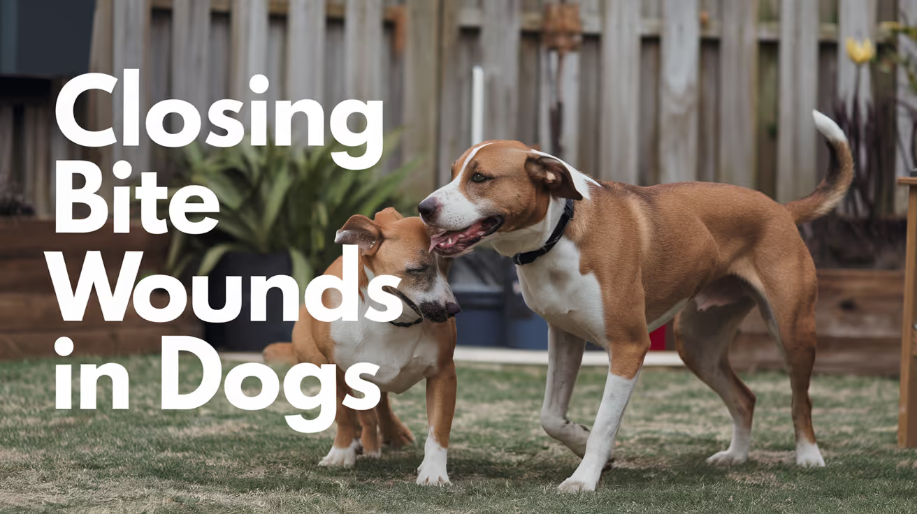
Bite wounds look smaller than they are. The surface puncture from a canine tooth may be half an inch wide, but the tissue damage underneath can extend several inches in every direction.
That's what makes bite wounds one of the more complex wound types in veterinary surgery. Whether and how to close them depends on contamination level, location, time since injury, and how much tissue was crushed.
Quick answer: All dog bite wounds are considered contaminated and require veterinary evaluation. Fresh, minimally contaminated wounds may be closed primarily after thorough lavage and debridement. Heavily contaminated, infected, or complex wounds are typically left open or treated with delayed closure. Antibiotics are routinely used. Most dogs require sedation or anesthesia for proper wound assessment and treatment.
Key takeaways
- All bite wounds are contaminated regardless of how small they appear on the surface.
- Immediate primary closure is appropriate only for clean, fresh wounds after debridement.
- Delayed primary closure is used for contaminated or infected wounds once the tissue stabilizes.
- Drains are often placed in closed bite wounds to prevent seroma and abscess formation.
- Antibiotics are typically prescribed to control or prevent infection in all bite wound cases.
- Surface size is misleading: puncture wounds can hide serious underlying damage to muscle, fascia, or organs.
Why bite wounds are different from other lacerations
A dog bite delivers two forces simultaneously: penetration and crushing. The teeth pierce the skin while the jaw compresses surrounding tissue. This creates tissue damage that extends far beyond the visible wound margins.
VCA Animal Hospitals explains: "Wounds that appear to be minor on the surface can be deceptive and may be life-threatening, depending on the location of the injury."
The American Animal Hospital Association (AAHA) emphasizes: "A clean fresh wound can be closed primarily, after appropriate lavage, debridement, and assessment of the potential need for a drain."
The key word is clean. Most bite wounds are not clean when they arrive.
When vets choose immediate (primary) closure
Primary closure means suturing the wound shut at the initial visit. This is appropriate when:
- The wound is less than 6 to 8 hours old
- The wound has been thoroughly lavaged and debrided
- There is minimal contamination (no gross debris, necrotic tissue, or obvious infection)
- The wound is in a cosmetically significant area (face) where open healing causes more problems than closure risk
Even primary closures in bite wounds are typically performed with drains placed to allow fluid escape. This reduces the risk of seroma and abscess formation.
For how drains interact with closure, see drains used in bite wound closure.
When vets delay closure
Delayed primary closure (waiting 3 to 5 days before suturing) is used when:
- The wound is heavily contaminated with debris, saliva, or infected material
- Tissue viability is uncertain after the initial crushing injury
- Infection is already established at presentation
- The wound is older than 6 to 8 hours with no prior treatment
The Merck Veterinary Manual states: "The time between initial debridement and final closure varies according to the extent of contamination or infection. Minimally contaminated wounds may be closed after 24 to 72 hours. Longer periods may be required for heavily infected wounds."
During the delay, the wound is treated as an open wound: cleaned, debrided, and bandaged daily. Once the tissue looks healthy and viable, closure is performed.
For broader guidance on the delayed closure decision, see delayed closure often used for bite wounds.
The closure procedure step by step
When the wound is ready for closure, your veterinarian will follow this sequence:
- Clip and clean the wound margins to create a sterile operating field
- Irrigate the wound under pressure with sterile saline to flush remaining bacteria
- Debride any remaining necrotic or non-viable tissue
- Assess depth: check for involvement of muscle, fascia, body cavities, or bones
- Place a drain if dead space is present or infection risk is high
- Close in layers: suture deep tissue first, then subcutaneous tissue, then skin
- Skin closure using interrupted or simple sutures or staples
For bite wounds on the head or face, the same process applies, but cosmetic outcome is a higher priority and skin flaps may be used to cover defects.
Infection risk in bite wound closure
Bite wounds carry significant infection risk from the bacteria in the biting animal's mouth. Common organisms include Pasteurella, Staphylococcus, Streptococcus, and various anaerobes.
VCA Animal Hospitals states: "Left untreated, the bacteria in an infected bite wound will cause a localized abscess or more generalized cellulitis that spreads through the surrounding area."
In rare cases, deep bite wounds can cause:
- Septic arthritis if near a joint
- Osteomyelitis if penetrating bone
- Pyothorax if penetrating the chest
Any of these requires hospitalization and intensive treatment beyond routine closure.
For how infection risk varies with closure technique, see infection risk in bite wound closure.
Owner care after bite wound closure
Your vet will provide specific instructions, but standard care after bite wound closure includes:
- Keep the E-collar on at all times to prevent licking, which rapidly introduces bacteria
- Limit activity to leash walks only during healing
- Check the wound twice daily for redness, swelling, discharge, or odor
- Complete the full antibiotic course even if the wound looks fine early
- Do not soak or bathe the wound area until the vet clears it
- Attend all scheduled rechecks so drain removal or suture removal can be performed safely
For how bite wound closure compares to cat bite wound management, see bite wound closure in cats for comparison. For how bite wounds are classified as contaminated wounds, see bite wounds as contaminated wounds.
Frequently asked questions
My dog was bitten but the wounds look small. Does it still need vet care?
Yes, always. Small puncture wounds from canine teeth close over quickly on the surface while bacteria are already tracking through the underlying tissue. VCA notes that minor-appearing wounds can be life-threatening depending on location. All bite wounds warrant prompt veterinary evaluation.
Will my dog need to be anesthetized to treat the wound?
Most bite wounds require at minimum heavy sedation for proper assessment. Complete wound exploration, irrigation, debridement, and closure all require the dog to be still and comfortable. Your vet will recommend the appropriate level of anesthesia for your dog's specific wounds.
How long does healing take after bite wound closure?
Uncomplicated closed bite wounds typically heal in 10 to 14 days. Wounds managed open, with delayed closure, or complicated by infection may take 3 to 6 weeks or longer. Your vet will monitor healing at recheck appointments.
Bite wounds demand more respect than their surface appearance suggests. The crush injury, contamination, and hidden tissue damage make them fundamentally different from simple lacerations. Early evaluation, thorough debridement, and appropriate closure timing are what keep a manageable wound from becoming a serious infection.
Resources
- VCA Animal Hospitals. Bite Wounds in Dogs. vcahospitals.com
- Merck Veterinary Manual. Management of Specific Wounds in Small Animals. merckvetmanual.com
- AAHA. Bite Wound Treatment Refresher. aaha.org
- Merck Veterinary Manual. Initial Wound Management in Small Animals. merckvetmanual.com
Isolation Protocols for Infectious Cats
Learn effective isolation protocols for infectious cats to prevent disease spread and protect your home and pets.
Sustainable Vet Group
When a cat arrives at a veterinary clinic with a contagious disease or is suspected of having one isolation is the most important immediate action.
Every minute a contagious patient spends in the general population is an opportunity for transmission.
Isolation protocols protect other patients in the hospital, protect staff, and protect the community's cats whose owners have not yet vaccinated them against the pathogens circulating in the clinic.
Quick answer: Suspected infectious cats go directly to isolation on arrival. Isolation needs dedicated equipment, full PPE for all staff entering, daily disinfection, and airflow separation from general wards. FPV requires at least 14 days isolation; URI typically 3 to 7 days after clinical improvement.
Key takeaways
- Move suspected infectious cats directly to isolation on arrival do not route them through reception or general wards
- FPV requires at least 14 days isolation; the virus survives on surfaces for months and needs parvocidal disinfectants
- Feline URI spreads via droplets up to 5 feet and via fomites (hands, clothing, equipment) standard contact precautions are required
- Ringworm is zoonotic: staff handling ringworm-positive cats need full contact PPE; precautions extend to the home
- Dedicated equipment (stethoscope, thermometer, food dishes, bedding) stays in the isolation room; shared equipment brings contamination in and out
- Separate airflow from the general ward is recommended; if unavailable, keep the door closed and minimize ventilation exchange
Which diseases require cat isolation?
Feline panleukopenia (FPV)
The most serious feline infectious disease in hospital settings.
ASPCA Pro: "Cats with FPV must be moved to an isolation area separate from unaffected and unexposed populations of cats.
Skilled staff with clinical treatment experience and knowledge of biosecurity should be assigned to the isolation ward."
ASPCA Pro: "Cats under treatment should remain in isolation for at least 14 days."
FPV is transmitted through fecal-oral contact and contaminated fomites. The virus is extremely environmentally stable it can survive on surfaces for months.
Only specific disinfectants (accelerated hydrogen peroxide, bleach at appropriate dilution) are effective against parvovirus.
ASPCA Pro: "If isolation is not available, animals with panleukopenia should be removed from the facility for treatment or euthanized to curtail their suffering and minimize disease spread."
Feline upper respiratory infection (URI)
The most common feline contagious disease encountered in clinical settings.
ASPCA Pro: "Feline URI easily spreads in shelter environments via fomites including hands, scrub tops, stethoscopes, toys, and shoes.
Feline URI can also be transmitted by droplets over distances of 5 feet or less."
Causative agents include feline herpesvirus (FHV-1) and feline calicivirus (FCV). Both can be transmitted via fomites and direct contact.
Calicivirus can also cause virulent systemic disease (FCV-VSD) a more severe form with skin lesions that has been reported as a post-surgical complication.
Best Friends Animal Society protocol: "PPE/handling restrictions lifted after 3 days" of no clinical signs in mild URI cases. Moderate to severe cases require longer isolation.
Ringworm (dermatophytosis)
Ringworm is a fungal infection not a worm caused by Microsporum canis in most feline cases. It is highly contagious between cats, between cats and dogs, and between cats and humans.
Animal Urgent Care: "Ringworm spreads through direct contact as well as contaminated bedding, furniture, and grooming tools."
Ringworm is zoonotic.
Staff handling ringworm-positive cats must use full contact precautions and should be aware that environmental contamination is extensive spores shed from infected cats can survive in the environment for months.
UC Davis IDC Protocol: disinfection requires an accelerated hydrogen peroxide product or dilute bleach; many common disinfectants are ineffective against dermatophyte spores.
Feline leukemia virus (FeLV) and feline immunodeficiency virus (FIV)
FeLV spreads through close contact and bodily fluids; FIV primarily through bite wounds. These viruses are not environmentally stable and standard contact precautions (gloves, hand hygiene) are sufficient.
Full isolation is not required for FeLV/FIV-positive cats in most hospital settings, but they should not be housed with FeLV/FIV-negative cats. Separate housing or adequate physical separation is required.
Setting up the isolation space
PMC (Infection Control Programs): "Isolation areas for sick animals should be present and these should have separate airflow from areas that house healthy animals."
PMC: "Isolation rooms may be poorly visible and/or accessible and may not provide access to an oxygen source or be amenable to intensive monitoring."
Minimum requirements for a feline isolation room:
- Separate from all general ward areas
- Door kept closed at all times when the room is occupied
- Separate ventilation where possible; if not, close the HVAC vent serving the room
- Dedicated equipment that never leaves the room: stethoscope, thermometer, food and water dishes, bedding, litter box and scoop
- Hand hygiene station immediately outside the room (or inside if space allows)
- PPE supply stocked outside the door: gowns, gloves, shoe covers, and masks or face shields where respiratory transmission is possible
- Biohazard waste disposal within or immediately adjacent to the room
PPE requirements by disease category
Animal Urgent Care identifies the core elements as: wearing PPE when handling infectious patients, minimizing exposure within the facility, and maintaining separate isolation rooms sanitized after every patient.
Staff and workflow protocols
Assign dedicated staff to the isolation room whenever possible. Staff who have handled an isolation patient should not return to the general ward without removing PPE, performing hand hygiene, and changing contaminated scrubs.
UC Davis IDC Protocol: "Within Hospital: Move patient directly to examination room or cage in isolation. Notify the infectious disease control personnel."
Handling sequence:
- Put on full PPE before entering
- Complete all care for the isolation patient
- Remove PPE inside or immediately outside the room (remove gloves last)
- Perform hand hygiene
- Change contaminated scrubs before entering general ward
Visit isolation patients last in the daily ward round, not between general patients.
Disinfection and cleaning
PMC: "Cages should be cleaned and disinfected at least daily." For FPV and ringworm cases, more frequent cleaning may be required.
Disinfectant selection by pathogen:
Standard quaternary ammonium compounds (common hospital disinfectants) are NOT effective against parvovirus or ringworm spores. Choosing the correct disinfectant is critical.
Terminal cleaning after discharge: a thorough cleaning of all surfaces, including walls, floor, ceiling fixtures, and all equipment. For parvovirus and ringworm cases, this requires two full cleaning and disinfection cycles.
Isolation duration guidelines
ASPCA Pro (FPV): "Cats under treatment should remain in isolation for at least 14 days."
Best Friends protocol: "PPE/handling restrictions lifted after 3 days" for mild URI cases.
For isolation protocols for infectious dogs, see isolation protocols for infectious dogs. For the PPE selection guide that applies to isolation work, see PPE use and barrier protection in veterinary clinics.
Frequently asked questions
My cat has a suspected URI. Does it need to be in a separate room at the vet clinic?
Yes, ideally. Feline URI spreads via droplets and fomites within 5 feet.
Cats with respiratory signs should be examined in a dedicated room or a drape-separated exam space, not in an open area near other cats.
Can I visit my cat while it is in isolation?
Policies vary by clinic. Many hospitals allow owner visits with PPE and staff supervision. The visit is typically brief and conducted in the isolation room itself.
Ask your clinic what their isolation visiting policy is.
How long does FPV survive in the environment?
Parvovirus is extremely hardy. ASPCA Pro notes it can survive for extended periods on contaminated surfaces.
This is why terminal cleaning with a parvocidal disinfectant, not just standard cleaning, is essential after a confirmed FPV case.
Is feline calicivirus dangerous to staff?
Feline calicivirus does not infect humans. However, staff can transmit it to other cats via their hands, clothing, and equipment. Standard contact precautions protect other feline patients, not necessarily staff health.
My cat was diagnosed with ringworm. What precautions should I take at home?
Wear gloves when handling your cat and cleaning the litter box. Wash hands after contact. Avoid sharing bedding or brushes. Vacuum and disinfect regularly with a product effective against ringworm spores.
Does a confirmed FeLV or FIV-positive cat need to be in a separate isolation room?
Not usually. FeLV/FIV-positive cats need physical separation from negative cats, not a full isolation room. Standard contact precautions apply. Full isolation is reserved for acutely contagious diseases like FPV and URI.
Resources
- ASPCA Pro. Feline Panleukopenia. aspcapro.org
- ASPCA Pro. Feline Upper Respiratory Infection. aspcapro.org
- PMC. Infection Control Programs for Dogs and Cats. ncbi.nlm.nih.gov
- Animal Urgent Care. Isolation Protocols: A Necessary Safeguard for Common Infectious Diseases. animalurgentcare.vet
- UC Davis. Small Animal Infectious Disease Control/Biosecurity Protocol. safety.vetmed.ucdavis.edu
Preventing Post-Operative Infections in Cats
Learn effective strategies for preventing post-operative infections in cats to ensure safe recovery and optimal healing.
Sustainable Vet Group
Cats are exceptional at hiding discomfort, and they're equally determined to groom themselves the moment you look away. Both of these traits make post-operative infection prevention in cats more demanding than in dogs.
The good news: most post-surgical infections in cats are preventable. The majority require the same foundational measures: keeping the wound protected, monitoring it daily, and getting your cat to their follow-up appointments.
Quick answer: Preventing post-operative infections in cats comes down to three owner responsibilities: keeping the E-collar on at all times to prevent licking and grooming of the incision, checking the surgical site daily for redness, swelling, or discharge, and following all medication and follow-up instructions from your vet. Cats that lick their incisions develop secondary infections at a much higher rate than those kept properly protected.
Key takeaways
- Licking is the leading preventable cause of post-operative infection in cats at home.
- E-collar compliance is non-negotiable until the vet confirms the incision is fully healed.
- Daily incision checks help you catch early infection signs before they become serious.
- Cats hide illness well: behavioral changes (hiding, reduced appetite) may be the first sign of trouble.
- FIV or FeLV positive cats carry higher infection risk and need closer post-surgical monitoring.
- Normal healing vs. early infection has clear distinguishing signs covered in the monitoring section below.
Why cats are at particular risk after surgery
Cats present specific challenges in post-surgical infection prevention that differ from dogs.
Grooming instinct: cats are compulsive self-groomers. The moment an E-collar is removed, most cats will attempt to access the incision site. Their tongue is rough enough to pull sutures and introduce oral bacteria directly into the healing wound.
Hiding pain: cats conceal discomfort as a survival instinct. By the time a cat is visibly unwell, the infection may be more established than it would be in a dog who shows pain more obviously. This is why daily visual inspection of the wound matters more than watching for behavioral signs alone.
FIV and FeLV: cats with feline immunodeficiency virus or feline leukemia virus have compromised immune systems that make infection more likely and harder to resolve. Post-surgical monitoring must be more intensive for these cats.
PetMD confirms: "Grooming and licking the surgical site can cause secondary complications such as infections, irritation, and damage to the sutures."
The E-collar: your most important tool
No single measure prevents more post-operative infections in cats than consistent E-collar use.
Revel Vet explains: "You should generally keep an e-collar on your cat until the incision has healed, unless you receive other instructions from your veterinarian."
When the E-collar must be worn
- At all times when unsupervised
- During the night
- When you're in the same room but not actively watching the cat
- During the full healing period, typically 10 to 14 days for routine incisions
The E-collar comes off for eating and drinking if you can watch directly and return it immediately after. It does not come off because your cat hates it or seems distressed. Cats adjust to E-collars far faster than owners expect, particularly if the alternative (infection and re-suturing) is considered.
E-collar alternatives
If your cat is extremely distressed by a traditional cone-style E-collar, alternatives include:
- Inflatable recovery collars: softer and more tolerable for some cats, but verify the cat can't access the wound
- Recovery body suits/onesies: cover abdominal and some thoracic incisions; not suitable for all wound locations
- Neck collars: a softer version that prevents head rotation to reach the body
Confirm any alternative with your vet before substituting, as the incision location determines which option adequately protects it.
Daily wound monitoring: what to look for
Check the incision every day at the same time, in good lighting.
Normal healing appearance
Signs that require a vet call
Contact your vet the same day if you notice:
- Redness spreading beyond the immediate wound margin
- Yellow, green, or opaque discharge
- Foul smell from the wound site
- Increasing rather than decreasing swelling after day 3
- The wound opening or sutures separating
- The cat obsessively pawing at or trying to reach the wound despite the E-collar
Behavioral signs to watch for
Because cats hide pain, watch for:
- Reduced appetite or refusing food for more than 24 hours
- Hiding more than usual or unwillingness to engage
- Increased or decreased grooming elsewhere (compensatory behavior)
- Vocalizing when the wound area is touched
- Fever (temperature above 39.5°C / 103.1°F)
For how to distinguish healing from infection in feline surgical wounds, see distinguishing healing from infection after surgery.
Home environment and wound care
Activity restriction
Cats recovering from surgery need restricted movement. Jumping, climbing, and running all place mechanical stress on incisions that can cause wound breakdown.
Practical measures:
- Confine to one room or a large crate during the initial recovery period
- Remove access to furniture they jump from
- Block stairs if the incision could be strained by stair use
- Keep children and other pets away to prevent accidental trauma to the wound
Wound hygiene
Unless your vet specifically instructs otherwise:
- Do not clean the incision with antiseptics, hydrogen peroxide, or any product not explicitly recommended by your vet
- Keep the wound dry: no bathing until fully healed
- Don't apply creams or ointments unless prescribed
- Keep bedding clean: change it regularly to reduce environmental bacterial load
Pooler Veterinary Hospital advises: "Keep the incision site clean and dry at all times. Avoid bathing your dog (or cat) until your vet gives approval."
Medications
Give all medications for the full prescribed course. Stopping antibiotics early when the cat appears improved is one of the most common owner errors that leads to recurrence and, in resistant bacteria, to selection pressure that makes retreatment harder.
Comparing cat and dog post-operative infection prevention
Cat post-operative infection prevention shares core principles with dogs but has cat-specific considerations:
For post-operative infection prevention in dogs specifically, see post-operative infection prevention in dogs for comparison. For the timing of when infections typically appear, see when to watch for post-operative infections in cats.
When to seek emergency care
Go to an emergency vet immediately if:
- Your cat is unresponsive or collapses
- The wound has opened significantly with visible tissue
- Rapidly spreading redness or warmth covers a large area
- Your cat has a high fever and severe lethargy together
- Bleeding from the wound site that doesn't stop within 5 minutes
These signs indicate the infection or wound complication has progressed beyond what can wait for a regular appointment.
For the specific surgical site infection prevention protocols vets use in cats, see surgical site infection prevention specifically in cats.
Frequently asked questions
My cat is acting normally but the wound looks a little red. Should I be worried?
Mild redness at the wound margin is normal for the first 2 to 3 days. If it's fading rather than spreading, and there's no discharge or odor, it's likely normal healing. If redness is spreading beyond the wound margin or appearing on day 5 or later when it should be diminishing, call your vet.
Can I take the E-collar off at night if I'm watching my cat sleep?
No. Cats move during sleep and can access wounds in positions you won't notice. The E-collar must stay on during sleeping hours. Remove it only for eating and drinking under direct supervision.
My cat had surgery two days ago and hasn't eaten much. Is that normal?
Reduced appetite for 24 to 48 hours post-surgery is common due to the effects of anesthesia and the stress of the procedure. By day 2 to 3, most cats return to normal or near-normal eating. If your cat is still refusing food at 72 hours post-surgery, contact your vet: this may indicate pain, nausea from medications, or early infection.
The owner's role in preventing post-operative infection in cats is more active than many people expect. The surgeon controls the intraoperative environment. You control everything that happens once your cat comes home, and for most preventable infections, that's where the critical work is done.
Resources
- PetMD. Cat Surgery Aftercare FAQs. petmd.com
- Revel Vet. Signs of Infection in Cats After Surgery. revelvet.com
- Pooler Veterinary Hospital. Pet Wound Care at Home. poolervet.com
- The Pet Vet. Post-Surgical Infection in Cats. thepetvet.com

Surgical Closure Protocol in Dogs and Cats
Learn the detailed surgical closure protocol in dogs and cats to ensure safe, effective healing after surgery.
Sustainable Vet Group
Every surgery ends with the same critical phase: closing the wound. How well that closure is performed determines whether the patient heals cleanly, develops complications, or ends up back in surgery.
A standardized closure protocol takes the guesswork out of this phase. It defines which layers to close, what materials to use, and in what sequence, adapted to the specific surgery and patient.
Quick answer: The surgical closure protocol in dogs and cats involves sequentially closing each tissue layer using appropriate suture materials, from deep to superficial. Muscle and fascia are closed with absorbable monofilament sutures, subcutaneous tissue is approximated to eliminate dead space, and skin is closed with sutures or staples. The protocol is adapted based on procedure type, patient size, and tissue condition.
Key takeaways
- Layered closure is the foundation of all surgical wound closure in small animal surgery.
- Each tissue layer requires its own suture material and pattern for optimal healing.
- Dead space elimination at every layer is as important as the skin closure itself.
- Absorbable sutures are used for internal layers; skin may use absorbable or non-absorbable.
- Suture pattern selection depends on tissue type, tension, and desired healing profile.
- Postoperative monitoring is an extension of the closure protocol, not an afterthought.
Why a standardized closure protocol matters
Surgical closure is not a single action. It is a sequence of decisions made for each layer of tissue from the deepest plane outward.
A consistent, evidence-based protocol:
- Reduces operator variability between surgeons
- Minimizes complications like seroma, dehiscence, and infection
- Adapts reproducibly to different procedure types
The protocol begins with understanding the anatomy of the wound and ends with a checklist confirming that every layer has been addressed. For the formal checklist approach, see checklist version of the protocol.
The core wound closure principles
Before selecting materials or patterns, every closure must satisfy four biological requirements:
- Tissue apposition: edges brought together without overlap or gap
- Tension distribution: no single suture bearing excessive load
- Dead space elimination: no empty pockets where fluid can pool
- Tissue handling: atraumatic technique preserving blood supply
These principles apply regardless of procedure. For a full exploration of each, see core wound closure principles.
The layered closure sequence
Deep tissue layers: muscle and fascia
Muscle layers are closed first using absorbable monofilament sutures (PDS or Biosyn). These provide long-term holding strength while the tissue heals.
The pattern used depends on tissue thickness and tension:
- Simple continuous: rapid, even tension distribution
- Interrupted: more secure if one suture fails, more time-consuming
- Ford interlocking: for longer wounds needing speed without sacrificing strength
For muscle-specific technique details, see layered closure within the protocol.
Subcutaneous tissue
The subcutaneous layer is closed using fine absorbable sutures in a simple continuous or interrupted pattern. This layer:
- Brings tissue planes together to eliminate dead space
- Reduces tension on the skin closure
- Prevents seroma formation
In dogs with significant subcutaneous fat (obese animals), this layer requires more care and sometimes additional walking sutures to prevent fluid accumulation.
Skin closure
The final layer. Multiple closure methods are available:
For a complete comparison of methods, see tissue-based technique selection.
Suture material selection overview
Size scales down in small dogs and cats. A 4-0 or 5-0 suture is often used in cats and toy breeds where a 2-0 or 3-0 would be used in medium to large dogs.
Adapting the protocol by procedure
The baseline sequence stays the same, but specific steps vary by surgery:
- Spay surgery in dogs: see spay-specific closure protocol in dogs
- Spay surgery in cats: see spay-specific closure protocol in cats
- Neuter surgery: see neuter-specific closure protocol
Each procedure has its own anatomical requirements that modify suture material choice, layer sequence, and skin closure selection.
Postoperative care: the owner's role in the protocol
The closure protocol does not end when the patient leaves the surgery table. Owner compliance with aftercare is a direct extension of what the surgeon started.
Essential owner actions:
- E-collar immediately: licking disrupts sutures and introduces bacteria within hours
- Activity restriction: movement prevents tissue planes from adhering and promotes fluid accumulation
- Wound checks twice daily: look for redness, swelling, discharge, or odor
- No bathing until the vet confirms healing is complete
- Attend all rechecks: especially suture removal and wound assessment visits
Frequently asked questions
What happens if a layer is not closed properly?
Each layer serves a function. If the subcutaneous layer is omitted, dead space forms and seromas develop. If fascial closure is too tight, it strangulates tissue. If the skin is under excessive tension, it tears through sutures. Each error has a predictable consequence.
How does suture material choice affect healing?
Different materials degrade at different rates and cause different levels of tissue reaction. An absorbable suture losing strength too early before healing is complete can allow wound breakdown. A non-absorbable suture left too long can cause a sinus tract. Your vet matches the material to the tissue's healing timeline.
Why do some dogs get dissolvable sutures while others need suture removal?
Internal layers always use dissolvable (absorbable) sutures. Skin closure may use either. Absorbable sutures for skin are used when minimizing trauma at recheck is a priority, or when the dog is difficult to handle. Non-absorbable skin sutures require removal at 10 to 14 days but allow precise monitoring of healing.
A surgical closure protocol is only as good as the consistency with which it is applied. Every layer closed correctly, with appropriate materials and tension, reduces the risk of the complications that bring dogs back for second procedures. The protocol exists because leaving anything to chance in wound closure has predictable, unpleasant consequences.
Resources
- Veterinary Surgery Online. Wound Closure: Continued. vetsurgeryonline.com
- Merck Veterinary Manual. Initial Wound Management in Small Animals. merckvetmanual.com
- VCA Animal Hospitals. Bite Wounds in Dogs. vcahospitals.com

Subcutaneous Closure Techniques in Cats
Learn about subcutaneous closure techniques in cats, including methods, materials, and best practices for optimal healing and minimal complications.
Sustainable Vet Group
Subcutaneous closure techniques in cats are essential for proper wound healing after surgery or injury. These methods help reduce tension on the skin, minimize infection risk, and improve cosmetic outcomes. Understanding these techniques can help you care better for your cat post-operation.
This article explains the most common subcutaneous closure methods, the materials used, and tips for ensuring your cat recovers well. You will learn how veterinarians choose the right technique and what to expect during healing.
What is subcutaneous closure in cats?
Subcutaneous closure refers to stitching the tissue layer just beneath the skin in cats. This layer contains fat and connective tissue and helps support the skin edges during healing.
Closing this layer properly reduces tension on the skin surface and promotes faster, cleaner healing. It is a standard step in many feline surgeries.
- Definition of subcutaneous closure: It is the suturing of the tissue layer under the skin to support wound edges and reduce skin tension during healing.
- Purpose in surgery: It stabilizes the wound, decreases dead space, and helps prevent skin suture line complications like dehiscence or infection.
- Common in feline surgeries: Used in spays, tumor removals, and trauma repairs to improve healing and cosmetic results.
- Difference from skin closure: Subcutaneous closure is beneath the skin, while skin closure involves suturing the outer skin layer for final wound sealing.
Proper subcutaneous closure is critical for successful wound healing in cats. It supports the skin and reduces complications.
What materials are used for subcutaneous closure in cats?
Choosing the right suture material is important for subcutaneous closure in cats. The material must be strong enough to hold tissue but absorbable to avoid removal.
Veterinarians typically use absorbable sutures that break down safely inside the body over time.
- Absorbable sutures preferred: Materials like poliglecaprone or polydioxanone dissolve naturally, eliminating the need for suture removal.
- Suture size selection: Sizes 3-0 or 4-0 are common to balance strength and minimal tissue reaction in cats.
- Monofilament vs. multifilament: Monofilament sutures reduce infection risk due to less bacterial trapping compared to braided multifilament sutures.
- Suture needle type: Taper-point needles are used to minimize tissue trauma when passing through delicate subcutaneous tissue.
Using appropriate suture materials helps ensure the closure holds during healing and reduces complications like irritation or infection.
What are the common subcutaneous closure techniques in cats?
Several stitching techniques are used for subcutaneous closure in cats. The choice depends on wound size, location, and surgeon preference.
Each technique aims to approximate tissue edges securely while minimizing tension and trauma.
- Simple continuous suture: A single running stitch that is fast and distributes tension evenly along the wound.
- Interrupted sutures: Individual stitches placed separately, allowing precise tension control and easier adjustment if one fails.
- Subcuticular suture: Placed just under the skin surface, this technique provides excellent cosmetic results with minimal scarring.
- Mattress sutures: Horizontal or vertical mattress stitches provide strong tissue apposition and are useful for wounds under tension.
Veterinarians select the technique based on wound characteristics and the cat’s condition to optimize healing.
How does subcutaneous closure benefit wound healing in cats?
Subcutaneous closure plays a key role in supporting the skin and promoting faster recovery after surgery or injury in cats.
It reduces complications and improves the overall healing process.
- Tension reduction: By closing the deeper tissue, it lowers tension on the skin edges, preventing wound opening or tearing.
- Dead space elimination: It closes gaps under the skin that could fill with fluid and cause infection or delayed healing.
- Improved cosmetic outcome: Proper closure results in less scarring and a smoother skin surface after healing.
- Lower infection risk: Stabilizing tissues reduces movement and irritation, which helps prevent bacterial invasion.
These benefits make subcutaneous closure a standard practice in feline surgery to ensure the best healing results.
What are the risks or complications of subcutaneous closure in cats?
While subcutaneous closure is generally safe, some risks exist if the technique or materials are not used properly.
Understanding these helps you recognize problems early and seek veterinary care if needed.
- Suture reaction: Some cats may develop inflammation or irritation around the suture material causing swelling or discomfort.
- Wound dehiscence: If sutures fail or are too loose, the wound can reopen, requiring additional treatment or surgery.
- Infection risk: Improper technique or contamination can introduce bacteria leading to abscess or delayed healing.
- Seroma formation: Fluid accumulation under the skin can occur if dead space is not adequately closed during suturing.
Close monitoring and proper surgical technique reduce these risks significantly.
How should you care for your cat after subcutaneous closure?
Post-operative care is vital to ensure your cat’s wound heals well after subcutaneous closure. You play an important role in this process.
Following veterinary instructions and monitoring the wound helps prevent complications.
- Limit activity: Restrict your cat’s movement to avoid stress on the wound and allow proper healing.
- Prevent licking: Use an Elizabethan collar or other methods to stop your cat from licking or biting the sutures.
- Keep wound clean: Follow your vet’s advice on cleaning the area and avoid bathing until fully healed.
- Watch for signs: Monitor for redness, swelling, discharge, or opening of the wound and contact your vet if noticed.
Good aftercare supports the surgical closure and helps your cat recover quickly and comfortably.
What alternatives exist to traditional subcutaneous suturing in cats?
Besides traditional sutures, some alternative closure methods are available for subcutaneous tissue in cats. These may offer benefits in certain cases.
Understanding these options can help you discuss the best approach with your veterinarian.
- Absorbable staples: Used occasionally for faster closure but may cause more tissue reaction than sutures.
- Tissue adhesives: Medical glues can close small wounds but are less common for deep subcutaneous layers.
- Barbed sutures: These self-anchoring sutures eliminate knots and may reduce closure time and tissue trauma.
- Combination techniques: Some surgeons combine sutures with adhesives or staples to optimize closure strength and healing.
While traditional suturing remains the most common, alternatives may be suitable depending on the wound and cat’s needs.
Conclusion
Subcutaneous closure techniques in cats are crucial for successful surgical wound healing. They support the skin, reduce tension, and minimize complications like infection or wound opening.
Choosing the right suture materials and closure methods, along with proper aftercare, ensures your cat recovers well. Understanding these techniques helps you support your cat’s healing and recognize any issues early.
What suture materials are best for subcutaneous closure in cats?
Absorbable monofilament sutures like poliglecaprone or polydioxanone in sizes 3-0 or 4-0 are preferred for strength and minimal tissue reaction.
How long does it take for subcutaneous sutures to absorb in cats?
Absorbable sutures typically dissolve within 2 to 4 weeks, depending on the material, allowing the tissue to heal without suture removal.
Can cats remove subcutaneous sutures by licking?
Cats cannot remove subcutaneous sutures by licking because they are beneath the skin, but licking can irritate the skin closure and cause complications.
When should I contact a vet after my cat’s subcutaneous closure?
Contact your vet if you notice redness, swelling, discharge, wound opening, or if your cat shows signs of pain or lethargy near the surgical site.
Are there risks if subcutaneous closure is not done properly?
Improper closure can lead to wound dehiscence, infection, seroma formation, and delayed healing, increasing the need for further treatment.
Intradermal Closure in Dogs: Techniques & Benefits
Learn about intradermal closure in dogs, its benefits, techniques, and aftercare for optimal healing and cosmetic results.
Sustainable Vet Group
Intradermal closure in dogs is a surgical technique used to close skin wounds with minimal scarring and faster healing. This method places sutures within the skin layers, avoiding external stitches that can irritate your dog or require removal. Understanding intradermal closure helps you make informed decisions about your pet's surgical care.
This article explains what intradermal closure is, how it works, its advantages, and how to care for your dog after surgery. You will learn about the materials used, common techniques, and potential complications to watch for during recovery.
What is intradermal closure in dogs?
Intradermal closure is a suturing technique where stitches are placed inside the skin layers, specifically within the dermis. This method hides the sutures beneath the skin surface, reducing irritation and improving cosmetic outcomes. It is commonly used in veterinary surgery for skin incisions and lacerations.
The goal is to bring the skin edges together securely while minimizing external suture exposure. This technique helps wounds heal with less inflammation and reduces the risk of infection or self-trauma by the dog.
- Hidden sutures: Sutures are placed beneath the skin surface, preventing your dog from licking or scratching them, which promotes safer healing.
- Cosmetic benefit: Intradermal closure results in less visible scarring compared to traditional external stitches, improving your dog's appearance after surgery.
- Reduced suture removal: Because sutures are buried, they often do not require removal, reducing stress for both you and your dog.
- Secure wound closure: This technique provides strong wound edge apposition, which helps prevent wound opening and infection.
Intradermal closure is a preferred method in many veterinary surgeries due to these benefits. It requires skill and appropriate suture materials to ensure success.
How is intradermal closure performed in dogs?
The procedure involves placing a continuous or interrupted suture pattern within the dermis layer of the skin. The surgeon carefully aligns the wound edges and uses absorbable sutures to close the incision beneath the surface.
Proper technique is essential to avoid tension on the skin and to ensure the wound edges heal evenly. The choice of suture material and needle type also affects the outcome.
- Suture placement: Sutures are inserted horizontally or vertically within the dermis to bring skin edges together without external exposure.
- Absorbable sutures: Materials like poliglecaprone or polydioxanone are used, which dissolve over time, eliminating the need for removal.
- Needle type: A reverse cutting needle is often preferred to minimize tissue trauma during suture placement.
- Continuous pattern: A running intradermal suture provides even tension distribution and faster closure compared to interrupted stitches.
After suturing, the skin surface appears smooth with no visible stitches. This technique requires careful handling to avoid puckering or uneven closure.
What are the benefits of intradermal closure for dogs?
Intradermal closure offers several advantages over traditional external suturing methods. These benefits improve healing, reduce complications, and enhance cosmetic results for your dog.
Understanding these benefits can help you discuss surgical options with your veterinarian confidently.
- Less irritation: Buried sutures prevent your dog from chewing or scratching the wound, reducing inflammation and discomfort.
- Improved healing: The technique promotes better skin edge alignment, which supports faster and stronger wound healing.
- Lower infection risk: With no external suture exposure, there is less chance for bacteria to enter the wound site.
- Better appearance: The cosmetic outcome is superior, with minimal scarring and no visible stitches after healing.
These benefits make intradermal closure a preferred choice for elective surgeries and wounds in dogs, especially in visible areas like the face or limbs.
When is intradermal closure recommended for dogs?
Veterinarians choose intradermal closure based on the wound type, location, and patient factors. It is most suitable for clean, straight incisions where cosmetic outcome is important.
Not all wounds are ideal for this technique, so your vet will assess the situation before deciding.
- Clean surgical incisions: Intradermal closure is ideal for planned surgeries with minimal contamination risk.
- Superficial wounds: Wounds involving only the skin layers without deep tissue damage are good candidates.
- Areas needing cosmetic care: Locations like the face, neck, or limbs benefit from less visible scarring.
- Stable patients: Dogs that can tolerate anesthesia and have no skin infection are suitable for this closure method.
Your veterinarian will evaluate your dog's specific case to determine if intradermal closure is the best option.
What materials are used for intradermal closure in dogs?
Choosing the right suture material is crucial for successful intradermal closure. Absorbable sutures are preferred to avoid the need for removal and reduce irritation.
The needle type also affects how easily the sutures pass through the skin and how much tissue damage occurs.
- Poliglecaprone sutures: These absorbable sutures dissolve within 90-120 days and cause minimal tissue reaction.
- Polydioxanone sutures: Longer-lasting absorbable sutures that maintain strength for several weeks, ideal for slow-healing wounds.
- Reverse cutting needles: Designed to cut through tough skin with less trauma, improving suture placement accuracy.
- Monofilament sutures: Smooth, single-strand sutures reduce bacterial colonization and tissue drag during closure.
Your veterinarian selects materials based on wound characteristics and healing needs to optimize outcomes.
How should you care for your dog after intradermal closure?
Proper aftercare is essential to ensure your dog's wound heals well after intradermal closure. You must prevent infection, avoid trauma, and monitor for complications.
Following your vet's instructions carefully will help your dog recover quickly and comfortably.
- Limit activity: Restrict your dog's movement to prevent stress on the wound and avoid suture disruption.
- Prevent licking: Use an Elizabethan collar or other devices to stop your dog from licking or chewing the wound area.
- Keep wound clean: Follow your vet’s advice on cleaning the site gently without soaking or harsh chemicals.
- Watch for signs: Monitor for redness, swelling, discharge, or pain, and contact your vet if these occur.
Most wounds heal within 10-14 days with proper care. Absorbable sutures dissolve on their own, so no removal is needed unless complications arise.
What complications can occur with intradermal closure in dogs?
While intradermal closure is generally safe, some complications can happen. Knowing what to expect helps you act quickly if problems arise.
Early detection and veterinary care can prevent minor issues from becoming serious.
- Wound dehiscence: The wound may reopen if sutures fail or if your dog stresses the area excessively.
- Infection risk: Though lower than external sutures, infections can still occur and require prompt treatment.
- Allergic reaction: Some dogs may react to suture materials, causing redness or swelling.
- Excessive scarring: Improper technique or healing can lead to thick or raised scars, affecting appearance and function.
If you notice any unusual changes in your dog's wound, contact your veterinarian immediately for assessment and care.
Conclusion
Intradermal closure in dogs is a valuable surgical technique that offers strong wound closure with minimal scarring and less irritation. It uses absorbable sutures placed beneath the skin surface, improving healing and cosmetic results.
Understanding how this method works, when it is recommended, and how to care for your dog afterward helps ensure the best recovery. Always follow your veterinarian’s advice and watch for any signs of complications to keep your dog safe and comfortable.
FAQs
Is intradermal closure painful for dogs?
The procedure itself is painless due to anesthesia during surgery. Postoperative discomfort is usually mild and manageable with pain medication prescribed by your vet.
How long do intradermal sutures last in dogs?
Absorbable intradermal sutures typically dissolve within 10 to 14 days, depending on the suture material and your dog’s healing rate.
Can intradermal closure be used on infected wounds?
Intradermal closure is generally not recommended for infected or heavily contaminated wounds, as buried sutures may trap bacteria and worsen infection.
Do dogs need a cone after intradermal closure?
Yes, an Elizabethan collar is usually recommended to prevent licking or chewing the wound, which can disrupt sutures and delay healing.
When should I contact my vet after intradermal closure?
Contact your vet if you notice redness, swelling, discharge, foul odor, or if your dog shows signs of pain or the wound opens unexpectedly.

Closure Protocol for Spay Surgery in Cats
Learn the detailed closure protocol for spay surgery in cats, including step-by-step wound closure and post-op care tips.
Sustainable Vet Group
Spay surgery in cats is a common procedure that requires careful closure to ensure proper healing and prevent complications. The closure protocol involves multiple layers of suturing to close the abdominal wall, subcutaneous tissue, and skin. Proper technique reduces infection risk and promotes faster recovery.
This article explains the step-by-step closure protocol for feline spay surgery. You will learn the types of sutures used, the order of closure, and tips for post-operative care to keep your cat safe and comfortable.
What is the standard closure protocol for spay surgery in cats?
The standard closure protocol for feline spay surgery involves closing three main layers: the abdominal wall, the subcutaneous tissue, and the skin. Each layer requires specific suture materials and techniques to ensure strength and minimize irritation.
Following the correct closure sequence helps prevent wound dehiscence and infection. It also supports proper healing and reduces discomfort for your cat.
- Abdominal wall closure: Use absorbable sutures like polydioxanone (PDS) in a simple continuous or interrupted pattern to securely close the linea alba.
- Subcutaneous layer closure: Close with absorbable sutures such as polyglactin 910 (Vicryl) in a simple continuous pattern to reduce dead space and support skin edges.
- Skin closure: Use non-absorbable sutures like nylon or absorbable monofilaments in interrupted or cruciate patterns for skin apposition.
- Suture removal timing: Remove skin sutures 10 to 14 days post-surgery to allow adequate healing.
Each closure layer plays a vital role in wound integrity and healing after spay surgery.
Why is multilayer closure important in cat spay surgeries?
Multilayer closure distributes tension evenly across the wound, reducing the risk of suture pull-through or wound opening. It also minimizes dead space where fluid can accumulate, lowering infection risk.
Each tissue layer has different strength and healing properties. Closing them separately supports natural tissue repair and reduces complications.
- Tension distribution: Closing multiple layers prevents excessive tension on skin sutures, reducing wound dehiscence risk.
- Dead space elimination: Subcutaneous closure reduces spaces where fluid or blood can collect, preventing seromas or hematomas.
- Layer-specific healing: Abdominal wall closure restores strength to the linea alba, essential for internal organ support.
- Infection prevention: Proper closure limits bacterial entry and promotes faster tissue recovery.
Multilayer closure is a surgical best practice that improves outcomes in feline spay procedures.
What suture materials are best for each closure layer in cat spays?
Choosing the right suture material is critical for effective closure and healing. Absorbable sutures are preferred for internal layers, while skin closure can use absorbable or non-absorbable sutures depending on surgeon preference.
Material choice affects tissue reaction, strength duration, and ease of removal.
- Abdominal wall sutures: Polydioxanone (PDS) or polyglyconate provide long-lasting strength and minimal tissue reaction for linea alba closure.
- Subcutaneous sutures: Polyglactin 910 (Vicryl) or poliglecaprone 25 (Monocryl) absorb within 2-3 weeks, suitable for soft tissue support.
- Skin sutures: Nylon or polypropylene offer good tensile strength and are easy to remove; absorbable monofilaments reduce the need for removal.
- Suture size: Typically 3-0 or 4-0 sutures balance strength and minimal tissue trauma for cats.
Using appropriate suture materials tailored to each layer supports optimal healing and reduces complications.
How should the abdominal wall be closed during a cat spay surgery?
The abdominal wall closure is the most critical step to restore the integrity of the linea alba and prevent herniation. It requires precise technique and strong sutures.
Surgeons usually use absorbable monofilament sutures in a simple continuous or interrupted pattern to ensure secure closure.
- Linea alba identification: Accurately identify and align the linea alba edges before suturing to avoid muscle incorporation.
- Suture pattern: Simple continuous sutures provide even tension and faster closure, while interrupted sutures allow better tension adjustment.
- Suture bite size: Take 5-7 mm bites from the edge to ensure strong tissue purchase without tearing.
- Suture spacing: Place sutures 4-6 mm apart to maintain wound strength and prevent gaps.
Proper abdominal wall closure is essential to prevent post-operative complications like hernias or wound breakdown.
What is the recommended technique for subcutaneous tissue closure in cats?
Subcutaneous closure reduces dead space and supports skin edges, promoting better healing and minimizing fluid accumulation. It uses absorbable sutures placed in a simple continuous pattern.
Careful handling of tissue and appropriate suture tension are important to avoid tissue strangulation or necrosis.
- Tissue handling: Gently handle subcutaneous tissue to avoid trauma and preserve blood supply for healing.
- Suture pattern: Simple continuous sutures evenly close the layer and reduce operative time.
- Suture tension: Apply moderate tension to approximate tissue without causing ischemia or puckering.
- Dead space closure: Ensure all pockets are closed to prevent seroma or hematoma formation.
Effective subcutaneous closure enhances overall wound strength and comfort for your cat.
How should the skin be closed after a cat spay surgery?
Skin closure is the final step and important for wound protection and cosmetic appearance. The choice of suture and pattern affects healing and ease of post-op care.
Options include interrupted sutures, cruciate patterns, or skin staples depending on surgeon preference.
- Suture type: Non-absorbable nylon sutures are common for easy removal; absorbable monofilaments reduce the need for suture removal visits.
- Suture pattern: Interrupted or cruciate sutures provide good skin edge apposition and allow drainage if needed.
- Suture spacing: Place sutures 4-6 mm apart to balance wound strength and minimize scarring.
- Suture removal: Remove skin sutures 10-14 days post-op to prevent irritation and allow full healing.
Proper skin closure protects the wound and helps your cat recover comfortably.
What post-operative care is needed after spay surgery closure in cats?
After closure, proper post-operative care is vital to ensure healing and reduce complications. Monitoring the incision and preventing self-trauma are key.
Follow your veterinarian’s instructions carefully to support your cat’s recovery.
- Incision monitoring: Check daily for redness, swelling, discharge, or opening that may indicate infection or dehiscence.
- Prevent licking: Use an Elizabethan collar or alternative to stop your cat from licking or biting the incision site.
- Limit activity: Restrict jumping and running for 10-14 days to avoid stress on the wound closure.
- Follow-up visits: Attend scheduled veterinary checks for suture removal and wound assessment.
Good post-op care helps your cat heal quickly and comfortably after spay surgery.
Conclusion
The closure protocol for spay surgery in cats involves careful multilayer suturing of the abdominal wall, subcutaneous tissue, and skin. Each layer requires specific suture types and patterns to ensure strong, secure closure.
Following proper closure techniques and post-operative care reduces complications and promotes fast healing. Understanding this protocol helps you support your cat’s recovery after spay surgery.
FAQs
How long does it take for a cat’s spay incision to heal?
Typically, the skin incision heals within 10 to 14 days, but internal healing may take several weeks. Follow-up care is essential to monitor healing progress.
Can I bathe my cat after spay surgery?
Avoid bathing your cat until the incision is fully healed and sutures are removed, usually after 10-14 days, to prevent infection and wound opening.
What signs indicate a spay incision infection?
Signs include redness, swelling, discharge, foul odor, warmth, or your cat showing pain when the area is touched. Contact your vet if these occur.
Is it normal for a small lump to form near the incision?
A small lump or swelling can be normal due to tissue reaction or fluid accumulation but should be monitored. Persistent or worsening lumps need veterinary evaluation.
When should skin sutures be removed after spay surgery?
Skin sutures are usually removed 10 to 14 days after surgery, once the incision has healed sufficiently to maintain closure without support.

Sustainable Vet | Copyright ©2026 | All Rights Reserved

