

Protecting
Pets, People & Planet
Join a group of veterinarians leveraging the latest technologies to deliver excellent care to their patients while being a responsible and positive force for their local and global communities.
100% secure. We do not share your information


Recent Articles
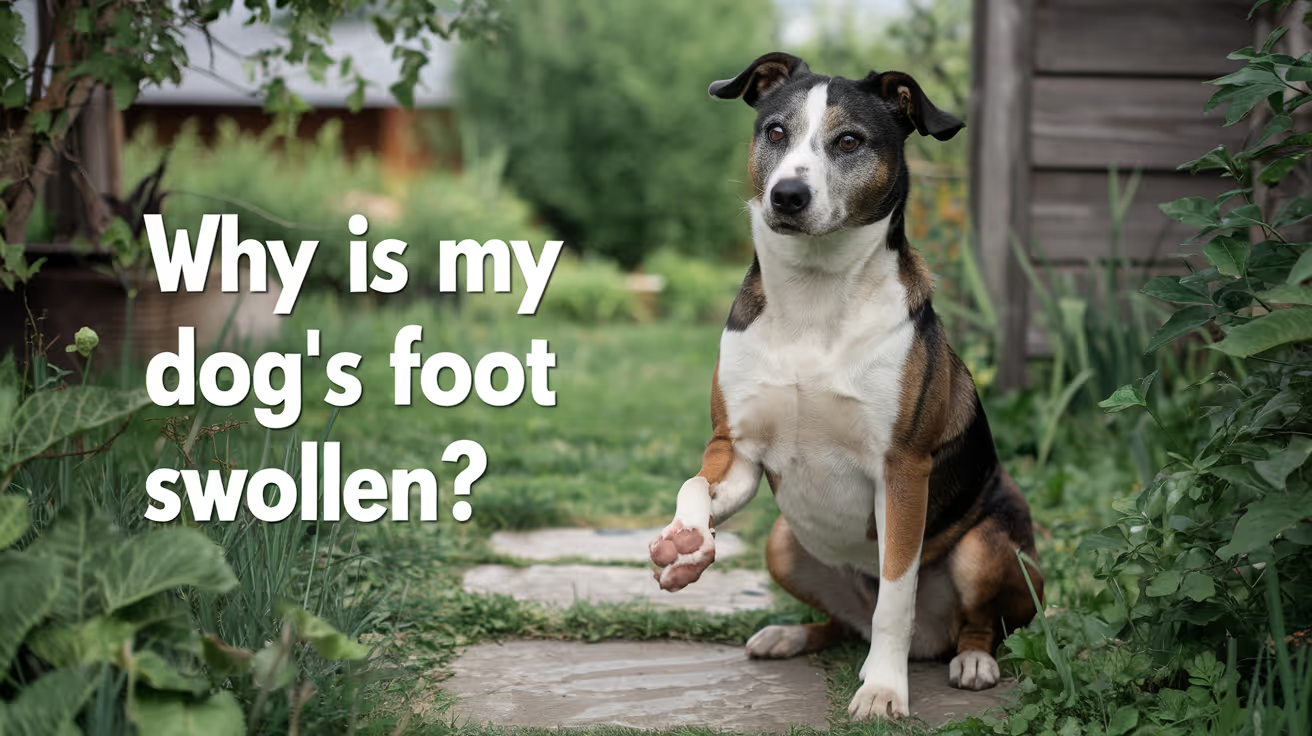
Why Is My Dog's Foot Swollen?
Discover why your dog's foot is swollen, common causes, treatments, and when to see a vet for proper care.
A swollen dog foot is one of the most common reasons owners call their vet. It can be as simple as a bee sting or as serious as a deep infection or bone tumor. The location, speed of onset, and other symptoms all help narrow down the cause.
This guide covers every common cause, how to assess severity at home, and the exact signs that mean your dog needs same-day care.
Quick answer: A swollen dog foot usually has a local cause: injury, insect sting, foreign object, or infection. Warm, increasing, or discharging swelling needs same-day vet care. Multiple swollen paws suggest allergies.
Key takeaways
- Single-paw swelling almost always has a local cause: injury, sting, foreign body, or infection
- Interdigital furunculosis (infected hair follicle cysts between toes) is among the most misidentified causes of recurring paw swelling
- Foreign objects like foxtails and glass splinters can migrate deep into tissue if left untreated
- Pododermatitis is the clinical term for inflamed paw skin; allergies are the most common underlying driver
- Swelling spreading up the leg or paired with face or neck swelling is an emergency requiring immediate care
- Most mild swelling from a sting or minor sprain improves within 24 to 48 hours with rest and monitoring
How to assess your dog's swollen foot before calling the vet
Before looking up causes, do a quick home assessment. This takes two minutes and tells you how urgent the situation is.
Step 1: Which paw?One paw = likely local cause. All four paws = likely allergic or systemic.
Step 2: Feel for heat.Warm or hot tissue means active inflammation or infection.
Step 3: Look between every toe.Redness, swelling between the digits, or visible nodules between the toes points to interdigital furunculosis or a foreign body.
Step 4: Check the pads.Burns appear as red, blistered, or peeling pad tissue. Cuts and punctures are often visible on the pad surface.
Step 5: Look at each nail.A broken nail or swollen nail bed causes localized pain and swelling at the toe tip.
VCA Animal Hospitals: "A deeper infection, like an abscess, will appear as a warm, soft to mildly firm swelling under the skin."
8 common causes of a swollen foot in dogs
1. Injury or trauma
Sprains, cuts, fractured toes, torn nails, and pad burns are the most frequent causes of sudden single-paw swelling. Dogs running on rough, hot, or icy terrain are especially prone.
PetMD notes that pad burns are particularly common in summer: check the pads after any walk on hot pavement. If your palm cannot hold against the pavement for five seconds, it is too hot for your dog's feet.
Signs: sudden lameness after activity, localized swelling, visible wound or bruising.
2. Foreign object
Thorns, glass, foxtails, and grass awns lodge between the toes or penetrate the pad. The body mounts an inflammatory response around the object, causing swelling that worsens over days.
SpectrumCare: "Foxtails and grass awns can lodge between the toes or in the pad and trigger pain, swelling, and infection."
Foxtails are particularly dangerous because they are barbed and can migrate deeper into tissue over days, eventually requiring surgical removal. If you cannot see and safely remove the object, do not probe. See a vet.
3. Insect sting or bite
Bees, wasps, fire ants, and spiders cause rapid single-paw swelling that appears within minutes of the sting. The paw may look puffy and your dog may lick or hold it up.
Dyer Animal Clinic advises watching for anaphylaxis signs: hives, difficulty breathing, excessive swelling spreading beyond the paw. These require emergency care immediately.
Localized sting swelling that stays in the paw and is not worsening can be monitored at home for 24 to 48 hours.
4. Infection: bacterial and fungal
Bacterial or fungal infections enter through cuts, puncture wounds, or damaged skin. Infected paws are typically warm, red, swollen, and often have an odor or discharge. VCA lists the visual signs: "Skin infections on the feet may result in red, moist lesions between the toes."
Pododermatitis (inflammation of the paw skin) is the umbrella clinical term. Common causes include:
- Secondary bacterial infection from chronic licking
- Yeast overgrowth in skin folds between toes
- Fungal infections (ringworm can affect paws)
- Demodectic mange (mite overgrowth)
Pododermatitis does not resolve without treatment. Antibiotics, antifungal medication, or both are typically required.
5. Interdigital furunculosis
This is one of the most commonly missed causes of recurring paw swelling. Interdigital furunculosis occurs when hair follicles between the toes become infected, forming painful reddish-purple nodules that may rupture and drain.
AKC notes that the condition is especially prevalent in short-coated, heavyset breeds: Bulldogs, Labrador Retrievers, and Chinese Shar-Pei are among the most commonly affected. Chronic licking due to allergies is a major driver.
Signs: swollen, painful nodules between the toes, draining tracts, recurrent swelling in the same location.
For how furuncles between the toes connect to abscess formation, see abscess as a cause of limb swelling.
6. Allergic reaction and pododermatitis
Environmental allergens (pollen, grass, lawn chemicals, road salt) and food allergies frequently cause paw inflammation. Dogs with allergies lick their feet chronically, creating secondary infection on top of the allergic reaction.
Wakefield Pet Vet: "Allergic reactions typically cause itching, redness, and sometimes blistering between the toes or on paw pads."
Allergic paw swelling usually affects multiple paws. It tends to recur seasonally or after contact with the trigger substance. Dogs with seasonal allergies often have their worst paw symptoms in spring and fall.
7. Bursitis
Bursae are small fluid-filled sacs that cushion joints. Repeated pressure on bony prominences, especially the elbow and hock, can cause bursitis. Affected joints may appear as soft, fluctuant swellings near a joint.
For how bursitis specifically produces foot and joint swelling, see bursitis as a common cause of foot swelling.
8. Cysts, tumors, and nail bed disease
Cysts, mast cell tumors, and subungual (under-nail) tumors can all present as localized swelling on or near the foot. Petcube notes that toenail tumors are more prevalent in large black-coated breeds such as Standard Poodles, Gordon Setters, and Schnauzers.
A lump that is growing, firm, or pigmented should always be evaluated by a vet rather than monitored at home.
For an overview of lumps that cause swelling on the legs, see lumps that can cause swelling.
Severity triage: what to do right now
Home care for mild cases
For minor swelling without infection signs:
- Keep the dog calm and limit walking
- Soak the paw in warm (not hot) water with Epsom salts for 10 minutes. PetMD recommends this as an excellent short-term measure regardless of cause.
- Gently clean any visible wound with mild soap and warm water
- Apply a cool damp cloth for 10 to 15 minutes to reduce swelling from sprains or stings
- Do not apply antibiotic ointments without vet guidance some formulations are toxic to dogs if licked
For pressure-related paw and leg swelling in dogs that rest in one position for extended periods, see pressure-related swelling in dogs.
Frequently asked questions
How do I treat my dog's swollen paw at home?
For mild swelling with no wound or discharge, rest the dog and soak the paw in warm Epsom salt water for 10 minutes. Check carefully for a foreign object or insect stinger. Do not use human antibiotic creams without vet guidance. If swelling has not reduced within 24 to 48 hours or is getting worse, contact your vet.
When should I be worried about my dog's swollen paw?
Be concerned immediately if the swelling is warm, increasing, or has discharge or odor. Call the vet the same day if your dog refuses to bear weight, if the swelling is spreading up the leg, or if there is any sign of an allergic reaction such as facial swelling or breathing difficulty.
What can I give my dog for a swollen paw?
Do not give human pain medications (ibuprofen, acetaminophen, aspirin) to dogs; many are toxic. For minor swelling, warm Epsom salt soaks are safe and effective for short-term relief. Your vet can prescribe appropriate anti-inflammatory medication if needed after examining the paw.
My dog's foot was normal this morning and is swollen now. What happened?
Sudden single-paw swelling most often means an insect sting, a foreign object picked up during activity, or a minor sprain. Check each toe carefully for a stinger, thorn, or visible wound. If swelling is mild and localized, monitor for 24 hours. If increasing or painful, call the vet the same day.
Can swelling in a dog's foot go away on its own?
Minor swelling from a bee sting or small sprain typically resolves within 24 to 48 hours with rest. Infections, foreign objects, interdigital furunculosis, and bursitis do not resolve without treatment. If swelling has not improved within 48 hours, veterinary assessment is needed.
My dog's foot smells bad and is swollen. Is that serious?
Yes. Odor from a swollen paw strongly indicates infection. A foul smell means bacteria are actively present. Common sources include interdigital furunculosis, a nail bed infection, or an abscess. This requires prompt veterinary treatment and should not be left to resolve on its own.
Resources
- PetMD. Dogs Swollen Paws: Causes and Treatments. petmd.com
- VCA Animal Hospitals. First Aid for Limping Dogs. vcahospitals.com
- AKC. Pododermatitis on Dog Paw: Causes, Symptoms and Treatment. akc.org
- SpectrumCare. Paw Swelling in Dogs. spectrumcare.pet
- Dyer Animal Clinic. Reasons Your Dog's Paw is Swollen. dyeranimalclinic.com
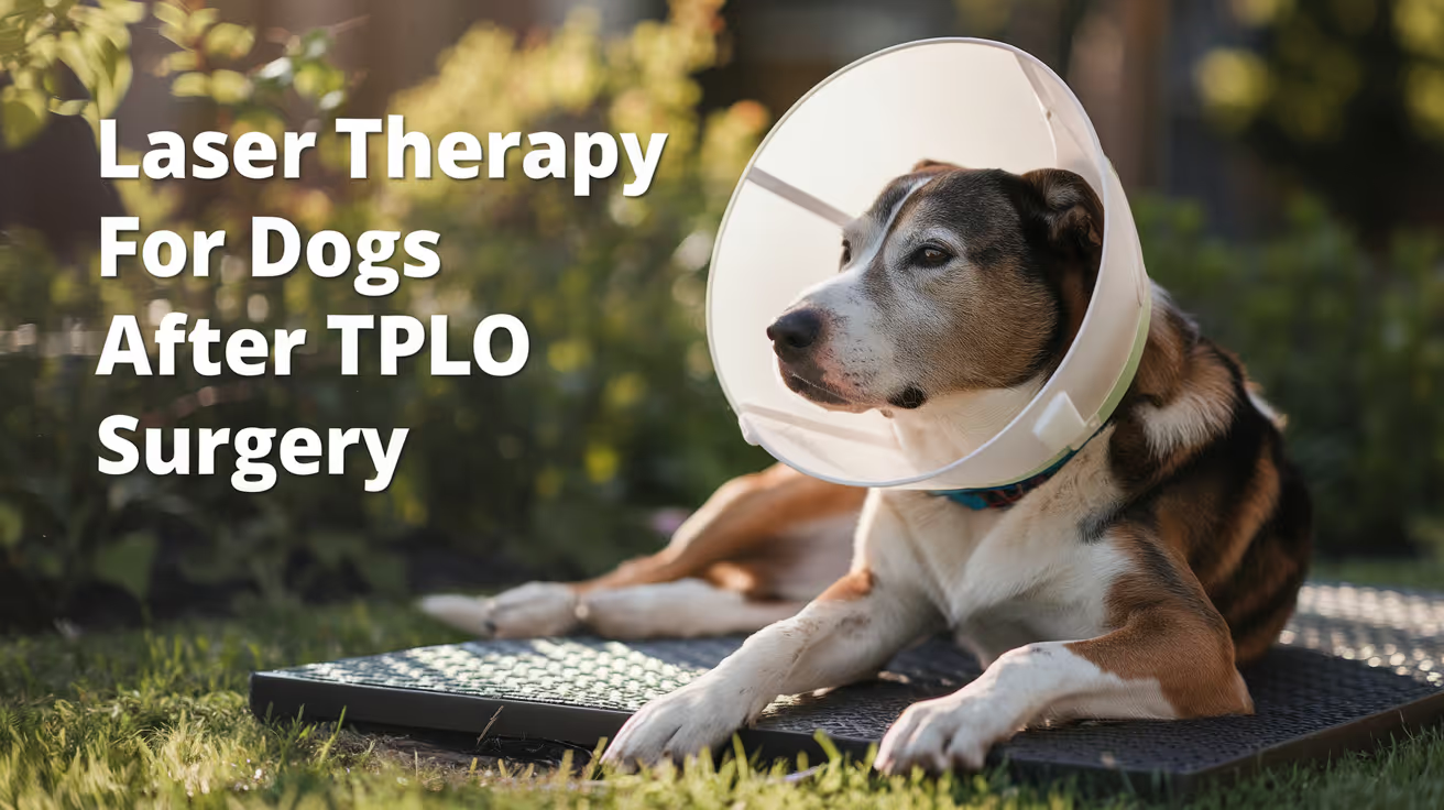
Laser Therapy for Dogs After TPLO Surgery
Learn how laser therapy helps dogs recover faster and with less pain after TPLO surgery for cruciate ligament repair.
Laser therapy also called photobiomodulation (PBMT) or low-level laser therapy (LLLT) is one of the most commonly offered adjunct treatments during TPLO recovery. Many specialist and rehabilitation centres include it routinely.
But the evidence for its benefits is more nuanced than the marketing suggests, and owners deserve an honest picture of what it does and does not reliably achieve.
Quick answer: Laser therapy after TPLO uses specific light wavelengths to reduce inflammation and support tissue healing. Evidence for early pain reduction is moderately supported; evidence for improved radiographic bone healing is weak. It is a safe adjunct but should not replace rehabilitation exercises, pain medication, or activity restriction.
Key takeaways
- Laser therapy reduces postoperative inflammation and may improve gait scores: a TPLO study found better hindlimb function at 8 weeks in treated dogs
- Evidence for improving radiographic bone healing is weak: three controlled studies found no statistically significant difference in healing time
- The 2024 AVMA randomized trial found no significant difference in CRP, weight bearing, pain scores, or SSI rates between PBMT and sham groups
- Sessions typically begin within the first few days of surgery and continue through the rehabilitation phase
- Laser therapy is safe with few contraindications: avoid eyes and active tumour sites; safe over the TPLO incision once closed
- It works best as part of a multimodal plan: exercise therapy and pain medication carry stronger evidence than laser alone
What laser therapy does
Laser therapy for dogs, also known as photobiomodulation, involves using specific wavelengths of light to penetrate tissues and promote cellular regeneration and healing. The laser light stimulates the production of ATP (adenosine triphosphate), enhancing cell repair and growth, reducing inflammation, and increasing blood circulation.
Photobiomodulation therapy has been shown to decrease inflammation, and increase analgesia, vascularization, and tissue healing after musculoskeletal injury or surgery.
The mechanism is photochemical: light energy at specific wavelengths (typically 630 to 980 nm) is absorbed by mitochondria.
This increases ATP production, modulates reactive oxygen species, and influences gene expression related to inflammation and healing.
The effects are local confined to the tissue depth the light reaches.
For post-TPLO use, the targets are: the surgical incision, the osteotomy site in the proximal tibia, and the surrounding periarticular soft tissues.
What the clinical evidence shows
Pain and function
Research following TPLO surgery showed that dogs receiving LLLT had better hindlimb function and gait scores at 8 weeks compared to controls. This is especially valuable in orthopedic recovery, where early weight-bearing can prevent muscle atrophy and joint stiffness.
In a controlled veterinary study, dogs with surgical incisions treated with laser therapy exhibited significantly less inflammatory cell infiltration and tissue necrosis within the first week post-op compared to untreated controls.
The 2024 randomized trial
54 client-owned dogs with CCL rupture undergoing unilateral TPLO surgery were enrolled. The study population was randomly assigned to either a treatment group receiving PBMT (24 dogs) or a control group (30 dogs). PBMT was performed immediately after induction, and at 6 hours, 24 hours, 48 hours, and 8 weeks postoperatively. Evaluation of CRP, pain scores, evidence of SSI, and percentage weight bearing were assessed at all time points.
The trial found the therapy showed promise but no statistically significant difference between groups on any primary outcome measure.
Bone healing
Three studies compared LLLT to a control and concluded that LLLT treatment did not make a significant difference in improving radiographic bone healing. The studies collectively provide weak evidence for this outcome.
This is an important distinction: laser therapy may support soft tissue healing, pain, and early function but it does not appear to accelerate the osteotomy healing visible on radiographs.
When to start and how often
Laser therapy uses focused light energy on the surgical site to support healing.
Most rehabilitation programmes begin laser therapy within the first 1 to 3 days after TPLO surgery, often at the surgical centre before discharge or at the first rehabilitation visit.
Typical post-TPLO laser protocol:
- Frequency: 3 to 5 sessions per week in the first 2 to 3 weeks
- Frequency: 1 to 2 sessions per week from weeks 3 to 8
- Session duration: 5 to 15 minutes depending on the laser system and dosing protocol
- Total sessions: typically 6 to 12 in the first 8-week recovery phase
The protocol varies by laser system, power output, and the individual patient's response.
Realistic expectations
Laser therapy is a useful adjunct in TPLO recovery. It is not a substitute for the treatments with stronger evidence: pain medication, activity restriction, and structured rehabilitation exercises.
Laser therapy could be particularly helpful for dogs with weight-bearing and gait issues while recovering from TPLO surgery after a cruciate injury.
Dogs with significant early swelling, wound sensitivity, or slow initial weight-bearing progress may benefit most. Dogs recovering well with standard multimodal analgesia and rehabilitation may show less measurable difference.
For the bone healing timeline that laser therapy supports during recovery, see TPLO bone healing time in dogs explained.
For the full recovery plan that laser therapy fits into, see 10 essential TPLO recovery tips for pet owners.
For the physical therapy that is the primary evidence-based adjunct, see when to start physical therapy after TPLO surgery.
For swelling management in the recovery period, see how long does swelling last after TPLO surgery.
Frequently asked questions
Is laser therapy safe over the TPLO incision?
Yes, once the incision is closed. Laser therapy is safe over sutured incisions and can be applied at the surgical site from the first post-operative day in most protocols.
Avoid direct application over open wounds or actively infected tissue.
How many laser sessions does a dog need after TPLO?
Typically 6 to 12 sessions across the first 8 weeks.
Start at 3 to 5 sessions per week for the first 2 to 3 weeks, tapering to 1 to 2 per week through the rehabilitation phase.
The exact protocol depends on the laser system and rehabilitation plan.
Does laser therapy replace pain medication after TPLO?
No. Laser therapy is an adjunct to pharmaceutical pain management, not a replacement. Post-TPLO pain management requires NSAIDs, and often gabapentin or other analgesics.
Laser therapy may reduce the pain burden and support earlier mobility but does not provide sufficient analgesia on its own.
Can I do laser therapy at home with a consumer device?
Consumer-grade red light therapy devices exist but operate at lower power densities than veterinary therapeutic lasers. The clinical evidence discussed in this article relates to veterinary-grade PBMT devices.
Home devices may offer some benefit but cannot replicate the dosing of professional equipment. Discuss with your rehabilitation veterinarian before purchasing.
Will laser therapy prevent my dog from needing more medication?
Possibly. If laser therapy reduces post-operative inflammation and pain, some dogs may need lower doses of pain medication or taper off sooner.
This should be guided by your veterinarian based on your dog's individual recovery trajectory.
Resources
- AVMA Journal. Photobiomodulation Therapy in Dogs Undergoing TPLO After Cranial Cruciate Ligament Rupture. avmajournals.avma.org
- Veterinary Evidence. Does LLLT Improve Radiographic Healing for Dogs with CCL Rupture Undergoing TPLO Surgery? veterinaryevidence.org
- AKC. Laser Therapy For Dogs: Uses, Side Effects, and Alternatives. akc.org
- Erchonia. Laser Therapy for Post-Surgical Recovery in Pets. erchonia.com
All Articles

Side Effects of Spaying a Female Dog Explained
Learn the common and rare side effects of spaying a female dog, including weight gain, hormonal issues, and recovery risks.
Sustainable Vet Group
Spaying is one of the most commonly performed veterinary procedures in the US. Most conversations about it focus on the benefits. The side effects get less attention.
That is worth correcting. Understanding what side effects are normal, which ones require monitoring, and which ones are breed and timing-dependent helps you make better decisions and know what to watch for after surgery.
This guide covers both the short-term effects that appear immediately after surgery and the long-term effects that develop over months or years.
Quick answer: Short-term side effects of spaying include temporary fatigue, reduced appetite, mild soreness, and incision swelling, all of which resolve within days. Long-term effects that affect some dogs include modest weight gain, urinary incontinence in larger breed females, and possible orthopedic risks in large breeds spayed early. Most long-term risks are manageable through appropriate timing, diet, and exercise.
Key takeaways
- Short-term side effects are mild and temporary: Fatigue, soreness, and reduced appetite typically resolve within 48 to 72 hours.
- Weight gain is the most common long-term side effect: Metabolism slows slightly after spaying; diet adjustment prevents it.
- Urinary incontinence affects a meaningful subset of spayed females: Estimated at 5 to 20%, with higher risk in larger breeds.
- Coat changes occur in some dogs: A softer, thicker coat (spay coat) can develop in certain breeds.
- Joint risks are breed and timing dependent: Large breed females spayed early face higher orthopedic risk; delayed spaying reduces this.
- Benefits outweigh risks for most dogs: Pyometra prevention, mammary cancer reduction, and eliminated heat cycles are significant health gains.
Short-term side effects (first 10 to 14 days)
These effects are directly related to the surgery and anesthesia. They are expected and resolve with time and proper care.
Fatigue and lethargy
Your dog will be groggy for the first 24 to 48 hours as anesthesia clears her system. Reduced energy for two to three days after surgery is completely normal.
Lethargy that persists beyond 48 hours, or a dog that is completely unresponsive, warrants a call to your vet.
Reduced appetite
Most female dogs show little interest in food the first evening after surgery. Appetite typically returns to normal by day two.
A dog still refusing food at 48 hours post-surgery needs veterinary evaluation.
Soreness and discomfort
Spaying involves abdominal surgery. Mild soreness at the incision site and general post-operative discomfort are expected.
Pain medication prescribed by your vet manages this effectively in most cases. Signs that pain is not being adequately controlled include:
- Panting or trembling without exertion
- Hunching the back or reluctance to lie down
- Whining or vocalizing when moving
- Persistent guarding of the belly
Incision swelling and redness
Mild swelling and pinkness at the incision site in the first three days are normal parts of the inflammatory healing response.
Swelling that increases after day three, discharge that is yellow or green, or any foul odor are not normal and require veterinary attention.
Nausea and vomiting
One or two episodes of vomiting the evening of surgery can be a normal response to anesthesia.
Vomiting that continues beyond 24 hours warrants a vet call.
Behavioral changes during recovery
Some dogs become clingy, withdrawn, or mildly irritable during the recovery period. This is related to pain and the stress of surgery, not permanent personality change.
For complete guidance on managing recovery safely, how to manage side effects during recovery covers every element of the post-operative window.
Long-term side effects
These are effects that develop over weeks, months, or years after spaying. They vary by individual dog, breed, and timing of surgery.
Weight gain
This is the most commonly observed long-term side effect and the one most within your control.
Spaying causes a modest reduction in metabolism because estrogen plays a role in regulating caloric burning. Dogs that continue eating the same amount after surgery tend to gradually gain weight.
The surgery does not cause obesity. Overfeeding does. A simple caloric adjustment of approximately 10 to 20% after spaying, combined with consistent exercise, prevents weight gain in most dogs.
Managing weight after spaying:
- Discuss post-spay caloric needs with your vet at the follow-up appointment
- Measure portions rather than free-feeding
- Maintain regular daily exercise
- Transition to a lower-calorie maintenance food if needed
Urinary incontinence
Urinary incontinence is one of the most underreported long-term side effects of spaying in female dogs.
Estrogen plays a role in maintaining the tone of the urethral sphincter. After spaying, estrogen levels drop permanently. In a subset of spayed females, the sphincter weakens enough to cause urine leakage, most commonly during sleep or rest.
Estimates of incidence range from 5% to 20% of spayed females, with the highest rates in females over 33 pounds. The condition is called hormone-responsive incontinence or spay-related incontinence.
It is manageable with medication in most cases and does not typically worsen dramatically over time. If you notice your spayed female leaving urine spots when she sleeps, mention it at your next vet visit.
For a comprehensive view of surgical risks versus expected side effects and how incontinence fits within the broader risk picture, that guide covers the full range.
Coat changes
Some breeds, particularly those with double coats such as Golden Retrievers, Labrador Retrievers, and similar breeds, develop what owners and groomers call a "spay coat" after spaying.
The coat may become softer, fluffier, and less well-defined than before surgery. This occurs because sex hormones influence coat texture in certain breeds.
Not all dogs are affected. The change is cosmetic and does not affect health.
Orthopedic risks in large breeds spayed early
Research from UC Davis and other institutions has established that large and giant breed females spayed before full skeletal maturity face elevated risk of certain joint disorders, including hip dysplasia and cranial cruciate ligament rupture.
Sex hormones signal growth plates to close at the appropriate time. Removing estrogen before the growth plates have closed can delay closure and affect long-term joint development and stability.
This risk is specific to large and giant breeds and is most significant when spaying occurs before 12 months of age. The same risk profile does not apply to small breed females.
Current guidelines recommend that large breed females be spayed between 5 and 15 months of age, with many veterinarians leaning toward the later end of that range. The specific recommendation varies by breed and individual health factors.
Some cancer associations in specific breeds
For certain large breeds, particularly Golden Retrievers and some other purebreds, early spaying has been associated in research with elevated rates of specific cancers including lymphoma and hemangiosarcoma.
These associations are breed-specific and do not apply universally. For the majority of female dogs, spaying reduces overall cancer risk by eliminating mammary tumors and uterine cancer.
The net cancer impact of spaying depends heavily on breed, timing, and individual risk profile.
Short-term vs. long-term side effects: at a glance
Side effects that are NOT caused by spaying
Several things are commonly attributed to spaying that are not actually caused by the procedure.
Does not cause:
- Permanent laziness or loss of energy (activity levels recover fully once healed)
- Personality changes (core temperament is not affected)
- All forms of aggression (hormone-related aggression may decrease; other types are not affected)
- Universal joint problems (this risk is specific to large breeds spayed before skeletal maturity)
Understanding behavior changes specific to female dogs after spaying helps clarify which changes are genuinely attributable to spaying and which are myths. For a broader view of behavioral changes in all dogs after spaying or neutering, including how male and female responses compare, that guide puts the female-specific picture in context.
When side effects warrant veterinary contact
Call your vet same day if:
- Lethargy continues beyond 48 hours
- Vomiting continues beyond 24 hours post-surgery
- Incision shows yellow or green discharge, increasing swelling, or foul odor
- Urination appears painful or your dog is straining to urinate
Long-term, discuss at your next regular appointment:
- Weight gain that is not responding to portion control
- Urine leakage during sleep (possible urinary incontinence)
- Coat changes that concern you
- Any signs of joint stiffness or pain in large breed dogs
For a detailed list of when side effects become warning signs that need prompt attention, understanding the difference between expected side effects and complications prevents both unnecessary worry and dangerous delays.
Frequently asked questions
Will my dog gain weight after being spayed?
Many dogs do gain weight if their diet is not adjusted, because spaying causes a modest metabolic slowdown. The weight gain itself is preventable with portion control and exercise. Talk to your vet at the follow-up appointment about whether a caloric adjustment is appropriate for your dog.
How long does soreness last after a spay?
Mild soreness typically peaks in the first two to three days and improves gradually through day five to seven. Some dogs show residual sensitivity at the incision site until the full 10 to 14 day healing window is complete. Pain medication prescribed by your vet manages this.
Can spaying cause urinary incontinence?
Yes, in some females. The condition is called hormone-responsive incontinence and is caused by the drop in estrogen after spaying, which affects urethral sphincter tone. It affects an estimated 5 to 20% of spayed females and is more common in larger breeds. It is manageable with medication in most cases.
Is a spay coat permanent?
The coat texture change that occurs in some double-coated breeds is generally long-lasting. However, it is purely cosmetic and does not affect your dog's health or comfort. Some owners find that regular grooming helps manage the texture change.
Are the long-term side effects serious enough to avoid spaying?
For the vast majority of dogs, no. The health benefits of spaying, including elimination of pyometra risk, significant reduction in mammary tumor risk, and elimination of heat cycle complications, substantially outweigh the side effects for most females. For large breed dogs, timing the surgery appropriately minimizes the orthopedic risks that represent the most significant long-term concern.
Side effects of spaying are real, and you should know about all of them. But understanding them in context makes the picture clearer: most short-term effects resolve within days, and most long-term effects are either manageable or timing-dependent. The side effect profile of spaying is well-understood, which is precisely what allows veterinarians and owners to plan around it effectively.
Resources
The following sources were used as reference and background for this article:
- American Veterinary Medical Association (AVMA). Spaying and Neutering. avma.org
- North Oatlands Animal Hospital. Common Side Effects of Spaying Your Dog Explained. noahvets.com
- PetsCare. Side Effects of Spaying a Female Dog: A Guide. petscare.com
- Kutzler, M.A. (2020). Possible Relationship between Long-Term Adverse Health Effects of Gonad-Removing Surgical Sterilization and Luteinizing Hormone in Dogs. Animals, 10(4), 599. ncbi.nlm.nih.gov
- Hart, B.L., Hart, L.A., Thigpen, A.P., Willits, N.H. (2020). Assisting Decision-Making on Age of Neutering for 35 Breeds of Dogs. Frontiers in Veterinary Science, 7: 388.
When Should You Consider Arthroscopy for Lameness in Dogs?
Learn when to consider arthroscopy for lameness in dogs. Discover key signs, conditions, and factors that show surgery may be the right choice
Sustainable Vet Group
Lameness in a dog is common. Most cases resolve with rest, pain management, and time. But some don't, and some require a look inside the joint to understand what's actually happening.
Arthroscopy moves from something to consider to something to pursue when imaging leaves questions unanswered, when a specific condition is confirmed that arthroscopy treats well, or when conservative management has been tried and hasn't worked. For a full explanation of what arthroscopy is, see what arthroscopy is.
Quick answer: Consider arthroscopy when a dog has persistent joint lameness that doesn't fully respond to conservative management, when imaging suggests a condition that arthroscopy treats (OCD, elbow dysplasia, meniscal tear, shoulder instability), or when a definitive diagnosis requires direct joint visualization. Early treatment through arthroscopy before arthritis establishes produces the best long-term outcomes.
Key takeaways
- Persistent lameness not responding to conservative care is the primary clinical trigger.
- Specific diagnoses drive arthroscopy more than symptom severity: OCD, FMCP, UAP, meniscal tears.
- Early intervention is better: treating before arthritis develops produces better outcomes.
- Arthroscopy is both diagnostic and therapeutic in most cases; two procedures often become one.
- Large active breeds with elbow or shoulder lameness are the most common arthroscopy candidates.
- Hip dysplasia as a cause of lameness has different management than arthroscopic conditions.
When conservative management alone isn't enough
Most lameness in dogs starts with conservative management: rest, anti-inflammatory medication, and restricted activity. For mild soft tissue injuries and minor joint flare-ups, this often resolves the problem.
The signals that conservative management needs to be re-evaluated:
- Lameness persisting beyond 4 to 6 weeks despite appropriate rest and medication
- Lameness recurring after a period of apparent improvement
- Worsening rather than improving lameness on a conservative management protocol
- A dog whose lameness isn't explained by imaging (X-rays show nothing remarkable but the dog continues to limp)
- Bilateral joint involvement where the dog is lame on both front legs, suggesting a systemic developmental condition rather than a simple injury
For most dogs with persistent front leg lameness in large breeds, elbow dysplasia is a strong diagnostic consideration. For young large-breed dogs with shoulder lameness, OCD is common.
Conditions that indicate arthroscopy
Elbow dysplasia (FMCP, UAP, OCD of elbow)
Elbow dysplasia is the most common indication for arthroscopy in dogs. It typically presents as front leg lameness in young large-breed dogs, often starting at 5 to 9 months of age. Breeds commonly affected: Labrador Retrievers, Golden Retrievers, Rottweilers, Bernese Mountain Dogs, German Shepherds.
When to consider arthroscopy: A young large-breed dog with persistent front leg lameness, elbow pain on palpation, and imaging suggesting elbow dysplasia. CT scan before arthroscopy is increasingly standard, as it provides better visualization of the coronoid and cartilage than radiographs.
Why early intervention matters: Metropolitan Veterinary Associates: "Early detection and removal is ideal." Arthritis accumulates at the fragmented coronoid site over time. Treating before significant arthritis develops produces better long-term outcomes.
For how these conditions are addressed, see conditions arthroscopy addresses.
OCD of the shoulder, stifle, or tarsus
OCD (osteochondritis dissecans) is a developmental condition in which cartilage fails to ossify correctly, forming a loose flap. It presents as lameness in young growing dogs, typically 5 to 14 months.
When to consider arthroscopy: OCD confirmed or strongly suspected by imaging in a young dog with corresponding lameness. Shoulder OCD in particular benefits from early arthroscopic treatment.
Typical dog: young, large breed, front leg lameness, shoulder pain on range-of-motion testing. Radiograph or CT confirms the lesion.
Meniscal tears associated with cruciate disease
When a dog undergoes TPLO or other cruciate surgery, the meniscus is routinely assessed arthroscopically. Meniscal tears are also assessed in dogs who develop new stifle lameness weeks or months after cruciate surgery.
When to consider arthroscopy: A dog developing (or returning with) stifle lameness after cruciate surgery. The meniscus that was intact at the time of the original surgery may have subsequently torn.
Shoulder instability and biceps tendon pathology
Medial shoulder instability and biceps tendon disease typically present as shoulder lameness in middle-aged to older active dogs. Both are difficult to diagnose definitively on radiographs and CT.
When to consider arthroscopy: Persistent shoulder lameness in an active dog where imaging is inconclusive. Arthroscopy of the shoulder confirms soft tissue pathology invisible to other imaging and allows treatment in the same procedure.
When diagnostic arthroscopy is warranted
Sometimes the indication for arthroscopy isn't a specific confirmed diagnosis but an unexplained joint problem.
Situations where diagnostic arthroscopy is appropriate:
- Persistent joint lameness where radiographs and CT show nothing conclusive
- Suspected joint infection where fluid sampling is needed for culture
- Suspected inflammatory joint disease requiring synovial biopsy
- A dog with joint effusion (fluid in the joint) of unknown cause
VCA Animal Hospitals: "Ultimately, arthroscopy can be used as a superior diagnostic tool, as the inside of the joint can be seen by your veterinarian and an appropriate treatment plan can be made."
The advantage is that diagnostic arthroscopy can transition to therapeutic arthroscopy in the same anesthetic event if treatment is needed. Finding a fragmented coronoid or OCD lesion during diagnostic arthroscopy means it can be addressed immediately.
The role of timing: why earlier is usually better
For the conditions arthroscopy treats, earlier intervention before arthritis establishes consistently produces better long-term outcomes.
Why delay is costly for arthroscopic conditions:
- OCD: every day the cartilage flap remains, more underlying bone damage occurs. The longer the OCD goes untreated, the more extensive the defect and the worse the prognosis.
- FMCP: ongoing fragmentation and bone contact continues to damage the medial cartilage of the elbow with each step. Earlier removal means less cartilage damage to recover from.
- Shoulder instability: progressive ligament stretching worsens with time; earlier stabilization prevents further laxity.
The practical implication: if your dog has a suspected arthroscopic condition confirmed by imaging, discussion with a specialist about timing should happen promptly rather than after months of continued conservative management.
Signs in the dog that suggest arthroscopy should be considered
Clinical signs:
- Persistent lameness on one specific front or rear leg beyond 4 to 6 weeks
- Swelling of the joint on the lame leg
- Pain on palpation or range-of-motion testing of a specific joint
- Young large-breed dog with front leg lameness (elbow dysplasia suspect)
- Dog with cruciate repair who develops new lameness (late meniscal tear)
Imaging findings that suggest arthroscopy:
- Elbow dysplasia changes on radiograph or CT (subchondral bone lesion, joint effusion, osteophytes)
- OCD lesion confirmed on radiograph or CT
- Joint effusion without obvious cause on imaging
- Shoulder changes suggesting OCD or soft tissue pathology
Response to treatment:
- Lameness that has been managed conservatively for 4 to 6 weeks and remains significant
- Lameness that returns after periods of apparent improvement
- A dog on NSAIDs who is comfortable on medication but immediately lame without it (masking rather than resolving the problem)
For how hip dysplasia as a cause of lameness is managed differently, see hip dysplasia as a cause of lameness.
When arthroscopy may not be the right next step
Arthroscopy is not appropriate for all lame dogs:
- Soft tissue injuries (muscle strains, ligament sprains) that need rest, not surgery
- Bone fractures requiring open fixation
- Neurological conditions causing apparent lameness: intervertebral disc disease, degenerative myelopathy
- Arthritis-only cases where the cartilage loss is too advanced for debridement to help significantly; medical management or THR may be more appropriate
Your vet or specialist will differentiate these through physical examination, imaging, and sometimes advanced diagnostics.
For the cost considerations when deciding on arthroscopy, see cost to consider when deciding. For the full comparison between arthroscopy and open surgery as decision factors, see comparing arthroscopy to open surgery alternatives.
Frequently asked questions
My dog is limping on and off. Should I push for arthroscopy?
Intermittent lameness that fully resolves between episodes may not yet require arthroscopy. The first step is a full orthopedic examination with appropriate imaging to determine whether a structural problem (like OCD or FMCP) is the cause. If imaging supports a structural diagnosis, arthroscopy becomes relevant. If nothing structural is found, conservative management and monitoring are appropriate.
My vet says to wait and see. Is that always the right approach?
For mild, possibly self-resolving lameness, watching and waiting is appropriate. For lameness in a young large-breed dog with imaging changes suggesting elbow dysplasia or OCD, waiting has real costs: more arthritis accumulates, cartilage damage worsens, and outcomes decline. Ask your vet specifically whether the condition being monitored is one where waiting worsens the prognosis.
Should I see a specialist or can my vet perform arthroscopy?
Arthroscopy requires specialized equipment and specific training. Most general practitioners don't have the equipment or the case volume to produce specialist-level outcomes. For elbow dysplasia, shoulder OCD, and other common arthroscopic conditions, a referral to a board-certified veterinary surgeon with regular arthroscopy experience is strongly recommended.
The decision to consider arthroscopy comes from the intersection of a persistent or worsening joint problem, an imaging picture that points toward a condition arthroscopy addresses, and a dog who would benefit from the definitive diagnosis and treatment that a direct look inside the joint provides. For young large-breed dogs with elbow or shoulder lameness, getting to that conversation with a specialist sooner rather than later is often in the dog's best interest.
Resources
- VCA Animal Hospitals. Arthroscopy. vcahospitals.com
- BluePearl Pet Hospital. Arthroscopic Surgery. bluepearlvet.com
- Metropolitan Veterinary Associates. Arthroscopy in Canine Orthopedic Disease. metro-vet.com
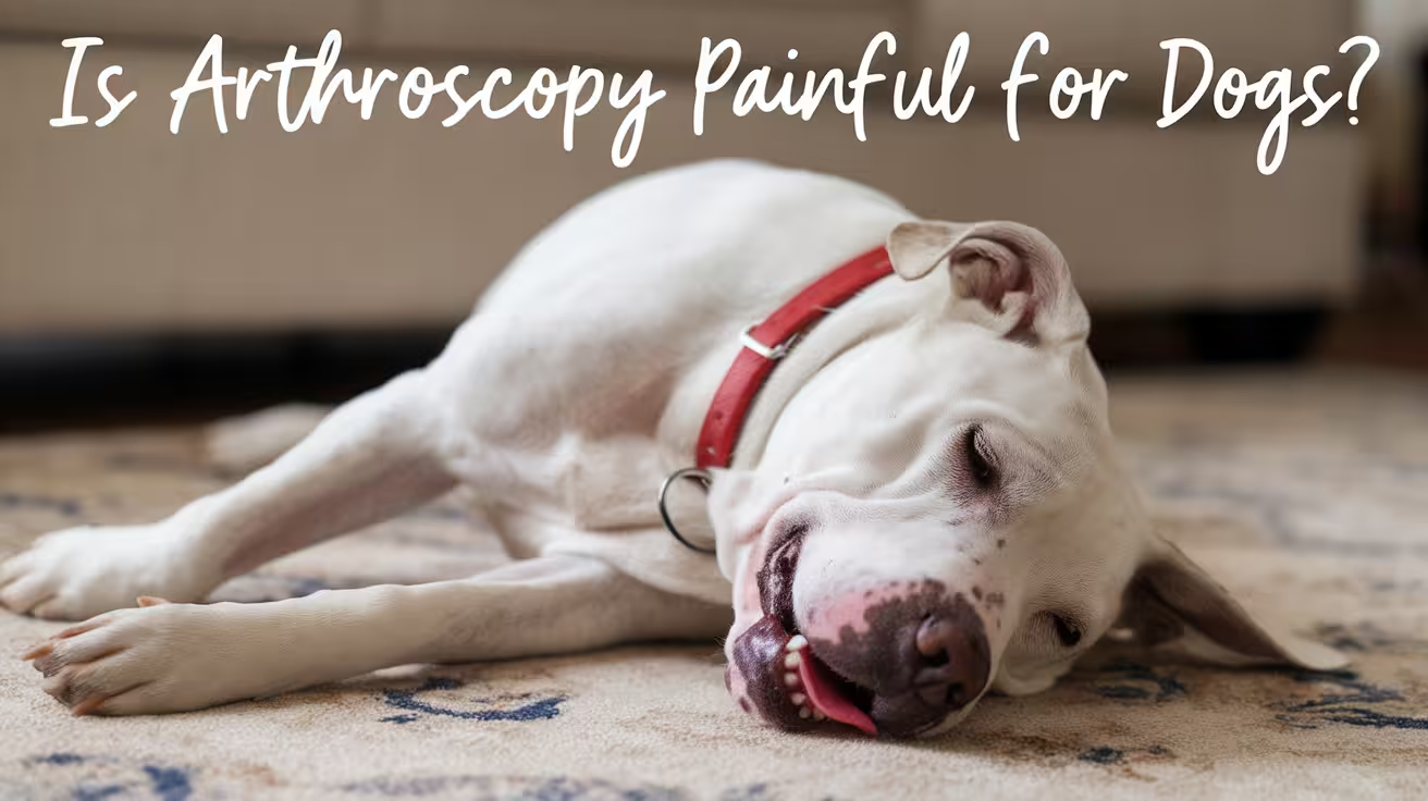
Is Arthroscopy Painful for Dogs?
Arthroscopy in dogs causes less pain than open surgery. Learn how much discomfort to expect, recovery timeline, and pain management after the procedure
Sustainable Vet Group
Pain is one of the first concerns owners raise when arthroscopy is recommended. The honest answer: during the procedure, your dog feels nothing. Afterward, there is some discomfort, and it's significantly less than open joint surgery. For a full explanation of what arthroscopy is and how it works, see what the procedure involves.
Understanding the pain experience, what to expect, and how it's managed helps you know what your dog is going through and what your role is in keeping them comfortable.
Quick answer: Dogs feel no pain during arthroscopy because they are under general anesthesia. Post-operative pain is mild to moderate for most dogs and is well-managed with NSAIDs and analgesics. Most dogs improve noticeably within 24 to 48 hours and begin weight-bearing within 1 to 2 days. Post-surgical pain from arthroscopy is significantly less than equivalent open joint surgery because the tissue disruption is far smaller.
Key takeaways
- No pain during the procedure: general anesthesia ensures complete unconsciousness.
- Mild to moderate post-operative soreness is expected for the first 24 to 48 hours.
- NSAIDs are the primary pain medication after arthroscopy; short opioid courses may supplement.
- Most dogs are noticeably more comfortable within 48 hours compared to pre-surgery chronic pain.
- Arthroscopy causes less post-surgical pain than open surgery for equivalent conditions.
- Stifle arthroscopy causes more soreness than elbow or shoulder, due to weight-bearing demands.
During the procedure: no pain
Dogs undergoing arthroscopy receive general anesthesia. This renders them fully unconscious and incapable of feeling pain throughout the procedure. Pre-anesthetic medications (sedation and often pre-emptive analgesia) are given before anesthesia induction.
In some cases, local anesthesia (intra-articular bupivacaine or similar) is injected into the joint at the conclusion of the procedure. This provides an additional layer of pain control that takes effect as the dog begins to wake up.
Fusion Veterinary Orthopedics: "Reduced pain is a key advantage of arthroscopy. Smaller incisions mean less pain for the pet during and after the procedure."
Post-operative pain: what to expect
The first 24 to 48 hours
Mild to moderate soreness at the operated joint is expected and normal in the first day or two. Sources of this pain include:
- The portal incisions: small but they do disrupt skin and the joint capsule
- The joint itself: the irrigation, instrument manipulation, and treatment create some inflammation
- Fluid pressure changes: the joint was distended with sterile fluid during the procedure; this can cause mild stiffness and aching as the fluid is absorbed
Most dogs show obvious soreness in the first 12 to 24 hours. By 24 to 48 hours, the trend should be clearly toward improvement.
Signs of expected post-operative pain:
- Limping on the operated leg
- Reluctance to bear full weight initially
- Mild restlessness or seeking comfortable positions
- Reduced activity and increased sleep
Days 3 to 7
Most dogs are noticeably more comfortable by day 3 to 5. Weight-bearing on the operated leg becomes more consistent. The trend of improvement should be clear.
For many dogs, particularly those who had chronic pre-surgical joint pain, the days following arthroscopy are actually more comfortable than the weeks before surgery, because the source of chronic pain (an OCD lesion, fragmented coronoid, loose fragment) has been removed.
How post-operative pain is managed
NSAIDs (anti-inflammatory medications)
NSAIDs are the primary post-arthroscopy pain medication. Carprofen, meloxicam, or grapiprant reduce joint inflammation and provide pain relief. These are typically prescribed for 7 to 14 days post-procedure.
Give NSAIDs with food to reduce GI effects. Don't skip doses even if your dog seems comfortable; consistent blood levels maintain effective pain control.
Gabapentin
Gabapentin is a nerve pain modulator that is sometimes added for the first 5 to 7 days post-procedure, particularly for stifle arthroscopy or when more significant intra-articular work was performed.
Short-course opioids
For more substantial arthroscopic procedures (extensive cartilage debridement, meniscal work), a 2 to 3 day supply of a mild opioid (tramadol, buprenorphine) may be sent home to cover the most acute phase.
Intra-articular analgesia
Local anesthetic injected into the joint at the conclusion of surgery provides several hours of additional pain relief as the dog wakes up. This reduces the severity of early post-operative pain.
Pain that varies by joint and procedure
Not all arthroscopy procedures produce the same pain level.
Elbow and shoulder arthroscopy: generally mild post-operative soreness. The elbow and shoulder are not primary weight-bearing joints during normal gait. Most dogs are comfortable within 48 to 72 hours.
Stifle arthroscopy: more soreness is expected, particularly when combined with meniscal work or performed alongside cruciate surgery. The stifle bears significant load with every step. Soreness may persist for 1 to 2 weeks at a mild level.
Extent of surgical work: a diagnostic arthroscopy with minimal treatment produces less pain than a therapeutic arthroscopy involving extensive cartilage debridement, bone work, or fragment removal.
Pre-existing arthritis: dogs with established joint disease before arthroscopy may have a slightly longer period of post-operative discomfort as the inflamed joint settles following the procedure.
Recognizing inadequate pain control
Signs that pain medication is not adequately controlling your dog's discomfort:
- Constant vocalization (whimpering, crying) beyond the first 12 to 24 hours
- Refusing to move or bear any weight on the operated leg by days 3 to 5
- Significant trembling or panting at rest
- Aggressive response when the operated area is touched (more than expected)
- Not eating by day 2 to 3
If these signs are present, contact your vet. Additional pain management options are available, and dogs don't need to suffer through inadequate control.
How arthroscopy pain compares to open surgery
This is where arthroscopy's advantage is most practical for owners.
Open joint surgery requires large incisions through skin, fascia, and muscle. Muscle retraction during surgery disrupts significant tissue mass. All these structures are inflamed post-operatively and must heal over weeks.
Arthroscopy uses 2 to 3 mm portals. Minimal muscle disruption occurs. The joint capsule is entered through tiny portals rather than a large incision.
The result: arthroscopy patients require less pain medication for a shorter duration than equivalent open surgery patients. Most arthroscopy dogs are comfortable and moving within 48 to 72 hours. Most open joint surgery dogs require several days of more intensive pain management before they begin comfortable weight-bearing.
WSAVA's evidence review confirms: "The advantages of minimally invasive arthroscopy compared to standard open arthrotomy are evident and proved clinically by reduced postoperative pain and accelerated recovery."
For the full comparison across recovery factors, see pain comparison with open surgery.
Long-term: is joint pain eliminated by arthroscopy?
Arthroscopy addresses the immediate source of joint pain (the OCD lesion, the fragmented coronoid, the loose fragment, the meniscal tear). Whether joint pain returns long-term depends on the degree of pre-existing arthritis.
- Dogs treated early, before significant arthritis: excellent long-term pain outcomes; many remain comfortable without long-term medication
- Dogs treated with established arthritis: arthroscopy reduces pain and slows progression but doesn't eliminate arthritis; long-term joint support medication (NSAIDs, Adequan, supplements) may still be appropriate
Metropolitan Veterinary Associates: "Many dogs will need periodic treatment with anti-inflammatories and/or joint injections for the remainder of their life, even after fragment removal."
For the full post-arthroscopy experience, see full arthroscopy experience. For how recovery duration relates to pain progression, see recovery timeline alongside pain management.
Frequently asked questions
My dog is crying after arthroscopy. Is that normal?
Some dogs vocalize while coming out of anesthesia, particularly as they become aware of the surgical site before pain medication has fully taken effect. This is usually brief. If vocalizing persists beyond the first few hours at home, or if your dog is crying at rest despite medication, contact your vet: the pain management protocol may need adjustment.
Can I give my dog human pain medication?
No. Ibuprofen, naproxen, and acetaminophen are toxic to dogs. Never give human pain medications to dogs, even in small amounts. Use only what your vet has prescribed.
When does pain from arthroscopy typically peak?
Pain is typically at its highest in the first 12 to 24 hours post-surgery. It should improve noticeably by day 2 to 3. If pain is increasing rather than decreasing by day 3, contact your vet.
The honest picture of arthroscopy pain: it exists, it's less than open surgery, and it's manageable with appropriate medication. For most dogs, the weeks of chronic pre-surgical pain that arthroscopy was intended to address were worse than the post-operative soreness. The recovery is measured in days of real discomfort, not weeks.
Resources
- Fusion Veterinary Orthopedics. Understanding Arthroscopy in Pets. fusionvetortho.com
- WSAVA 2010. Benefits and Limits of Arthroscopy. vin.com
- Metropolitan Veterinary Associates. Arthroscopy in Canine Orthopedic Disease. metro-vet.com
How Long Is Recovery After Arthroscopy in Dogs?
Dog arthroscopy recovery takes 8–12 weeks. Learn the timeline, phases, and factors that affect healing after joint surgery in dogs
Sustainable Vet Group
One of the most appealing features of arthroscopy over open joint surgery is the recovery time. For most conditions and most dogs, returning to normal activity takes weeks rather than months.
The precise timeline depends on the joint treated, the condition addressed, the dog's size and age, and how consistently the rehabilitation protocol is followed. For a complete guide to what happens during and after the procedure itself, see what the full recovery involves.
Quick answer: Most dogs begin weight-bearing within 1 to 2 days of arthroscopy. Full recovery, including return to normal activity, typically takes 4 to 6 weeks for most arthroscopic procedures. Stifle (knee) arthroscopy may extend to 6 to 8 weeks due to greater joint loading. This is substantially shorter than equivalent open surgery recovery.
Key takeaways
- Weight-bearing starts within 1 to 2 days for most dogs after arthroscopy.
- Full recovery: 4 to 6 weeks for most conditions and joints.
- Stifle arthroscopy recovery: 6 to 8 weeks due to higher mechanical demands.
- Activity restriction in weeks 1 to 4 is essential; early overloading risks setbacks.
- Physical therapy from weeks 2 to 3 accelerates muscle rebuilding and return to function.
- Recovery is significantly faster than open surgery, which typically takes 8 to 16 weeks for equivalent procedures.
How recovery length compares to open surgery
The difference in recovery between arthroscopy and open joint surgery is one of the strongest arguments for arthroscopy when both options are available.
Open joint surgery involves large incisions through multiple tissue layers: skin, subcutaneous fat, fascia, muscle or retraction of muscle groups, and joint capsule. All these layers must heal. The soft tissue disruption causes pain and swelling that extend recovery to 8 to 16 weeks for most major open joint procedures.
Arthroscopy accesses the joint through 2 to 3 mm portals. There is minimal soft tissue disruption. The small portal incisions heal within a week or two. The joint itself begins responding to treatment almost immediately.
Most dogs walk within 1 to 2 days after arthroscopy. Most dogs take 3 to 7 days after open joint surgery to begin comfortable weight-bearing.
For the full direct comparison, see how recovery compares to open surgery.
Recovery timeline by phase
Phase 1 (days 1 to 7): early healing
What's happening in the body: The portal incisions are healing. Joint inflammation from the procedure is settling. The joint begins responding to the treatment performed.
Activity level: Leash-only bathroom walks. No running, jumping, stairs, or off-leash activity.
Signs of normal recovery:
- Limping on the operated leg, improving day by day
- Mild swelling at portal sites, resolving within a few days
- Tentative but increasing weight-bearing by days 2 to 3
First vet recheck: typically at 10 to 14 days for suture/staple removal and progress assessment.
Phase 2 (weeks 2 to 4): controlled exercise
What's happening in the body: Portal incisions are healed. The joint is less inflamed. Muscle mass is beginning to stabilize. Tissue healing within the joint is progressing.
Activity level: Short controlled leash walks, increasing progressively. Most protocols begin with 5-minute walks 2 to 3 times daily, adding 5 minutes per week.
Ridge Referrals elbow arthroscopy protocol: "6 to 12 weeks: start to increase time on lead to maximum 10 minutes three to four times daily."
Rehabilitation starts: gentle passive range-of-motion exercises, light massage, and controlled walking. These prevent excessive scar tissue formation and begin muscle rebuilding.
Still prohibited: running, jumping, off-leash activity, rough play.
Phase 3 (weeks 4 to 6): return to activity
What's happening in the body: Internal joint healing is substantially complete for most arthroscopic procedures. Muscle mass is rebuilding with increasing activity.
Activity level: Most conditions allow progressive return to normal activity by week 6. Walk duration increases toward normal. Light play and activity on secure ground may be permitted after vet clearance.
Hydrotherapy: if available, underwater treadmill sessions are excellent for muscle rebuilding without joint impact. Often introduced from week 3 to 4 when portal incisions are confirmed healed.
Full recovery: Most owners report their dog moving normally and freely by week 4 to 6 for elbow and shoulder procedures.
How the joint treated affects recovery length
Not all joints recover at the same rate after arthroscopy.
Elbow and shoulder: faster recovery
Elbow and shoulder joints are not the primary weight-bearing joints. Dogs shift some load to other limbs during early recovery. The mechanical demands on these joints during the healing phase are lower than on the stifle or hip.
Most elbow and shoulder arthroscopy cases reach full activity by 4 to 6 weeks.
Stifle: longer recovery
The stifle (knee) is a major weight-bearing joint that bears significant load with every step. Stifle arthroscopy (typically for meniscal assessment and treatment, or OCD of the stifle) takes longer than elbow or shoulder arthroscopy.
Expect 6 to 8 weeks for stifle arthroscopy recovery. When stifle arthroscopy is combined with open cruciate surgery (TPLO), the TPLO recovery protocol drives the timeline, typically 10 to 14 weeks total.
VCA Animal Hospitals confirms: "The recovery time will depend on the extent of the injury, and the specific joint affected."
Factors that extend recovery
- Degree of pre-existing arthritis: dogs with established joint disease before arthroscopy take longer to respond
- Extent of surgical work: more debridement, bone work, or meniscal treatment means more internal healing needed
- Dog's age and size: older dogs and large breeds typically recover more slowly
- Rehabilitation compliance: dogs whose owners follow the exercise protocol consistently recover faster
The role of physical therapy in recovery speed
Physical therapy materially shortens recovery time and improves functional outcomes after arthroscopy.
Key exercises during recovery:
- Passive range-of-motion (PROM): gently flexing and extending the operated joint during weeks 1 to 4 prevents stiffness and scar tissue contracture
- Controlled leash walking: slow walking that encourages weight-bearing builds muscle and joint stability
- Hydrotherapy: underwater treadmill or pool work allows full muscle engagement without joint impact; ideal from week 3 to 4 onward
- Sit-to-stands: builds the hip extensors and quadriceps supporting the operated joint
VCA Animal Hospitals: "Rehabilitation therapy or animal physiotherapy may be recommended to expedite a return to function."
When to call the vet during recovery
The following signs during arthroscopy recovery warrant a vet call rather than waiting for the scheduled recheck:
- Dog is still not weight-bearing by day 3 to 5
- Progressive worsening of lameness after an initial period of improvement
- Spreading redness, discharge, or odor at the portal incision sites
- Significant joint swelling that is increasing rather than settling
- Fever, lethargy, or reduced appetite beyond day 2
For what normal vs. concerning signs look like in more detail, see pain management during recovery.
When faster recovery justifies arthroscopy
For owners weighing whether arthroscopy's higher cost is worth it compared to open surgery for the same condition, the recovery timeline is a meaningful factor:
- 4 to 6 weeks of restricted activity vs. 8 to 16 weeks
- Same-day discharge vs. 1 to 3 days hospitalization
- Walking within 1 to 2 days vs. 3 to 7 days
- Lower medication burden (less pain = less medication needed)
- Faster return to work for working or service dogs
For the full comparison of when the faster recovery justifies arthroscopy, see when faster recovery justifies arthroscopy.
Frequently asked questions
My dog seems completely normal at week 3. Can I stop the restrictions?
No. The internal joint healing continues even when the dog looks and acts completely normal. Removing restrictions at week 3 risks re-injuring the treated tissue before it's fully healed. Follow the protocol from your vet's discharge instructions, not your dog's apparent activity level.
Can both elbows be arthroscoped in the same procedure?
Yes. Because arthroscopy is minimally invasive with low morbidity, bilateral elbow arthroscopy in the same anesthetic event is commonly performed. Irish Vet Ortho Referrals notes: "Both stifle joints can be examined and treated arthroscopically at one surgery."
My dog had FMCP arthroscopy. The surgeon said some dogs need anti-inflammatories long-term. Is that normal?
Yes. Metropolitan Veterinary Associates notes: "Many dogs will need periodic treatment with anti-inflammatories and/or joint injections for the remainder of their life, even after fragment removal." This is because FMCP typically involves existing cartilage damage that the arthroscopy addressed but didn't erase. Long-term joint support is appropriate.
Arthroscopy recovery is measured in weeks, not months. For owners who have experienced the 12 to 16 week recovery of TPLO or another major open surgery, the 4 to 6 week arthroscopy recovery is a genuine relief. The key is maintaining discipline through that shorter period, because recovery that seems fast still needs to be managed correctly to produce the best long-term outcome.
Resources
- VCA Animal Hospitals. Arthroscopy. vcahospitals.com
- Ridge Referrals. Post Elbow Arthroscopy Rehabilitation Protocol. ridgereferrals.co.uk
- Metropolitan Veterinary Associates. Arthroscopy in Canine Orthopedic Disease. metro-vet.com
What to Expect During and After Arthroscopic Surgery
Learn what to expect during and after arthroscopic surgery in dogs, from the procedure and recovery timeline to aftercare, risks, and long-term outcomes
Sustainable Vet Group
Arthroscopy is a minimally invasive surgery used to diagnose and treat joint problems in dogs. It involves using a small camera and instruments through tiny incisions, allowing precise care with less trauma than open surgery. Many owners want to know what their dog will go through during and after this procedure. Understanding the steps, recovery timeline, and safety helps set realistic expectations.
This guide explains what happens before, during, and after arthroscopy so you can feel confident about your dog’s treatment and healing process.
What Happens During Arthroscopic Surgery
Arthroscopic surgery is a carefully planned procedure performed under full anesthesia to ensure the dog is safe and pain-free. The technique uses small incisions and advanced instruments to diagnose and treat joint conditions with minimal trauma.
- General anesthesia and preparation: Dogs are fully anesthetized to prevent movement, ensure pain control, and allow precise work inside delicate joint structures. The surgical area is shaved, cleaned, and prepared to maintain a sterile field.
- Small incisions for scope and tools: The surgeon makes tiny cuts, usually 2–3 millimeters wide, to introduce the arthroscope and specialized surgical instruments.
- Joint distension with sterile fluid: The joint is filled with sterile saline to widen the space, flush debris, and improve visualization for the surgeon.
- Real-time imaging: The arthroscope projects magnified, angled images of cartilage, ligaments, and bone onto a monitor, giving the surgeon a clear and detailed view.
- Specialized surgical tools: Graspers, shavers, scissors, and burrs are inserted through other small incisions to remove cartilage flaps, collect biopsies, or smooth rough bone surfaces.
- Surgical duration: Depending on the condition and joint, the procedure usually takes 30–90 minutes.
This combination of magnified visualization and precision tools makes arthroscopy highly effective for both diagnosis and treatment, while minimizing damage to surrounding tissues.
Immediately After Surgery
After arthroscopy, the immediate focus is on safe anesthesia recovery, pain control, and wound protection. Dogs are monitored closely until they are stable enough to go home.
- Anesthesia recovery: Most dogs wake up within an hour but may remain groggy, disoriented, or wobbly for several hours. Veterinary staff monitor breathing, heart rate, and body temperature throughout.
- Post-surgical monitoring: Dogs remain in the hospital for observation to ensure no complications such as bleeding, swelling, or difficulty standing. Intravenous fluids may be used if needed.
- Bandages and wound care: Small bandages are applied to cover the tiny incision sites, reducing the risk of contamination and supporting healing.
- Pain management: Anti-inflammatories and analgesics are administered to keep the dog comfortable. Some dogs may also receive antibiotics depending on the condition treated.
- Discharge timing: Most dogs are able to return home within 12–24 hours once they are alert, walking short distances, and have stable vital signs.
By the time of discharge, owners receive detailed instructions on medication, wound care, and activity restriction, which are crucial for smooth recovery.
Early Recovery: First Few Days
The first few days after arthroscopy are critical for healing, as the body adjusts to the procedure and begins repairing tissue. Dogs usually recover faster than with open surgery, but careful management is still needed.
- Weight-bearing: Many dogs start placing weight on the affected limb within 3–5 days. Although they may limp slightly, this is expected and improves with time. Early weight-bearing helps prevent muscle wasting and stiffness.
- Incision appearance: Mild swelling, bruising, or fluid accumulation around the incision sites is normal. These changes usually resolve within a week and are not signs of complications unless redness or discharge develops.
- Keeping wounds clean: Owners must ensure the incisions remain clean and dry. Licking or chewing can cause infections, so the use of an Elizabethan collar is strongly recommended.
- Restricted activity: Strict rest is necessary during the early phase. Only short leash walks for bathroom breaks should be allowed, avoiding stairs, running, or jumping.
- Medication adherence: Pain relievers and, when prescribed, antibiotics must be given on schedule to reduce discomfort and prevent complications.
Close monitoring during this stage sets the foundation for smooth healing and prevents setbacks that could prolong recovery.
Recovery Timeline in Weeks
Recovery after arthroscopy follows clear phases, with gradual return to function over several weeks. While healing is faster than open surgery, structured management is key.
- 2–3 weeks: Incisions usually heal by this stage, and sutures or staples are removed during a follow-up appointment. Dogs can begin short, controlled leash walks beyond bathroom breaks.
- 3–4 weeks: Depending on the condition treated, physiotherapy or hydrotherapy may be introduced. These exercises strengthen muscles, restore range of motion, and support joint stability.
- 4–6 weeks: Activity is gradually increased. Dogs may tolerate longer walks and mild play, though off-leash exercise is still restricted.
- 8–12 weeks: Most dogs regain full mobility and return to their normal lifestyle. Sporting or working dogs may require a tailored rehabilitation plan to resume high activity levels.
This timeline may vary depending on the joint treated and the extent of disease. Following veterinary guidance ensures safe, long-term improvement.
Risks and Safety Considerations
Arthroscopy is considered very safe, but as with all surgeries, potential risks should be understood. Fortunately, complications are uncommon and typically mild.
- Low infection rate: Small incisions reduce exposure, making joint infections rare compared to open surgery. When infections occur, they are usually superficial and treatable with antibiotics.
- Fluid leakage: Sterile saline used to distend the joint can sometimes leak into nearby tissue, causing temporary swelling. This resolves naturally without long-term issues.
- Conversion to open surgery: In some cases, if damage is extensive or visualization is limited, surgeons may switch to open surgery for effective treatment.
- Anesthesia risks: Though rare with modern monitoring, anesthesia can pose risks, especially in senior dogs or those with heart, lung, or kidney conditions. Pre-operative screening minimizes these dangers.
- Post-operative discomfort: Mild pain, bruising, or swelling are normal but manageable with prescribed medications.
When performed by experienced surgeons in a specialty setting, arthroscopy has an excellent safety record and is well tolerated by most dogs.
What Owners Should Do at Home
Owner participation is essential to ensure healing and prevent complications. Diligent care at home directly affects long-term outcomes.
- Strict exercise control: Limit activity to short, leash-only walks until cleared by your veterinarian. Unrestricted play can delay healing or damage the joint.
- Incision monitoring: Check daily for redness, swelling, discharge, or separation of sutures. Contact the vet immediately if signs of infection appear.
- Keep incisions dry: No bathing, swimming, or grooming should be done until the vet confirms complete healing of the surgical sites.
- Medication compliance: Give all prescribed pain medications and anti-inflammatories on schedule. Missing doses can lead to unnecessary pain or delayed recovery.
- Follow-up visits: Attend every scheduled check-up for wound assessment, suture removal, and rehabilitation advice.
Consistent, careful home care ensures the benefits of arthroscopy are fully realized and reduces the chance of setbacks.
Long-Term Expectations
Arthroscopy often provides excellent long-term results, especially when performed early in the disease process. Dogs usually recover fully and return to active, comfortable lives.
- Quick return to activity: Most dogs regain mobility within 8–12 weeks, with many showing significant improvement earlier.
- Slowing arthritis progression: By removing fragments, smoothing cartilage, or addressing early joint lesions, arthroscopy delays degenerative changes and prolongs joint function.
- Improved quality of life: Pain relief and restored mobility allow dogs to return to playing, exercising, and working without chronic discomfort.
- Supportive therapies: Physiotherapy, hydrotherapy, weight management, and joint supplements can further improve outcomes and extend joint health.
- Limitations in advanced disease: In severe arthritis cases, arthroscopy provides pain relief but may not stop progression entirely. Long-term management strategies may still be needed.
Overall, most dogs achieve lasting improvements in comfort, activity, and quality of life after arthroscopy.
Conclusion
Arthroscopy is one of the safest and most effective ways to diagnose and treat joint conditions in dogs. Unlike open surgery, it requires only small incisions, causes less trauma, and provides a magnified view of the joint for precise treatment. Most dogs recover comfortably within weeks, especially when owners follow strict aftercare instructions on rest, medication, and incision monitoring.
- Safe and effective: Minimally invasive with low complication rates.
- Faster recovery: Dogs regain mobility much sooner than with traditional surgery.
- Specialist guidance: Consulting a veterinary orthopedic surgeon ensures accurate diagnosis, proper case selection, and the best treatment plan.
With timely intervention and professional care, arthroscopy restores mobility, reduces pain, and helps protect long-term joint health, giving dogs a better quality of life and allowing them to stay active for years.
FAQs
How long will my dog stay at the clinic after arthroscopy?
Most dogs go home the same day or within 24 hours after arthroscopy. They are monitored until they are awake, stable, and comfortable. Some may stay longer if the joint treated was complex or if extra observation is needed. Clear discharge instructions are always given to support safe recovery at home.
When can my dog walk normally again?
Many dogs begin walking with partial weight-bearing within 2–5 days after surgery. While some limping is expected, mobility improves quickly. Normal walking usually returns within 2–3 weeks, depending on the joint and condition treated. Controlled activity, like leash walking, is encouraged, but full unrestricted movement must wait until the vet approves it.
What signs after surgery should worry me?
Concerning signs include excessive redness, swelling, or discharge at the incision site, refusal to bear weight after several days, persistent pain despite medication, or signs of infection such as fever or lethargy. Any sudden worsening of lameness or chewing at the stitches should be reported to the veterinarian immediately for timely intervention.
Can my dog play or run after arthroscopy?
Not right away. Play and running must be restricted during the first 4–6 weeks to allow proper healing. Controlled leash walks are permitted early, but off-leash activity is only allowed once your veterinarian clears it, often after 8–12 weeks. Premature play or running risks damaging the joint and delaying recovery.
Is arthroscopy safer than open surgery?
Yes, arthroscopy is generally safer because it uses very small incisions, reducing infection risk, pain, and tissue trauma. Dogs recover faster and more comfortably compared to open joint surgery. However, both procedures are safe when performed by skilled surgeons, and the best choice depends on the dog’s condition, disease severity, and availability.
How long does full recovery take?
Most dogs achieve full recovery within 8–12 weeks after arthroscopy, although improvements are often seen much sooner. The timeline depends on the joint treated, the condition’s severity, and how well aftercare instructions are followed. Rehabilitation therapies, such as physiotherapy or hydrotherapy, can further speed healing and improve long-term mobility and comfort.
Arthroscopy vs Open Surgery: Guide for Dog Owners
Compare arthroscopy vs open surgery in dogs. Learn differences in recovery, cost, risks, and when vets recommend each option for joint problems
Sustainable Vet Group
When your vet or specialist recommends joint surgery for your dog, you may hear both "arthroscopy" and "open surgery" mentioned. Understanding the difference, and why the choice matters, helps you ask better questions and understand what your dog is going through. For a full explanation of what arthroscopy is and how it works, see what arthroscopy is.
Quick answer: Arthroscopy uses tiny incisions (2 to 3 mm) and a small camera to access the joint, causing far less tissue disruption than open surgery. It offers superior visualization, less post-operative pain, faster recovery, and lower complication rates for most joint conditions. Open surgery is still used when the joint requires direct physical access that arthroscopy can't provide, or when the surgeon needs to place implants, stabilize structures, or address complex anatomy. For many common conditions, arthroscopy is the preferred approach at referral hospitals.
Key takeaways
- Arthroscopy uses 2 to 3 mm incisions compared to the much larger cuts of open surgery.
- Arthroscopy provides superior visualization: magnified, illuminated view of all joint surfaces.
- Less post-operative pain is consistently demonstrated with arthroscopy vs. open joint surgery.
- Recovery is faster: most dogs walk the same day and return to activity within weeks rather than months.
- Complication rates are lower with arthroscopy for most joint conditions.
- Open surgery is still needed for ligament reconstruction, implant placement, and complex structural repair.
The fundamental difference
Open surgery (arthrotomy)
Open surgery involves making an incision large enough to directly expose the joint. The surgeon can see and touch the joint structures directly, without a camera. This direct access is necessary when:
- Physical manipulation of structures is required (placing a screw, cutting bone, repairing a ligament)
- The repair requires working at a scale too large for arthroscopic instruments
- The joint anatomy makes arthroscopic access impractical
Open joint surgery typically requires cutting through multiple tissue layers to reach the joint, including skin, subcutaneous tissue, muscle or fascia, and the joint capsule. All of these layers must be sutured closed afterward. The resulting soft tissue disruption causes more post-operative pain and takes longer to heal.
Arthroscopy
Arthroscopy accesses the joint through portals of 2 to 3 mm each. One portal accepts the arthroscope (camera and light). Additional portals accept surgical instruments. The joint is distended with sterile fluid to create working space.
WSAVA clinical evidence review: "The advantages of minimally invasive arthroscopy compared to standard open arthrotomy are evident and proved clinically by reduced postoperative pain and accelerated recovery. Arthroscopy improves visualization of articular structures by magnification and illumination, and constant irrigation with fluids."
The key insight: less tissue disruption doesn't mean less visibility. It means more visibility, through a magnified, brightly lit camera system, with the ability to examine all joint surfaces including areas that would be difficult to reach with open surgery.
Visualization comparison
This is the area where arthroscopy's advantage is most counterintuitive.
Open surgery visualization
The surgeon sees the joint through the incision. The view is limited by the angle of the incision and the depth of the joint. Hidden surfaces, tight recesses, and subtle findings may be missed. The Joint surface visible depends on how much tissue is retracted and how deeply the surgeon can see.
Arthroscopy visualization
The camera is placed directly inside the joint. It captures magnified, high-resolution images of every cartilage surface, ligament, meniscus, and synovial membrane. The image is projected on a monitor in real time.
MedVet: "Arthroscopy allows a thorough examination of the joint." Dallas Veterinary Surgical Center's arthroscopy images show the level of detail achievable: clear visualization of fragmented coronoid processes, OCD cartilage flaps, and meniscal tears with surgical precision.
For conditions like meniscal tears, partial cruciate tears, and early OCD, arthroscopy reveals pathology that open surgery may miss because the surgeon can't see around corners without the camera.
Pain and recovery comparison
Post-operative pain
Arthroscopy consistently produces less post-operative pain than open surgery for equivalent joint procedures. The reason is straightforward: less tissue was cut. Fewer nerves were disrupted. Less muscle was retracted or incised.
Most dogs undergoing arthroscopy can walk on the operated leg the same day. Most dogs undergoing open joint surgery require several days before weight-bearing is expected.
BluePearl Vet Hospital: "Due to the small surgical sites of 3 mm diameter, there is greatly reduced post-operative pain compared to open joint surgery, so most dogs are walking on the operated leg right away."
Recovery timeline
Complication rates
Arthroscopy carries a lower overall complication rate than open joint surgery for the conditions it treats. The main reasons are smaller incisions (less surface area for infection), less tissue disruption (less inflammation), and shorter anesthetic time.
VCA Animal Hospitals: "Arthroscopy is considered a low-risk procedure with a low rate of complications."
When arthroscopy is the better choice
Arthroscopy is preferred when the condition involves:
- Removing a loose fragment or cartilage flap (OCD lesion, coronoid fragment)
- Assessing or treating the meniscus
- Diagnosing and treating soft tissue pathology (biceps tendon, shoulder ligaments)
- Comprehensive joint assessment where all surfaces need to be evaluated
- A condition where open surgery would require significant muscle disruption
Specific conditions where arthroscopy is the standard of care:
- Fragmented medial coronoid process (FMCP)
- Osteochondritis dissecans (OCD) of the shoulder, elbow, or stifle
- Meniscal assessment during cruciate surgery
- Shoulder instability assessment
For the full list of conditions treated arthroscopically, see conditions where the comparison matters.
When open surgery is still the better choice
Open surgery remains necessary when:
- Bone needs to be cut or repositioned: TPLO (tibial plateau leveling osteotomy) for cruciate disease; pelvic osteotomy; tibial tuberosity transposition for MPL
- Implants need to be placed: bone plates, screws, pins, hip prostheses
- Ligament reconstruction is needed: lateral suture placement, other extra-articular stabilization procedures
- The joint is too small for arthroscopic instruments in a very small dog
- The condition requires a degree of physical manipulation that can't be done through instrument portals
TPLO for cruciate disease is the most common example: the stifle is often assessed arthroscopically for the meniscus, but the TPLO bone cut is then performed through open surgery. Both techniques are used in the same procedure for different purposes.
For the arthroscopy recovery process that follows when arthroscopy is the choice, see arthroscopy recovery expectations.
For how recovery duration compares between arthroscopy and open surgery in concrete terms, see recovery comparison with open surgery.
Cost comparison
Arthroscopy typically costs more upfront due to the specialized equipment and training required. However, the total cost may be similar to or lower than open surgery when accounting for recovery costs.
For specific cost figures, see cost comparison with open surgery.
Limitations of arthroscopy
Arthroscopy is not superior in every situation:
- Surgeon learning curve: arthroscopy requires specific training and regular practice. An experienced open surgeon without arthroscopy training may produce better outcomes with open surgery than an inexperienced arthroscopist.
- Equipment requirements: specialist facilities only; not available at general practices.
- Certain conditions still require open surgery: no arthroscopic technique can cut bone or place implants.
- Advanced arthritis: arthroscopy can debride and lavage but can't regenerate cartilage; in end-stage disease, open surgery for joint replacement may be the only effective option.
Frequently asked questions
My vet recommended open surgery for my dog's elbow. Should I seek an arthroscopy opinion?
For elbow dysplasia (FMCP or UAP), arthroscopy is the standard of care at referral hospitals and has largely replaced open elbow surgery. If your regular vet has recommended open elbow surgery, a consultation with a board-certified veterinary surgeon who performs arthroscopy is entirely reasonable to explore whether the arthroscopic approach is appropriate for your dog's specific case.
Can a surgeon switch from arthroscopy to open surgery during the same procedure?
Yes, and this happens. If arthroscopic evaluation reveals a condition that requires open access, the surgeon can convert to open surgery in the same anesthetic event. This is planned for occasionally and discussed pre-operatively when the specific findings aren't yet fully known.
Is arthroscopy available for all dog sizes?
Mostly. Very small joints in very small dogs may be too small for standard arthroscopic instruments. However, equipment for small patients is available and continues to improve. Most dogs from medium breeds upward are suitable for arthroscopy of the major joints. Your specialist can advise on whether your dog's size is a limiting factor.
How do I know if the surgeon recommending open surgery has considered arthroscopy?
Ask directly: "Is arthroscopy an option for this condition? If not, why not?" A surgeon who routinely performs both will be able to explain clearly why one is preferred for your dog's specific case. If you're uncertain, a second opinion from a board-certified orthopedic surgeon is always appropriate for significant joint surgery.
The choice between arthroscopy and open surgery is rarely black and white. For many common joint conditions in dogs, arthroscopy is now the preferred option because it delivers better outcomes with less disruption. For conditions requiring physical reconstruction, open surgery remains essential. The best answer depends on the specific diagnosis, the individual dog's anatomy, and the surgeon's experience with both techniques.
Resources
- WSAVA 2010. Benefits and Limits of Arthroscopy. vin.com
- BluePearl Pet Hospital. Arthroscopic Surgery. bluepearlvet.com
- VCA Animal Hospitals. Arthroscopy. vcahospitals.com
- MedVet. Understanding the Use of Arthroscopy in Dogs. blog.medvet.com
Common Conditions Treated with Arthroscopy in Dogs
Discover the most common conditions treated with arthroscopy in dogs, including OCD, elbow dysplasia, ligament injuries, and shoulder problems
Sustainable Vet Group
Arthroscopy was once a specialized technique used for a narrow range of joint conditions in dogs. Over the past two decades, it has become the standard of care for many of the most common orthopedic problems that affect canine joints. For a full explanation of what arthroscopy is and how it works, see what arthroscopy is.
Understanding which conditions are treated arthroscopically helps owners recognize when their dog's diagnosis is one that arthroscopy addresses, and what the procedure is expected to achieve.
Quick answer: The most common conditions treated arthroscopically in dogs are osteochondritis dissecans (OCD) of the shoulder, elbow, or stifle; fragmented medial coronoid process (FMCP) and ununited anconeal process (UAP) as part of elbow dysplasia; and meniscal assessment and treatment in dogs with cruciate ligament surgery. Shoulder instability and biceps tendon pathology are also commonly addressed. Arthroscopy is the standard of care for elbow dysplasia treatment at referral hospitals.
Key takeaways
- OCD is treated arthroscopically across multiple joints: shoulder, elbow, stifle, and tarsus.
- Elbow dysplasia (FMCP and UAP) is the most common arthroscopy indication in dogs.
- Meniscal evaluation and treatment is routinely performed arthroscopically during cruciate surgery.
- Shoulder OCD and biceps tendon pathology are diagnosed and treated in the same procedure.
- Arthroscopy for elbow dysplasia has replaced open surgery at most referral hospitals.
- Early treatment through arthroscopy before arthritis establishes produces the best long-term outcomes.
Osteochondritis dissecans (OCD)
OCD is a developmental condition in which a segment of cartilage fails to properly ossify, separates from the underlying bone, and may form a loose flap or fragment within the joint. It primarily affects young large-breed dogs in the shoulder, elbow, stifle, and tarsus.
The detached cartilage causes pain, joint inflammation, and if untreated, progressive arthritis. Arthroscopy allows the surgeon to visualize the OCD lesion, remove the loose cartilage flap, and debride the underlying bone surface.
OCD of the shoulder
This is one of the most common shoulder arthroscopy indications. The OCD lesion typically occurs on the caudal (back) aspect of the humeral head. Metropolitan Veterinary Associates describes: "Osteochondrosis dissecans is seen most commonly in the shoulder on the caudal humeral head, although it also occurs in the tarsus, stifle, elbow, and other joints."
The arthroscopic approach allows complete visualization of the lesion, thorough removal of the cartilage flap, and debridement of the bone bed, all through portals of a few millimeters. Recovery is faster than open shoulder surgery and the joint is disturbed less.
OCD of the elbow
Less common than shoulder OCD but addressed arthroscopically as part of comprehensive elbow evaluation. Dallas Veterinary Surgical Center notes elbow arthroscopy indications include "OCD of humeral head" (within the elbow context).
OCD of the stifle and tarsus
Stifle OCD typically occurs on the lateral femoral condyle. BluePearl Vet Hospital: "Stifle OCD is a relatively uncommon disease that is often operated on arthroscopically. The loose cartilage flap generally occurs in the lateral femoral condyle."
Tarsal OCD is managed similarly when it occurs, with arthroscopic flap removal and bone surface debridement.
Elbow dysplasia: FMCP and UAP
Elbow dysplasia encompasses a group of developmental conditions causing elbow joint abnormality in large-breed dogs. It is the most common cause of front leg lameness in dogs, particularly in Labrador Retrievers, Golden Retrievers, Rottweilers, and Newfoundlands.
Fragmented medial coronoid process (FMCP)
FMCP is the most common elbow dysplasia condition and the most common single indication for arthroscopy in dogs. Part of the medial coronoid process (a small projection of the ulna within the elbow joint) fractures and may remain in place or displace, causing pain and cartilage damage.
BluePearl Vet Hospital: "By far the most common indication is the fragmented medial coronoid process. Arthroscopy for FMCP allows for more complete exploration of the joint, more rapid recovery, and easy retrieval of the coronoid fragment. In most referral hospitals, elbow arthrotomy for FMCP is largely obsolete because of the additional trauma to the joint and morbidity associated with the open procedure."
Key point: arthroscopy has replaced open surgery as the standard of care for FMCP at referral hospitals.
The arthroscopic treatment involves:
- Complete visualization of the medial compartment
- Identification and removal of the fragmented coronoid fragment
- Assessment of cartilage damage on the radial head and humeral condyle
- Debridement (smoothing) of damaged cartilage surfaces
Metropolitan Veterinary Associates notes: "Outcomes following arthroscopic removal of the fragment depend greatly on the degree of arthritis and cartilage loss present at the time of treatment. Early detection and removal is ideal."
Ununited anconeal process (UAP)
UAP is a condition in which the anconeal process (another projection within the elbow joint) fails to properly fuse to the ulna during development. It causes elbow pain and degenerative joint disease if left untreated.
Arthroscopy is used to assess the anconeal process, evaluate the degree of joint damage, and guide treatment decisions, which may involve fragment removal or lag screw fixation depending on the age of the dog and degree of non-union.
Meniscal evaluation and treatment
The menisci are C-shaped fibrocartilage pads within the stifle (knee) that act as shock absorbers between the femur and tibia. Meniscal tears are common in dogs with cruciate ligament disease.
MedVet: "Stifle arthroscopy is a very effective tool to evaluate the stifle joint, not only to confirm the diagnosis of a cranial cruciate ligament joint, but also to evaluate and treat meniscal tears, as we see them develop frequently in conjunction with CrCL tears."
During cruciate surgery
When dogs undergo TPLO or TTA for cruciate ligament rupture, the meniscus is routinely evaluated arthroscopically. Meniscal tears are identified and the damaged portion removed (partial meniscectomy) through the instrument portal.
Late meniscal tears
Dogs who have already had cruciate surgery may develop meniscal tears in the weeks or months afterward. Arthroscopy allows the meniscus to be assessed and treated through minimal incisions rather than requiring full re-opening of the stifle.
BluePearl: "Arthroscopy may be used in conjunction with TPLO or other stifle repair techniques or to debride late meniscal tears in dogs that have already had stifle surgery."
Shoulder instability and biceps tendon pathology
The shoulder joint is inherently mobile, and instability from ligament laxity is increasingly recognized as a cause of chronic shoulder lameness in active adult dogs. Biceps tendon pathology (inflammation, partial tears, or avulsion) is also common.
Medial shoulder instability (MSI)
Medial shoulder instability occurs when the medial glenohumeral ligament and subscapularis tendon stretch or tear, allowing abnormal range of motion. It's difficult to detect on radiographs and CT.
Arthroscopy allows direct visualization of the medial compartment structures, confirms the instability diagnosis, and guides stabilization treatment.
Biceps tendon pathology
The biceps tendon originates inside the shoulder joint and is subject to inflammation and tearing, especially in working and active dogs. MedVet: "In addition to the conditions discussed above, arthroscopy is used to help diagnose cruciate disease and meniscal injury, treat biceps tendon injury, and monitor progression of healing."
Arthroscopic biceps tendon release (tenotomy) is a common and effective treatment for biceps tendon disease, with faster recovery than open surgery.
For when these conditions lead to an arthroscopy recommendation, see when these conditions indicate arthroscopy.
Hip dysplasia assessment
While hip arthroscopy is less commonly performed than elbow or shoulder procedures, it is used to assess cartilage quality and early joint changes in dogs with hip dysplasia before advanced arthritis develops.
Arthroscopic assessment allows direct grading of cartilage damage and may guide management decisions, including whether conservative management or surgical intervention is most appropriate at that stage.
For how these conditions compare to hip dysplasia management, see hip dysplasia as a treatable condition.
Cartilage damage and joint lavage
Beyond specific named conditions, arthroscopy is used for:
- Cartilage debridement: smoothing irregular or damaged cartilage surfaces in joints with early-to-moderate osteoarthritis
- Loose body removal: retrieving chip fractures or free fragments of bone or cartilage that cause pain and joint irritation
- Joint lavage: thoroughly flushing the joint to remove inflammatory debris, fibrin, and bacteria
- Synovial biopsy: obtaining tissue samples for histopathology or culture when infection or inflammatory joint disease is suspected
How arthroscopy compares to open surgery for these conditions
For the conditions listed above, arthroscopy typically offers:
- Better visualization (magnified view of all joint surfaces rather than line-of-sight access)
- Less post-operative pain
- Faster return to function
- Lower complication rate
For the full direct comparison between arthroscopy and open surgery, see how arthroscopy compares to open surgery for these conditions.
Frequently asked questions
My dog has been diagnosed with FMCP. Will they definitely need surgery?
Not automatically. The decision depends on the severity of symptoms, the degree of cartilage damage visible on imaging, and the dog's age. Dogs with FMCP that causes significant lameness typically benefit from arthroscopic fragment removal. Dogs with mild symptoms and minimal cartilage damage may be managed conservatively in some cases. A veterinary orthopedic specialist can assess the specific case.
How soon after OCD diagnosis should arthroscopy happen?
For OCD, earlier treatment generally produces better outcomes. Metropolitan Veterinary Associates: "Early detection and removal is ideal." Untreated OCD allows progressive cartilage damage and arthritis to develop. Once the diagnosis is confirmed, discussion of timing with a specialist is appropriate. Waiting months is not advisable for symptomatic cases.
Can arthroscopy treat arthritis?
Arthroscopy can slow arthritis progression by removing loose fragments, smoothing irregular cartilage, and lavaging inflammatory debris. It doesn't reverse established arthritis or regenerate cartilage. For end-stage arthritis, arthroscopy is a palliative tool rather than a curative one.
The range of conditions arthroscopy addresses in dogs has expanded significantly as the technique has matured and specialist training has spread. For the conditions covered here, it is now often the preferred approach rather than an alternative, because it consistently delivers better visibility, less tissue trauma, and faster recovery than the open surgical alternatives it replaced.
Resources
- BluePearl Pet Hospital. Arthroscopic Surgery. bluepearlvet.com
- MedVet. Understanding the Use of Arthroscopy in Dogs. blog.medvet.com
- Metropolitan Veterinary Associates. Arthroscopy in Canine Orthopedic Disease. metro-vet.com
- Dallas Veterinary Surgical Center. Arthroscopy. dvsc.com
What Is Arthroscopy and When Is It Used in Dogs?
Learn what arthroscopy in dogs is, how it works, and when vets use it for joint issues like elbow dysplasia, OCD, or ligament injuries
Sustainable Vet Group
Arthroscopy is one of the most significant advances in veterinary orthopedic surgery of the past two decades. It allows surgeons to see inside a joint and treat problems there with far less disruption to surrounding tissue than traditional open surgery.
For many of the most common joint conditions in dogs, arthroscopy has become the standard of care rather than an alternative to it.
Quick answer: Arthroscopy is a minimally invasive surgical technique where a small camera (arthroscope) is inserted into a joint through a tiny incision. The surgeon views the joint interior on a monitor and can simultaneously treat problems through additional small instrument ports. It's used for diagnosis and treatment of OCD lesions, fragmented coronoid process, ununited anconeal process, meniscal tears, and other joint conditions. Recovery is faster and less painful than open joint surgery.
Key takeaways
- Arthroscopy uses tiny incisions (a few millimeters) rather than the large incisions of open surgery.
- The arthroscope is a thin tube with a camera and light that projects magnified joint images onto a monitor.
- Diagnosis and treatment can occur in the same procedure rather than requiring separate surgeries.
- The elbow is the most commonly arthroscoped joint in dogs; the shoulder, stifle, and hip follow.
- Recovery is faster than open surgery: most dogs go home the same day and walk immediately.
- Arthroscopy is considered low-risk with a low complication rate according to VCA Animal Hospitals.
What arthroscopy actually is
Arthroscopy comes from the Greek "arthro" (joint) and "skopein" (to look). The procedure was first described in human medicine in 1912 by Danish surgeon Severin Nordentoft. Its use in veterinary medicine has expanded dramatically since the 1990s and is now standard practice at referral hospitals for many joint conditions.
VCA Animal Hospitals defines it: "Arthroscopy is a minimally invasive therapy that is used to examine, diagnose, and treat diseases and conditions that affect joints."
The equipment
The core instrument is the arthroscope: a thin rigid tube (typically 1.9 to 4 mm in diameter) containing:
- A camera chip or fiber-optic lens at the tip
- A light source to illuminate the joint interior
- A channel for sterile irrigation fluid
The arthroscope connects to a camera unit that projects magnified, high-resolution images of the joint interior onto a monitor in the operating room. The surgeon operates by watching the monitor rather than looking directly into the joint.
How it works
- The dog is anesthetized and the joint is aseptically prepared
- Sterile fluid (typically saline) is used to distend (expand) the joint, creating the working space the arthroscope needs
- A small stab incision (2 to 3 mm) is made and the arthroscope is introduced into the joint
- Additional 2 to 3 mm portals are created for instrument insertion
- The surgeon examines all internal joint structures on the monitor: cartilage surfaces, synovial membrane, ligaments, menisci
- Surgical treatment is performed through the instrument portals using specialized miniature tools
- The joint is lavaged and the small incisions are closed with a single suture or staple
The SustainableVet arthroscopy guide notes: "Sterile fluid is used to expand the joint and improve visibility. Small instruments can also be inserted through these openings to remove loose tissue, repair damage, or take samples for testing."
What arthroscopy can do: diagnostic and therapeutic uses
Diagnostic use
Arthroscopy provides superior diagnostic detail compared to other imaging. Conditions that are subtle or invisible on radiographs, CT, or MRI may be clearly visible arthroscopically.
Advantages over imaging alone:
- Directly visualizes cartilage surface quality (grading, fissuring, erosion)
- Identifies loose fragments not visible on radiograph
- Assesses ligament integrity (tension, tears, partial tears)
- Evaluates synovial membrane health
- Allows synovial biopsy for culture or histopathology
In many cases, arthroscopy is both the definitive diagnosis and the treatment in the same anesthetic event.
Therapeutic use
The surgical treatments most commonly performed through an arthroscope in dogs:
- Removal of OCD lesions: The detached or loose cartilage flap is removed through the instrument portal; the underlying bone surface is debrided
- Retrieval of loose fragments (joint mice): Fragments of bone or cartilage that broke free into the joint are removed before they cause progressive damage
- Fragmented coronoid process treatment: Removal of the abnormal coronoid fragment in dogs with elbow dysplasia
- Meniscal assessment and treatment: In TPLO surgery, the meniscus is often examined arthroscopically for tears
- Synovial biopsy: Taking tissue samples from the joint lining for laboratory analysis
- Joint lavage: Flushing the joint to remove inflammatory debris
Which joints are treated arthroscopically in dogs
The elbow
The elbow is the most frequently arthroscoped joint in veterinary practice. BluePearl Vet Hospital: "Arthroscopic procedures of the elbow are among the most common arthroscopic procedures performed."
Elbow arthroscopy is indicated for:
- Fragmented medial coronoid process (FMCP): The most common elbow arthroscopy indication; the fragment is removed and the joint is assessed
- Osteochondritis dissecans (OCD) of the elbow: Cartilage flap removal
- Ununited anconeal process (UAP): Assessment and treatment of this elbow growth abnormality
- Diagnostic exploration and biopsy
BluePearl notes: "In most referral hospitals, elbow arthrotomy (open surgery) for FMCP is largely obsolete because of the additional trauma to the joint and morbidity associated with the open procedure." Arthroscopy has replaced open surgery as the standard of care for elbow dysplasia treatment.
The shoulder
Shoulder arthroscopy is used for:
- OCD of the shoulder: Cartilage flap removal; one of the most common shoulder arthroscopy indications
- Biceps tendon pathology: Assessment and treatment of inflammation or tears
- Medial shoulder instability: Confirmation and potential repair of ligament damage
The shoulder is particularly well-suited to arthroscopy because X-rays and CT often underestimate soft tissue pathology. Arthroscopy reveals what imaging can't.
The stifle (knee)
Stifle arthroscopy is used to:
- Assess the meniscus in dogs undergoing cruciate ligament surgery (TPLO or TTA)
- Treat OCD of the stifle (less common than shoulder or elbow)
- Diagnose joint pathology causing stifle pain not fully explained by imaging
The hip
Hip arthroscopy is less commonly performed than elbow or shoulder but is used for:
- Assessment and treatment of hip OCD
- Diagnostic evaluation of painful hip conditions
- Lavage and synovial biopsy when infection or inflammatory joint disease is suspected
For how hip dysplasia is managed compared to arthroscopic conditions, see hip dysplasia as a treatable condition.
For the full range of specific conditions arthroscopy addresses across all joints, see conditions arthroscopy treats.
Other joints
Carpus (wrist) and tarsus (hock) arthroscopy is performed less frequently but is used for OCD lesions and chip fractures in these joints.
How arthroscopy compares to open surgery
For the full comparison between arthroscopy and open surgery, see how arthroscopy compares to open surgery.
Recovery after arthroscopy
Most dogs go home the same day as the procedure. VCA Animal Hospitals: "Arthroscopy is considered a low-risk procedure with a low rate of complications."
BluePearl: "Most dogs with arthroscopic procedures can go home the same day as the procedure and are able to walk on the operated leg right away."
Typical recovery:
- Initial restricted activity: 2 to 3 weeks
- Full recovery: 6 to 8 weeks depending on the condition treated
- No large incision to manage; tiny portal sites heal quickly
For what to expect during and after the procedure, see what the procedure involves. For when these conditions indicate arthroscopy, see when arthroscopy is the right choice.
Frequently asked questions
Is my dog a candidate for arthroscopy?
Candidacy depends on the joint affected, the condition causing lameness, the dog's size (very small joints may be technically difficult to arthroscope), and the surgeon's experience. A board-certified veterinary surgeon who performs arthroscopy can assess whether your dog's specific condition is suitable.
Is arthroscopy available at my regular vet?
Generally no. Arthroscopy requires specialized equipment (arthroscopes, camera systems, irrigation pumps, miniature surgical instruments) and specific training. Most arthroscopic procedures in dogs are performed at veterinary referral hospitals and teaching hospitals.
What happens if arthroscopy reveals more damage than imaging suggested?
This is common. Arthroscopy regularly reveals pathology invisible on radiographs. In most cases, the surgeon proceeds with appropriate treatment in the same anesthetic event since the joint is already open and the necessary instruments are available.
Does arthroscopy hurt dogs?
For information on pain management during and after arthroscopy, see pain management during and after arthroscopy.
Arthroscopy represents a genuine advance in how joint disease is managed in dogs. The ability to see inside a joint clearly, diagnose precisely, and treat effectively, all through incisions smaller than a pencil, has made procedures that once required extensive open surgery into day procedures with faster recovery and fewer complications. For the right condition, it's not just better than open surgery; in many cases, it's replaced it entirely.
Resources
- VCA Animal Hospitals. Arthroscopy. vcahospitals.com
- BluePearl Pet Hospital. Arthroscopic Surgery. bluepearlvet.com
- Fusion Veterinary Orthopedics. Understanding Arthroscopy in Pets. fusionvetortho.com
Is Anal Gland Removal Safe for Older Dogs?
Find out if anal gland removal is safe for older dogs. Learn risks, pre-surgery checks, recovery tips, and how surgery can improve quality of life
Sustainable Vet Group
Age is the most common concern owners raise when anal sacculectomy is recommended for a senior dog. The concern is understandable older dogs have more health complexity. But age alone does not determine whether surgery is safe.
The real question is not how old the dog is. It is what the dog's organ function, cardiovascular status, and overall health look like information that only a thorough pre-operative workup can provide.
Quick answer: Anal sacculectomy is safe for older dogs when pre-operative screening confirms adequate organ function. Age alone does not contraindicate surgery. Geriatric anesthesia mortality is 1.8% versus under 0.5% in adults, but appropriate precautions minimize this risk.
Key takeaways
- Age alone does not determine anesthetic safety: organ function, cardiovascular status, and overall health are what matter
- Geriatric anesthesia mortality is 1.8% versus under 0.5% in adult dogs; manageable with appropriate precautions
- Pre-operative screening for senior dogs typically includes bloodwork, chest X-ray, ECG, and urinalysis
- Drug selection and dosing must be adjusted: reduced hepatic and renal clearance makes standard doses produce stronger effects
- Multimodal pain management is especially important in senior dogs, who have heightened pain sensitivity and slower recovery
- Quality of life from untreated disease must be weighed: chronic pain and infection also take a toll on older dogs
Why age raises concerns about anesthesia
Maryland Veterinary Surgical Services: "Senior dogs may have slower healing times, reduced organ function, and an increased risk of complications. Common age-related health issues such as heart disease, kidney disease, arthritis, or hormonal imbalances can affect how a dog tolerates anesthesia and surgery."
The biological changes that matter most:
Reduced hepatic clearance: the liver metabolizes most anesthetic drugs. Aging reduces liver enzyme activity and blood flow, meaning drugs are cleared more slowly. Standard doses become effectively higher doses in senior dogs.
Reduced renal clearance: the kidneys eliminate many drugs and their metabolites. Reduced GFR (glomerular filtration rate) in older dogs even with normal-appearing creatinine alters drug kinetics.
Decreased cardiac output: the aging heart has reduced reserve capacity. Hemodynamic changes under anesthesia are less well-tolerated.
Reduced respiratory reserve: lung compliance decreases with age, making ventilation support more important.
Parnell (Geriatric Anesthesia review): "Geriatric dogs often have decreased hepatic and renal clearance, even when blood work appears normal. These changes, combined with potentially reduced cardiac output, mean that many anesthetic drugs have an increased effect on geriatric dogs."
The statistics: what the literature says
Parnell cites "a large-scale study on anesthetic mortality in dogs" finding that "the risk of dying during or in the 48 hours after surgery was higher in geriatric dogs (1.8%) than in adult dogs (less than 0.5%). However, veterinary professionals who take appropriate precautions can minimize this risk."
1.8% versus under 0.5% is a meaningful elevation. However, framed correctly:
- The vast majority of geriatric dogs over 98% survive anesthesia without mortality
- This figure includes all procedures of all complexity levels across all health statuses
- A healthy senior dog with normal organ function undergoing a routine sacculectomy is at the lower end of this risk range
- MedVet: "Age alone does not determine the safety of anesthesia. Instead, the overall health of the pet and breed are important factors."
Pre-operative screening for senior dogs
Standard pre-operative bloodwork (CBC + chemistry panel) is the minimum. For senior dogs, Maryland MVSS recommends a more comprehensive workup:
- Bloodwork: CBC and chemistry panel to assess organ function and red and white blood cell status
- Chest X-rays: evaluate cardiac size and lung fields; identify early heart disease or pulmonary changes
- Electrocardiogram (ECG): assess heart rhythm and identify arrhythmias that require management before anesthesia
- Urinalysis: assess kidney function; detect concurrent urinary tract infection or proteinuria that may indicate renal disease
SustainableVet: "Pre-anesthetic bloodwork checks liver, kidney, and blood cell function to confirm your dog can safely undergo anesthesia."
Treatable conditions identified in pre-operative screening can often be stabilized before surgery, converting a higher-risk patient into a manageable one. This is why pre-operative workup is not just paperwork it genuinely changes outcomes.
How anesthesia is modified for older dogs
Maryland MVSS: "Modern veterinary medicine offers safer anesthesia protocols than ever before, but the plan must be tailored to the dog's age, weight, breed, and medical history."
Drug selection: short-acting anesthetic agents that metabolize predictably are preferred over longer-acting drugs. Parnell: "Thoughtful drug selection and conservative dosing can moderate" the increased drug effect in geriatric dogs.
Dose reduction: the same drug at the same weight-based dose will produce a more pronounced effect in an older dog with reduced clearance. Starting at lower doses and titrating to effect reduces the risk of prolonged recovery.
IV fluid support: maintaining blood pressure under anesthesia is more challenging in dogs with reduced cardiac reserve; IV fluid therapy throughout the procedure is standard.
Enhanced monitoring: Maryland MVSS: "Advanced monitoring equipment to track heart rate, oxygen levels, and blood pressure." Temperature monitoring is also critical older dogs are more susceptible to hypothermia under anesthesia.
Pre-medication strategy: Parnell: "Reduce sedative dosing and use acepromazine" with caution or avoid it. Multi-modal pre-medication (combining low doses of different drug classes) reduces the amount of each individual agent needed.
Pain management considerations for older dogs
Maryland MVSS: "Older dogs may experience heightened pain sensitivity, and untreated pain can slow recovery. Veterinary teams now prioritize multimodal pain management combining medications, nerve blocks, and sometimes alternative therapies like laser therapy or acupuncture to keep pets comfortable and encourage faster healing."
For anal sacculectomy specifically, WagWalking notes that the pain management program may include "a combination of general anesthesia including an anti-inflammatory drug, oral analgesics, epidural analgesia, and/or injectable analgesics."
Epidural analgesia (local anesthetic delivered near the spinal cord) is particularly useful for perineal surgery, providing excellent regional pain control and reducing the amount of systemic anesthesia required directly benefiting senior dogs.
Recovery considerations for older dogs
Maryland MVSS: "Recovery for senior dogs often requires more time and attention."
What this means in practice:
- Longer anesthesia clearance time: senior dogs may be slower to wake from anesthesia and may be more disoriented during recovery
- Slower wound healing: reduced immune function and circulation affect healing speed; 10 to 14 days for surface healing may extend to 14 to 21 days
- More intensive monitoring at home: daily incision checks, prompt reporting of any changes
- More conservative activity restriction: no running or jumping for longer than a younger dog would require
The SustainableVet anal-gland-removal-older-dogs-safe article notes: "Most dogs return to their normal lifestyle within two to three weeks. In more complex cases, such as tumor removal, recovery may take longer."
Weighing surgery against the cost of not operating
For an older dog with severe, recurrent anal sac disease or adenocarcinoma, the alternative to surgery is not health it is continued pain, repeated infections, and antibiotic resistance accumulation, or in the case of adenocarcinoma, progressive cancer spread.
MedVet: "As we often hear, anesthesia is not without any risk, but in many of our senior patients, the benefits of the procedure in removing a painful stimulus significantly outweighs the risk associated with anesthesia."
The surgical conversation for a senior dog should include:
- Current quality of life: how much does the anal sac disease affect daily comfort?
- Pre-operative screening results: what do the numbers actually show?
- Surgeon's assessment of risk: is this low, moderate, or high risk for this specific dog?
- Realistic alternatives: what does ongoing non-surgical management involve for this dog specifically?
For what the surgery itself involves, see what anal sacculectomy involves. For signs that surgery is indicated, see signs that surgery is needed. For full recovery expectations, see recovery after anal sacculectomy.
Frequently asked questions
Is a 12-year-old dog too old for anal sacculectomy?
No age cutoff exists for sacculectomy. A healthy 12-year-old with normal bloodwork and no significant cardiac disease can be an acceptable anesthetic candidate. Pre-operative screening, not age, determines fitness.
Will my senior dog take longer to recover from anal sacculectomy?
Likely yes. Slower wound healing and longer anesthesia clearance are expected. Budget 3 to 4 weeks rather than 2 to 3 weeks for full recovery, and monitor more closely than you would a young dog.
What happens if we don't do surgery on a senior dog with severe anal gland disease?
Continued pain, repeated infections, antibiotic courses, and reduced quality of life. For adenocarcinoma, disease progression with lymph node spread. The risk of untreated disease is a real counterweight to surgical risk.
Does my senior dog need a heart scan before anal sacculectomy?
An ECG is typically recommended. A full echocardiogram is added if the ECG shows abnormalities or if the dog has a murmur, exercise intolerance, or cough. Maryland MVSS lists ECG as standard for senior pre-operative workup.
Can the anesthesia be shortened to reduce risk for an older dog?
Yes. Shorter anesthesia reduces overall risk. Some vets stage bilateral sacculectomy removing one side first for very high-risk patients. Discuss this if pre-operative workup reveals significant concerns.
What should I tell the vet about my senior dog before anal sacculectomy?
Bring a complete medication list (all drugs and supplements), prior surgery records, any known organ disease, and vaccination history. Note prior anesthetic reactions, exercise intolerance, or coughing all affect anesthesia planning.
Anal Gland Problems: When Surgery Becomes Necessary
Learn when anal gland problems in dogs require surgery. Discover key symptoms, failed treatments, and how surgery can provide lasting relief
Sustainable Vet Group
Most dogs with anal gland problems never need surgery. Expression, dietary management, and antibiotics resolve the majority of cases. Surgery becomes necessary when these approaches stop working when the cycle of impaction, infection, and abscess keeps returning despite appropriate treatment.
Recognizing where a dog sits on that spectrum is the key clinical and owner-decision challenge.
Quick answer: Anal gland surgery is needed when expression is required more than every 4 to 6 weeks, two or more infections occur in 12 months, or adenocarcinoma is diagnosed. Complication rates are 3 to 32%; most are mild.
Key takeaways
- Surgery is a last resort for most anal gland conditions, reserved when conservative management consistently fails
- Complication rates in studies range from 3 to 32% but the majority of complications are mild and self-limiting
- Risk factors for recurrent disease include small breed, obesity, allergies, and chronic soft stools
- Anal sac adenocarcinoma requires urgent surgery not the standard wait-and-see approach
- Emergency surgery (not elective) is indicated for ruptured abscesses with fever, persistent bleeding, or extreme swelling
- Closed surgical technique is associated with fewer complications than the open technique
Conservative management options that precede surgery
Before surgery is considered, these approaches should be tried:
Manual expression: the simplest intervention. The vet or technician compresses the anal sacs from the outside to empty them. Appropriate for occasional impaction with normal secretion consistency.
Dietary fiber supplementation: firmer stools increase the natural pressure during defecation that empties the glands. Pumpkin, psyllium, and prescription high-fiber diets are commonly used.
Weight management: overweight dogs produce softer stools. Reaching a healthy body weight often reduces expression frequency significantly.
Antibiotic treatment: for infections, culture-guided antibiotics for 3 to 4 weeks. SustainableVet: "Medical management may offer short-term relief, but surgery provides a lasting solution" when infections keep recurring.
Gland flushing: under veterinary sedation, the anal sac duct is cannulated and the sac is flushed. Used for thickened or impacted secretion resistant to external expression.
Allergy management: for dogs whose chronic inflammation is allergy-driven, addressing the allergy (Apoquel, Cytopoint, elimination diet) reduces anal sac inflammation and can significantly extend the interval between needed expression.
The conditions that cross into surgical territory
Chronic impaction
Expression needed every 2 to 4 weeks consistently is not a cure it is management. When dietary changes and weight management have not extended this interval, the structural factors driving the problem are not amenable to further conservative intervention. SustainableVet: "When these problems become frequent, quality of life is significantly reduced."
Recurrent infection
Two or more anal sac infections requiring antibiotics within 12 months particularly when each infection follows appropriate treatment indicate that the structural or anatomical conditions in that dog's anal sacs reliably promote bacterial growth. Antibiotics will keep resolving the active infection, but they cannot change the underlying anatomy.
Recurrent abscess
An anal gland abscess is painful, disfiguring, and a significant welfare concern. A first abscess is treated acutely and monitored. A second abscess within 12 months, or any abscess that is the product of a chronic infection that never truly resolved, is a strong indication for surgical discussion.
SustainableVet: "Repeated infections, abscesses, or tumors indicate that conservative care is no longer enough to manage the disease."
Anal sac adenocarcinoma
This is the only indication where surgery is recommended immediately, regardless of prior history. Adenocarcinoma spreads early to regional lymph nodes and is the most serious anal sac diagnosis. Signs include a firm mass within the sac (not just swelling from infection), elevated blood calcium causing increased thirst and urination, and enlarged sublumbar lymph nodes visible on ultrasound.
SustainableVet: "Emergency cases ruptured abscesses, fever, or extreme swelling require immediate intervention, sometimes leading directly to surgery."
Risk factors that drive recurrence
Understanding why some dogs need surgery and others do not helps owners anticipate and advocate for their dog's care.
Small breed dogs: Clinician's Brief identifies Cavalier King Charles Spaniels and Labrador-type dogs as overrepresented in surgical populations. Vet Help Direct: "Particularly small breeds like Pugs, Chihuahuas, and Shih Tzus seem predisposed to problems."
Obesity: Overweight dogs produce softer stools with less defecation pressure. SustainableVet: "Obesity and poor diet overweight dogs or those fed low-quality diets may have softer stools, which do not naturally express the glands."
Food or skin allergies: SustainableVet: "Food or skin allergies cause chronic inflammation in the skin and anal sacs, leading to swelling, blockage, and recurrent infections." Many dogs with recurrent anal gland disease have underlying atopic or food allergic disease driving it.
Chronic diarrhea or soft stools: any ongoing stool abnormality reduces the mechanical emptying pressure during defecation.
Anatomical predisposition: some dogs have an anal sac duct opening that is unusually small or tortuous, making natural emptying inherently difficult regardless of stool consistency.
Complication rates: what the literature says
SustainableVet: "Studies report complication rates between 3% and 32%, depending on the case."
The Clinician's Brief review of a key surgical study found: "Postoperative complication rate was 32.3%, with defecatory complications at 14.5%... The majority of these complications resolved without specific treatment by suture removal. No dogs developed permanent fecal incontinence."
Wiley (Anal Sac Excision textbook chapter): "Long-term complications are reported in 15% of dogs. The most commonly reported long-term complication is persistent infection or fistula formation reported in 6.3% of dogs."
The key message: complication rates sound alarming in isolation, but most complications are mild, temporary, and resolve without intervention. Permanent serious complications are rare when surgery is performed by an experienced surgeon.
Surgical technique matters
Tier 1 Vet: "The open technique carries a higher risk of postoperative complications, including contamination, infection, and trauma to the external anal sphincter muscles. There is also a higher incidence of long-term complications such as fecal incontinence and stricture formation."
The closed technique dissecting the anal sac free without opening it has a lower contamination risk and is associated with fewer long-term complications. Confirming which technique your surgeon uses is a reasonable pre-surgical question.
Quality of life: the ultimate criterion
Medical management may provide acceptable control for some dogs indefinitely. For others, the repeated cycles of pain, antibiotics, and veterinary visits significantly reduce quality of life. SustainableVet: "Dogs are often more playful, active, and relaxed once they are free of the constant irritation caused by diseased anal sacs."
The surgical conversation should happen proactively when a pattern is recognized rather than reactively during the third or fourth crisis.
For the full description of what surgery involves, see what anal sacculectomy involves. For the signs that specifically indicate surgery is needed, see signs that surgery is needed. For recovery expectations, see recovery after anal sacculectomy.
Frequently asked questions
Can diet alone fix recurrent anal gland problems?
For mild cases driven by soft stools, dietary fiber often helps. For dogs with structural problems, allergies, or established infection cycles, diet alone is insufficient but remains worth trying first.
Is anal gland surgery a last resort?
For non-cancerous disease, yes. Conservative management (expression, diet, antibiotics, allergy treatment) is always tried first. Surgery is considered when conservative approaches consistently fail or when quality of life is severely impacted.
My dog had surgery and still gets infections. Why?
Most post-surgical infections indicate retained anal sac tissue small remnants of the sac lining left during removal. These form draining tracts that require surgical re-exploration.
How do I know if my dog has adenocarcinoma vs. a regular abscess?
Adenocarcinoma presents as a firm mass within the sac, elevated blood calcium, and often enlarged lymph nodes on ultrasound. Abscesses are warm, painful, and fluctuant. FNA and histopathology confirm the diagnosis definitively.
Should both anal sacs be removed even if only one is affected?
If only one side has recurrent disease, unilateral sacculectomy is appropriate. WagWalking: "If only one anal gland is affected, the surgeon may choose to leave the healthy gland intact unilateral sacculectomy is less commonly associated with incontinence."
How do allergies connect to anal gland surgery?
Allergies cause chronic anal sac inflammation, reducing emptying efficiency and promoting infection. Addressing the allergy can significantly extend expression intervals and in some dogs may prevent surgery entirely. Always evaluate for allergic disease before scheduling elective sacculectomy.
Resources
- SustainableVet. Anal Gland Problems: When Surgery Becomes Necessary. sustainablevet.org
- Clinician's Brief. Anal Sacculectomy Complication Rates. cliniciansbrief.com
- Tier 1 Veterinary Medical Center. Anal Sacculectomy Unilateral and Bilateral. tier1vet.com
- Vet Help Direct. Anal Sac Removal (Sacculectomy) in Dogs. vethelpdirect.com
Recovery and Potential Complications After Anal Sacculectomy
Learn what to expect after your dog’s anal sacculectomy. Recovery tips, healing timeline, and potential complications every owner should know
Sustainable Vet Group
Most dogs recover from anal sacculectomy within 2 to 3 weeks. The procedure eliminates the source of chronic pain, infection, and scooting and the vast majority of dogs adapt quickly to life without anal sacs.
That said, the incision location creates a unique recovery challenge: every bowel movement is a potential contamination event. Knowing what normal recovery looks like, and what to watch for, makes the difference between an uncomplicated healing process and a preventable complication.
Quick answer: Anal sacculectomy recovery takes 2 to 3 weeks; the incision heals in 10 to 14 days. Temporary fecal incontinence occurs in up to 33% of cases but nearly always resolves. Fecal contamination is the key infection risk.
Key takeaways
- Incision healing takes 10 to 14 days with suture removal at a recheck appointment
- Temporary fecal incontinence occurs in 14 to 33% of cases; nearly all resolve within weeks
- No dogs in the key Clinician's Brief study developed permanent fecal incontinence
- Infection is the most owner-preventable complication: hygiene after every bowel movement is essential
- Fistula formation (persistent draining tract) usually indicates retained anal sac tissue and may require a second procedure
- Stricture formation (narrowing of the anal opening) is rare and typically presents as progressive difficulty defecating
Recovery timeline: week by week
Day of surgery
The dog returns home groggy from anesthesia. Pain medication will be dispensed the anal region is sensitive, and adequate pain control is essential for the dog's willingness to defecate. Without pain control, dogs may avoid defecating, leading to constipation and excessive straining when they do go both of which stress the healing incision.
Anicira: "By the morning after surgery we expect them to have eaten the equivalent of one meal."
Days 1 to 3
- Pain and swelling are at their peak
- Temporary fecal incontinence, if it occurs, typically appears in this window
- Check the incision after every bowel movement
- Clean gently with dilute chlorhexidine or saline; pat dry
- E-collar on without exception
- Leash walks only for bathroom access
- Stool may be softer or more frequent initially as the bowel adjusts
WagWalking: "Pain management is the main goal for a canine's aftercare, paired with infection prevention."
Days 4 to 7
- Pain improves significantly
- Appetite returns to normal
- Incontinence, if present, should be showing early improvement
- Continue E-collar, continue cleaning after every bowel movement
- Watch for signs of infection: increasing redness, yellow or green discharge, swelling that is not improving, odor
Days 7 to 14
- The incision surface heals over
- Most temporary incontinence has resolved by this point
- Recheck appointment at 10 to 14 days for suture removal and incision assessment
- Tier 1 Vet: "Most wounds heal within two weeks."
Weeks 2 to 3
- Full recovery expected by 3 weeks for most dogs
- Activity can gradually resume after suture removal and vet confirmation of healing
- Dogs typically return to normal bowel function, energy levels, and comfort
SustainableVet: "Most dogs recover within 2 to 3 weeks after anal sacculectomy. The incision usually heals in 10 to 14 days, while internal tissues take longer. Activity must be restricted during this period."
Complications: what to expect
Complication rates in the veterinary literature vary by study design and patient population. The Clinician's Brief review of a key study found an overall postoperative complication rate of 32.3%, but critically: "The majority of these complications resolved without specific treatment by suture removal. No dogs developed permanent fecal incontinence."
Dallas Veterinary Surgical Center provides a useful distinction: short-term complications (infection, scooting, skin inflammation, drainage, swelling) versus long-term complications (fecal incontinence, fistula formation, stricture formation).
Complication 1: Temporary fecal incontinence
The most frequently discussed complication, and the one that most worries owners. SustainableVet: "Temporary fecal incontinence studies show this can occur in 14 to 33% of cases. Most dogs regain normal bowel control within weeks as inflammation subsides."
Why it happens: the external anal sphincter and its nerve supply (pudendal nerve branches) lie directly adjacent to the anal sacs. During dissection, traction or edema around these structures temporarily disrupts sphincter coordination.
What it looks like: the dog drops small amounts of stool without appearing to intend to defecate; the dog cannot control gas; the dog does not fully empty or leaks after defecation.
Anicira: "If the tumor is only on one side, the incontinence is typically partial the dog has difficulty controlling bowel movements but not continuous dropping of stool."
WagWalking: "Studies show small dogs are more prone to the occurrence."
Prognosis: Clinician's Brief: "No dogs developed permanent fecal incontinence." Unilateral nerve damage typically causes transient incontinence lasting several days to 2 weeks (Wiley Anal Sac Excision complications chapter).
Complication 2: Surgical site infection
The most practically significant risk in owner management. The incision is directly adjacent to the anus, creating a contamination challenge with every bowel movement.
Vet Help Direct: "Due to the location of the surgical incision there is a high risk of fecal contamination and infection."
Signs: increasing redness after day 3, yellow or green discharge, swelling not improving after day 5, foul odor, pain when the area is touched.
Response: same-day veterinary contact. Wound infection caught early responds to antibiotics; untreated infection can cause wound breakdown.
Prevention: E-collar at all times, cleaning with dilute chlorhexidine after every bowel movement, keeping the area dry, completing the full antibiotic course.
Complication 3: Fistula formation
An abnormal draining tract that fails to close. Dallas DVSC: "Persistent infections with drainage are usually associated with incomplete removal of the anal sac or its duct. A second surgery may be required to remove the residual tissues."
Wiley (Anal Sac Excision chapter): "The most commonly reported long-term complication is persistent infection or fistula formation, reported in 6.3% of dogs."
If a drain was placed at surgery, the drain removal appointment is the point at which residual sac tissue non-closure typically becomes apparent. Early identification and surgical re-exploration if needed produces better outcomes than delayed recognition.
Complication 4: Anal stricture
Scar tissue formation at the surgical site can narrow the anal opening over time. Vet Help Direct: "Scar tissue formation that can cause narrowing of the anus (stricture), though this is uncommon."
Wiley: anal stricture occurred in 3.2% of dogs in the study population. Signs are progressive straining to defecate, ribbon-like stool, and eventual obstipation if severe. Surgical correction may be required if stricture is significant.
Complication 5: Wound dehiscence
The incision reopening. More likely if the dog accesses the wound (E-collar failure), if infection undermines the sutures, or if excessive straining during defecation puts mechanical tension on the closure. Monitor for gapping at the incision edges at every post-bowel-movement hygiene check.
What normal recovery looks like vs. call the vet
Long-term outlook
SustainableVet: "Once the anal sacs are removed, the cycle of impactions, infections, and abscesses comes to an end. Full recovery most dogs return to normal eating, playing, and stooling within weeks. Improved quality of life chronic pain, scooting, and discomfort resolve, leading to more energy and comfort. Low recurrence risk since the sacs are completely removed, infections almost never return."
Vet Help Direct: "Most pet owners report that their dog is happier, healthier, and more comfortable after the surgery."
For signs leading up to the decision for surgery, see signs that surgery was needed. For what the surgery itself involves, see what anal sacculectomy involves. For preparation before surgery, see preparing for anal sacculectomy.
Frequently asked questions
Is fecal incontinence after anal sacculectomy permanent?
Rarely. Clinician's Brief: no dogs in the key study developed permanent incontinence. Most cases resolve within days to 2 weeks as nerve inflammation subsides.
How do I clean the incision after a bowel movement?
Gently apply dilute chlorhexidine or saline with cotton balls; pat dry. Check for redness, swelling, or discharge while cleaning. Replace the E-collar immediately after.
Can my dog defecate normally after anal sacculectomy?
Yes. The anal sacs are not involved in defecation mechanics. WagWalking: "Surgically removing the anal glands has no impact on your pet's overall health."
When can my dog resume normal activity?
After suture removal at 10 to 14 days and veterinary confirmation of healing. No running, jumping, or off-leash play until the vet clears it.
My dog is still incontinent at 3 weeks. What now?
Contact the surgical team promptly. Most incontinence resolves before 3 weeks. Persistent cases may indicate more significant nerve involvement and warrant reassessment.
How do I know if a fistula is forming?
A persistent draining opening near the surgical site that does not close between bowel movements or a new draining point that appears after the incision seemed healed indicates a possible fistula. Contact the vet for assessment.
Resources
- SustainableVet. Recovery and Complications After Anal Sacculectomy. sustainablevet.org
- Clinician's Brief. Anal Sacculectomy Complication Rates. cliniciansbrief.com
- Dallas Veterinary Surgical Center. Anal Sac Removal. dvsc.com
- Vet Help Direct. Anal Sac Removal (Sacculectomy) in Dogs. vethelpdirect.com
- WagWalking. Anal Sacculectomy in Dogs. wagwalking.com
How to Prepare for Your Dog’s Anal Sacculectomy
Learn how to prepare your dog for anal sacculectomy. Pre-surgery steps, home prep, fasting tips, and recovery setup for a safe, stress-free experience
Sustainable Vet Group
Anal sacculectomy is a straightforward procedure when performed by an experienced veterinary surgeon but preparation on the owner's side makes a meaningful difference in how smoothly the surgery and recovery go.
Because the incision is directly adjacent to the anus, hygiene preparation and post-surgical wound management are more demanding than for most skin surgeries. Knowing what to do before and after reduces the risk of infection and helps your dog heal comfortably.
Quick answer: Before anal sacculectomy, fast your dog 8 to 12 hours, complete pre-surgical bloodwork, review medications, bathe the hindquarters, introduce the E-collar early, and set up a floor-level recovery space. Post-surgery hygiene is critical.
Key takeaways
- Fast for 8 to 12 hours before surgery: food must be withheld to prevent vomiting under anesthesia
- Bloodwork is essential: pre-anesthetic panel identifies any organ dysfunction before the procedure
- Medication review is non-negotiable: NSAIDs, supplements, and some other medications must be paused; cardiac meds typically continue
- Introduce the E-collar before surgery: familiarization reduces post-operative stress significantly
- Bathing the hindquarters the night before reduces skin bacteria near the surgical site
- Strict hygiene post-surgery is the most important owner role; fecal contamination is the primary infection risk
Step 1: The pre-surgical consultation
Use the consultation appointment to ensure you understand the full picture before surgery day.
Questions to ask:
- Will this be unilateral (one gland) or bilateral (both glands)?
- Which surgical technique will be used open or closed?
- Is the gland currently infected, and how does that affect the procedure?
- What anesthesia monitoring will be in place?
- What is the discharge plan same day or overnight?
- What are the post-operative hygiene instructions specific to this incision location?
- When will sutures be removed and what does healing normally look like?
Blue Valley Animal Hospital: "Understanding what type of surgery your dog is having helps you ask the right questions and set realistic expectations for recovery."
For the broader context on what the surgery involves, see what anal sacculectomy involves.
Step 2: Pre-surgical bloodwork
A complete blood count (CBC) and chemistry panel assess kidney and liver function, blood glucose, clotting factors, and overall health. Tier 1 Vet: "Ensure your pet undergoes a thorough pre-surgical examination to confirm they are healthy enough for anesthesia. This includes blood tests to check for any underlying health issues."
For dogs with chronic anal gland disease, pre-existing inflammation and repeated antibiotic use may affect organ function. Bloodwork identifies any concerns before anesthesia is induced.
Step 3: Fasting instructions
SustainableVet (sacculectomy article): "Fasting before surgery withholding food for 12 to 24 hours prevents vomiting and aspiration under anesthesia. Water access dogs may drink water until a few hours before surgery, unless told otherwise. Hydration helps stabilize circulation."
Standard fasting guidelines:
- Withhold food from midnight the night before surgery (Tier 1 Vet specific guidance)
- Or 8 to 12 hours before the scheduled procedure time
- Water is typically allowed until 2 to 4 hours before surgery
- SurgiPet: "Up to 1 hour prior to the scheduled start, your pet may be given free access to water"
- Remove food bowls to prevent accidental eating
Puppies and young dogs: may need different fasting protocols. Follow your vet's specific instructions young dogs can develop hypoglycemia with extended fasting.
Step 4: Medication review
SustainableVet: "Medication review all drugs and supplements, including natural remedies, should be reviewed. Some need to be paused, while others (such as cardiac meds) must be continued."
Medications typically paused before surgery:
- NSAIDs (carprofen, meloxicam, aspirin): pause 5 to 7 days before surgery due to effects on platelet function and kidney perfusion during anesthesia
- Fish oil, vitamin E, garlic, and other supplements affecting bleeding: disclose all supplements regardless of how natural they seem
- Herbal products: many have anticoagulant or anesthetic interaction effects
Medications that typically continue:
- Cardiac medications (must not be stopped abruptly)
- Seizure medications
- Thyroid hormone supplementation
SurgiPet: "Up to 4 hours prior to the scheduled appointment time, oral medications may be administered with a small amount (1 to 2 tablespoons) of wet food or pills coated in an edible paste."
Step 5: Bathing and hygiene preparation
SustainableVet (recovery article): "Bathing and nail trimming bathing lowers skin bacteria, while shorter nails reduce injury if your dog scratches near the surgical site."
Night before surgery:
- Bathe the hindquarters with mild dog shampoo, paying particular attention to the area around the tail and anus
- Ensure the dog is completely dry before sleeping
- Trim rear nails if they are sharp scratching near the incision with sharp nails during recovery causes trauma
Do not apply:
- Flea treatments, topical medications, or any product to the surgical area the night before
- Powder or spray near the hindquarters (interferes with sterile prep)
The veterinary team will aseptically prepare the surgical site on surgery day, but starting with cleaner skin reduces the baseline bacterial load.
Step 6: Introducing the E-collar
The anal sacculectomy incision is in a location almost every dog will attempt to lick or bite. The E-collar must be worn at all times until sutures are removed.
SustainableVet: "Introducing the E-collar early letting your dog wear the Elizabethan collar before surgery helps them adjust, making post-op recovery smoother."
How to introduce the E-collar:
- Put it on 2 to 3 days before surgery for short periods (30 minutes to an hour, increasing gradually)
- Let the dog eat and drink while wearing it so they learn it is manageable
- Use positive reinforcement (treats, calm praise) when the collar is on
An inflatable collar may be a more tolerable alternative for some dogs discuss with your vet whether it provides adequate protection for this specific incision location.
Step 7: Setting up the home recovery space
Set this up before surgery day not after you return with a groggy dog.
Required elements:
- Quiet, contained area away from stairs, children, and other pets
- Floor-level bedding (no couch or bed access to prevent jumping)
- Non-slip flooring or mats
- Food and water bowls accessible without jumping
- Easy bathroom access for leash walks outside
- Phone number of an after-hours emergency clinic
SurgiPet: "Getting your dog's crate or gates set up, moving dishes closer to their bedding, and washing your dog's bedding to lower the risk of infection are all good preparation steps."
Recovery diet considerations: your vet may recommend a bland diet (boiled chicken and rice) or a stool softener for the first few days after surgery to reduce straining during defecation and minimize pressure on the healing incision. SustainableVet (when surgery necessary article): "Safe stool softeners options like pumpkin or vet-prescribed medications may be used to keep stools comfortable."
Step 8: Morning of surgery
- No food after the fasting cutoff (no treats either)
- Give approved medications with a minimum of water as directed
- Leash walk for bathroom break before leaving home
- Keep the dog calm no rough play or excitement
- Arrive on time (late arrivals disrupt OR scheduling)
Blue Valley Animal Hospital: "Arrive on time late arrivals can disrupt scheduling for all patients. Bring your dog's health records if not already on file. Bring any medications your vet asked you to bring."
The post-surgical hygiene challenge
Anal sacculectomy has a uniquely demanding post-surgical hygiene requirement. The incision heals directly adjacent to the anus meaning every bowel movement is a contamination event.
What this means in practice:
- Clean the area gently with dilute chlorhexidine or saline after every bowel movement, as directed by your vet
- Pat dry (do not rub)
- Check the incision immediately after every cleaning for signs of infection (redness, swelling, discharge)
- E-collar must be on at all times licking introduces mouth bacteria to an already-challenged wound environment
- Leash walks only until sutures are removed
For the full complications guide after anal sacculectomy, see recovery and complications after sacculectomy. For signs that surgery was needed before preparing, see signs anal sac surgery is needed.
Frequently asked questions
My dog is on chronic antibiotics for anal gland infections. Should I stop them before surgery?
Discuss with your vet explicitly. Chronic antibiotic use is relevant to anesthetic planning and to intraoperative decision-making. Do not stop any medication without veterinary guidance. Your vet will make the specific recommendation based on which antibiotic is being used and how close to surgery you are.
How do I prevent the incision from getting contaminated after surgery?
Clean gently with dilute chlorhexidine or saline solution after every bowel movement. Keep the E-collar on without exception. Use leash walks only controlled elimination is easier to manage hygienically than free outdoor access. Check the incision every time you clean it. Call the vet if you see yellow or green discharge, increasing redness, or swelling that is not improving.
Should I change my dog's diet before surgery?
No specific dietary changes are needed before surgery. The more relevant dietary consideration is after surgery: a bland diet or stool softener for the first several days reduces straining that would put tension on the incision during defecation. Discuss post-surgical dietary management with your vet at the discharge appointment.
My dog will be miserable in an E-collar. What can I do?
Start E-collar familiarization 2 to 3 days before surgery. An inflatable donut collar may be more comfortable than the traditional plastic cone, but confirm with your vet that it will prevent access to the hindquarters for this specific incision location. Recovery suits (surgical onesies) that cover the hindquarters may be an alternative again, confirm with your vet. Do not use any alternative without veterinary approval: the consequences of licking this incision are serious.
How long does the recovery require strict hygiene management?
Most anal sacculectomy incisions are sutured and rechecked at 10 to 14 days. Strict hygiene management is most important for the first 10 days. After suture removal and veterinary confirmation of healing, the hygiene demands decrease. Full healing typically occurs within 3 to 4 weeks.
What should I bring to the surgery drop-off appointment?
Bring your dog's current medication list (all prescription drugs, supplements, and natural remedies), any prior health records or recent bloodwork if done separately, your payment method or insurance pre-authorization, and your contact number for updates during the procedure. Blue Valley Animal Hospital: "Bring your dog's health records if not already on file. Bring any medications your vet asked you to bring." Arrive on time the surgical schedule affects all patients, not just yours.
Resources
- SustainableVet. What Is Anal Sacculectomy in Dogs and Why Is It Performed? sustainablevet.org
- Tier 1 Veterinary Medical Center. Anal Sacculectomy Unilateral and Bilateral. tier1vet.com
- SurgiPet. Preparing Your Pet for Surgery. surgipet.com
- Blue Valley Animal Hospital. How to Prepare Your Dog for Surgery: Pre-Op Guide. bluevalleyanimalhospital.net
- Highlands Veterinary Hospital. How to Prepare Your Dog for Surgery: Pre-Op Guide. highlandsvethosp.com

Sustainable Vet | Copyright ©2026 | All Rights Reserved

