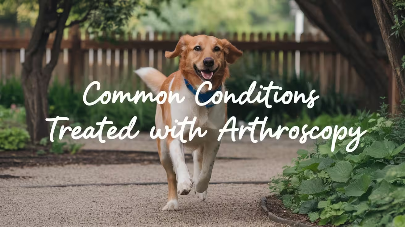
Common Conditions Treated with Arthroscopy in Dogs
Discover the most common conditions treated with arthroscopy in dogs, including OCD, elbow dysplasia, ligament injuries, and shoulder problems

This article is for informational purposes only and is not a substitute for professional veterinary advice. Every case is unique, so always consult your veterinarian for guidance specific to your pet.
This content is intended for veterinary professionals for educational purposes. It does not replace clinical judgment or tailored advice. Always rely on your training, expertise, and the specific context of your patients.
Arthroscopy was once a specialized technique used for a narrow range of joint conditions in dogs. Over the past two decades, it has become the standard of care for many of the most common orthopedic problems that affect canine joints. For a full explanation of what arthroscopy is and how it works, see what arthroscopy is.
Understanding which conditions are treated arthroscopically helps owners recognize when their dog's diagnosis is one that arthroscopy addresses, and what the procedure is expected to achieve.
Quick answer: The most common conditions treated arthroscopically in dogs are osteochondritis dissecans (OCD) of the shoulder, elbow, or stifle; fragmented medial coronoid process (FMCP) and ununited anconeal process (UAP) as part of elbow dysplasia; and meniscal assessment and treatment in dogs with cruciate ligament surgery. Shoulder instability and biceps tendon pathology are also commonly addressed. Arthroscopy is the standard of care for elbow dysplasia treatment at referral hospitals.
Key takeaways
- OCD is treated arthroscopically across multiple joints: shoulder, elbow, stifle, and tarsus.
- Elbow dysplasia (FMCP and UAP) is the most common arthroscopy indication in dogs.
- Meniscal evaluation and treatment is routinely performed arthroscopically during cruciate surgery.
- Shoulder OCD and biceps tendon pathology are diagnosed and treated in the same procedure.
- Arthroscopy for elbow dysplasia has replaced open surgery at most referral hospitals.
- Early treatment through arthroscopy before arthritis establishes produces the best long-term outcomes.
Osteochondritis dissecans (OCD)
OCD is a developmental condition in which a segment of cartilage fails to properly ossify, separates from the underlying bone, and may form a loose flap or fragment within the joint. It primarily affects young large-breed dogs in the shoulder, elbow, stifle, and tarsus.
The detached cartilage causes pain, joint inflammation, and if untreated, progressive arthritis. Arthroscopy allows the surgeon to visualize the OCD lesion, remove the loose cartilage flap, and debride the underlying bone surface.
OCD of the shoulder
This is one of the most common shoulder arthroscopy indications. The OCD lesion typically occurs on the caudal (back) aspect of the humeral head. Metropolitan Veterinary Associates describes: "Osteochondrosis dissecans is seen most commonly in the shoulder on the caudal humeral head, although it also occurs in the tarsus, stifle, elbow, and other joints."
The arthroscopic approach allows complete visualization of the lesion, thorough removal of the cartilage flap, and debridement of the bone bed, all through portals of a few millimeters. Recovery is faster than open shoulder surgery and the joint is disturbed less.
OCD of the elbow
Less common than shoulder OCD but addressed arthroscopically as part of comprehensive elbow evaluation. Dallas Veterinary Surgical Center notes elbow arthroscopy indications include "OCD of humeral head" (within the elbow context).
OCD of the stifle and tarsus
Stifle OCD typically occurs on the lateral femoral condyle. BluePearl Vet Hospital: "Stifle OCD is a relatively uncommon disease that is often operated on arthroscopically. The loose cartilage flap generally occurs in the lateral femoral condyle."
Tarsal OCD is managed similarly when it occurs, with arthroscopic flap removal and bone surface debridement.
Elbow dysplasia: FMCP and UAP
Elbow dysplasia encompasses a group of developmental conditions causing elbow joint abnormality in large-breed dogs. It is the most common cause of front leg lameness in dogs, particularly in Labrador Retrievers, Golden Retrievers, Rottweilers, and Newfoundlands.
Fragmented medial coronoid process (FMCP)
FMCP is the most common elbow dysplasia condition and the most common single indication for arthroscopy in dogs. Part of the medial coronoid process (a small projection of the ulna within the elbow joint) fractures and may remain in place or displace, causing pain and cartilage damage.
BluePearl Vet Hospital: "By far the most common indication is the fragmented medial coronoid process. Arthroscopy for FMCP allows for more complete exploration of the joint, more rapid recovery, and easy retrieval of the coronoid fragment. In most referral hospitals, elbow arthrotomy for FMCP is largely obsolete because of the additional trauma to the joint and morbidity associated with the open procedure."
Key point: arthroscopy has replaced open surgery as the standard of care for FMCP at referral hospitals.
The arthroscopic treatment involves:
- Complete visualization of the medial compartment
- Identification and removal of the fragmented coronoid fragment
- Assessment of cartilage damage on the radial head and humeral condyle
- Debridement (smoothing) of damaged cartilage surfaces
Metropolitan Veterinary Associates notes: "Outcomes following arthroscopic removal of the fragment depend greatly on the degree of arthritis and cartilage loss present at the time of treatment. Early detection and removal is ideal."
Ununited anconeal process (UAP)
UAP is a condition in which the anconeal process (another projection within the elbow joint) fails to properly fuse to the ulna during development. It causes elbow pain and degenerative joint disease if left untreated.
Arthroscopy is used to assess the anconeal process, evaluate the degree of joint damage, and guide treatment decisions, which may involve fragment removal or lag screw fixation depending on the age of the dog and degree of non-union.
Meniscal evaluation and treatment
The menisci are C-shaped fibrocartilage pads within the stifle (knee) that act as shock absorbers between the femur and tibia. Meniscal tears are common in dogs with cruciate ligament disease.
MedVet: "Stifle arthroscopy is a very effective tool to evaluate the stifle joint, not only to confirm the diagnosis of a cranial cruciate ligament joint, but also to evaluate and treat meniscal tears, as we see them develop frequently in conjunction with CrCL tears."
During cruciate surgery
When dogs undergo TPLO or TTA for cruciate ligament rupture, the meniscus is routinely evaluated arthroscopically. Meniscal tears are identified and the damaged portion removed (partial meniscectomy) through the instrument portal.
Late meniscal tears
Dogs who have already had cruciate surgery may develop meniscal tears in the weeks or months afterward. Arthroscopy allows the meniscus to be assessed and treated through minimal incisions rather than requiring full re-opening of the stifle.
BluePearl: "Arthroscopy may be used in conjunction with TPLO or other stifle repair techniques or to debride late meniscal tears in dogs that have already had stifle surgery."
Shoulder instability and biceps tendon pathology
The shoulder joint is inherently mobile, and instability from ligament laxity is increasingly recognized as a cause of chronic shoulder lameness in active adult dogs. Biceps tendon pathology (inflammation, partial tears, or avulsion) is also common.
Medial shoulder instability (MSI)
Medial shoulder instability occurs when the medial glenohumeral ligament and subscapularis tendon stretch or tear, allowing abnormal range of motion. It's difficult to detect on radiographs and CT.
Arthroscopy allows direct visualization of the medial compartment structures, confirms the instability diagnosis, and guides stabilization treatment.
Biceps tendon pathology
The biceps tendon originates inside the shoulder joint and is subject to inflammation and tearing, especially in working and active dogs. MedVet: "In addition to the conditions discussed above, arthroscopy is used to help diagnose cruciate disease and meniscal injury, treat biceps tendon injury, and monitor progression of healing."
Arthroscopic biceps tendon release (tenotomy) is a common and effective treatment for biceps tendon disease, with faster recovery than open surgery.
For when these conditions lead to an arthroscopy recommendation, see when these conditions indicate arthroscopy.
Hip dysplasia assessment
While hip arthroscopy is less commonly performed than elbow or shoulder procedures, it is used to assess cartilage quality and early joint changes in dogs with hip dysplasia before advanced arthritis develops.
Arthroscopic assessment allows direct grading of cartilage damage and may guide management decisions, including whether conservative management or surgical intervention is most appropriate at that stage.
For how these conditions compare to hip dysplasia management, see hip dysplasia as a treatable condition.
Cartilage damage and joint lavage
Beyond specific named conditions, arthroscopy is used for:
- Cartilage debridement: smoothing irregular or damaged cartilage surfaces in joints with early-to-moderate osteoarthritis
- Loose body removal: retrieving chip fractures or free fragments of bone or cartilage that cause pain and joint irritation
- Joint lavage: thoroughly flushing the joint to remove inflammatory debris, fibrin, and bacteria
- Synovial biopsy: obtaining tissue samples for histopathology or culture when infection or inflammatory joint disease is suspected
How arthroscopy compares to open surgery for these conditions
For the conditions listed above, arthroscopy typically offers:
- Better visualization (magnified view of all joint surfaces rather than line-of-sight access)
- Less post-operative pain
- Faster return to function
- Lower complication rate
For the full direct comparison between arthroscopy and open surgery, see how arthroscopy compares to open surgery for these conditions.
Frequently asked questions
My dog has been diagnosed with FMCP. Will they definitely need surgery?
Not automatically. The decision depends on the severity of symptoms, the degree of cartilage damage visible on imaging, and the dog's age. Dogs with FMCP that causes significant lameness typically benefit from arthroscopic fragment removal. Dogs with mild symptoms and minimal cartilage damage may be managed conservatively in some cases. A veterinary orthopedic specialist can assess the specific case.
How soon after OCD diagnosis should arthroscopy happen?
For OCD, earlier treatment generally produces better outcomes. Metropolitan Veterinary Associates: "Early detection and removal is ideal." Untreated OCD allows progressive cartilage damage and arthritis to develop. Once the diagnosis is confirmed, discussion of timing with a specialist is appropriate. Waiting months is not advisable for symptomatic cases.
Can arthroscopy treat arthritis?
Arthroscopy can slow arthritis progression by removing loose fragments, smoothing irregular cartilage, and lavaging inflammatory debris. It doesn't reverse established arthritis or regenerate cartilage. For end-stage arthritis, arthroscopy is a palliative tool rather than a curative one.
The range of conditions arthroscopy addresses in dogs has expanded significantly as the technique has matured and specialist training has spread. For the conditions covered here, it is now often the preferred approach rather than an alternative, because it consistently delivers better visibility, less tissue trauma, and faster recovery than the open surgical alternatives it replaced.
Resources
- BluePearl Pet Hospital. Arthroscopic Surgery. bluepearlvet.com
- MedVet. Understanding the Use of Arthroscopy in Dogs. blog.medvet.com
- Metropolitan Veterinary Associates. Arthroscopy in Canine Orthopedic Disease. metro-vet.com
- Dallas Veterinary Surgical Center. Arthroscopy. dvsc.com

Get a Free Poster
Enhance your workspace with a high-quality radiographs reference poster, designed for veterinary professionals. This free physical poster will be shipped directly to you—just fill out the form to request your copy.

Taking Great TPLO Radiographs
Click Below to Watch Live Video Demos


We'll send you a Free Wall Poster with all the steps
Step #1
Getting Ready
Ensuring a clean surgical field starts with proper skin preparation. This video demonstrates the best practices for:
- Shaving the patient – Achieving a close, even shave while minimizing skin irritation
- The Dirty Scrub – The initial skin prep step to remove surface debris and reduce bacterial load before the sterile scrub.
Following these techniques helps reduce infection risk and improve surgical outcomes. Watch the video to see how it’s done effectively!


Step #2
Reduce Your Risks
Many surgeons are shocked to find out that their patients are not protected from biofilms and resistant bacteria when they use saline and post-op antibiotics.
That’s Where Simini Comes In.
Why leave these risks and unmanaged? Just apply Simini Protect Lavage for one minute. Biofilms and resistant bacteria can be removed, and you can reduce two significant sources of infection.
Step #3
Take the Course
Preventing surgical infections is critical for patient safety and successful outcomes. This course covers:
- Aseptic techniques – Best practices to maintain a sterile field.
- Skin prep & draping – Proper methods to minimize contamination.
- Antibiotic stewardship – When and how to use perioperative antibiotics effectively.
Stay up to date with the latest evidence-based protocols. Click the link to start learning and earn CE credits!

Things to know
What Is Arthroscopy and When Is It Used in Dogs?
Learn what arthroscopy in dogs is, how it works, and when vets use it for joint issues like elbow dysplasia, OCD, or ligament injuries
Arthroscopy is one of the most significant advances in veterinary orthopedic surgery of the past two decades. It allows surgeons to see inside a joint and treat problems there with far less disruption to surrounding tissue than traditional open surgery.
For many of the most common joint conditions in dogs, arthroscopy has become the standard of care rather than an alternative to it.
Quick answer: Arthroscopy is a minimally invasive surgical technique where a small camera (arthroscope) is inserted into a joint through a tiny incision. The surgeon views the joint interior on a monitor and can simultaneously treat problems through additional small instrument ports. It's used for diagnosis and treatment of OCD lesions, fragmented coronoid process, ununited anconeal process, meniscal tears, and other joint conditions. Recovery is faster and less painful than open joint surgery.
Key takeaways
- Arthroscopy uses tiny incisions (a few millimeters) rather than the large incisions of open surgery.
- The arthroscope is a thin tube with a camera and light that projects magnified joint images onto a monitor.
- Diagnosis and treatment can occur in the same procedure rather than requiring separate surgeries.
- The elbow is the most commonly arthroscoped joint in dogs; the shoulder, stifle, and hip follow.
- Recovery is faster than open surgery: most dogs go home the same day and walk immediately.
- Arthroscopy is considered low-risk with a low complication rate according to VCA Animal Hospitals.
What arthroscopy actually is
Arthroscopy comes from the Greek "arthro" (joint) and "skopein" (to look). The procedure was first described in human medicine in 1912 by Danish surgeon Severin Nordentoft. Its use in veterinary medicine has expanded dramatically since the 1990s and is now standard practice at referral hospitals for many joint conditions.
VCA Animal Hospitals defines it: "Arthroscopy is a minimally invasive therapy that is used to examine, diagnose, and treat diseases and conditions that affect joints."
The equipment
The core instrument is the arthroscope: a thin rigid tube (typically 1.9 to 4 mm in diameter) containing:
- A camera chip or fiber-optic lens at the tip
- A light source to illuminate the joint interior
- A channel for sterile irrigation fluid
The arthroscope connects to a camera unit that projects magnified, high-resolution images of the joint interior onto a monitor in the operating room. The surgeon operates by watching the monitor rather than looking directly into the joint.
How it works
- The dog is anesthetized and the joint is aseptically prepared
- Sterile fluid (typically saline) is used to distend (expand) the joint, creating the working space the arthroscope needs
- A small stab incision (2 to 3 mm) is made and the arthroscope is introduced into the joint
- Additional 2 to 3 mm portals are created for instrument insertion
- The surgeon examines all internal joint structures on the monitor: cartilage surfaces, synovial membrane, ligaments, menisci
- Surgical treatment is performed through the instrument portals using specialized miniature tools
- The joint is lavaged and the small incisions are closed with a single suture or staple
The SustainableVet arthroscopy guide notes: "Sterile fluid is used to expand the joint and improve visibility. Small instruments can also be inserted through these openings to remove loose tissue, repair damage, or take samples for testing."
What arthroscopy can do: diagnostic and therapeutic uses
Diagnostic use
Arthroscopy provides superior diagnostic detail compared to other imaging. Conditions that are subtle or invisible on radiographs, CT, or MRI may be clearly visible arthroscopically.
Advantages over imaging alone:
- Directly visualizes cartilage surface quality (grading, fissuring, erosion)
- Identifies loose fragments not visible on radiograph
- Assesses ligament integrity (tension, tears, partial tears)
- Evaluates synovial membrane health
- Allows synovial biopsy for culture or histopathology
In many cases, arthroscopy is both the definitive diagnosis and the treatment in the same anesthetic event.
Therapeutic use
The surgical treatments most commonly performed through an arthroscope in dogs:
- Removal of OCD lesions: The detached or loose cartilage flap is removed through the instrument portal; the underlying bone surface is debrided
- Retrieval of loose fragments (joint mice): Fragments of bone or cartilage that broke free into the joint are removed before they cause progressive damage
- Fragmented coronoid process treatment: Removal of the abnormal coronoid fragment in dogs with elbow dysplasia
- Meniscal assessment and treatment: In TPLO surgery, the meniscus is often examined arthroscopically for tears
- Synovial biopsy: Taking tissue samples from the joint lining for laboratory analysis
- Joint lavage: Flushing the joint to remove inflammatory debris
Which joints are treated arthroscopically in dogs
The elbow
The elbow is the most frequently arthroscoped joint in veterinary practice. BluePearl Vet Hospital: "Arthroscopic procedures of the elbow are among the most common arthroscopic procedures performed."
Elbow arthroscopy is indicated for:
- Fragmented medial coronoid process (FMCP): The most common elbow arthroscopy indication; the fragment is removed and the joint is assessed
- Osteochondritis dissecans (OCD) of the elbow: Cartilage flap removal
- Ununited anconeal process (UAP): Assessment and treatment of this elbow growth abnormality
- Diagnostic exploration and biopsy
BluePearl notes: "In most referral hospitals, elbow arthrotomy (open surgery) for FMCP is largely obsolete because of the additional trauma to the joint and morbidity associated with the open procedure." Arthroscopy has replaced open surgery as the standard of care for elbow dysplasia treatment.
The shoulder
Shoulder arthroscopy is used for:
- OCD of the shoulder: Cartilage flap removal; one of the most common shoulder arthroscopy indications
- Biceps tendon pathology: Assessment and treatment of inflammation or tears
- Medial shoulder instability: Confirmation and potential repair of ligament damage
The shoulder is particularly well-suited to arthroscopy because X-rays and CT often underestimate soft tissue pathology. Arthroscopy reveals what imaging can't.
The stifle (knee)
Stifle arthroscopy is used to:
- Assess the meniscus in dogs undergoing cruciate ligament surgery (TPLO or TTA)
- Treat OCD of the stifle (less common than shoulder or elbow)
- Diagnose joint pathology causing stifle pain not fully explained by imaging
The hip
Hip arthroscopy is less commonly performed than elbow or shoulder but is used for:
- Assessment and treatment of hip OCD
- Diagnostic evaluation of painful hip conditions
- Lavage and synovial biopsy when infection or inflammatory joint disease is suspected
For how hip dysplasia is managed compared to arthroscopic conditions, see hip dysplasia as a treatable condition.
For the full range of specific conditions arthroscopy addresses across all joints, see conditions arthroscopy treats.
Other joints
Carpus (wrist) and tarsus (hock) arthroscopy is performed less frequently but is used for OCD lesions and chip fractures in these joints.
How arthroscopy compares to open surgery
For the full comparison between arthroscopy and open surgery, see how arthroscopy compares to open surgery.
Recovery after arthroscopy
Most dogs go home the same day as the procedure. VCA Animal Hospitals: "Arthroscopy is considered a low-risk procedure with a low rate of complications."
BluePearl: "Most dogs with arthroscopic procedures can go home the same day as the procedure and are able to walk on the operated leg right away."
Typical recovery:
- Initial restricted activity: 2 to 3 weeks
- Full recovery: 6 to 8 weeks depending on the condition treated
- No large incision to manage; tiny portal sites heal quickly
For what to expect during and after the procedure, see what the procedure involves. For when these conditions indicate arthroscopy, see when arthroscopy is the right choice.
Frequently asked questions
Is my dog a candidate for arthroscopy?
Candidacy depends on the joint affected, the condition causing lameness, the dog's size (very small joints may be technically difficult to arthroscope), and the surgeon's experience. A board-certified veterinary surgeon who performs arthroscopy can assess whether your dog's specific condition is suitable.
Is arthroscopy available at my regular vet?
Generally no. Arthroscopy requires specialized equipment (arthroscopes, camera systems, irrigation pumps, miniature surgical instruments) and specific training. Most arthroscopic procedures in dogs are performed at veterinary referral hospitals and teaching hospitals.
What happens if arthroscopy reveals more damage than imaging suggested?
This is common. Arthroscopy regularly reveals pathology invisible on radiographs. In most cases, the surgeon proceeds with appropriate treatment in the same anesthetic event since the joint is already open and the necessary instruments are available.
Does arthroscopy hurt dogs?
For information on pain management during and after arthroscopy, see pain management during and after arthroscopy.
Arthroscopy represents a genuine advance in how joint disease is managed in dogs. The ability to see inside a joint clearly, diagnose precisely, and treat effectively, all through incisions smaller than a pencil, has made procedures that once required extensive open surgery into day procedures with faster recovery and fewer complications. For the right condition, it's not just better than open surgery; in many cases, it's replaced it entirely.
Resources
- VCA Animal Hospitals. Arthroscopy. vcahospitals.com
- BluePearl Pet Hospital. Arthroscopic Surgery. bluepearlvet.com
- Fusion Veterinary Orthopedics. Understanding Arthroscopy in Pets. fusionvetortho.com
What to Expect During and After Arthroscopic Surgery
Learn what to expect during and after arthroscopic surgery in dogs, from the procedure and recovery timeline to aftercare, risks, and long-term outcomes
Arthroscopy is a minimally invasive surgery used to diagnose and treat joint problems in dogs. It involves using a small camera and instruments through tiny incisions, allowing precise care with less trauma than open surgery. Many owners want to know what their dog will go through during and after this procedure. Understanding the steps, recovery timeline, and safety helps set realistic expectations.
This guide explains what happens before, during, and after arthroscopy so you can feel confident about your dog’s treatment and healing process.
What Happens During Arthroscopic Surgery
Arthroscopic surgery is a carefully planned procedure performed under full anesthesia to ensure the dog is safe and pain-free. The technique uses small incisions and advanced instruments to diagnose and treat joint conditions with minimal trauma.
- General anesthesia and preparation: Dogs are fully anesthetized to prevent movement, ensure pain control, and allow precise work inside delicate joint structures. The surgical area is shaved, cleaned, and prepared to maintain a sterile field.
- Small incisions for scope and tools: The surgeon makes tiny cuts, usually 2–3 millimeters wide, to introduce the arthroscope and specialized surgical instruments.
- Joint distension with sterile fluid: The joint is filled with sterile saline to widen the space, flush debris, and improve visualization for the surgeon.
- Real-time imaging: The arthroscope projects magnified, angled images of cartilage, ligaments, and bone onto a monitor, giving the surgeon a clear and detailed view.
- Specialized surgical tools: Graspers, shavers, scissors, and burrs are inserted through other small incisions to remove cartilage flaps, collect biopsies, or smooth rough bone surfaces.
- Surgical duration: Depending on the condition and joint, the procedure usually takes 30–90 minutes.
This combination of magnified visualization and precision tools makes arthroscopy highly effective for both diagnosis and treatment, while minimizing damage to surrounding tissues.
Immediately After Surgery
After arthroscopy, the immediate focus is on safe anesthesia recovery, pain control, and wound protection. Dogs are monitored closely until they are stable enough to go home.
- Anesthesia recovery: Most dogs wake up within an hour but may remain groggy, disoriented, or wobbly for several hours. Veterinary staff monitor breathing, heart rate, and body temperature throughout.
- Post-surgical monitoring: Dogs remain in the hospital for observation to ensure no complications such as bleeding, swelling, or difficulty standing. Intravenous fluids may be used if needed.
- Bandages and wound care: Small bandages are applied to cover the tiny incision sites, reducing the risk of contamination and supporting healing.
- Pain management: Anti-inflammatories and analgesics are administered to keep the dog comfortable. Some dogs may also receive antibiotics depending on the condition treated.
- Discharge timing: Most dogs are able to return home within 12–24 hours once they are alert, walking short distances, and have stable vital signs.
By the time of discharge, owners receive detailed instructions on medication, wound care, and activity restriction, which are crucial for smooth recovery.
Early Recovery: First Few Days
The first few days after arthroscopy are critical for healing, as the body adjusts to the procedure and begins repairing tissue. Dogs usually recover faster than with open surgery, but careful management is still needed.
- Weight-bearing: Many dogs start placing weight on the affected limb within 3–5 days. Although they may limp slightly, this is expected and improves with time. Early weight-bearing helps prevent muscle wasting and stiffness.
- Incision appearance: Mild swelling, bruising, or fluid accumulation around the incision sites is normal. These changes usually resolve within a week and are not signs of complications unless redness or discharge develops.
- Keeping wounds clean: Owners must ensure the incisions remain clean and dry. Licking or chewing can cause infections, so the use of an Elizabethan collar is strongly recommended.
- Restricted activity: Strict rest is necessary during the early phase. Only short leash walks for bathroom breaks should be allowed, avoiding stairs, running, or jumping.
- Medication adherence: Pain relievers and, when prescribed, antibiotics must be given on schedule to reduce discomfort and prevent complications.
Close monitoring during this stage sets the foundation for smooth healing and prevents setbacks that could prolong recovery.
Recovery Timeline in Weeks
Recovery after arthroscopy follows clear phases, with gradual return to function over several weeks. While healing is faster than open surgery, structured management is key.
- 2–3 weeks: Incisions usually heal by this stage, and sutures or staples are removed during a follow-up appointment. Dogs can begin short, controlled leash walks beyond bathroom breaks.
- 3–4 weeks: Depending on the condition treated, physiotherapy or hydrotherapy may be introduced. These exercises strengthen muscles, restore range of motion, and support joint stability.
- 4–6 weeks: Activity is gradually increased. Dogs may tolerate longer walks and mild play, though off-leash exercise is still restricted.
- 8–12 weeks: Most dogs regain full mobility and return to their normal lifestyle. Sporting or working dogs may require a tailored rehabilitation plan to resume high activity levels.
This timeline may vary depending on the joint treated and the extent of disease. Following veterinary guidance ensures safe, long-term improvement.
Risks and Safety Considerations
Arthroscopy is considered very safe, but as with all surgeries, potential risks should be understood. Fortunately, complications are uncommon and typically mild.
- Low infection rate: Small incisions reduce exposure, making joint infections rare compared to open surgery. When infections occur, they are usually superficial and treatable with antibiotics.
- Fluid leakage: Sterile saline used to distend the joint can sometimes leak into nearby tissue, causing temporary swelling. This resolves naturally without long-term issues.
- Conversion to open surgery: In some cases, if damage is extensive or visualization is limited, surgeons may switch to open surgery for effective treatment.
- Anesthesia risks: Though rare with modern monitoring, anesthesia can pose risks, especially in senior dogs or those with heart, lung, or kidney conditions. Pre-operative screening minimizes these dangers.
- Post-operative discomfort: Mild pain, bruising, or swelling are normal but manageable with prescribed medications.
When performed by experienced surgeons in a specialty setting, arthroscopy has an excellent safety record and is well tolerated by most dogs.
What Owners Should Do at Home
Owner participation is essential to ensure healing and prevent complications. Diligent care at home directly affects long-term outcomes.
- Strict exercise control: Limit activity to short, leash-only walks until cleared by your veterinarian. Unrestricted play can delay healing or damage the joint.
- Incision monitoring: Check daily for redness, swelling, discharge, or separation of sutures. Contact the vet immediately if signs of infection appear.
- Keep incisions dry: No bathing, swimming, or grooming should be done until the vet confirms complete healing of the surgical sites.
- Medication compliance: Give all prescribed pain medications and anti-inflammatories on schedule. Missing doses can lead to unnecessary pain or delayed recovery.
- Follow-up visits: Attend every scheduled check-up for wound assessment, suture removal, and rehabilitation advice.
Consistent, careful home care ensures the benefits of arthroscopy are fully realized and reduces the chance of setbacks.
Long-Term Expectations
Arthroscopy often provides excellent long-term results, especially when performed early in the disease process. Dogs usually recover fully and return to active, comfortable lives.
- Quick return to activity: Most dogs regain mobility within 8–12 weeks, with many showing significant improvement earlier.
- Slowing arthritis progression: By removing fragments, smoothing cartilage, or addressing early joint lesions, arthroscopy delays degenerative changes and prolongs joint function.
- Improved quality of life: Pain relief and restored mobility allow dogs to return to playing, exercising, and working without chronic discomfort.
- Supportive therapies: Physiotherapy, hydrotherapy, weight management, and joint supplements can further improve outcomes and extend joint health.
- Limitations in advanced disease: In severe arthritis cases, arthroscopy provides pain relief but may not stop progression entirely. Long-term management strategies may still be needed.
Overall, most dogs achieve lasting improvements in comfort, activity, and quality of life after arthroscopy.
Conclusion
Arthroscopy is one of the safest and most effective ways to diagnose and treat joint conditions in dogs. Unlike open surgery, it requires only small incisions, causes less trauma, and provides a magnified view of the joint for precise treatment. Most dogs recover comfortably within weeks, especially when owners follow strict aftercare instructions on rest, medication, and incision monitoring.
- Safe and effective: Minimally invasive with low complication rates.
- Faster recovery: Dogs regain mobility much sooner than with traditional surgery.
- Specialist guidance: Consulting a veterinary orthopedic surgeon ensures accurate diagnosis, proper case selection, and the best treatment plan.
With timely intervention and professional care, arthroscopy restores mobility, reduces pain, and helps protect long-term joint health, giving dogs a better quality of life and allowing them to stay active for years.
FAQs
How long will my dog stay at the clinic after arthroscopy?
Most dogs go home the same day or within 24 hours after arthroscopy. They are monitored until they are awake, stable, and comfortable. Some may stay longer if the joint treated was complex or if extra observation is needed. Clear discharge instructions are always given to support safe recovery at home.
When can my dog walk normally again?
Many dogs begin walking with partial weight-bearing within 2–5 days after surgery. While some limping is expected, mobility improves quickly. Normal walking usually returns within 2–3 weeks, depending on the joint and condition treated. Controlled activity, like leash walking, is encouraged, but full unrestricted movement must wait until the vet approves it.
What signs after surgery should worry me?
Concerning signs include excessive redness, swelling, or discharge at the incision site, refusal to bear weight after several days, persistent pain despite medication, or signs of infection such as fever or lethargy. Any sudden worsening of lameness or chewing at the stitches should be reported to the veterinarian immediately for timely intervention.
Can my dog play or run after arthroscopy?
Not right away. Play and running must be restricted during the first 4–6 weeks to allow proper healing. Controlled leash walks are permitted early, but off-leash activity is only allowed once your veterinarian clears it, often after 8–12 weeks. Premature play or running risks damaging the joint and delaying recovery.
Is arthroscopy safer than open surgery?
Yes, arthroscopy is generally safer because it uses very small incisions, reducing infection risk, pain, and tissue trauma. Dogs recover faster and more comfortably compared to open joint surgery. However, both procedures are safe when performed by skilled surgeons, and the best choice depends on the dog’s condition, disease severity, and availability.
How long does full recovery take?
Most dogs achieve full recovery within 8–12 weeks after arthroscopy, although improvements are often seen much sooner. The timeline depends on the joint treated, the condition’s severity, and how well aftercare instructions are followed. Rehabilitation therapies, such as physiotherapy or hydrotherapy, can further speed healing and improve long-term mobility and comfort.
How Long Is Recovery After Arthroscopy in Dogs?
Dog arthroscopy recovery takes 8–12 weeks. Learn the timeline, phases, and factors that affect healing after joint surgery in dogs
One of the most appealing features of arthroscopy over open joint surgery is the recovery time. For most conditions and most dogs, returning to normal activity takes weeks rather than months.
The precise timeline depends on the joint treated, the condition addressed, the dog's size and age, and how consistently the rehabilitation protocol is followed. For a complete guide to what happens during and after the procedure itself, see what the full recovery involves.
Quick answer: Most dogs begin weight-bearing within 1 to 2 days of arthroscopy. Full recovery, including return to normal activity, typically takes 4 to 6 weeks for most arthroscopic procedures. Stifle (knee) arthroscopy may extend to 6 to 8 weeks due to greater joint loading. This is substantially shorter than equivalent open surgery recovery.
Key takeaways
- Weight-bearing starts within 1 to 2 days for most dogs after arthroscopy.
- Full recovery: 4 to 6 weeks for most conditions and joints.
- Stifle arthroscopy recovery: 6 to 8 weeks due to higher mechanical demands.
- Activity restriction in weeks 1 to 4 is essential; early overloading risks setbacks.
- Physical therapy from weeks 2 to 3 accelerates muscle rebuilding and return to function.
- Recovery is significantly faster than open surgery, which typically takes 8 to 16 weeks for equivalent procedures.
How recovery length compares to open surgery
The difference in recovery between arthroscopy and open joint surgery is one of the strongest arguments for arthroscopy when both options are available.
Open joint surgery involves large incisions through multiple tissue layers: skin, subcutaneous fat, fascia, muscle or retraction of muscle groups, and joint capsule. All these layers must heal. The soft tissue disruption causes pain and swelling that extend recovery to 8 to 16 weeks for most major open joint procedures.
Arthroscopy accesses the joint through 2 to 3 mm portals. There is minimal soft tissue disruption. The small portal incisions heal within a week or two. The joint itself begins responding to treatment almost immediately.
Most dogs walk within 1 to 2 days after arthroscopy. Most dogs take 3 to 7 days after open joint surgery to begin comfortable weight-bearing.
For the full direct comparison, see how recovery compares to open surgery.
Recovery timeline by phase
Phase 1 (days 1 to 7): early healing
What's happening in the body: The portal incisions are healing. Joint inflammation from the procedure is settling. The joint begins responding to the treatment performed.
Activity level: Leash-only bathroom walks. No running, jumping, stairs, or off-leash activity.
Signs of normal recovery:
- Limping on the operated leg, improving day by day
- Mild swelling at portal sites, resolving within a few days
- Tentative but increasing weight-bearing by days 2 to 3
First vet recheck: typically at 10 to 14 days for suture/staple removal and progress assessment.
Phase 2 (weeks 2 to 4): controlled exercise
What's happening in the body: Portal incisions are healed. The joint is less inflamed. Muscle mass is beginning to stabilize. Tissue healing within the joint is progressing.
Activity level: Short controlled leash walks, increasing progressively. Most protocols begin with 5-minute walks 2 to 3 times daily, adding 5 minutes per week.
Ridge Referrals elbow arthroscopy protocol: "6 to 12 weeks: start to increase time on lead to maximum 10 minutes three to four times daily."
Rehabilitation starts: gentle passive range-of-motion exercises, light massage, and controlled walking. These prevent excessive scar tissue formation and begin muscle rebuilding.
Still prohibited: running, jumping, off-leash activity, rough play.
Phase 3 (weeks 4 to 6): return to activity
What's happening in the body: Internal joint healing is substantially complete for most arthroscopic procedures. Muscle mass is rebuilding with increasing activity.
Activity level: Most conditions allow progressive return to normal activity by week 6. Walk duration increases toward normal. Light play and activity on secure ground may be permitted after vet clearance.
Hydrotherapy: if available, underwater treadmill sessions are excellent for muscle rebuilding without joint impact. Often introduced from week 3 to 4 when portal incisions are confirmed healed.
Full recovery: Most owners report their dog moving normally and freely by week 4 to 6 for elbow and shoulder procedures.
How the joint treated affects recovery length
Not all joints recover at the same rate after arthroscopy.
Elbow and shoulder: faster recovery
Elbow and shoulder joints are not the primary weight-bearing joints. Dogs shift some load to other limbs during early recovery. The mechanical demands on these joints during the healing phase are lower than on the stifle or hip.
Most elbow and shoulder arthroscopy cases reach full activity by 4 to 6 weeks.
Stifle: longer recovery
The stifle (knee) is a major weight-bearing joint that bears significant load with every step. Stifle arthroscopy (typically for meniscal assessment and treatment, or OCD of the stifle) takes longer than elbow or shoulder arthroscopy.
Expect 6 to 8 weeks for stifle arthroscopy recovery. When stifle arthroscopy is combined with open cruciate surgery (TPLO), the TPLO recovery protocol drives the timeline, typically 10 to 14 weeks total.
VCA Animal Hospitals confirms: "The recovery time will depend on the extent of the injury, and the specific joint affected."
Factors that extend recovery
- Degree of pre-existing arthritis: dogs with established joint disease before arthroscopy take longer to respond
- Extent of surgical work: more debridement, bone work, or meniscal treatment means more internal healing needed
- Dog's age and size: older dogs and large breeds typically recover more slowly
- Rehabilitation compliance: dogs whose owners follow the exercise protocol consistently recover faster
The role of physical therapy in recovery speed
Physical therapy materially shortens recovery time and improves functional outcomes after arthroscopy.
Key exercises during recovery:
- Passive range-of-motion (PROM): gently flexing and extending the operated joint during weeks 1 to 4 prevents stiffness and scar tissue contracture
- Controlled leash walking: slow walking that encourages weight-bearing builds muscle and joint stability
- Hydrotherapy: underwater treadmill or pool work allows full muscle engagement without joint impact; ideal from week 3 to 4 onward
- Sit-to-stands: builds the hip extensors and quadriceps supporting the operated joint
VCA Animal Hospitals: "Rehabilitation therapy or animal physiotherapy may be recommended to expedite a return to function."
When to call the vet during recovery
The following signs during arthroscopy recovery warrant a vet call rather than waiting for the scheduled recheck:
- Dog is still not weight-bearing by day 3 to 5
- Progressive worsening of lameness after an initial period of improvement
- Spreading redness, discharge, or odor at the portal incision sites
- Significant joint swelling that is increasing rather than settling
- Fever, lethargy, or reduced appetite beyond day 2
For what normal vs. concerning signs look like in more detail, see pain management during recovery.
When faster recovery justifies arthroscopy
For owners weighing whether arthroscopy's higher cost is worth it compared to open surgery for the same condition, the recovery timeline is a meaningful factor:
- 4 to 6 weeks of restricted activity vs. 8 to 16 weeks
- Same-day discharge vs. 1 to 3 days hospitalization
- Walking within 1 to 2 days vs. 3 to 7 days
- Lower medication burden (less pain = less medication needed)
- Faster return to work for working or service dogs
For the full comparison of when the faster recovery justifies arthroscopy, see when faster recovery justifies arthroscopy.
Frequently asked questions
My dog seems completely normal at week 3. Can I stop the restrictions?
No. The internal joint healing continues even when the dog looks and acts completely normal. Removing restrictions at week 3 risks re-injuring the treated tissue before it's fully healed. Follow the protocol from your vet's discharge instructions, not your dog's apparent activity level.
Can both elbows be arthroscoped in the same procedure?
Yes. Because arthroscopy is minimally invasive with low morbidity, bilateral elbow arthroscopy in the same anesthetic event is commonly performed. Irish Vet Ortho Referrals notes: "Both stifle joints can be examined and treated arthroscopically at one surgery."
My dog had FMCP arthroscopy. The surgeon said some dogs need anti-inflammatories long-term. Is that normal?
Yes. Metropolitan Veterinary Associates notes: "Many dogs will need periodic treatment with anti-inflammatories and/or joint injections for the remainder of their life, even after fragment removal." This is because FMCP typically involves existing cartilage damage that the arthroscopy addressed but didn't erase. Long-term joint support is appropriate.
Arthroscopy recovery is measured in weeks, not months. For owners who have experienced the 12 to 16 week recovery of TPLO or another major open surgery, the 4 to 6 week arthroscopy recovery is a genuine relief. The key is maintaining discipline through that shorter period, because recovery that seems fast still needs to be managed correctly to produce the best long-term outcome.
Resources
- VCA Animal Hospitals. Arthroscopy. vcahospitals.com
- Ridge Referrals. Post Elbow Arthroscopy Rehabilitation Protocol. ridgereferrals.co.uk
- Metropolitan Veterinary Associates. Arthroscopy in Canine Orthopedic Disease. metro-vet.com
Is Arthroscopy Painful for Dogs?
Arthroscopy in dogs causes less pain than open surgery. Learn how much discomfort to expect, recovery timeline, and pain management after the procedure
Pain is one of the first concerns owners raise when arthroscopy is recommended. The honest answer: during the procedure, your dog feels nothing. Afterward, there is some discomfort, and it's significantly less than open joint surgery. For a full explanation of what arthroscopy is and how it works, see what the procedure involves.
Understanding the pain experience, what to expect, and how it's managed helps you know what your dog is going through and what your role is in keeping them comfortable.
Quick answer: Dogs feel no pain during arthroscopy because they are under general anesthesia. Post-operative pain is mild to moderate for most dogs and is well-managed with NSAIDs and analgesics. Most dogs improve noticeably within 24 to 48 hours and begin weight-bearing within 1 to 2 days. Post-surgical pain from arthroscopy is significantly less than equivalent open joint surgery because the tissue disruption is far smaller.
Key takeaways
- No pain during the procedure: general anesthesia ensures complete unconsciousness.
- Mild to moderate post-operative soreness is expected for the first 24 to 48 hours.
- NSAIDs are the primary pain medication after arthroscopy; short opioid courses may supplement.
- Most dogs are noticeably more comfortable within 48 hours compared to pre-surgery chronic pain.
- Arthroscopy causes less post-surgical pain than open surgery for equivalent conditions.
- Stifle arthroscopy causes more soreness than elbow or shoulder, due to weight-bearing demands.
During the procedure: no pain
Dogs undergoing arthroscopy receive general anesthesia. This renders them fully unconscious and incapable of feeling pain throughout the procedure. Pre-anesthetic medications (sedation and often pre-emptive analgesia) are given before anesthesia induction.
In some cases, local anesthesia (intra-articular bupivacaine or similar) is injected into the joint at the conclusion of the procedure. This provides an additional layer of pain control that takes effect as the dog begins to wake up.
Fusion Veterinary Orthopedics: "Reduced pain is a key advantage of arthroscopy. Smaller incisions mean less pain for the pet during and after the procedure."
Post-operative pain: what to expect
The first 24 to 48 hours
Mild to moderate soreness at the operated joint is expected and normal in the first day or two. Sources of this pain include:
- The portal incisions: small but they do disrupt skin and the joint capsule
- The joint itself: the irrigation, instrument manipulation, and treatment create some inflammation
- Fluid pressure changes: the joint was distended with sterile fluid during the procedure; this can cause mild stiffness and aching as the fluid is absorbed
Most dogs show obvious soreness in the first 12 to 24 hours. By 24 to 48 hours, the trend should be clearly toward improvement.
Signs of expected post-operative pain:
- Limping on the operated leg
- Reluctance to bear full weight initially
- Mild restlessness or seeking comfortable positions
- Reduced activity and increased sleep
Days 3 to 7
Most dogs are noticeably more comfortable by day 3 to 5. Weight-bearing on the operated leg becomes more consistent. The trend of improvement should be clear.
For many dogs, particularly those who had chronic pre-surgical joint pain, the days following arthroscopy are actually more comfortable than the weeks before surgery, because the source of chronic pain (an OCD lesion, fragmented coronoid, loose fragment) has been removed.
How post-operative pain is managed
NSAIDs (anti-inflammatory medications)
NSAIDs are the primary post-arthroscopy pain medication. Carprofen, meloxicam, or grapiprant reduce joint inflammation and provide pain relief. These are typically prescribed for 7 to 14 days post-procedure.
Give NSAIDs with food to reduce GI effects. Don't skip doses even if your dog seems comfortable; consistent blood levels maintain effective pain control.
Gabapentin
Gabapentin is a nerve pain modulator that is sometimes added for the first 5 to 7 days post-procedure, particularly for stifle arthroscopy or when more significant intra-articular work was performed.
Short-course opioids
For more substantial arthroscopic procedures (extensive cartilage debridement, meniscal work), a 2 to 3 day supply of a mild opioid (tramadol, buprenorphine) may be sent home to cover the most acute phase.
Intra-articular analgesia
Local anesthetic injected into the joint at the conclusion of surgery provides several hours of additional pain relief as the dog wakes up. This reduces the severity of early post-operative pain.
Pain that varies by joint and procedure
Not all arthroscopy procedures produce the same pain level.
Elbow and shoulder arthroscopy: generally mild post-operative soreness. The elbow and shoulder are not primary weight-bearing joints during normal gait. Most dogs are comfortable within 48 to 72 hours.
Stifle arthroscopy: more soreness is expected, particularly when combined with meniscal work or performed alongside cruciate surgery. The stifle bears significant load with every step. Soreness may persist for 1 to 2 weeks at a mild level.
Extent of surgical work: a diagnostic arthroscopy with minimal treatment produces less pain than a therapeutic arthroscopy involving extensive cartilage debridement, bone work, or fragment removal.
Pre-existing arthritis: dogs with established joint disease before arthroscopy may have a slightly longer period of post-operative discomfort as the inflamed joint settles following the procedure.
Recognizing inadequate pain control
Signs that pain medication is not adequately controlling your dog's discomfort:
- Constant vocalization (whimpering, crying) beyond the first 12 to 24 hours
- Refusing to move or bear any weight on the operated leg by days 3 to 5
- Significant trembling or panting at rest
- Aggressive response when the operated area is touched (more than expected)
- Not eating by day 2 to 3
If these signs are present, contact your vet. Additional pain management options are available, and dogs don't need to suffer through inadequate control.
How arthroscopy pain compares to open surgery
This is where arthroscopy's advantage is most practical for owners.
Open joint surgery requires large incisions through skin, fascia, and muscle. Muscle retraction during surgery disrupts significant tissue mass. All these structures are inflamed post-operatively and must heal over weeks.
Arthroscopy uses 2 to 3 mm portals. Minimal muscle disruption occurs. The joint capsule is entered through tiny portals rather than a large incision.
The result: arthroscopy patients require less pain medication for a shorter duration than equivalent open surgery patients. Most arthroscopy dogs are comfortable and moving within 48 to 72 hours. Most open joint surgery dogs require several days of more intensive pain management before they begin comfortable weight-bearing.
WSAVA's evidence review confirms: "The advantages of minimally invasive arthroscopy compared to standard open arthrotomy are evident and proved clinically by reduced postoperative pain and accelerated recovery."
For the full comparison across recovery factors, see pain comparison with open surgery.
Long-term: is joint pain eliminated by arthroscopy?
Arthroscopy addresses the immediate source of joint pain (the OCD lesion, the fragmented coronoid, the loose fragment, the meniscal tear). Whether joint pain returns long-term depends on the degree of pre-existing arthritis.
- Dogs treated early, before significant arthritis: excellent long-term pain outcomes; many remain comfortable without long-term medication
- Dogs treated with established arthritis: arthroscopy reduces pain and slows progression but doesn't eliminate arthritis; long-term joint support medication (NSAIDs, Adequan, supplements) may still be appropriate
Metropolitan Veterinary Associates: "Many dogs will need periodic treatment with anti-inflammatories and/or joint injections for the remainder of their life, even after fragment removal."
For the full post-arthroscopy experience, see full arthroscopy experience. For how recovery duration relates to pain progression, see recovery timeline alongside pain management.
Frequently asked questions
My dog is crying after arthroscopy. Is that normal?
Some dogs vocalize while coming out of anesthesia, particularly as they become aware of the surgical site before pain medication has fully taken effect. This is usually brief. If vocalizing persists beyond the first few hours at home, or if your dog is crying at rest despite medication, contact your vet: the pain management protocol may need adjustment.
Can I give my dog human pain medication?
No. Ibuprofen, naproxen, and acetaminophen are toxic to dogs. Never give human pain medications to dogs, even in small amounts. Use only what your vet has prescribed.
When does pain from arthroscopy typically peak?
Pain is typically at its highest in the first 12 to 24 hours post-surgery. It should improve noticeably by day 2 to 3. If pain is increasing rather than decreasing by day 3, contact your vet.
The honest picture of arthroscopy pain: it exists, it's less than open surgery, and it's manageable with appropriate medication. For most dogs, the weeks of chronic pre-surgical pain that arthroscopy was intended to address were worse than the post-operative soreness. The recovery is measured in days of real discomfort, not weeks.
Resources
- Fusion Veterinary Orthopedics. Understanding Arthroscopy in Pets. fusionvetortho.com
- WSAVA 2010. Benefits and Limits of Arthroscopy. vin.com
- Metropolitan Veterinary Associates. Arthroscopy in Canine Orthopedic Disease. metro-vet.com
Arthroscopy vs Open Surgery: Guide for Dog Owners
Compare arthroscopy vs open surgery in dogs. Learn differences in recovery, cost, risks, and when vets recommend each option for joint problems
When your vet or specialist recommends joint surgery for your dog, you may hear both "arthroscopy" and "open surgery" mentioned. Understanding the difference, and why the choice matters, helps you ask better questions and understand what your dog is going through. For a full explanation of what arthroscopy is and how it works, see what arthroscopy is.
Quick answer: Arthroscopy uses tiny incisions (2 to 3 mm) and a small camera to access the joint, causing far less tissue disruption than open surgery. It offers superior visualization, less post-operative pain, faster recovery, and lower complication rates for most joint conditions. Open surgery is still used when the joint requires direct physical access that arthroscopy can't provide, or when the surgeon needs to place implants, stabilize structures, or address complex anatomy. For many common conditions, arthroscopy is the preferred approach at referral hospitals.
Key takeaways
- Arthroscopy uses 2 to 3 mm incisions compared to the much larger cuts of open surgery.
- Arthroscopy provides superior visualization: magnified, illuminated view of all joint surfaces.
- Less post-operative pain is consistently demonstrated with arthroscopy vs. open joint surgery.
- Recovery is faster: most dogs walk the same day and return to activity within weeks rather than months.
- Complication rates are lower with arthroscopy for most joint conditions.
- Open surgery is still needed for ligament reconstruction, implant placement, and complex structural repair.
The fundamental difference
Open surgery (arthrotomy)
Open surgery involves making an incision large enough to directly expose the joint. The surgeon can see and touch the joint structures directly, without a camera. This direct access is necessary when:
- Physical manipulation of structures is required (placing a screw, cutting bone, repairing a ligament)
- The repair requires working at a scale too large for arthroscopic instruments
- The joint anatomy makes arthroscopic access impractical
Open joint surgery typically requires cutting through multiple tissue layers to reach the joint, including skin, subcutaneous tissue, muscle or fascia, and the joint capsule. All of these layers must be sutured closed afterward. The resulting soft tissue disruption causes more post-operative pain and takes longer to heal.
Arthroscopy
Arthroscopy accesses the joint through portals of 2 to 3 mm each. One portal accepts the arthroscope (camera and light). Additional portals accept surgical instruments. The joint is distended with sterile fluid to create working space.
WSAVA clinical evidence review: "The advantages of minimally invasive arthroscopy compared to standard open arthrotomy are evident and proved clinically by reduced postoperative pain and accelerated recovery. Arthroscopy improves visualization of articular structures by magnification and illumination, and constant irrigation with fluids."
The key insight: less tissue disruption doesn't mean less visibility. It means more visibility, through a magnified, brightly lit camera system, with the ability to examine all joint surfaces including areas that would be difficult to reach with open surgery.
Visualization comparison
This is the area where arthroscopy's advantage is most counterintuitive.
Open surgery visualization
The surgeon sees the joint through the incision. The view is limited by the angle of the incision and the depth of the joint. Hidden surfaces, tight recesses, and subtle findings may be missed. The Joint surface visible depends on how much tissue is retracted and how deeply the surgeon can see.
Arthroscopy visualization
The camera is placed directly inside the joint. It captures magnified, high-resolution images of every cartilage surface, ligament, meniscus, and synovial membrane. The image is projected on a monitor in real time.
MedVet: "Arthroscopy allows a thorough examination of the joint." Dallas Veterinary Surgical Center's arthroscopy images show the level of detail achievable: clear visualization of fragmented coronoid processes, OCD cartilage flaps, and meniscal tears with surgical precision.
For conditions like meniscal tears, partial cruciate tears, and early OCD, arthroscopy reveals pathology that open surgery may miss because the surgeon can't see around corners without the camera.
Pain and recovery comparison
Post-operative pain
Arthroscopy consistently produces less post-operative pain than open surgery for equivalent joint procedures. The reason is straightforward: less tissue was cut. Fewer nerves were disrupted. Less muscle was retracted or incised.
Most dogs undergoing arthroscopy can walk on the operated leg the same day. Most dogs undergoing open joint surgery require several days before weight-bearing is expected.
BluePearl Vet Hospital: "Due to the small surgical sites of 3 mm diameter, there is greatly reduced post-operative pain compared to open joint surgery, so most dogs are walking on the operated leg right away."
Recovery timeline
Complication rates
Arthroscopy carries a lower overall complication rate than open joint surgery for the conditions it treats. The main reasons are smaller incisions (less surface area for infection), less tissue disruption (less inflammation), and shorter anesthetic time.
VCA Animal Hospitals: "Arthroscopy is considered a low-risk procedure with a low rate of complications."
When arthroscopy is the better choice
Arthroscopy is preferred when the condition involves:
- Removing a loose fragment or cartilage flap (OCD lesion, coronoid fragment)
- Assessing or treating the meniscus
- Diagnosing and treating soft tissue pathology (biceps tendon, shoulder ligaments)
- Comprehensive joint assessment where all surfaces need to be evaluated
- A condition where open surgery would require significant muscle disruption
Specific conditions where arthroscopy is the standard of care:
- Fragmented medial coronoid process (FMCP)
- Osteochondritis dissecans (OCD) of the shoulder, elbow, or stifle
- Meniscal assessment during cruciate surgery
- Shoulder instability assessment
For the full list of conditions treated arthroscopically, see conditions where the comparison matters.
When open surgery is still the better choice
Open surgery remains necessary when:
- Bone needs to be cut or repositioned: TPLO (tibial plateau leveling osteotomy) for cruciate disease; pelvic osteotomy; tibial tuberosity transposition for MPL
- Implants need to be placed: bone plates, screws, pins, hip prostheses
- Ligament reconstruction is needed: lateral suture placement, other extra-articular stabilization procedures
- The joint is too small for arthroscopic instruments in a very small dog
- The condition requires a degree of physical manipulation that can't be done through instrument portals
TPLO for cruciate disease is the most common example: the stifle is often assessed arthroscopically for the meniscus, but the TPLO bone cut is then performed through open surgery. Both techniques are used in the same procedure for different purposes.
For the arthroscopy recovery process that follows when arthroscopy is the choice, see arthroscopy recovery expectations.
For how recovery duration compares between arthroscopy and open surgery in concrete terms, see recovery comparison with open surgery.
Cost comparison
Arthroscopy typically costs more upfront due to the specialized equipment and training required. However, the total cost may be similar to or lower than open surgery when accounting for recovery costs.
For specific cost figures, see cost comparison with open surgery.
Limitations of arthroscopy
Arthroscopy is not superior in every situation:
- Surgeon learning curve: arthroscopy requires specific training and regular practice. An experienced open surgeon without arthroscopy training may produce better outcomes with open surgery than an inexperienced arthroscopist.
- Equipment requirements: specialist facilities only; not available at general practices.
- Certain conditions still require open surgery: no arthroscopic technique can cut bone or place implants.
- Advanced arthritis: arthroscopy can debride and lavage but can't regenerate cartilage; in end-stage disease, open surgery for joint replacement may be the only effective option.
Frequently asked questions
My vet recommended open surgery for my dog's elbow. Should I seek an arthroscopy opinion?
For elbow dysplasia (FMCP or UAP), arthroscopy is the standard of care at referral hospitals and has largely replaced open elbow surgery. If your regular vet has recommended open elbow surgery, a consultation with a board-certified veterinary surgeon who performs arthroscopy is entirely reasonable to explore whether the arthroscopic approach is appropriate for your dog's specific case.
Can a surgeon switch from arthroscopy to open surgery during the same procedure?
Yes, and this happens. If arthroscopic evaluation reveals a condition that requires open access, the surgeon can convert to open surgery in the same anesthetic event. This is planned for occasionally and discussed pre-operatively when the specific findings aren't yet fully known.
Is arthroscopy available for all dog sizes?
Mostly. Very small joints in very small dogs may be too small for standard arthroscopic instruments. However, equipment for small patients is available and continues to improve. Most dogs from medium breeds upward are suitable for arthroscopy of the major joints. Your specialist can advise on whether your dog's size is a limiting factor.
How do I know if the surgeon recommending open surgery has considered arthroscopy?
Ask directly: "Is arthroscopy an option for this condition? If not, why not?" A surgeon who routinely performs both will be able to explain clearly why one is preferred for your dog's specific case. If you're uncertain, a second opinion from a board-certified orthopedic surgeon is always appropriate for significant joint surgery.
The choice between arthroscopy and open surgery is rarely black and white. For many common joint conditions in dogs, arthroscopy is now the preferred option because it delivers better outcomes with less disruption. For conditions requiring physical reconstruction, open surgery remains essential. The best answer depends on the specific diagnosis, the individual dog's anatomy, and the surgeon's experience with both techniques.
Resources
- WSAVA 2010. Benefits and Limits of Arthroscopy. vin.com
- BluePearl Pet Hospital. Arthroscopic Surgery. bluepearlvet.com
- VCA Animal Hospitals. Arthroscopy. vcahospitals.com
- MedVet. Understanding the Use of Arthroscopy in Dogs. blog.medvet.com
Cost of Arthroscopy for Dog Joint Issues
Dog arthroscopy costs range from $1,500 to $10,000. Learn average prices, cost breakdown, and factors that affect surgery expenses for joint issues
Arthroscopy costs more upfront than many routine veterinary procedures. Understanding what drives the cost, what's typically included, and how to compare quotes accurately helps you plan for the real investment rather than just the headline number. For what arthroscopy involves, see what arthroscopy involves. For how arthroscopy and open surgery costs compare at the procedure level, see cost comparison with open surgery.
Quick answer: Arthroscopy in dogs typically costs $1,500 to $4,000 for most procedures, with complex or multi-joint cases reaching higher. WagWalking reports $1,500 to $4,000 as the standard range; SustainableVet's review notes $1,500 to $10,000 depending on complexity. The cost reflects specialized equipment, board-certified surgeon fees, and facility costs. Pre-surgical workup and post-operative care add $500 to $1,500.
Key takeaways
- Typical range: $1,500 to $4,000 for most single-joint arthroscopic procedures.
- OCD and shoulder procedures: $2,000 to $3,000 is commonly cited.
- Pre-surgical workup adds $300 to $600: bloodwork, imaging, consultation.
- Post-operative care adds $300 to $800: medications, recheck visits, physical therapy.
- Arthroscopy costs significantly less than open joint surgery for equivalent conditions when total recovery costs are included.
- Pet insurance can offset substantially if coverage was in place before the diagnosis.
What the cost range reflects
Specialized equipment
Arthroscopy requires purpose-built equipment: the arthroscope itself, a camera and imaging system, a light source, irrigation pumps, and a set of miniature surgical instruments specific to the joint being treated. This equipment costs tens of thousands of dollars and requires maintenance. Practices offering arthroscopy must recover these costs.
General practice vets don't carry this equipment. Arthroscopy is almost exclusively performed at veterinary referral hospitals and specialist centers, which carry higher baseline facility costs than general practices.
Board-certified surgeon fees
Arthroscopy requires specific training beyond general veterinary surgery. Board-certified veterinary surgeons (DACVS) who perform arthroscopy regularly produce better outcomes and fewer complications than those who perform it infrequently. Specialist fees are higher than general practice fees, reflecting additional years of training and certification.
Anesthesia and monitoring
General anesthesia with continuous monitoring (ECG, pulse oximetry, blood pressure, capnography) adds cost. Arthroscopy typically takes 30 to 90 minutes, which is shorter than open surgery but still requires a full anesthetic setup and monitoring team.
Cost by joint and condition
Costs vary by which joint is treated and what work is performed.
SustainableVet's review: "The cost of arthroscopy varies by joint and complexity. For OCD or shoulder injuries, costs may range from $2,000 to $3,000." WagWalking reports $1,500 to $4,000 as the standard range for arthroscopic surgery in dogs.
What's typically included in the quoted fee
Ask for an itemized estimate. Inclusions vary between practices.
Usually included:
- The arthroscopic procedure
- Anesthesia and monitoring
- Recovery room time until discharge
- Discharge medications (typically 7 to 14 days of NSAIDs)
- Initial post-operative recheck
Often not included:
- Pre-surgical bloodwork ($100 to $200)
- Pre-surgical radiographs or CT scan ($200 to $500)
- Specialist consultation fee ($100 to $200)
- Physical therapy or hydrotherapy sessions
- Additional rechecks beyond the initial visit
- Any revision procedure if a complication occurs
Always ask: "What is NOT included in this estimate?" before confirming. The difference between practices often comes down to what they include in the surgical package.
Factors that affect the cost
Geographic location
Specialist hospitals in major metropolitan areas and high cost-of-living regions charge substantially more than equivalent facilities in smaller markets. The same FMCP arthroscopy might cost $1,800 in a mid-sized Midwest city and $3,200 in San Francisco or New York.
The specific joint and condition
Diagnostic arthroscopy alone (camera only, no treatment) is less expensive than therapeutic arthroscopy (diagnosis plus treatment). More complex therapeutic work (extensive cartilage debridement, bilateral joint treatment, combined procedures) costs more.
Combined procedures
When arthroscopy is combined with open surgery in the same anesthetic event (for example, stifle arthroscopy for meniscal assessment plus TPLO for the cruciate), the total cost reflects both procedures but with some economy of scale for shared anesthesia.
Surgeon experience and institution type
Veterinary teaching hospitals at universities sometimes offer lower fees than private specialist practices, though availability and wait times vary. Prices at both settings tend to be higher than general practice.
Pre-surgical costs
Before arthroscopy proceeds, diagnostic workup is required:
CT scanning has become standard before elbow arthroscopy at many specialist centers because it provides better assessment of the coronoid and cartilage than radiographs alone. If CT is recommended, budget accordingly.
Total pre-surgical workup: approximately $300 to $1,000 depending on imaging required.
Post-operative care costs
Total post-operative care: approximately $300 to $800 for a standard uncomplicated recovery. Less than equivalent open surgery care because the recovery is shorter and less medication-intensive.
Arthroscopy cost vs. open surgery: the full comparison
Arthroscopy's higher upfront cost is partially offset by lower total treatment costs compared to equivalent open surgery:
For the conditions where arthroscopy is the preferred approach, see conditions that require arthroscopy.
Financial planning
Pet insurance
Pet insurance covers arthroscopy when the condition is not pre-existing at the time of policy purchase. For dogs not yet diagnosed with elbow dysplasia, OCD, or other arthroscopic conditions, insurance obtained before diagnosis is the strongest financial protection.
Check your policy for:
- Coverage of hereditary or developmental conditions
- Orthopedic procedure exclusions
- Annual and lifetime limits
- Waiting periods for orthopedic conditions (many policies have 6 to 12 month waiting periods)
Financing options
- CareCredit: widely accepted at specialty hospitals; 0% promotional periods available
- Scratchpay: veterinary-specific financing with flexible terms
- In-house payment plans: some practices offer installment arrangements
Getting an accurate estimate
Request an itemized written estimate before confirming. When comparing between practices, confirm both estimates include the same items (consultation, pre-surgical imaging, procedure, discharge medications, first recheck).
For when the cost is justified by the outcome, see when the cost is justified.
Frequently asked questions
Why does arthroscopy cost more than I expected?
The cost reflects specialized equipment that costs tens of thousands of dollars to purchase and maintain, board-certified surgeon fees, and a facility designed for advanced procedures. It's a specialist procedure at a specialist facility, and the pricing reflects that.
Is it worth paying for the better-equipped specialist?
For arthroscopic procedures, generally yes. The technique requires consistent practice to perform at the quality that produces good outcomes. A surgeon who performs elbow arthroscopy several times a week will produce better outcomes than one who performs it several times a year. The equipment quality also matters for image clarity and instrument precision.
Can I get arthroscopy done at a regular vet to save money?
Generally no. Standard general practice doesn't have the equipment. If a general practitioner offers arthroscopy, ask specifically how frequently they perform it and what outcomes they see. For common procedures like FMCP or shoulder OCD, a referral hospital with high case volume is the appropriate setting.
Arthroscopy's cost is real, and it's higher than some owners expect for a minimally invasive procedure. The cost reflects not the size of the incisions but the sophistication of the equipment and the training required to use it well. When it's the right procedure for the condition, it produces better outcomes than open surgery at a lower total treatment cost when the full recovery is factored in.
Resources
- WagWalking. Arthroscopic Surgery in Dogs. wagwalking.com
- Simon Vet Surgical. Preparing Your Dog for Arthritis Surgery. simonvetsurgical.com
- VCA Animal Hospitals. Arthroscopy. vcahospitals.com
Is Arthroscopy Painful for Dogs?
Arthroscopy in dogs causes less pain than open surgery. Learn how much discomfort to expect, recovery timeline, and pain management after the procedure
Pain is one of the first concerns owners raise when arthroscopy is recommended. The honest answer: during the procedure, your dog feels nothing. Afterward, there is some discomfort, and it's significantly less than open joint surgery. For a full explanation of what arthroscopy is and how it works, see what the procedure involves.
Understanding the pain experience, what to expect, and how it's managed helps you know what your dog is going through and what your role is in keeping them comfortable.
Quick answer: Dogs feel no pain during arthroscopy because they are under general anesthesia. Post-operative pain is mild to moderate for most dogs and is well-managed with NSAIDs and analgesics. Most dogs improve noticeably within 24 to 48 hours and begin weight-bearing within 1 to 2 days. Post-surgical pain from arthroscopy is significantly less than equivalent open joint surgery because the tissue disruption is far smaller.
Key takeaways
- No pain during the procedure: general anesthesia ensures complete unconsciousness.
- Mild to moderate post-operative soreness is expected for the first 24 to 48 hours.
- NSAIDs are the primary pain medication after arthroscopy; short opioid courses may supplement.
- Most dogs are noticeably more comfortable within 48 hours compared to pre-surgery chronic pain.
- Arthroscopy causes less post-surgical pain than open surgery for equivalent conditions.
- Stifle arthroscopy causes more soreness than elbow or shoulder, due to weight-bearing demands.
During the procedure: no pain
Dogs undergoing arthroscopy receive general anesthesia. This renders them fully unconscious and incapable of feeling pain throughout the procedure. Pre-anesthetic medications (sedation and often pre-emptive analgesia) are given before anesthesia induction.
In some cases, local anesthesia (intra-articular bupivacaine or similar) is injected into the joint at the conclusion of the procedure. This provides an additional layer of pain control that takes effect as the dog begins to wake up.
Fusion Veterinary Orthopedics: "Reduced pain is a key advantage of arthroscopy. Smaller incisions mean less pain for the pet during and after the procedure."
Post-operative pain: what to expect
The first 24 to 48 hours
Mild to moderate soreness at the operated joint is expected and normal in the first day or two. Sources of this pain include:
- The portal incisions: small but they do disrupt skin and the joint capsule
- The joint itself: the irrigation, instrument manipulation, and treatment create some inflammation
- Fluid pressure changes: the joint was distended with sterile fluid during the procedure; this can cause mild stiffness and aching as the fluid is absorbed
Most dogs show obvious soreness in the first 12 to 24 hours. By 24 to 48 hours, the trend should be clearly toward improvement.
Signs of expected post-operative pain:
- Limping on the operated leg
- Reluctance to bear full weight initially
- Mild restlessness or seeking comfortable positions
- Reduced activity and increased sleep
Days 3 to 7
Most dogs are noticeably more comfortable by day 3 to 5. Weight-bearing on the operated leg becomes more consistent. The trend of improvement should be clear.
For many dogs, particularly those who had chronic pre-surgical joint pain, the days following arthroscopy are actually more comfortable than the weeks before surgery, because the source of chronic pain (an OCD lesion, fragmented coronoid, loose fragment) has been removed.
How post-operative pain is managed
NSAIDs (anti-inflammatory medications)
NSAIDs are the primary post-arthroscopy pain medication. Carprofen, meloxicam, or grapiprant reduce joint inflammation and provide pain relief. These are typically prescribed for 7 to 14 days post-procedure.
Give NSAIDs with food to reduce GI effects. Don't skip doses even if your dog seems comfortable; consistent blood levels maintain effective pain control.
Gabapentin
Gabapentin is a nerve pain modulator that is sometimes added for the first 5 to 7 days post-procedure, particularly for stifle arthroscopy or when more significant intra-articular work was performed.
Short-course opioids
For more substantial arthroscopic procedures (extensive cartilage debridement, meniscal work), a 2 to 3 day supply of a mild opioid (tramadol, buprenorphine) may be sent home to cover the most acute phase.
Intra-articular analgesia
Local anesthetic injected into the joint at the conclusion of surgery provides several hours of additional pain relief as the dog wakes up. This reduces the severity of early post-operative pain.
Pain that varies by joint and procedure
Not all arthroscopy procedures produce the same pain level.
Elbow and shoulder arthroscopy: generally mild post-operative soreness. The elbow and shoulder are not primary weight-bearing joints during normal gait. Most dogs are comfortable within 48 to 72 hours.
Stifle arthroscopy: more soreness is expected, particularly when combined with meniscal work or performed alongside cruciate surgery. The stifle bears significant load with every step. Soreness may persist for 1 to 2 weeks at a mild level.
Extent of surgical work: a diagnostic arthroscopy with minimal treatment produces less pain than a therapeutic arthroscopy involving extensive cartilage debridement, bone work, or fragment removal.
Pre-existing arthritis: dogs with established joint disease before arthroscopy may have a slightly longer period of post-operative discomfort as the inflamed joint settles following the procedure.
Recognizing inadequate pain control
Signs that pain medication is not adequately controlling your dog's discomfort:
- Constant vocalization (whimpering, crying) beyond the first 12 to 24 hours
- Refusing to move or bear any weight on the operated leg by days 3 to 5
- Significant trembling or panting at rest
- Aggressive response when the operated area is touched (more than expected)
- Not eating by day 2 to 3
If these signs are present, contact your vet. Additional pain management options are available, and dogs don't need to suffer through inadequate control.
How arthroscopy pain compares to open surgery
This is where arthroscopy's advantage is most practical for owners.
Open joint surgery requires large incisions through skin, fascia, and muscle. Muscle retraction during surgery disrupts significant tissue mass. All these structures are inflamed post-operatively and must heal over weeks.
Arthroscopy uses 2 to 3 mm portals. Minimal muscle disruption occurs. The joint capsule is entered through tiny portals rather than a large incision.
The result: arthroscopy patients require less pain medication for a shorter duration than equivalent open surgery patients. Most arthroscopy dogs are comfortable and moving within 48 to 72 hours. Most open joint surgery dogs require several days of more intensive pain management before they begin comfortable weight-bearing.
WSAVA's evidence review confirms: "The advantages of minimally invasive arthroscopy compared to standard open arthrotomy are evident and proved clinically by reduced postoperative pain and accelerated recovery."
For the full comparison across recovery factors, see pain comparison with open surgery.
Long-term: is joint pain eliminated by arthroscopy?
Arthroscopy addresses the immediate source of joint pain (the OCD lesion, the fragmented coronoid, the loose fragment, the meniscal tear). Whether joint pain returns long-term depends on the degree of pre-existing arthritis.
- Dogs treated early, before significant arthritis: excellent long-term pain outcomes; many remain comfortable without long-term medication
- Dogs treated with established arthritis: arthroscopy reduces pain and slows progression but doesn't eliminate arthritis; long-term joint support medication (NSAIDs, Adequan, supplements) may still be appropriate
Metropolitan Veterinary Associates: "Many dogs will need periodic treatment with anti-inflammatories and/or joint injections for the remainder of their life, even after fragment removal."
For the full post-arthroscopy experience, see full arthroscopy experience. For how recovery duration relates to pain progression, see recovery timeline alongside pain management.
Frequently asked questions
My dog is crying after arthroscopy. Is that normal?
Some dogs vocalize while coming out of anesthesia, particularly as they become aware of the surgical site before pain medication has fully taken effect. This is usually brief. If vocalizing persists beyond the first few hours at home, or if your dog is crying at rest despite medication, contact your vet: the pain management protocol may need adjustment.
Can I give my dog human pain medication?
No. Ibuprofen, naproxen, and acetaminophen are toxic to dogs. Never give human pain medications to dogs, even in small amounts. Use only what your vet has prescribed.
When does pain from arthroscopy typically peak?
Pain is typically at its highest in the first 12 to 24 hours post-surgery. It should improve noticeably by day 2 to 3. If pain is increasing rather than decreasing by day 3, contact your vet.
The honest picture of arthroscopy pain: it exists, it's less than open surgery, and it's manageable with appropriate medication. For most dogs, the weeks of chronic pre-surgical pain that arthroscopy was intended to address were worse than the post-operative soreness. The recovery is measured in days of real discomfort, not weeks.
Resources
- Fusion Veterinary Orthopedics. Understanding Arthroscopy in Pets. fusionvetortho.com
- WSAVA 2010. Benefits and Limits of Arthroscopy. vin.com
- Metropolitan Veterinary Associates. Arthroscopy in Canine Orthopedic Disease. metro-vet.com
Arthroscopy vs Open Surgery: Guide for Dog Owners
Compare arthroscopy vs open surgery in dogs. Learn differences in recovery, cost, risks, and when vets recommend each option for joint problems
When your vet or specialist recommends joint surgery for your dog, you may hear both "arthroscopy" and "open surgery" mentioned. Understanding the difference, and why the choice matters, helps you ask better questions and understand what your dog is going through. For a full explanation of what arthroscopy is and how it works, see what arthroscopy is.
Quick answer: Arthroscopy uses tiny incisions (2 to 3 mm) and a small camera to access the joint, causing far less tissue disruption than open surgery. It offers superior visualization, less post-operative pain, faster recovery, and lower complication rates for most joint conditions. Open surgery is still used when the joint requires direct physical access that arthroscopy can't provide, or when the surgeon needs to place implants, stabilize structures, or address complex anatomy. For many common conditions, arthroscopy is the preferred approach at referral hospitals.
Key takeaways
- Arthroscopy uses 2 to 3 mm incisions compared to the much larger cuts of open surgery.
- Arthroscopy provides superior visualization: magnified, illuminated view of all joint surfaces.
- Less post-operative pain is consistently demonstrated with arthroscopy vs. open joint surgery.
- Recovery is faster: most dogs walk the same day and return to activity within weeks rather than months.
- Complication rates are lower with arthroscopy for most joint conditions.
- Open surgery is still needed for ligament reconstruction, implant placement, and complex structural repair.
The fundamental difference
Open surgery (arthrotomy)
Open surgery involves making an incision large enough to directly expose the joint. The surgeon can see and touch the joint structures directly, without a camera. This direct access is necessary when:
- Physical manipulation of structures is required (placing a screw, cutting bone, repairing a ligament)
- The repair requires working at a scale too large for arthroscopic instruments
- The joint anatomy makes arthroscopic access impractical
Open joint surgery typically requires cutting through multiple tissue layers to reach the joint, including skin, subcutaneous tissue, muscle or fascia, and the joint capsule. All of these layers must be sutured closed afterward. The resulting soft tissue disruption causes more post-operative pain and takes longer to heal.
Arthroscopy
Arthroscopy accesses the joint through portals of 2 to 3 mm each. One portal accepts the arthroscope (camera and light). Additional portals accept surgical instruments. The joint is distended with sterile fluid to create working space.
WSAVA clinical evidence review: "The advantages of minimally invasive arthroscopy compared to standard open arthrotomy are evident and proved clinically by reduced postoperative pain and accelerated recovery. Arthroscopy improves visualization of articular structures by magnification and illumination, and constant irrigation with fluids."
The key insight: less tissue disruption doesn't mean less visibility. It means more visibility, through a magnified, brightly lit camera system, with the ability to examine all joint surfaces including areas that would be difficult to reach with open surgery.
Visualization comparison
This is the area where arthroscopy's advantage is most counterintuitive.
Open surgery visualization
The surgeon sees the joint through the incision. The view is limited by the angle of the incision and the depth of the joint. Hidden surfaces, tight recesses, and subtle findings may be missed. The Joint surface visible depends on how much tissue is retracted and how deeply the surgeon can see.
Arthroscopy visualization
The camera is placed directly inside the joint. It captures magnified, high-resolution images of every cartilage surface, ligament, meniscus, and synovial membrane. The image is projected on a monitor in real time.
MedVet: "Arthroscopy allows a thorough examination of the joint." Dallas Veterinary Surgical Center's arthroscopy images show the level of detail achievable: clear visualization of fragmented coronoid processes, OCD cartilage flaps, and meniscal tears with surgical precision.
For conditions like meniscal tears, partial cruciate tears, and early OCD, arthroscopy reveals pathology that open surgery may miss because the surgeon can't see around corners without the camera.
Pain and recovery comparison
Post-operative pain
Arthroscopy consistently produces less post-operative pain than open surgery for equivalent joint procedures. The reason is straightforward: less tissue was cut. Fewer nerves were disrupted. Less muscle was retracted or incised.
Most dogs undergoing arthroscopy can walk on the operated leg the same day. Most dogs undergoing open joint surgery require several days before weight-bearing is expected.
BluePearl Vet Hospital: "Due to the small surgical sites of 3 mm diameter, there is greatly reduced post-operative pain compared to open joint surgery, so most dogs are walking on the operated leg right away."
Recovery timeline
Complication rates
Arthroscopy carries a lower overall complication rate than open joint surgery for the conditions it treats. The main reasons are smaller incisions (less surface area for infection), less tissue disruption (less inflammation), and shorter anesthetic time.
VCA Animal Hospitals: "Arthroscopy is considered a low-risk procedure with a low rate of complications."
When arthroscopy is the better choice
Arthroscopy is preferred when the condition involves:
- Removing a loose fragment or cartilage flap (OCD lesion, coronoid fragment)
- Assessing or treating the meniscus
- Diagnosing and treating soft tissue pathology (biceps tendon, shoulder ligaments)
- Comprehensive joint assessment where all surfaces need to be evaluated
- A condition where open surgery would require significant muscle disruption
Specific conditions where arthroscopy is the standard of care:
- Fragmented medial coronoid process (FMCP)
- Osteochondritis dissecans (OCD) of the shoulder, elbow, or stifle
- Meniscal assessment during cruciate surgery
- Shoulder instability assessment
For the full list of conditions treated arthroscopically, see conditions where the comparison matters.
When open surgery is still the better choice
Open surgery remains necessary when:
- Bone needs to be cut or repositioned: TPLO (tibial plateau leveling osteotomy) for cruciate disease; pelvic osteotomy; tibial tuberosity transposition for MPL
- Implants need to be placed: bone plates, screws, pins, hip prostheses
- Ligament reconstruction is needed: lateral suture placement, other extra-articular stabilization procedures
- The joint is too small for arthroscopic instruments in a very small dog
- The condition requires a degree of physical manipulation that can't be done through instrument portals
TPLO for cruciate disease is the most common example: the stifle is often assessed arthroscopically for the meniscus, but the TPLO bone cut is then performed through open surgery. Both techniques are used in the same procedure for different purposes.
For the arthroscopy recovery process that follows when arthroscopy is the choice, see arthroscopy recovery expectations.
For how recovery duration compares between arthroscopy and open surgery in concrete terms, see recovery comparison with open surgery.
Cost comparison
Arthroscopy typically costs more upfront due to the specialized equipment and training required. However, the total cost may be similar to or lower than open surgery when accounting for recovery costs.
For specific cost figures, see cost comparison with open surgery.
Limitations of arthroscopy
Arthroscopy is not superior in every situation:
- Surgeon learning curve: arthroscopy requires specific training and regular practice. An experienced open surgeon without arthroscopy training may produce better outcomes with open surgery than an inexperienced arthroscopist.
- Equipment requirements: specialist facilities only; not available at general practices.
- Certain conditions still require open surgery: no arthroscopic technique can cut bone or place implants.
- Advanced arthritis: arthroscopy can debride and lavage but can't regenerate cartilage; in end-stage disease, open surgery for joint replacement may be the only effective option.
Frequently asked questions
My vet recommended open surgery for my dog's elbow. Should I seek an arthroscopy opinion?
For elbow dysplasia (FMCP or UAP), arthroscopy is the standard of care at referral hospitals and has largely replaced open elbow surgery. If your regular vet has recommended open elbow surgery, a consultation with a board-certified veterinary surgeon who performs arthroscopy is entirely reasonable to explore whether the arthroscopic approach is appropriate for your dog's specific case.
Can a surgeon switch from arthroscopy to open surgery during the same procedure?
Yes, and this happens. If arthroscopic evaluation reveals a condition that requires open access, the surgeon can convert to open surgery in the same anesthetic event. This is planned for occasionally and discussed pre-operatively when the specific findings aren't yet fully known.
Is arthroscopy available for all dog sizes?
Mostly. Very small joints in very small dogs may be too small for standard arthroscopic instruments. However, equipment for small patients is available and continues to improve. Most dogs from medium breeds upward are suitable for arthroscopy of the major joints. Your specialist can advise on whether your dog's size is a limiting factor.
How do I know if the surgeon recommending open surgery has considered arthroscopy?
Ask directly: "Is arthroscopy an option for this condition? If not, why not?" A surgeon who routinely performs both will be able to explain clearly why one is preferred for your dog's specific case. If you're uncertain, a second opinion from a board-certified orthopedic surgeon is always appropriate for significant joint surgery.
The choice between arthroscopy and open surgery is rarely black and white. For many common joint conditions in dogs, arthroscopy is now the preferred option because it delivers better outcomes with less disruption. For conditions requiring physical reconstruction, open surgery remains essential. The best answer depends on the specific diagnosis, the individual dog's anatomy, and the surgeon's experience with both techniques.
Resources
- WSAVA 2010. Benefits and Limits of Arthroscopy. vin.com
- BluePearl Pet Hospital. Arthroscopic Surgery. bluepearlvet.com
- VCA Animal Hospitals. Arthroscopy. vcahospitals.com
- MedVet. Understanding the Use of Arthroscopy in Dogs. blog.medvet.com
What to Expect During and After Arthroscopic Surgery
Learn what to expect during and after arthroscopic surgery in dogs, from the procedure and recovery timeline to aftercare, risks, and long-term outcomes
Arthroscopy is a minimally invasive surgery used to diagnose and treat joint problems in dogs. It involves using a small camera and instruments through tiny incisions, allowing precise care with less trauma than open surgery. Many owners want to know what their dog will go through during and after this procedure. Understanding the steps, recovery timeline, and safety helps set realistic expectations.
This guide explains what happens before, during, and after arthroscopy so you can feel confident about your dog’s treatment and healing process.
What Happens During Arthroscopic Surgery
Arthroscopic surgery is a carefully planned procedure performed under full anesthesia to ensure the dog is safe and pain-free. The technique uses small incisions and advanced instruments to diagnose and treat joint conditions with minimal trauma.
- General anesthesia and preparation: Dogs are fully anesthetized to prevent movement, ensure pain control, and allow precise work inside delicate joint structures. The surgical area is shaved, cleaned, and prepared to maintain a sterile field.
- Small incisions for scope and tools: The surgeon makes tiny cuts, usually 2–3 millimeters wide, to introduce the arthroscope and specialized surgical instruments.
- Joint distension with sterile fluid: The joint is filled with sterile saline to widen the space, flush debris, and improve visualization for the surgeon.
- Real-time imaging: The arthroscope projects magnified, angled images of cartilage, ligaments, and bone onto a monitor, giving the surgeon a clear and detailed view.
- Specialized surgical tools: Graspers, shavers, scissors, and burrs are inserted through other small incisions to remove cartilage flaps, collect biopsies, or smooth rough bone surfaces.
- Surgical duration: Depending on the condition and joint, the procedure usually takes 30–90 minutes.
This combination of magnified visualization and precision tools makes arthroscopy highly effective for both diagnosis and treatment, while minimizing damage to surrounding tissues.
Immediately After Surgery
After arthroscopy, the immediate focus is on safe anesthesia recovery, pain control, and wound protection. Dogs are monitored closely until they are stable enough to go home.
- Anesthesia recovery: Most dogs wake up within an hour but may remain groggy, disoriented, or wobbly for several hours. Veterinary staff monitor breathing, heart rate, and body temperature throughout.
- Post-surgical monitoring: Dogs remain in the hospital for observation to ensure no complications such as bleeding, swelling, or difficulty standing. Intravenous fluids may be used if needed.
- Bandages and wound care: Small bandages are applied to cover the tiny incision sites, reducing the risk of contamination and supporting healing.
- Pain management: Anti-inflammatories and analgesics are administered to keep the dog comfortable. Some dogs may also receive antibiotics depending on the condition treated.
- Discharge timing: Most dogs are able to return home within 12–24 hours once they are alert, walking short distances, and have stable vital signs.
By the time of discharge, owners receive detailed instructions on medication, wound care, and activity restriction, which are crucial for smooth recovery.
Early Recovery: First Few Days
The first few days after arthroscopy are critical for healing, as the body adjusts to the procedure and begins repairing tissue. Dogs usually recover faster than with open surgery, but careful management is still needed.
- Weight-bearing: Many dogs start placing weight on the affected limb within 3–5 days. Although they may limp slightly, this is expected and improves with time. Early weight-bearing helps prevent muscle wasting and stiffness.
- Incision appearance: Mild swelling, bruising, or fluid accumulation around the incision sites is normal. These changes usually resolve within a week and are not signs of complications unless redness or discharge develops.
- Keeping wounds clean: Owners must ensure the incisions remain clean and dry. Licking or chewing can cause infections, so the use of an Elizabethan collar is strongly recommended.
- Restricted activity: Strict rest is necessary during the early phase. Only short leash walks for bathroom breaks should be allowed, avoiding stairs, running, or jumping.
- Medication adherence: Pain relievers and, when prescribed, antibiotics must be given on schedule to reduce discomfort and prevent complications.
Close monitoring during this stage sets the foundation for smooth healing and prevents setbacks that could prolong recovery.
Recovery Timeline in Weeks
Recovery after arthroscopy follows clear phases, with gradual return to function over several weeks. While healing is faster than open surgery, structured management is key.
- 2–3 weeks: Incisions usually heal by this stage, and sutures or staples are removed during a follow-up appointment. Dogs can begin short, controlled leash walks beyond bathroom breaks.
- 3–4 weeks: Depending on the condition treated, physiotherapy or hydrotherapy may be introduced. These exercises strengthen muscles, restore range of motion, and support joint stability.
- 4–6 weeks: Activity is gradually increased. Dogs may tolerate longer walks and mild play, though off-leash exercise is still restricted.
- 8–12 weeks: Most dogs regain full mobility and return to their normal lifestyle. Sporting or working dogs may require a tailored rehabilitation plan to resume high activity levels.
This timeline may vary depending on the joint treated and the extent of disease. Following veterinary guidance ensures safe, long-term improvement.
Risks and Safety Considerations
Arthroscopy is considered very safe, but as with all surgeries, potential risks should be understood. Fortunately, complications are uncommon and typically mild.
- Low infection rate: Small incisions reduce exposure, making joint infections rare compared to open surgery. When infections occur, they are usually superficial and treatable with antibiotics.
- Fluid leakage: Sterile saline used to distend the joint can sometimes leak into nearby tissue, causing temporary swelling. This resolves naturally without long-term issues.
- Conversion to open surgery: In some cases, if damage is extensive or visualization is limited, surgeons may switch to open surgery for effective treatment.
- Anesthesia risks: Though rare with modern monitoring, anesthesia can pose risks, especially in senior dogs or those with heart, lung, or kidney conditions. Pre-operative screening minimizes these dangers.
- Post-operative discomfort: Mild pain, bruising, or swelling are normal but manageable with prescribed medications.
When performed by experienced surgeons in a specialty setting, arthroscopy has an excellent safety record and is well tolerated by most dogs.
What Owners Should Do at Home
Owner participation is essential to ensure healing and prevent complications. Diligent care at home directly affects long-term outcomes.
- Strict exercise control: Limit activity to short, leash-only walks until cleared by your veterinarian. Unrestricted play can delay healing or damage the joint.
- Incision monitoring: Check daily for redness, swelling, discharge, or separation of sutures. Contact the vet immediately if signs of infection appear.
- Keep incisions dry: No bathing, swimming, or grooming should be done until the vet confirms complete healing of the surgical sites.
- Medication compliance: Give all prescribed pain medications and anti-inflammatories on schedule. Missing doses can lead to unnecessary pain or delayed recovery.
- Follow-up visits: Attend every scheduled check-up for wound assessment, suture removal, and rehabilitation advice.
Consistent, careful home care ensures the benefits of arthroscopy are fully realized and reduces the chance of setbacks.
Long-Term Expectations
Arthroscopy often provides excellent long-term results, especially when performed early in the disease process. Dogs usually recover fully and return to active, comfortable lives.
- Quick return to activity: Most dogs regain mobility within 8–12 weeks, with many showing significant improvement earlier.
- Slowing arthritis progression: By removing fragments, smoothing cartilage, or addressing early joint lesions, arthroscopy delays degenerative changes and prolongs joint function.
- Improved quality of life: Pain relief and restored mobility allow dogs to return to playing, exercising, and working without chronic discomfort.
- Supportive therapies: Physiotherapy, hydrotherapy, weight management, and joint supplements can further improve outcomes and extend joint health.
- Limitations in advanced disease: In severe arthritis cases, arthroscopy provides pain relief but may not stop progression entirely. Long-term management strategies may still be needed.
Overall, most dogs achieve lasting improvements in comfort, activity, and quality of life after arthroscopy.
Conclusion
Arthroscopy is one of the safest and most effective ways to diagnose and treat joint conditions in dogs. Unlike open surgery, it requires only small incisions, causes less trauma, and provides a magnified view of the joint for precise treatment. Most dogs recover comfortably within weeks, especially when owners follow strict aftercare instructions on rest, medication, and incision monitoring.
- Safe and effective: Minimally invasive with low complication rates.
- Faster recovery: Dogs regain mobility much sooner than with traditional surgery.
- Specialist guidance: Consulting a veterinary orthopedic surgeon ensures accurate diagnosis, proper case selection, and the best treatment plan.
With timely intervention and professional care, arthroscopy restores mobility, reduces pain, and helps protect long-term joint health, giving dogs a better quality of life and allowing them to stay active for years.
FAQs
How long will my dog stay at the clinic after arthroscopy?
Most dogs go home the same day or within 24 hours after arthroscopy. They are monitored until they are awake, stable, and comfortable. Some may stay longer if the joint treated was complex or if extra observation is needed. Clear discharge instructions are always given to support safe recovery at home.
When can my dog walk normally again?
Many dogs begin walking with partial weight-bearing within 2–5 days after surgery. While some limping is expected, mobility improves quickly. Normal walking usually returns within 2–3 weeks, depending on the joint and condition treated. Controlled activity, like leash walking, is encouraged, but full unrestricted movement must wait until the vet approves it.
What signs after surgery should worry me?
Concerning signs include excessive redness, swelling, or discharge at the incision site, refusal to bear weight after several days, persistent pain despite medication, or signs of infection such as fever or lethargy. Any sudden worsening of lameness or chewing at the stitches should be reported to the veterinarian immediately for timely intervention.
Can my dog play or run after arthroscopy?
Not right away. Play and running must be restricted during the first 4–6 weeks to allow proper healing. Controlled leash walks are permitted early, but off-leash activity is only allowed once your veterinarian clears it, often after 8–12 weeks. Premature play or running risks damaging the joint and delaying recovery.
Is arthroscopy safer than open surgery?
Yes, arthroscopy is generally safer because it uses very small incisions, reducing infection risk, pain, and tissue trauma. Dogs recover faster and more comfortably compared to open joint surgery. However, both procedures are safe when performed by skilled surgeons, and the best choice depends on the dog’s condition, disease severity, and availability.
How long does full recovery take?
Most dogs achieve full recovery within 8–12 weeks after arthroscopy, although improvements are often seen much sooner. The timeline depends on the joint treated, the condition’s severity, and how well aftercare instructions are followed. Rehabilitation therapies, such as physiotherapy or hydrotherapy, can further speed healing and improve long-term mobility and comfort.
When Should You Consider Arthroscopy for Lameness in Dogs?
Learn when to consider arthroscopy for lameness in dogs. Discover key signs, conditions, and factors that show surgery may be the right choice
Lameness in a dog is common. Most cases resolve with rest, pain management, and time. But some don't, and some require a look inside the joint to understand what's actually happening.
Arthroscopy moves from something to consider to something to pursue when imaging leaves questions unanswered, when a specific condition is confirmed that arthroscopy treats well, or when conservative management has been tried and hasn't worked. For a full explanation of what arthroscopy is, see what arthroscopy is.
Quick answer: Consider arthroscopy when a dog has persistent joint lameness that doesn't fully respond to conservative management, when imaging suggests a condition that arthroscopy treats (OCD, elbow dysplasia, meniscal tear, shoulder instability), or when a definitive diagnosis requires direct joint visualization. Early treatment through arthroscopy before arthritis establishes produces the best long-term outcomes.
Key takeaways
- Persistent lameness not responding to conservative care is the primary clinical trigger.
- Specific diagnoses drive arthroscopy more than symptom severity: OCD, FMCP, UAP, meniscal tears.
- Early intervention is better: treating before arthritis develops produces better outcomes.
- Arthroscopy is both diagnostic and therapeutic in most cases; two procedures often become one.
- Large active breeds with elbow or shoulder lameness are the most common arthroscopy candidates.
- Hip dysplasia as a cause of lameness has different management than arthroscopic conditions.
When conservative management alone isn't enough
Most lameness in dogs starts with conservative management: rest, anti-inflammatory medication, and restricted activity. For mild soft tissue injuries and minor joint flare-ups, this often resolves the problem.
The signals that conservative management needs to be re-evaluated:
- Lameness persisting beyond 4 to 6 weeks despite appropriate rest and medication
- Lameness recurring after a period of apparent improvement
- Worsening rather than improving lameness on a conservative management protocol
- A dog whose lameness isn't explained by imaging (X-rays show nothing remarkable but the dog continues to limp)
- Bilateral joint involvement where the dog is lame on both front legs, suggesting a systemic developmental condition rather than a simple injury
For most dogs with persistent front leg lameness in large breeds, elbow dysplasia is a strong diagnostic consideration. For young large-breed dogs with shoulder lameness, OCD is common.
Conditions that indicate arthroscopy
Elbow dysplasia (FMCP, UAP, OCD of elbow)
Elbow dysplasia is the most common indication for arthroscopy in dogs. It typically presents as front leg lameness in young large-breed dogs, often starting at 5 to 9 months of age. Breeds commonly affected: Labrador Retrievers, Golden Retrievers, Rottweilers, Bernese Mountain Dogs, German Shepherds.
When to consider arthroscopy: A young large-breed dog with persistent front leg lameness, elbow pain on palpation, and imaging suggesting elbow dysplasia. CT scan before arthroscopy is increasingly standard, as it provides better visualization of the coronoid and cartilage than radiographs.
Why early intervention matters: Metropolitan Veterinary Associates: "Early detection and removal is ideal." Arthritis accumulates at the fragmented coronoid site over time. Treating before significant arthritis develops produces better long-term outcomes.
For how these conditions are addressed, see conditions arthroscopy addresses.
OCD of the shoulder, stifle, or tarsus
OCD (osteochondritis dissecans) is a developmental condition in which cartilage fails to ossify correctly, forming a loose flap. It presents as lameness in young growing dogs, typically 5 to 14 months.
When to consider arthroscopy: OCD confirmed or strongly suspected by imaging in a young dog with corresponding lameness. Shoulder OCD in particular benefits from early arthroscopic treatment.
Typical dog: young, large breed, front leg lameness, shoulder pain on range-of-motion testing. Radiograph or CT confirms the lesion.
Meniscal tears associated with cruciate disease
When a dog undergoes TPLO or other cruciate surgery, the meniscus is routinely assessed arthroscopically. Meniscal tears are also assessed in dogs who develop new stifle lameness weeks or months after cruciate surgery.
When to consider arthroscopy: A dog developing (or returning with) stifle lameness after cruciate surgery. The meniscus that was intact at the time of the original surgery may have subsequently torn.
Shoulder instability and biceps tendon pathology
Medial shoulder instability and biceps tendon disease typically present as shoulder lameness in middle-aged to older active dogs. Both are difficult to diagnose definitively on radiographs and CT.
When to consider arthroscopy: Persistent shoulder lameness in an active dog where imaging is inconclusive. Arthroscopy of the shoulder confirms soft tissue pathology invisible to other imaging and allows treatment in the same procedure.
When diagnostic arthroscopy is warranted
Sometimes the indication for arthroscopy isn't a specific confirmed diagnosis but an unexplained joint problem.
Situations where diagnostic arthroscopy is appropriate:
- Persistent joint lameness where radiographs and CT show nothing conclusive
- Suspected joint infection where fluid sampling is needed for culture
- Suspected inflammatory joint disease requiring synovial biopsy
- A dog with joint effusion (fluid in the joint) of unknown cause
VCA Animal Hospitals: "Ultimately, arthroscopy can be used as a superior diagnostic tool, as the inside of the joint can be seen by your veterinarian and an appropriate treatment plan can be made."
The advantage is that diagnostic arthroscopy can transition to therapeutic arthroscopy in the same anesthetic event if treatment is needed. Finding a fragmented coronoid or OCD lesion during diagnostic arthroscopy means it can be addressed immediately.
The role of timing: why earlier is usually better
For the conditions arthroscopy treats, earlier intervention before arthritis establishes consistently produces better long-term outcomes.
Why delay is costly for arthroscopic conditions:
- OCD: every day the cartilage flap remains, more underlying bone damage occurs. The longer the OCD goes untreated, the more extensive the defect and the worse the prognosis.
- FMCP: ongoing fragmentation and bone contact continues to damage the medial cartilage of the elbow with each step. Earlier removal means less cartilage damage to recover from.
- Shoulder instability: progressive ligament stretching worsens with time; earlier stabilization prevents further laxity.
The practical implication: if your dog has a suspected arthroscopic condition confirmed by imaging, discussion with a specialist about timing should happen promptly rather than after months of continued conservative management.
Signs in the dog that suggest arthroscopy should be considered
Clinical signs:
- Persistent lameness on one specific front or rear leg beyond 4 to 6 weeks
- Swelling of the joint on the lame leg
- Pain on palpation or range-of-motion testing of a specific joint
- Young large-breed dog with front leg lameness (elbow dysplasia suspect)
- Dog with cruciate repair who develops new lameness (late meniscal tear)
Imaging findings that suggest arthroscopy:
- Elbow dysplasia changes on radiograph or CT (subchondral bone lesion, joint effusion, osteophytes)
- OCD lesion confirmed on radiograph or CT
- Joint effusion without obvious cause on imaging
- Shoulder changes suggesting OCD or soft tissue pathology
Response to treatment:
- Lameness that has been managed conservatively for 4 to 6 weeks and remains significant
- Lameness that returns after periods of apparent improvement
- A dog on NSAIDs who is comfortable on medication but immediately lame without it (masking rather than resolving the problem)
For how hip dysplasia as a cause of lameness is managed differently, see hip dysplasia as a cause of lameness.
When arthroscopy may not be the right next step
Arthroscopy is not appropriate for all lame dogs:
- Soft tissue injuries (muscle strains, ligament sprains) that need rest, not surgery
- Bone fractures requiring open fixation
- Neurological conditions causing apparent lameness: intervertebral disc disease, degenerative myelopathy
- Arthritis-only cases where the cartilage loss is too advanced for debridement to help significantly; medical management or THR may be more appropriate
Your vet or specialist will differentiate these through physical examination, imaging, and sometimes advanced diagnostics.
For the cost considerations when deciding on arthroscopy, see cost to consider when deciding. For the full comparison between arthroscopy and open surgery as decision factors, see comparing arthroscopy to open surgery alternatives.
Frequently asked questions
My dog is limping on and off. Should I push for arthroscopy?
Intermittent lameness that fully resolves between episodes may not yet require arthroscopy. The first step is a full orthopedic examination with appropriate imaging to determine whether a structural problem (like OCD or FMCP) is the cause. If imaging supports a structural diagnosis, arthroscopy becomes relevant. If nothing structural is found, conservative management and monitoring are appropriate.
My vet says to wait and see. Is that always the right approach?
For mild, possibly self-resolving lameness, watching and waiting is appropriate. For lameness in a young large-breed dog with imaging changes suggesting elbow dysplasia or OCD, waiting has real costs: more arthritis accumulates, cartilage damage worsens, and outcomes decline. Ask your vet specifically whether the condition being monitored is one where waiting worsens the prognosis.
Should I see a specialist or can my vet perform arthroscopy?
Arthroscopy requires specialized equipment and specific training. Most general practitioners don't have the equipment or the case volume to produce specialist-level outcomes. For elbow dysplasia, shoulder OCD, and other common arthroscopic conditions, a referral to a board-certified veterinary surgeon with regular arthroscopy experience is strongly recommended.
The decision to consider arthroscopy comes from the intersection of a persistent or worsening joint problem, an imaging picture that points toward a condition arthroscopy addresses, and a dog who would benefit from the definitive diagnosis and treatment that a direct look inside the joint provides. For young large-breed dogs with elbow or shoulder lameness, getting to that conversation with a specialist sooner rather than later is often in the dog's best interest.
Resources
- VCA Animal Hospitals. Arthroscopy. vcahospitals.com
- BluePearl Pet Hospital. Arthroscopic Surgery. bluepearlvet.com
- Metropolitan Veterinary Associates. Arthroscopy in Canine Orthopedic Disease. metro-vet.com
What Is Arthroscopy and When Is It Used in Dogs?
Learn what arthroscopy in dogs is, how it works, and when vets use it for joint issues like elbow dysplasia, OCD, or ligament injuries
Arthroscopy is one of the most significant advances in veterinary orthopedic surgery of the past two decades. It allows surgeons to see inside a joint and treat problems there with far less disruption to surrounding tissue than traditional open surgery.
For many of the most common joint conditions in dogs, arthroscopy has become the standard of care rather than an alternative to it.
Quick answer: Arthroscopy is a minimally invasive surgical technique where a small camera (arthroscope) is inserted into a joint through a tiny incision. The surgeon views the joint interior on a monitor and can simultaneously treat problems through additional small instrument ports. It's used for diagnosis and treatment of OCD lesions, fragmented coronoid process, ununited anconeal process, meniscal tears, and other joint conditions. Recovery is faster and less painful than open joint surgery.
Key takeaways
- Arthroscopy uses tiny incisions (a few millimeters) rather than the large incisions of open surgery.
- The arthroscope is a thin tube with a camera and light that projects magnified joint images onto a monitor.
- Diagnosis and treatment can occur in the same procedure rather than requiring separate surgeries.
- The elbow is the most commonly arthroscoped joint in dogs; the shoulder, stifle, and hip follow.
- Recovery is faster than open surgery: most dogs go home the same day and walk immediately.
- Arthroscopy is considered low-risk with a low complication rate according to VCA Animal Hospitals.
What arthroscopy actually is
Arthroscopy comes from the Greek "arthro" (joint) and "skopein" (to look). The procedure was first described in human medicine in 1912 by Danish surgeon Severin Nordentoft. Its use in veterinary medicine has expanded dramatically since the 1990s and is now standard practice at referral hospitals for many joint conditions.
VCA Animal Hospitals defines it: "Arthroscopy is a minimally invasive therapy that is used to examine, diagnose, and treat diseases and conditions that affect joints."
The equipment
The core instrument is the arthroscope: a thin rigid tube (typically 1.9 to 4 mm in diameter) containing:
- A camera chip or fiber-optic lens at the tip
- A light source to illuminate the joint interior
- A channel for sterile irrigation fluid
The arthroscope connects to a camera unit that projects magnified, high-resolution images of the joint interior onto a monitor in the operating room. The surgeon operates by watching the monitor rather than looking directly into the joint.
How it works
- The dog is anesthetized and the joint is aseptically prepared
- Sterile fluid (typically saline) is used to distend (expand) the joint, creating the working space the arthroscope needs
- A small stab incision (2 to 3 mm) is made and the arthroscope is introduced into the joint
- Additional 2 to 3 mm portals are created for instrument insertion
- The surgeon examines all internal joint structures on the monitor: cartilage surfaces, synovial membrane, ligaments, menisci
- Surgical treatment is performed through the instrument portals using specialized miniature tools
- The joint is lavaged and the small incisions are closed with a single suture or staple
The SustainableVet arthroscopy guide notes: "Sterile fluid is used to expand the joint and improve visibility. Small instruments can also be inserted through these openings to remove loose tissue, repair damage, or take samples for testing."
What arthroscopy can do: diagnostic and therapeutic uses
Diagnostic use
Arthroscopy provides superior diagnostic detail compared to other imaging. Conditions that are subtle or invisible on radiographs, CT, or MRI may be clearly visible arthroscopically.
Advantages over imaging alone:
- Directly visualizes cartilage surface quality (grading, fissuring, erosion)
- Identifies loose fragments not visible on radiograph
- Assesses ligament integrity (tension, tears, partial tears)
- Evaluates synovial membrane health
- Allows synovial biopsy for culture or histopathology
In many cases, arthroscopy is both the definitive diagnosis and the treatment in the same anesthetic event.
Therapeutic use
The surgical treatments most commonly performed through an arthroscope in dogs:
- Removal of OCD lesions: The detached or loose cartilage flap is removed through the instrument portal; the underlying bone surface is debrided
- Retrieval of loose fragments (joint mice): Fragments of bone or cartilage that broke free into the joint are removed before they cause progressive damage
- Fragmented coronoid process treatment: Removal of the abnormal coronoid fragment in dogs with elbow dysplasia
- Meniscal assessment and treatment: In TPLO surgery, the meniscus is often examined arthroscopically for tears
- Synovial biopsy: Taking tissue samples from the joint lining for laboratory analysis
- Joint lavage: Flushing the joint to remove inflammatory debris
Which joints are treated arthroscopically in dogs
The elbow
The elbow is the most frequently arthroscoped joint in veterinary practice. BluePearl Vet Hospital: "Arthroscopic procedures of the elbow are among the most common arthroscopic procedures performed."
Elbow arthroscopy is indicated for:
- Fragmented medial coronoid process (FMCP): The most common elbow arthroscopy indication; the fragment is removed and the joint is assessed
- Osteochondritis dissecans (OCD) of the elbow: Cartilage flap removal
- Ununited anconeal process (UAP): Assessment and treatment of this elbow growth abnormality
- Diagnostic exploration and biopsy
BluePearl notes: "In most referral hospitals, elbow arthrotomy (open surgery) for FMCP is largely obsolete because of the additional trauma to the joint and morbidity associated with the open procedure." Arthroscopy has replaced open surgery as the standard of care for elbow dysplasia treatment.
The shoulder
Shoulder arthroscopy is used for:
- OCD of the shoulder: Cartilage flap removal; one of the most common shoulder arthroscopy indications
- Biceps tendon pathology: Assessment and treatment of inflammation or tears
- Medial shoulder instability: Confirmation and potential repair of ligament damage
The shoulder is particularly well-suited to arthroscopy because X-rays and CT often underestimate soft tissue pathology. Arthroscopy reveals what imaging can't.
The stifle (knee)
Stifle arthroscopy is used to:
- Assess the meniscus in dogs undergoing cruciate ligament surgery (TPLO or TTA)
- Treat OCD of the stifle (less common than shoulder or elbow)
- Diagnose joint pathology causing stifle pain not fully explained by imaging
The hip
Hip arthroscopy is less commonly performed than elbow or shoulder but is used for:
- Assessment and treatment of hip OCD
- Diagnostic evaluation of painful hip conditions
- Lavage and synovial biopsy when infection or inflammatory joint disease is suspected
For how hip dysplasia is managed compared to arthroscopic conditions, see hip dysplasia as a treatable condition.
For the full range of specific conditions arthroscopy addresses across all joints, see conditions arthroscopy treats.
Other joints
Carpus (wrist) and tarsus (hock) arthroscopy is performed less frequently but is used for OCD lesions and chip fractures in these joints.
How arthroscopy compares to open surgery
For the full comparison between arthroscopy and open surgery, see how arthroscopy compares to open surgery.
Recovery after arthroscopy
Most dogs go home the same day as the procedure. VCA Animal Hospitals: "Arthroscopy is considered a low-risk procedure with a low rate of complications."
BluePearl: "Most dogs with arthroscopic procedures can go home the same day as the procedure and are able to walk on the operated leg right away."
Typical recovery:
- Initial restricted activity: 2 to 3 weeks
- Full recovery: 6 to 8 weeks depending on the condition treated
- No large incision to manage; tiny portal sites heal quickly
For what to expect during and after the procedure, see what the procedure involves. For when these conditions indicate arthroscopy, see when arthroscopy is the right choice.
Frequently asked questions
Is my dog a candidate for arthroscopy?
Candidacy depends on the joint affected, the condition causing lameness, the dog's size (very small joints may be technically difficult to arthroscope), and the surgeon's experience. A board-certified veterinary surgeon who performs arthroscopy can assess whether your dog's specific condition is suitable.
Is arthroscopy available at my regular vet?
Generally no. Arthroscopy requires specialized equipment (arthroscopes, camera systems, irrigation pumps, miniature surgical instruments) and specific training. Most arthroscopic procedures in dogs are performed at veterinary referral hospitals and teaching hospitals.
What happens if arthroscopy reveals more damage than imaging suggested?
This is common. Arthroscopy regularly reveals pathology invisible on radiographs. In most cases, the surgeon proceeds with appropriate treatment in the same anesthetic event since the joint is already open and the necessary instruments are available.
Does arthroscopy hurt dogs?
For information on pain management during and after arthroscopy, see pain management during and after arthroscopy.
Arthroscopy represents a genuine advance in how joint disease is managed in dogs. The ability to see inside a joint clearly, diagnose precisely, and treat effectively, all through incisions smaller than a pencil, has made procedures that once required extensive open surgery into day procedures with faster recovery and fewer complications. For the right condition, it's not just better than open surgery; in many cases, it's replaced it entirely.
Resources
- VCA Animal Hospitals. Arthroscopy. vcahospitals.com
- BluePearl Pet Hospital. Arthroscopic Surgery. bluepearlvet.com
- Fusion Veterinary Orthopedics. Understanding Arthroscopy in Pets. fusionvetortho.com

