
Protecting
Pets, People & Planet
Join a group of veterinarians leveraging the latest technologies to deliver excellent care to their patients while being a responsible and positive force for their local and global communities.
100% secure. We do not share your information
Recent Articles
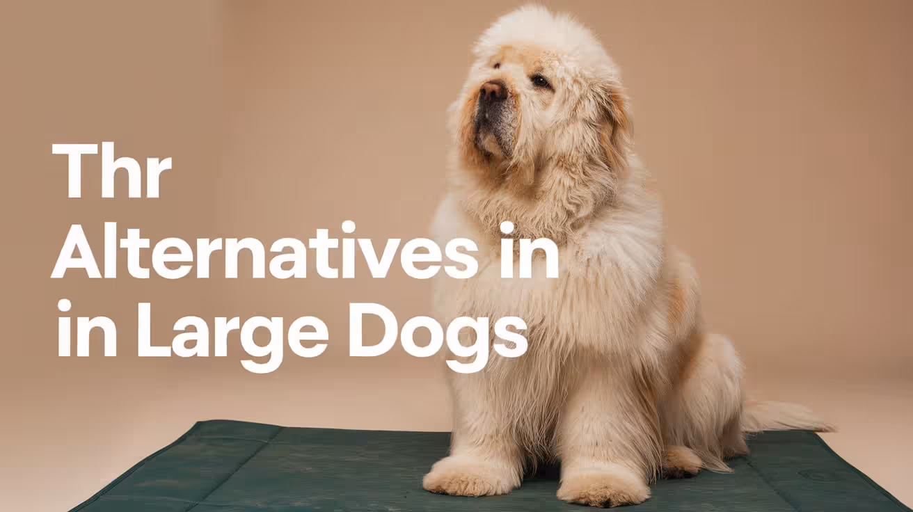
Alternatives to Hip Replacement Surgery in Large Dogs
Explore effective alternatives to hip replacement surgery in large dogs, including surgical options, non-surgical care, regenerative therapies, and long-term outcomes
Why Look for Alternatives to Hip Replacement in Large Dogs?
Total hip replacement (THR) is considered the gold standard for treating severe hip disease, but it may not be the right choice for every dog. Large breeds, in particular, require careful consideration because of their size, weight, and the demands placed on their joints.
- High Cost of Surgery – THR often ranges from $4,000 to $10,000 per hip, which may be unaffordable for some owners.
- Age and Health Concerns – Senior dogs or those with systemic illnesses may face higher surgical risks and longer recovery times.
- Owner Preference – Some families prefer less invasive options, especially when managing comfort rather than pursuing full joint restoration.
- Unique Large-Breed Challenges – Heavier dogs place more stress on implants, increasing the importance of evaluating alternatives that can still reduce pain and improve mobility.
Considering alternatives ensures that every dog receives the most practical and safe solution for its individual needs.
Surgical Alternatives to Hip Replacement
While THR is the most effective treatment, certain surgical alternatives may provide relief when hip replacement is not possible. These procedures are usually chosen based on the dog’s age, size, and stage of hip disease.
1. Femoral Head Ostectomy (FHO) in Large Dogs
FHO involves removing the femoral head, which allows a false joint to form. It can relieve pain by stopping bone-on-bone contact, but in large breeds, outcomes are less predictable because body weight and muscle mass place more stress on the joint.
Many large dogs develop an abnormal gait or limited strength post-FHO. Still, it may be useful when THR is not affordable or possible, especially in cases of severe fractures or chronic dislocations where pain relief is the priority over restoring full mobility.
2. Triple or Double Pelvic Osteotomy (TPO/DPO)
TPO and DPO surgeries involve cutting and rotating sections of the pelvis to improve coverage of the femoral head. These procedures are designed for younger dogs—typically under one year—who show early signs of hip dysplasia but have not yet developed arthritis.
When performed at the right stage, TPO/DPO can help preserve the natural hip joint and delay the need for more advanced surgeries. However, once arthritis sets in, results are poor. Large breed dogs diagnosed early may benefit, but the strict age and condition requirements limit its use.
3. Juvenile Pubic Symphysiodesis (JPS)
JPS is a preventive surgery done in puppies younger than 5 months. It works by altering the growth of the pelvis so the hip socket develops better coverage of the femoral head. This reduces the chance of severe hip dysplasia later in life.
However, timing is critical—if the puppy is older or already showing signs of arthritis, JPS is no longer effective. In large dogs, JPS is rarely used once hip disease is established, but it may help breeders or owners of high-risk puppies by reducing future problems.
Non-Surgical Alternatives for Large Dogs
When hip replacement is not an option, non-surgical approaches can still improve comfort and mobility in large dogs. These methods focus on managing pain, protecting joints, and supporting long-term quality of life.
1. Weight Management and Exercise Control
Excess weight puts enormous strain on already weak hips, especially in large dogs. Careful weight management through calorie-controlled diets is one of the most effective ways to reduce pain. Controlled, low-impact exercise such as leash walking and swimming keeps joints flexible without overloading them.
Activities like running, jumping, or rough play should be avoided. Consistency is key, as even moderate weight loss can significantly reduce hip stress. With proper exercise control, large dogs maintain strength while avoiding activities that accelerate joint degeneration.
2. Pain Management and Medications
Medications are often essential for long-term comfort in large dogs with hip disease. Non-steroidal anti-inflammatory drugs (NSAIDs) help reduce both pain and inflammation.
Additional medications like gabapentin or tramadol may be used for nerve or breakthrough pain. Joint supplements containing glucosamine, chondroitin, and omega-3s support joint health and reduce stiffness.
These treatments do not cure hip disease but provide meaningful relief and improve daily function. Long-term veterinary monitoring is needed to adjust dosages and minimize potential side effects.
3. Physical Therapy and Hydrotherapy
Physical therapy builds strength in the muscles surrounding the hip, providing extra stability to weakened joints. Exercises like controlled sit-to-stand routines, gentle stretching, and balance training can improve mobility.
Hydrotherapy, including swimming and underwater treadmill sessions, allows dogs to exercise without putting weight on painful joints. These methods increase circulation, reduce stiffness, and improve confidence in movement.
Structured rehabilitation under professional guidance can extend mobility and significantly improve quality of life. For many large dogs, consistent physiotherapy delays the need for surgery and provides safer long-term management.
Regenerative and Advanced Therapies
Regenerative medicine is increasingly being explored as an alternative for managing hip disease in large dogs. These advanced therapies focus on reducing pain and promoting healing within the joint.
- Stem Cell Therapy – Mesenchymal stem cells, often harvested from fat or bone marrow, are injected into the hip joint to reduce inflammation and encourage tissue repair. Some dogs show improved comfort and mobility, though results can be variable.
- Platelet-Rich Plasma (PRP) – PRP injections deliver concentrated growth factors that help decrease inflammation and promote healing. These can be combined with other treatments for better results.
- Prolotherapy – Involves injecting irritant solutions around ligaments to stimulate new tissue growth, potentially increasing joint stability.
- Limitations in Large Dogs – While promising, these therapies often provide partial improvement and may not restore full function, especially in heavier breeds.
Regenerative therapies can reduce pain and delay surgery but are rarely permanent solutions. For large dogs, they are best considered as part of a broader treatment plan.
Supportive Devices and Lifestyle Adjustments
Supportive devices and home adjustments play a major role in improving comfort and independence for large dogs with hip disease. These simple interventions protect joints and make daily living safer.
- Hip Braces and Harnesses – Braces stabilize the hip joint and reduce pain during movement. Harnesses give owners better control when assisting dogs with walking or climbing.
- Orthopedic Beds – Provide cushioning and joint support, reducing stiffness after rest and encouraging proper posture.
- Ramps and Stairs Alternatives – Installing ramps for vehicles, furniture, or outdoor steps prevents jumping, which can worsen hip pain.
- Non-Slip Flooring – Mats or rugs prevent slipping on tile or hardwood, reducing the risk of falls.
- Owner’s Role – Monitoring activity, creating a safe space, and maintaining routine adjustments are vital for long-term success.
With the right support devices and home care, many large dogs can remain active and comfortable despite hip disease.
Combination Approaches for Better Outcomes
No single alternative can fully match the success of hip replacement, especially in large dogs. However, combining multiple strategies often leads to better long-term results and improved quality of life.
- Braces Plus Weight Control – Using hip braces alongside strict weight management reduces stress on the joint and supports stability.
- Exercise with Therapy – Low-impact exercise, combined with hydrotherapy or physiotherapy, strengthens muscles and helps compensate for joint weakness.
- Medications with Supplements – Long-term NSAID use, supported by glucosamine, chondroitin, and omega-3s, provides layered pain control and joint support.
- Advanced Therapies as Add-Ons – Stem cell or PRP injections can be used in combination with physical therapy to extend mobility further.
- Setting Expectations – While these methods improve comfort, they rarely restore full athletic ability in large dogs. The goal is pain reduction and better daily function.
Multi-modal management maximizes results by addressing hip disease from several angles. With realistic expectations, dogs can enjoy meaningful improvements in quality of life.
Cost Comparison of Hip Replacement Alternatives
When total hip replacement is not possible, cost often becomes a major deciding factor in choosing alternatives. Each option carries its own expense range, depending on the procedure, follow-up care, and the dog’s size.
- Femoral Head Ostectomy (FHO) – Typically costs $2,000–$4,500. While less expensive, results are less predictable in large dogs and may lead to gait changes.
- Triple or Double Pelvic Osteotomy (TPO/DPO) – Priced around $3,000–$6,000. Best for younger dogs before arthritis develops, making it less commonly applicable in adults.
- Stem Cell Therapy or PRP Injections – Range from $2,000–$5,000. Provide temporary pain relief but usually require repeat treatments.
- Hip Braces – Cost $200–$800. Affordable and non-invasive, but they provide only partial support.
- Conservative Management – Includes medications, supplements, and physiotherapy. While monthly costs may seem low, they accumulate significantly over a dog’s lifetime.
Though less costly upfront, many alternatives may require repeat treatments or ongoing care. Owners should balance immediate affordability with long-term outcomes.
Long-Term Expectations Without Hip Replacement
Alternatives to hip replacement can provide significant relief, but they rarely restore full hip function in large dogs. Owners should understand what outcomes are realistic.
- Pain Reduction – Surgeries like FHO or therapies such as PRP can decrease pain, but results may not equal the stability of a prosthetic joint.
- Mobility Improvements – Physiotherapy, hydrotherapy, and braces help maintain function, though gait abnormalities may persist in heavier dogs.
- Limitations in Large Breeds – Because of their size, large dogs often place more stress on affected joints, making results less predictable compared to smaller breeds.
- Importance of Early Diagnosis – Interventions like TPO or JPS are only effective when started early, before arthritis permanently damages the joint.
- Ongoing Management – Consistent weight control, supplements, and veterinary monitoring are required for long-term comfort.
While alternatives can extend mobility and reduce suffering, they cannot fully replicate hip replacement. Realistic goals focus on comfort, daily function, and slowing disease progression.
Conclusion
Total hip replacement (THR) remains the gold standard for treating severe hip disease, offering the best chance for restoring full mobility and long-term comfort. However, it is not always possible for every large dog due to cost, health, or owner preference. In such cases, alternatives like FHO, TPO/DPO, regenerative therapies, and conservative management can still provide meaningful relief.
Large dogs benefit most from tailored solutions that combine surgery, rehabilitation, weight management, and supportive lifestyle adjustments. While these options may not match the complete outcomes of THR, they can extend mobility, reduce pain, and improve quality of life.
The final step should always be consulting a veterinary orthopedic specialist. With expert guidance, owners can make the safest, most effective choice for their dog’s unique needs.
FAQs
What are the main alternatives to hip replacement in large dogs?
Alternatives include femoral head ostectomy (FHO), triple or double pelvic osteotomy (TPO/DPO), juvenile pubic symphysiodesis (JPS), regenerative therapies like stem cells or PRP, supportive devices such as hip braces, and conservative management with medications, weight control, and physiotherapy. The best option depends on the dog’s age, size, and stage of disease.
Is femoral head ostectomy (FHO) effective for large dogs?
FHO can relieve pain by removing the femoral head, but outcomes are less predictable in large breeds due to their weight and muscle mass. Many large dogs develop an altered gait or reduced strength after FHO. It may still be considered when hip replacement is not possible, especially for pain relief.
Can younger large-breed dogs benefit from pelvic osteotomy?
Yes, triple or double pelvic osteotomy (TPO/DPO) can help younger dogs diagnosed with hip dysplasia before arthritis develops. It realigns the hip socket to improve stability. However, the procedure must be performed early, usually under one year of age, making it unsuitable for older large dogs with advanced joint damage.
How do regenerative therapies help dogs with hip disease?
Stem cell therapy, platelet-rich plasma (PRP) injections, and prolotherapy reduce inflammation, promote healing, and may improve comfort. These therapies are less invasive than surgery and can delay progression of hip disease. However, results vary, and in large dogs, they often provide partial improvement rather than restoring full mobility or joint function.
Are braces and lifestyle changes enough for large dogs with hip problems?
Braces, ramps, orthopedic bedding, and non-slip flooring can make daily life safer and more comfortable. While these adjustments do not cure hip disease, they reduce stress on joints and prevent further injury. When combined with weight control and low-impact exercise, they can significantly improve quality of life for large dogs.
How do costs of alternatives compare to hip replacement?
Alternatives are generally less expensive than total hip replacement. FHO costs $2,000–$4,500, TPO/DPO around $3,000–$6,000, and regenerative therapies $2,000–$5,000. Braces range from $200–$800, while long-term conservative care builds ongoing monthly costs. While more affordable, these options may require repeat treatments and usually don’t match the lasting results of THR.
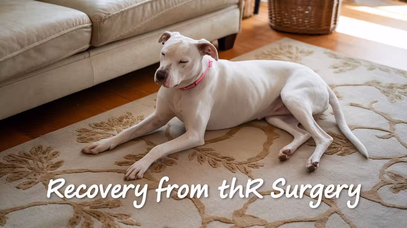
What to Expect During Recovery from Total Hip Replacement in Dogs
Learn what to expect during recovery from total hip replacement (THR) in dogs, including activity restrictions, rehab milestones, follow-ups, and long-term outcomes
Immediate Post-THR Surgery Care for Dogs
The first days after total hip replacement are critical for preventing complications and ensuring proper healing. Careful management of the surgical site and effective pain control set the foundation for recovery.
- Incision Protection – The surgical site must stay clean and dry at all times. Prevent your dog from licking or chewing at the incision to avoid infection.
- Elizabethan Collar – An e-collar or protective barrier should be worn until the incision heals fully, usually for 10–14 days.
- Pain Management – Veterinarians prescribe NSAIDs, opioids, or pain patches to control discomfort and support early mobility.
- Hospital Monitoring – Dogs are closely observed for bleeding, swelling, or complications before being safely discharged home.
Proper immediate care reduces risks and helps your dog transition smoothly into the next phase of recovery.
Activity Restrictions After THR Surgery
Activity control is the most important part of recovery after total hip replacement. In the first weeks, even small mistakes like a jump or slip can damage the implant. Owners must provide a safe and structured environment.
- Strict Confinement – Dogs should stay in a crate or small enclosed area with non-slip flooring. This prevents running or sudden twisting movements that can harm the hip.
- No High-Impact Activity – Jumping on furniture, climbing stairs, or playing with other pets must be avoided. These actions place stress on the implant and may cause loosening.
- Leash-Controlled Breaks – Only short bathroom walks on a leash are allowed. Owners should guide movements carefully to avoid slips or overexertion.
- Constant Supervision – Dogs cannot be left unsupervised during recovery. Owners must watch closely to prevent sudden activity or accidents.
While these restrictions may feel difficult, they are temporary. Following them closely gives the implant time to heal securely and ensures long-term success.
THR Recovery Milestones and Mobility Timeline
Recovery from hip replacement follows a predictable timeline, with steady improvements in comfort and activity. Knowing these milestones helps owners track progress and stay encouraged.
- Early Weight Bearing – Within the first week, many dogs cautiously use the operated limb, showing implant stability and reduced pain.
- 6–8 Weeks – Lameness improves significantly, with dogs walking more normally and using the leg with increasing confidence.
- 12 Weeks – By this stage, most dogs can resume near-normal activities such as walking around the home, climbing short steps, and enjoying longer leash walks.
- 3–4 Months – Full function is typically regained. Dogs often return to running, swimming, and even athletic play with veterinary approval.
Recovery takes patience, but each step forward builds strength and stability. By following the timeline, most dogs achieve lasting, pain-free mobility.
Exercise and Walking Plan During THR Recovery
Exercise must be carefully structured after total hip replacement. A gradual walking plan builds strength and coordination while protecting the implant.
- Starting Walks – Begin with short 10–15 minute leash walks, two to three times per day. Keep surfaces flat and steady, avoiding hills or uneven ground.
- Progressive Increases – Add about 5 minutes to each walk weekly if your dog shows no signs of soreness or limping. This steady buildup strengthens muscles without straining the hip.
- Avoid Slippery Surfaces – For at least 6 weeks, dogs should stay off slick floors like tile or hardwood. These surfaces increase the risk of slips that can damage the implant.
- Transitioning to Longer Walks – Once dogs are comfortable with 30–40 minute walks, they can begin light, supervised play in a safe area.
- Off-Leash Activity – Running or free play should be introduced only at 8–12 weeks, and only after veterinary clearance confirms stability.
A structured walking plan ensures safe recovery. With time and patience, dogs rebuild muscle strength and return to their normal activities without risk.
Rehabilitation and Physiotherapy After THR in Dogs
Rehabilitation is one of the most important parts of recovery after hip replacement. It helps rebuild strength, restore range of motion, and ensures the implant is fully supported by healthy muscles.
- Hydrotherapy for Early Support – Around 4 weeks post-op, underwater treadmill or swimming sessions can begin. Water reduces weight on the new joint while allowing safe movement, which helps improve circulation and flexibility without stressing the implant.
- Controlled Strengthening Exercises – Sit-to-stand drills, slow leash walking on gentle inclines, and treadmill sessions help retrain balance and coordination. These exercises are done in short, controlled sessions to prevent fatigue.
- Muscle Rebuilding Phase – From 6–8 weeks, the focus shifts to strengthening the hip, gluteal, and quadriceps muscles, which are key for long-term joint stability.
- Flexibility and Range of Motion – Targeted stretches and guided movements prevent stiffness and encourage smooth joint function as healing continues.
- Benefits of Structured Rehab – Dogs in physiotherapy programs typically achieve faster recovery, lower complication rates, and better long-term outcomes.
Rehab is not optional—it’s a structured process that greatly improves both the comfort and durability of the hip replacement.
Veterinary Follow-Up After THR Surgery
Follow-up care is a lifelong commitment after hip replacement. These visits ensure healing is progressing properly and catch issues before they become serious.
- Initial Post-Surgery Recheck (10–14 Days) – At this appointment, the incision is inspected, sutures or staples are removed, and any early wound concerns are addressed.
- Six-Week Assessment with X-Rays – Imaging confirms that the implant is seated correctly, while gait assessments show how well the dog is regaining normal movement.
- Intermediate Recovery Checks – Some dogs may require additional visits to adjust pain medication, track muscle development, or refine exercise plans.
- Annual or Biannual Radiographs – Once healing is complete, routine imaging is recommended to confirm implant stability and identify any early changes in bone support.
- Value of Consistent Monitoring – Regular visits allow quick responses to potential problems such as implant loosening, helping preserve long-term success.
These follow-ups ensure the hip replacement remains secure for life and give owners reassurance that recovery is on track.
Diet, Supplements, and Medications After THR
Long-term hip health depends on more than surgery alone. Nutrition, supplements, and proper medication use all support healing and protect the new implant.
- Medication Compliance – Prescribed NSAIDs, opioids, or antibiotics must be given exactly as directed. These drugs control pain, prevent infection, and create the conditions for steady healing.
- Weight Control as a Priority – Every extra pound adds stress to the hip joint. Maintaining an ideal body weight through portion-controlled meals and regular monitoring is one of the best ways to protect the implant.
- Support from Supplements – Omega-3 fatty acids reduce inflammation, while glucosamine and chondroitin nourish cartilage and support surrounding joints. Though the replaced hip does not need cartilage, these supplements benefit the entire musculoskeletal system.
- Balanced Recovery Diet – High-quality proteins repair muscle tissue, while vitamins and minerals strengthen bones. A balanced diet is critical during the rebuilding phase.
- Long-Term Lifestyle – Ongoing weight control, anti-inflammatory support, and proper nutrition ensure that the replaced hip remains functional for a lifetime.
Combining surgery with lifelong diet and supplement management ensures stronger, healthier outcomes that last.
Home Modifications to Support THR Recovery
A safe home environment is crucial for protecting the new hip during recovery. Making small but thoughtful adjustments can prevent accidents and help your dog heal more comfortably.
- Ramps Instead of Stairs – Install ramps for staircases, furniture, or vehicles to reduce jumping or climbing, which can strain the implant.
- Supportive Harnesses – A rehabilitation harness allows owners to assist dogs with standing, walking, and navigating difficult areas without stressing the hip.
- Raised Feeding Stations – Elevated food and water bowls reduce strain on the hips, making mealtimes easier and more comfortable.
- Orthopedic Bedding – Soft, supportive bedding cushions the joints and provides a designated rest area that encourages proper healing.
- Slip-Free Flooring – Use rugs, mats, or non-slip coverings on tile and hardwood to prevent dangerous falls during the recovery phase.
Simple modifications make the home safer and support smoother recovery. By reducing physical stress, you give your dog the best chance for a strong return to mobility.
Complications During THR Recovery: Warning Signs to Watch
Though most dogs recover smoothly, complications can occur. Recognizing early warning signs allows owners to seek help before problems become severe.
- Incision Concerns – Redness, swelling, discharge, or foul odor from the surgical site may indicate infection and require prompt treatment.
- Weight-Bearing Issues – Reluctance to use the operated limb or sudden worsening of lameness may signal implant instability or dislocation.
- Excessive Pain – Pain that seems more severe or lasts longer than expected should not be ignored, as it may point to complications.
- Behavioral Changes – Refusing food, restlessness, or hiding may reflect discomfort or underlying infection.
- Immediate Veterinary Attention – Any suspected complication should be reported to the surgeon immediately for evaluation and treatment.
Close observation at home is vital. Quick action ensures complications are addressed early, protecting both the implant and your dog’s long-term comfort.
Long-Term Expectations After THR in Dogs
Total hip replacement offers dogs the chance to return to an active, pain-free lifestyle. Long-term expectations are excellent when recovery protocols are followed closely.
- Full Mobility Restored – Most dogs regain normal walking ability within weeks and return to running, swimming, and playing by 3–4 months.
- Implant Longevity – Modern prosthetic implants are designed to last the lifetime of the dog, with very few requiring revision surgery.
- Lifelong Comfort – Successful THR eliminates the chronic pain of hip disease, improving energy, mood, and overall quality of life.
- Owner Commitment – Long-term success depends on maintaining a healthy weight, scheduling annual check-ups, and preventing high-impact injuries.
- Return to Normal Lifestyle – Family pets can resume daily activities, and even working or sporting dogs often return to their previous roles.
With proper aftercare and monitoring, THR provides reliable, lasting results. For most dogs, this surgery represents not just recovery but a true return to a pain-free life.
FAQs
How long is recovery after total hip replacement in dogs?
Recovery usually takes 8–12 weeks. Dogs begin bearing weight within the first week, lameness improves by 6–8 weeks, and most resume near-normal activity by 12 weeks. Full athletic ability, such as running and swimming, often returns by 3–4 months, provided rehabilitation and follow-up guidelines are strictly followed.
What activity restrictions are needed after THR surgery?
Dogs must remain on strict crate rest or confined in a non-slip area. No running, jumping, climbing, or stairs are allowed in the early weeks. Only short, controlled leash walks for bathroom breaks are permitted. Owner supervision is essential, as even one slip or jump can compromise the new implant’s stability.
When can my dog start physiotherapy after THR?
Rehabilitation usually begins around 4 weeks post-surgery. Hydrotherapy, sit-to-stand drills, incline walking, and treadmill therapy are gradually introduced. From 6–8 weeks, exercises focus on muscle rebuilding and hip flexibility. A structured rehabilitation program greatly improves long-term outcomes, helping dogs regain strength and confidence without overloading the new hip joint.
What signs of complications should I watch for during recovery?
Warning signs include redness, swelling, or discharge at the incision, worsening lameness, or reluctance to bear weight on the operated leg. Excessive pain beyond the expected recovery timeline also signals potential issues. If any of these occur, immediate veterinary attention is critical to protect the implant and ensure safe recovery.
How can I make my home safer for recovery?
Simple modifications support healing. Use ramps for stairs, furniture, or cars to prevent jumping. Place non-slip mats on slick floors to avoid falls. Provide raised food bowls, orthopedic bedding, and a rehabilitation harness for added support. These changes create a safe, comfortable environment that reduces stress on the new hip.
Will my dog return to a normal lifestyle after THR?
Yes, most dogs regain full mobility and quality of life within 3–4 months. Once healed, they can walk, run, swim, and play without pain. With proper care, implants usually last a lifetime, giving dogs the chance to enjoy daily family activities and, in many cases, even return to work or sport.
All Articles

Signs Your Dog Might Need Femoral Head Ostectomy
Learn the common signs your dog may need femoral head ostectomy surgery to relieve hip pain and improve mobility for a better quality of life
If your dog is showing signs of pain or trouble moving their hip, it might be time to consider a surgery called Femoral Head Ostectomy (FHO). This surgery helps dogs with serious hip problems feel better and walk more easily. Knowing the signs your dog needs FHO can help you act early and improve their quality of life.
Signs Your Dog Might Need Femoral Head Ostectomy
Recognizing the signs early helps ensure your dog receives timely treatment for painful hip conditions that may require Femoral Head Ostectomy (FHO) surgery.
1. Difficulty Walking or Limping
Limping or difficulty walking is one of the most obvious signs your dog might need FHO surgery. When the hip joint is painful or damaged, your dog will try to avoid putting weight on the affected leg.
- Your dog may limp continuously or only after exercise.
- Favoring one leg over the other helps reduce pressure and pain in the hip.
- Difficulty walking may present as slow, hesitant steps or reluctance to move.
This limping indicates that the hip joint is not functioning properly due to pain, arthritis, fracture, or other damage. Persistent limping despite rest or medication is a strong sign that surgical intervention like FHO could be necessary.
2. Abnormal Gait or Favoring One Leg
An abnormal gait means your dog’s walking pattern changes because of discomfort or weakness. Dogs with hip problems may shift their weight unevenly, causing visible changes in how they move.
- Your dog may “swing” the affected leg or take shorter steps on that side.
- Uneven weight distribution may cause limping or skipping.
- The gait may look unbalanced or awkward, especially after activity.
These changes result from pain or instability in the hip joint. Abnormal gait often worsens over time and is a clear signal to consult your vet for possible surgery.
3. Loss of Weight-Bearing on the Affected Leg
Loss of weight-bearing means your dog refuses to put any weight on the painful leg. This is a severe sign of hip dysfunction and pain.
- Your dog may hold the leg completely off the ground while standing or walking.
- This can occur suddenly or develop gradually with worsening discomfort.
- Loss of weight-bearing leads to muscle weakness and further mobility issues.
When your dog stops using the leg due to pain, it often indicates advanced joint damage requiring surgical treatment such as FHO for pain relief and function restoration.
Read more:
4. Persistent Hip Pain or Discomfort
Persistent hip pain affects your dog’s quality of life and is a common reason for FHO surgery.
- Your dog may show signs of discomfort such as whining or reluctance to move.
- Pain might be constant or worsen after activity.
- Hip pain reduces interest in walks, play, and normal activities.
Ongoing pain means conservative treatments are not controlling the condition, and surgery may be the best option to restore comfort and mobility.
5. Decreased Activity or Reluctance to Play
Changes in activity level are often one of the first signs owners notice when their dog is in pain.
- Your dog may avoid running, jumping, or climbing stairs.
- Reduced playfulness and slower movements indicate discomfort.
- Reluctance to exercise can lead to weight gain and muscle loss.
This behavior change shows your dog is trying to avoid pain, which may signal the need for surgical intervention like FHO.
6. Stiffness in the Hip Joint, Especially After Rest
Stiffness after rest or sleep is common in dogs with hip joint problems.
- Your dog may be slow to stand or walk after naps.
- The hip may feel tight or rigid, limiting movement temporarily.
- Stiffness often improves with gentle activity but returns after resting.
This stiffness indicates joint inflammation or damage, which may require surgery if it significantly impacts mobility.
7. Pain When the Hip Is Manipulated or Touched
During veterinary exams, pain responses when the hip is moved or touched can confirm joint problems.
- Vets check for tenderness by gently manipulating the hip joint.
- Pain during these tests often correlates with arthritis, fractures, or hip dysplasia.
- A painful response supports the need for further treatment or surgery.
This exam finding helps vets decide if FHO surgery is appropriate for your dog’s condition.
8. Limited Range of Motion in the Hip
A reduced ability to move the hip joint shows stiffness and pain.
- Your dog may struggle to fully extend or flex the leg.
- Limited motion causes difficulty in walking, running, or climbing.
- This restriction often worsens over time without treatment.
Limited range of motion is a key symptom indicating severe joint damage that may benefit from FHO surgery.
9. Muscle Loss or Atrophy in the Affected Leg
Muscle wasting happens when a dog stops using the painful leg regularly.
- You may notice the leg looks thinner or weaker compared to the other side.
- Muscle loss reduces joint support and slows recovery.
- Atrophy often signals chronic discomfort and long-term mobility issues.
Muscle loss is a serious sign that conservative care is no longer enough, and surgical options should be considered.
Read more:
10. Chronic Lameness Not Improving Over Time
Lameness that does not improve with rest or treatment indicates the need for further evaluation.
- Persistent limping despite medication or physical therapy shows worsening hip disease.
- Chronic lameness reduces your dog’s activity and quality of life.
- This symptom often leads vets to recommend FHO surgery for pain relief.
Ignoring ongoing lameness can cause further joint damage and pain.
11. Joint Instability or Looseness Detected by the Vet
During exams, vets may find instability or looseness in the hip joint.
- The hip may feel unstable or shift abnormally during manipulation.
- Joint instability increases pain and risk of further injury.
- This finding supports surgical intervention to stabilize the joint and relieve pain.
Hip instability is often a clear reason to consider FHO surgery.
12. Reduced Quality of Life Due to Hip Issues
Chronic hip pain and mobility problems can greatly reduce your dog’s happiness.
- Dogs may become withdrawn, less playful, and reluctant to exercise.
- Pain and difficulty moving affect daily activities like walking or climbing stairs.
- Improving quality of life is a primary goal of FHO surgery.
If hip problems interfere with your dog’s enjoyment of life, surgery may provide relief and restore activity.
When to Consult Your Veterinarian About FHO Surgery
Early consultation with your veterinarian is essential if you notice signs of severe hip pain or mobility issues in your dog. Prompt veterinary evaluation helps diagnose the problem accurately and determine if Femoral Head Ostectomy (FHO) surgery is necessary.
- Importance of early consultation: Early vet visits can catch hip conditions before they worsen, improving treatment options and outcomes. Waiting too long may lead to increased pain, joint damage, and muscle loss.
- Diagnostic steps: Your vet will perform a physical exam to assess pain, joint stability, and range of motion. They will also recommend diagnostic imaging like X-rays to evaluate the hip joint’s condition.
- Treatment planning: Based on findings, the vet discusses conservative care options and, if needed, explains the benefits and risks of FHO surgery.
- Ongoing monitoring: Even if surgery isn’t immediately required, regular vet check-ups help track progression and adjust treatment plans.
Consulting your vet early ensures your dog receives timely care, reducing discomfort and helping maintain a good quality of life. Early diagnosis and intervention are key to successful management of hip problems.
Read more:
How Veterinarians Diagnose the Need for FHO
Veterinarians use a combination of diagnostic tools and physical exams to determine if Femoral Head Ostectomy (FHO) surgery is the best option for your dog.
- Physical exams: The vet assesses your dog’s hip for pain, swelling, decreased range of motion, and instability. They watch how your dog walks and moves to identify signs of discomfort or limited mobility.
- X-rays: Radiographs provide a clear image of the hip joint, showing bone damage, arthritis, fractures, or deformities. X-rays are essential to confirm the severity of the problem and help plan treatment.
- Additional imaging: In some cases, vets may use advanced imaging such as CT scans or MRI to get detailed views of the joint and surrounding tissues.
- Evaluating symptoms: Vets also consider your dog’s history, including persistent limping, pain levels, and response to previous treatments.
Based on these findings, the vet decides if FHO surgery will relieve pain and improve your dog’s quality of life. Early diagnosis ensures timely intervention and better recovery outcomes. Working closely with your vet helps develop a tailored treatment plan that meets your dog’s specific needs.
What to Expect From Femoral Head Ostectomy Surgery
Femoral Head Ostectomy (FHO) surgery is a valuable procedure designed to relieve severe hip pain and improve mobility in dogs with damaged hip joints. By removing the femoral head and neck, FHO stops painful bone-on-bone contact, allowing your dog to move more comfortably.
- Surgery benefits: The main benefit is significant pain relief, which helps your dog regain use of the affected leg. FHO can improve quality of life, especially for dogs suffering from arthritis, fractures, or hip dysplasia.
- Surgical process: The surgery typically takes one to two hours under general anesthesia. Your dog will be closely monitored during and after the procedure to ensure safety.
- Recovery expectations: Recovery usually takes 6 to 12 weeks and involves rest, pain management, and physical therapy. Gradual weight-bearing and muscle strengthening are important for success.
- Owner’s role: Following your vet’s post-operative care instructions and attending follow-up visits are critical to a smooth recovery.
Most dogs adapt well to the changes and regain comfortable mobility, making FHO a highly effective solution for many painful hip conditions.
Read more:
FAQs About Femoral Head Ostectomy (FHO) Surgery
How long does it take for a dog to recover from FHO surgery?
Recovery from FHO surgery typically takes 6 to 12 weeks. During this time, your dog needs restricted activity and physical therapy to rebuild muscle strength and improve joint mobility. Full recovery may continue over several months depending on the dog’s size, health, and rehabilitation efforts.
Is FHO surgery painful for dogs?
FHO surgery is performed under general anesthesia, so your dog won’t feel pain during the operation. After surgery, veterinarians prescribe pain medications to keep your dog comfortable while healing. Proper pain management is essential for recovery and helps your dog remain calm and active during rehabilitation.
What dogs are good candidates for FHO surgery?
Small to medium-sized dogs with severe hip pain caused by arthritis, fractures, hip dysplasia, or trauma are good candidates for FHO. Dogs who do not respond well to conservative treatments or other surgeries may also benefit. Your vet will evaluate your dog’s overall health and mobility to decide if FHO is suitable.
Can large dogs undergo FHO surgery?
Large dogs can have FHO surgery, but recovery can be more challenging due to their weight putting extra pressure on the new joint. For bigger dogs, vets often recommend alternatives like total hip replacement, which may provide better long-term mobility and comfort.
What are the risks of FHO surgery?
Risks include infection, muscle atrophy, decreased joint motion, and persistent limping. These risks are minimized by following post-operative care instructions closely, including medication, activity restriction, and physical therapy. Regular vet check-ups help identify and manage any complications early.
How does FHO surgery help improve mobility?
FHO surgery removes the damaged femoral head, eliminating painful bone-on-bone contact. The body forms a fibrous “false joint” that cushions the hip and allows pain-free movement. Combined with rehab, this improves your dog’s ability to walk, run, and enjoy daily activities comfortably.

When Is FHO Surgery Recommended for Dogs?
Discover when Femoral Head Ostectomy (FHO) surgery is recommended for dogs, including common conditions and signs needing surgical care
Understanding FHO Surgery
Femoral Head Ostectomy (FHO) is a surgical procedure where the head and neck of the thigh bone (femur) are removed. This surgery is done to relieve severe pain caused by hip problems like arthritis, fractures, or hip dysplasia.
FHO helps dogs by eliminating the painful bone-on-bone contact inside the hip joint. After surgery, the body forms a “false joint” made of fibrous tissue, which cushions the area and allows more comfortable movement. Although it’s not a normal joint, this new structure reduces pain and improves mobility.
This surgery is often recommended when other treatments like medication or physical therapy have not worked. It allows dogs to regain the use of their leg and live with less discomfort. FHO can be especially helpful for dogs with severe hip damage or those who cannot have more complex surgeries like total hip replacement.
How Veterinarians Diagnose the Need for FHO Surgery
Veterinarians use a combination of physical exams and diagnostic imaging to determine if Femoral Head Ostectomy (FHO) surgery is needed for your dog.
- Physical exam: The vet will assess your dog’s hip joint by checking for pain, swelling, decreased range of motion, and signs of lameness or instability. They will observe how your dog walks and moves to identify discomfort or limited mobility.
- Diagnostic imaging: X-rays are essential to see the condition of the hip joint, including bone damage, arthritis, fractures, or deformities. These images help confirm the diagnosis and guide treatment decisions.
- Signs and symptoms: Dogs showing severe hip pain, persistent limping, difficulty standing or walking, and poor response to medication or physical therapy are often evaluated for surgery.
- Additional tests: In some cases, advanced imaging like CT or MRI may be used for detailed views of the joint structures.
After evaluating these findings along with your dog’s age, size, and overall health, the vet decides if FHO surgery is the best option to relieve pain and improve mobility. Early diagnosis helps plan effective treatment and improve outcomes.
Read more:
Medical Conditions That Indicate FHO Surgery
Femoral Head Ostectomy (FHO) surgery is often recommended for dogs suffering from specific medical conditions that cause severe hip pain and joint damage.
- Hip dysplasia and severe arthritis: Hip dysplasia is a genetic condition where the hip joint forms abnormally, leading to arthritis and chronic pain. When arthritis becomes severe, FHO can relieve pain by removing the damaged femoral head.
- Traumatic hip injuries: Fractures or dislocations of the femoral head or neck caused by accidents or trauma may require FHO if the bones cannot be repaired. This surgery helps restore comfort and function.
- Legg-Calve-Perthes disease: This condition causes the femoral head to deteriorate (necrosis) due to poor blood supply. FHO removes the damaged bone to eliminate pain and improve mobility.
- Degenerative joint disease in older dogs: Age-related joint wear and tear can cause chronic hip pain. When other treatments fail, FHO can improve quality of life by reducing discomfort and increasing mobility.
Your vet will assess these conditions through exams and imaging to determine if FHO is the best surgical option for your dog’s specific needs.
When Conservative Treatments Are Not Enough
Conservative treatments like medication, rest, and physical therapy are often the first steps in managing hip problems in dogs. However, these approaches may not always provide enough relief.
- Medication failure: Pain relievers and anti-inflammatory drugs can reduce discomfort temporarily, but if your dog continues to show pain or limping, medication alone may not be sufficient.
- Rest and activity modification: Limiting activity can help reduce stress on the hip joint, but persistent discomfort despite rest indicates the need for further intervention.
- Physical therapy limitations: While rehab exercises strengthen muscles and improve joint function, some dogs do not respond well enough to prevent ongoing pain or mobility issues.
When pain and limited mobility continue despite these treatments, it suggests the hip joint damage is severe. At this stage, Femoral Head Ostectomy (FHO) surgery may be recommended to relieve pain and improve quality of life.
Your vet will carefully evaluate your dog’s response to conservative care before suggesting surgery, ensuring that FHO is the best option for lasting relief and better mobility. Early surgical intervention can prevent further decline and discomfort.
Read more:
Ideal Candidates for FHO Surgery
Femoral Head Ostectomy (FHO) surgery is best suited for certain groups of dogs based on their size, health, and response to other treatments.
- Small to medium-sized dogs: FHO works especially well for dogs under 50 pounds. Their lighter weight allows the “false joint” formed after surgery to support movement effectively, leading to better recovery and mobility.
- Dogs with poor response to previous treatments: If your dog has had medication, physical therapy, or other hip surgeries without sufficient pain relief or improvement, FHO may be the next best step to address ongoing discomfort.
- Dogs with significant reduction in quality of life: When hip pain severely limits your dog’s ability to walk, run, play, or enjoy daily activities, surgery can greatly improve comfort and function.
Vets consider factors like your dog’s age, overall health, activity level, and severity of joint damage when recommending FHO. While FHO may not be suitable for every dog, it offers excellent pain relief and improved mobility for many. Discussing your dog’s specific situation with your vet helps ensure the best treatment choice.
FHO Surgery as an Alternative to Total Hip Replacement
Total hip replacement (THR) is a highly effective surgery that replaces the entire hip joint with an artificial implant. However, THR may not always be feasible or affordable for every dog and owner.
- When THR is not feasible: Some dogs, especially smaller breeds or those with certain health issues, may not be good candidates for THR due to surgical complexity or recovery demands.
- Cost considerations: THR is usually more expensive than Femoral Head Ostectomy (FHO), making FHO a practical option for many owners seeking pain relief for their dogs.
FHO is a less invasive salvage procedure that removes only the damaged femoral head and neck, reducing pain and improving mobility without the need for artificial implants.
- Benefits of FHO: It generally has a shorter surgery time, fewer risks, and a good success rate, especially in smaller dogs. The formation of a “false joint” allows dogs to regain comfortable movement.
While THR can provide better long-term joint function for some, FHO remains a valuable, effective alternative for dogs needing pain relief with fewer surgical demands.
Read more:
Expected Outcomes and Quality of Life Improvements
Femoral Head Ostectomy (FHO) surgery significantly improves pain and mobility in dogs suffering from severe hip problems. By removing the damaged femoral head and neck, FHO eliminates painful bone-on-bone contact, allowing dogs to move more comfortably.
- Pain relief: Most dogs experience a noticeable reduction in hip pain soon after surgery, which helps them regain willingness to walk, run, and play.
- Improved mobility: As muscles strengthen and the “false joint” forms, dogs regain better use of their leg, leading to a more active and happier life.
- Adaptation: Dogs naturally adjust to the new joint structure, often returning to normal activities with minimal discomfort.
Post-surgery rehabilitation plays a crucial role in maximizing these benefits. Physical therapy helps rebuild muscle strength, improve flexibility, and support joint stability. Controlled exercise and guided rehab reduce stiffness and prevent muscle loss, speeding recovery.
With proper care and rehab, most dogs enjoy a significant improvement in quality of life after FHO surgery, living comfortably and actively without the pain caused by their damaged hip joint.
Conclusion
Femoral Head Ostectomy (FHO) is a valuable surgical option for dogs suffering from severe hip pain caused by conditions like arthritis, fractures, or hip dysplasia. It relieves pain by removing the damaged femoral head, stopping painful bone-on-bone contact. This surgery improves mobility and helps dogs regain comfort, especially when conservative treatments have failed.
FHO is often recommended for small to medium-sized dogs or those that cannot undergo more complex surgeries like total hip replacement. The formation of a “false joint” allows dogs to move with less pain and better function.
Recovery and rehabilitation are important for the best results. If your dog shows signs of hip pain or difficulty moving, consulting your vet early is key. Personalized advice ensures the right treatment plan for your dog’s specific needs, helping them live a happier, more comfortable life.
Read more:
FAQs
What dogs are the best candidates for FHO surgery?
Small to medium-sized dogs are the best candidates for FHO surgery because their lighter weight allows easier adaptation to the “false joint.” Dogs that have severe hip pain from arthritis, fractures, or hip dysplasia, and those who do not respond to conservative treatments, are also good candidates.
How long does recovery take after FHO surgery?
Recovery after FHO surgery usually takes 6 to 12 weeks. During this time, dogs need restricted activity and physical therapy to rebuild muscle strength and improve joint mobility. Full recovery can take several months depending on the dog’s size and health.
Can large dogs benefit from FHO surgery?
Large dogs can have FHO surgery, but recovery is often more challenging due to their weight. The “false joint” must support more load, which may affect mobility. Alternative treatments or total hip replacement may be better for larger dogs with severe hip issues.
Is FHO surgery painful for dogs?
FHO surgery is performed under general anesthesia, so dogs don’t feel pain during the operation. Post-surgery, pain is managed with medications prescribed by the vet to keep your dog comfortable during healing.
What are the risks of not treating severe hip problems?
Untreated severe hip problems can lead to chronic pain, arthritis, decreased mobility, muscle loss, and a poor quality of life. Over time, the condition worsens, causing more discomfort and difficulty with normal activities.
How does FHO compare to total hip replacement?
FHO removes the femoral head to stop pain, forming a “false joint,” while total hip replacement replaces the entire hip joint with an artificial implant. THR may offer better long-term function but is more complex and costly. FHO is less invasive and often preferred for smaller dogs or when THR isn’t feasible.

What Is Femoral Head Ostectomy in Dogs?
Learn about femoral head ostectomy (FHO) surgery in dogs, its purpose to relieve hip pain, and how it helps improve mobility and quality of life
What Is Femoral Head Ostectomy (FHO)?
Femoral Head Ostectomy (FHO) is a surgical procedure for dogs where the head of the thigh bone (femur) is removed. This surgery is done to relieve severe pain caused by hip problems. Without the femoral head, the painful bone-on-bone contact in the hip joint stops, which reduces discomfort.
FHO is usually performed when other treatments, like medication or therapy, have not helped. Common reasons for FHO include hip dysplasia, severe arthritis, fractures, or injury that damage the hip joint. The main goal of the surgery is to improve your dog’s comfort and quality of life by stopping the pain from a damaged hip.
After surgery, the body forms a “false joint” where the femoral head was removed. This new joint allows your dog to move more comfortably without the usual pain caused by the damaged bone. FHO can be very effective in helping dogs regain mobility.
Why Do Dogs Need FHO Surgery?
Femoral Head Ostectomy (FHO) surgery is recommended when dogs have severe hip problems that cause pain and limit movement. It is often chosen when other treatments like medication or physical therapy do not provide enough relief.
Common causes that lead to FHO surgery include:
- Hip dysplasia: A genetic condition where the hip joint develops abnormally, causing pain and arthritis.
- Severe arthritis: Wear and tear in the hip joint that causes inflammation, stiffness, and chronic pain.
- Trauma or injury: Damage from accidents, such as fractures or dislocations, that severely affect the hip joint.
- Fractures: Broken bones in the femoral head or neck that cannot be repaired easily.
FHO is usually recommended when these conditions cause persistent pain and reduce your dog’s quality of life. It is often chosen over hip replacement surgery for smaller dogs, or when the cost or complexity of other surgeries is not suitable.
The surgery helps relieve pain by removing the damaged bone, allowing your dog to regain mobility and live more comfortably. Your vet will decide if FHO is the best option based on your dog’s age, size, and condition severity.
Read more:
How Does FHO Surgery Work?
Femoral Head Ostectomy (FHO) surgery involves removing the femoral head and neck—the round top part of the thigh bone that fits into the hip socket. By removing these damaged bone parts, the surgery eliminates painful bone-on-bone contact in the hip joint.
After surgery, the body naturally forms a “false joint” made of fibrous tissue and muscle in place of the removed bone. This false joint acts like a cushion, allowing the leg to move smoothly without bone grinding or pain. Although it’s not a normal joint, this fibrous tissue provides enough support for comfortable movement.
Inside the body, muscles and connective tissue around the hip adapt to stabilize the new joint area. Over weeks to months, scar tissue strengthens and improves flexibility, helping your dog regain use of the leg.
Because the bone is removed, the hip joint no longer causes pain from arthritis, fractures, or injury. The false joint helps your dog move more freely and reduces discomfort, improving quality of life. Proper post-surgery rehab is essential to build muscle and support this new joint.
Who Is a Good Candidate for FHO?
Femoral Head Ostectomy (FHO) is a good option for many dogs, especially those who suffer from severe hip pain that limits their mobility. Typically, smaller dogs weighing less than 50 pounds are ideal candidates because their lighter weight makes it easier for the false joint to support movement. However, dogs of any size or age may benefit from FHO depending on their condition.
Good candidates usually include dogs with:
- Severe hip arthritis causing chronic pain
- Hip dysplasia with joint damage
- Fractures or trauma to the femoral head or neck that cannot be repaired
- Poor response to medication or conservative treatments
FHO is often preferred over more complex surgeries like total hip replacement for younger dogs, smaller breeds, or when cost or health concerns make other options less suitable.
Your vet will evaluate your dog’s age, weight, activity level, and overall health to determine if FHO is the best solution. The goal is to relieve pain and improve mobility, giving your dog a better quality of life.
Read more:
What to Expect During the Surgery
Femoral Head Ostectomy (FHO) surgery is performed under general anesthesia to ensure your dog is completely unconscious and pain-free. Before the procedure, your vet will conduct a full health check and blood tests to make sure your dog is fit for anesthesia.
During surgery, the vet makes an incision near the hip to access the joint. The femoral head and neck—the top part of the thigh bone—are carefully removed. The area is cleaned, and the muscles and tissues around the hip are sutured to help form the “false joint.” The skin is then closed with stitches or staples.
The surgery usually takes about one to two hours, depending on the dog’s size and condition. After surgery, your dog will be monitored closely as they wake up from anesthesia.
Immediate post-surgery care includes pain management, preventing infection, and limiting movement to protect the surgical site. Your vet will provide detailed instructions on medications and activity restrictions to help your dog heal safely and comfortably.
Recovery and Rehabilitation After FHO
Recovery after Femoral Head Ostectomy (FHO) requires patience and careful management. Physical therapy and gradual weight-bearing are key to helping your dog regain strength and mobility.
- Physical therapy: Helps rebuild muscles around the hip and improve joint movement. Without it, muscle loss and stiffness can slow recovery.
- Gradual weight-bearing: Start with short, gentle walks as your dog feels comfortable. Slowly increase activity following your vet’s guidance.
- Recovery timeline: Most dogs heal significantly within 6 to 12 weeks, but full improvement may take several months. Regular vet visits track progress.
- Owner’s role: Follow all activity restrictions and medication schedules. Assist with home exercises and provide a safe, calm environment.
Your care and attention during recovery greatly influence the outcome. By supporting your dog with controlled activity and rehab, you help ensure a smoother, faster recovery and improve their long-term quality of life.
Read more:
Expected Outcomes and Benefits
Femoral Head Ostectomy (FHO) surgery offers significant benefits for dogs suffering from severe hip pain. The primary outcome is pain relief by removing the damaged femoral head, which stops the painful bone-on-bone contact in the hip joint.
- Pain relief: Most dogs experience a noticeable reduction in pain soon after surgery, allowing them to move more comfortably.
- Improved mobility: As the false joint forms and muscles strengthen, dogs regain better use of the leg, improving walking, running, and daily activities.
- Adaptation: Dogs naturally adjust to the new joint structure, often returning to an active and happy lifestyle.
The long-term prognosis for most dogs after FHO is excellent, especially when combined with proper rehabilitation and care. While the false joint is not a true hip replacement, it provides enough support for normal movement without pain. Small to medium-sized dogs usually recover faster and have better outcomes, but dogs of all sizes can benefit.
Overall, FHO improves quality of life by eliminating chronic pain and increasing mobility, allowing dogs to enjoy their favorite activities again.
Potential Risks and Complications
While Femoral Head Ostectomy (FHO) surgery is generally successful, there are potential risks and complications to be aware of during recovery.
- Muscle atrophy: Reduced use of the leg after surgery can lead to muscle loss around the hip. Without proper physical therapy, this can slow recovery and reduce strength.
- Reduced range of motion: Scar tissue and stiffness may limit how much your dog can move the leg if rehab is insufficient.
- Infection: Like any surgery, there is a risk of infection at the surgical site, which requires prompt veterinary treatment.
- Lameness or limping: Some dogs may continue to limp or show uneven weight-bearing after surgery, especially if healing is slow or if complications occur.
- Nerve or blood vessel injury: Though rare, surgery may affect nearby nerves or vessels, causing additional issues.
Follow-up care is essential to minimize these risks. Regular veterinary visits allow early detection and management of problems. Strict adherence to activity restrictions, medication schedules, and rehabilitation exercises supports healing and reduces complications. With careful monitoring and care, most dogs recover well and regain comfortable mobility.
Alternatives to FHO Surgery
Femoral Head Ostectomy (FHO) is one option for treating severe hip problems, but there are alternatives depending on your dog’s condition and needs.
- Conservative treatments: These include pain medications, anti-inflammatory drugs, physical therapy, weight management, and joint supplements. Conservative care is often tried first for mild to moderate cases or when surgery isn’t an option due to age or health. While it may help reduce pain, it usually does not fix severe joint damage.
- Total hip replacement (THR): This surgery replaces the entire hip joint with an artificial implant. THR is more complex and expensive but can provide better long-term function and mobility, especially for larger or active dogs. It is often preferred when joint damage is severe but the dog is healthy enough for major surgery.
Choosing between FHO and alternatives depends on factors like your dog’s size, age, activity level, and overall health. FHO may be better for smaller dogs or those who cannot undergo extensive surgery. THR can offer improved outcomes for some dogs but requires a longer recovery. Your vet will help determine the best option based on your dog’s specific situation.
Read more:
How Veterinarians Diagnose the Need for FHO
Veterinarians use a combination of physical exams and diagnostic tools to determine if Femoral Head Ostectomy (FHO) surgery is necessary for your dog.
- Physical examination: The vet assesses your dog’s hip joint by checking for pain, swelling, range of motion, and signs of instability or lameness. They observe how your dog walks and moves to identify discomfort or limited mobility.
- X-rays: Radiographs help vets see the condition of the hip joint, including bone damage, arthritis, fractures, or deformities. X-rays are essential for confirming the diagnosis and planning treatment.
- Additional tests: In some cases, vets may use advanced imaging like CT scans or MRI to get a detailed view of the joint structures.
After evaluating the exam results and imaging, the vet considers your dog’s age, size, overall health, and lifestyle. They weigh the benefits and risks of surgery versus other treatments.
If pain and joint damage are severe, and conservative treatments haven’t helped, the vet may recommend FHO as the best option to relieve pain and improve mobility. Clear communication with your vet helps ensure the right decision for your dog’s care.
Conclusion
Femoral Head Ostectomy (FHO) is a valuable surgery that helps dogs suffering from severe hip pain caused by conditions like arthritis, fractures, or hip dysplasia. By removing the damaged femoral head, FHO stops painful bone-on-bone contact and allows the formation of a new “false joint.” This procedure significantly reduces pain and improves mobility, helping dogs regain comfort and quality of life.
Recovery requires patience, physical therapy, and owner support, but most dogs adapt well and return to active lives. FHO is often recommended for smaller dogs or when other treatments fail. If your dog shows signs of hip pain or mobility issues, consult your vet early. Understanding FHO can help you make informed decisions to give your dog the best care and a happier, pain-free future.
FAQs About Femoral Head Ostectomy (FHO) in Dogs
What is Femoral Head Ostectomy (FHO) surgery?
FHO is a procedure where the top part of the thigh bone (femoral head) is removed to relieve pain caused by hip joint damage. It stops bone-on-bone contact and helps dogs move more comfortably by forming a “false joint” made of fibrous tissue.
When is FHO surgery recommended for dogs?
FHO is usually recommended for dogs with severe hip pain from arthritis, fractures, hip dysplasia, or trauma, especially when conservative treatments don’t help. It’s often preferred for smaller dogs or when total hip replacement is not suitable.
How long does it take for a dog to recover from FHO?
Recovery typically takes 6 to 12 weeks. During this time, physical therapy and gradual weight-bearing help rebuild muscle and improve mobility. Full recovery may take several months, depending on the dog’s size and health.
What are the risks of FHO surgery?
Possible risks include muscle loss, reduced joint motion, infection, and continued limping. Following post-surgery care and rehabilitation instructions closely helps minimize these risks and improves outcomes.
Can large dogs have FHO surgery?
Yes, large dogs can have FHO, but recovery may be more challenging because of their weight. Vets may recommend alternative treatments or total hip replacement for better long-term results in bigger dogs.
How does the “false joint” work after FHO?
After surgery, fibrous tissue forms where the femoral head was removed, creating a “false joint.” This tissue cushions the hip, allowing pain-free movement even without a true ball-and-socket joint, helping dogs regain function.
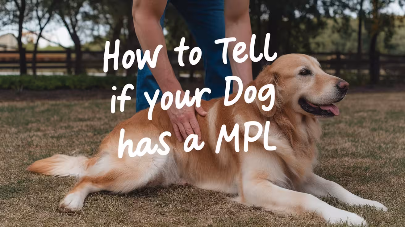
How to Tell If Your Dog Has a Medially Luxating Patella
Learn how to recognize signs of medial patellar luxation in dogs early, including limping, clicking sounds, and changes in gait for timely treatment
What Is Medial Patellar Luxation in Dogs?
Medial Patellar Luxation (MPL) is a condition where the kneecap (patella) slips out of its normal groove toward the inside of a dog’s leg. This causes the knee joint to become unstable, leading to pain, limping, and difficulty walking. MPL is especially common in small and toy dog breeds but can affect dogs of any size.
MPL matters because if left untreated, it can cause long-term joint damage, arthritis, and chronic pain. The slipping kneecap puts extra stress on the knee, making movement uncomfortable and sometimes causing your dog to avoid using the leg.
Early detection is very important for your dog’s health and comfort. Recognizing signs like limping, skipping steps, or holding up a leg allows you to seek veterinary care sooner. Early treatment can reduce pain, prevent further damage, and improve your dog’s quality of life. Keeping a close eye on your dog’s movement helps catch MPL before it worsens.
Common Signs That Your Dog May Have MPL
Here are some common signs that can help you identify if your dog may be suffering from medial patellar luxation (MPL).
1. Limping or Skipping Steps
Limping or skipping steps is one of the earliest signs your dog might have medial patellar luxation (MPL). When the kneecap slips out of place, your dog may feel pain or instability, causing uneven movement.
- Watch for your dog lifting a leg briefly while walking or running.
- Notice if your dog skips steps or hops on one leg suddenly.
- Limping can be subtle at first and may only happen after exercise or prolonged activity.
If you see your dog favoring one leg or walking unevenly, it’s a sign to check with your vet. Early limping or skipping indicates discomfort or instability in the knee, which can worsen if untreated. Tracking these patterns helps you catch MPL early and get your dog the care needed to prevent long-term damage.
2. Intermittent or Persistent Lameness
Lameness means your dog is limping or not using a leg normally. It can be intermittent (comes and goes) or persistent (constant). Both can be warning signs of MPL.
- Intermittent lameness often appears after exercise or activity when the kneecap slips out temporarily.
- Persistent lameness means ongoing pain or instability in the knee, which needs urgent attention.
- Your dog may hold up the leg or limp constantly if the condition is severe.
Recognizing when lameness changes from occasional to frequent is important. Early veterinary diagnosis can help treat mild lameness before it becomes severe. Persistent lameness might indicate worsening MPL or other complications. Watching how often your dog limps and seeking veterinary advice quickly improves treatment outcomes.
3. Abnormal Gait or Skipping Leg Movement
An abnormal gait means your dog’s walk or run looks different from normal. In dogs with MPL, this often shows as unusual skipping or jerky leg movement.
- Your dog may appear to skip or hop instead of walking smoothly.
- The affected leg might move differently, seeming stiff or shaky.
- This irregular movement happens because the kneecap slips, causing discomfort or instability.
This gait change may be subtle at first and can be mistaken for other issues. Watching carefully during walks or play helps spot unusual leg movements. Early detection allows for prompt treatment to restore normal walking patterns and reduce pain.
4. Audible Clicking or Popping Sounds
Clicking or popping sounds from your dog’s knee are signs that the kneecap is moving abnormally. These sounds happen when the patella slips out of its groove and snaps back.
- You might hear a faint “click” when your dog walks, runs, or moves the leg.
- These noises show joint instability and possible damage to soft tissues.
- Not all dogs make these sounds, but if you hear them often, it’s a sign to get a vet check.
Listening for these sounds during activity or when your dog moves the leg gently can provide clues about MPL. Early veterinary diagnosis helps prevent joint damage from frequent slipping.
5. Visible or Palpable Slipping of the Kneecap
Sometimes, you can see or feel the kneecap slipping out of place. This popping or luxation happens when the kneecap moves out of its normal groove on the thigh bone.
- Gently feel your dog’s knee when the leg is bent and straightened to check for slipping.
- You might see the kneecap visibly move or pop to the side during leg movement.
- If unsure, have a vet perform this test to avoid causing pain or injury.
Feeling or seeing the patella slip is a clear sign of MPL. If you notice this, it’s important to visit your vet for a full examination and diagnosis.
6. Holding Up the Leg or Avoiding Weight Bearing
Dogs with MPL often hold up the affected leg or avoid putting weight on it to relieve pain or discomfort.
- Your dog may lift the leg while standing or walking, especially after activity.
- Avoidance of weight bearing is a common way dogs protect an injured or painful knee.
- This behavior can be temporary or frequent depending on MPL severity.
If your dog regularly holds up a leg or refuses to walk on it, it’s a strong sign of knee pain. Early vet evaluation helps manage discomfort and improves healing.
7. Difficulty or Reluctance to Jump, Run, or Climb Stairs
Changes in your dog’s activity level, like difficulty or unwillingness to jump, run, or climb stairs, often point to knee problems like MPL.
- Your dog may avoid stairs or hesitate before jumping onto furniture or into cars.
- Running or playing less than usual can indicate discomfort during high-impact activities.
- These behavioral changes help protect the painful knee from stress.
Noticing reluctance to be active is important for early MPL detection. Discussing these changes with your vet can lead to timely diagnosis and treatment.
8. Swelling or Pain Around the Knee Joint
Swelling or pain near the knee joint may develop with MPL due to inflammation from repeated kneecap slipping.
- Look for visible swelling, warmth, or tenderness around the knee.
- Your dog might lick or chew the knee area more than usual.
- Pain signs include limping, whining, or reluctance to move.
Swelling and pain indicate irritation or early joint damage. Prompt veterinary care can reduce inflammation and prevent progression.
9. Changes in Behavior Like Reluctance to Play or Exercise
Discomfort from MPL often causes changes in your dog’s behavior, such as reduced playfulness or exercise reluctance.
- Your dog may become less active or hide more than usual.
- Decreased interest in walks, toys, or interaction can signal pain.
- These subtle mood changes are important clues to underlying knee issues.
Recognizing these behavior shifts early helps you seek veterinary care and improve your dog’s comfort and quality of life.
How MPL Symptoms Can Worsen Over Time
If medial patellar luxation (MPL) is not treated, your dog’s symptoms can get worse and cause more serious problems.
- More frequent kneecap slipping: The patella may move out of place more often, causing pain and joint instability.
- Joint damage and arthritis: Repeated slipping can wear down cartilage and bones, leading to arthritis. This causes swelling, stiffness, and long-term pain.
- Increased limping or holding up the leg: Your dog may limp more or avoid using the affected leg due to discomfort.
- Posture and gait changes: To reduce pain, your dog might change how they stand or walk, which can cause muscle loss and strain on other legs.
- Both knees affected: Sometimes MPL develops in both legs, worsening mobility and quality of life.
Monitoring your dog’s symptoms closely helps catch these changes early. Watch for increased limping, reduced activity, or changes in behavior. Early vet care can reduce pain, prevent joint damage, and improve your dog’s chances of a happy, active life.
How to Monitor Your Dog’s Mobility and Pain at Home
Monitoring your dog’s mobility and pain at home helps you track their condition and notice any worsening signs early. Regular observation lets you provide important information to your vet for better care.
- Watch your dog’s walking: Look for limping, skipping steps, or difficulty standing up. Notice if your dog favors one leg or hesitates to move.
- Observe activity levels: Pay attention to changes in how much your dog wants to play, run, or climb stairs. Reduced activity can signal pain or discomfort.
- Check for stiffness: Notice if your dog is stiff or slow to get moving after resting or sleeping.
- Look for pain behaviors: Whining, licking the knee, or sudden stops during movement may show discomfort.
- Examine posture: Watch for changes in how your dog holds their leg or stands.
Keep a simple journal to record daily observations, noting any new or worsening symptoms. Include details about when symptoms appear, their severity, and any triggers like exercise. Sharing this information with your vet helps tailor treatment and improves your dog’s care. Regular monitoring is key to managing MPL effectively.
When to Seek Veterinary Care for Suspected MPL
Knowing when to seek veterinary care for suspected medial patellar luxation (MPL) is crucial to protect your dog’s health and comfort. Early veterinary attention can prevent worsening damage and reduce pain.
- Sudden or severe limping: If your dog starts limping suddenly or cannot put weight on a leg, it needs immediate vet care.
- Persistent or worsening lameness: Continuous limping or increasing difficulty walking are signs of serious knee issues.
- Visible swelling or redness: Swelling, heat, or redness around the knee may indicate inflammation or infection.
- Audible clicking or popping: Hearing frequent clicking sounds from the knee can signal instability needing professional evaluation.
- Reluctance to move or play: A sudden decrease in activity or reluctance to jump, run, or climb stairs suggests discomfort.
Early diagnosis allows your vet to assess the severity of MPL and recommend the best treatment, whether conservative care or surgery. Prompt treatment reduces pain, slows joint damage, and improves your dog’s quality of life. Don’t wait for symptoms to worsen—early veterinary care is key to a better outcome.
FAQs About How to Tell If Your Dog Has a Medially Luxating Patella
What is medial patellar luxation in dogs?
Medial patellar luxation (MPL) occurs when the kneecap slips out of its normal position toward the inside of the leg. It causes pain, limping, and joint instability. It’s common in small breeds but can affect all dogs. Early detection is key to preventing long-term damage.
How can I spot limping caused by MPL?
Limping or skipping steps may happen suddenly or after activity. Your dog might lift or favor one leg, showing discomfort. Limping can be subtle at first, so careful observation during walks is important to catch early signs of MPL.
What does an abnormal gait look like in dogs with MPL?
An abnormal gait may appear as skipping, hopping, or uneven leg movement. The affected leg might move stiffly or jerk unexpectedly due to the kneecap slipping out of place, causing discomfort and instability while walking or running.
Why does my dog’s knee make clicking sounds?
Clicking or popping noises occur when the kneecap moves out and back into its groove. These sounds indicate joint instability and frequent slipping of the patella. Hearing this often suggests your dog should be checked by a vet.
How can I check if my dog’s kneecap is slipping?
Gently feel your dog’s knee while moving the leg to detect popping or slipping of the kneecap. Be careful to avoid causing pain. If unsure, let a vet perform the test safely for an accurate diagnosis.
When should I take my dog to the vet for suspected MPL?
Visit the vet if your dog shows limping, skipping steps, leg holding, swelling, or audible knee clicks. Early veterinary care is important to diagnose MPL, start treatment, and prevent worsening symptoms and joint damage.

Recovery After Medial Patellar Luxation Surgery in Dogs
Discover the typical recovery timeline, care tips, and pain management after medial patellar luxation surgery to help your dog heal safely and comfortably
Typical Recovery Timeline After MPL Surgery
After medial patellar luxation surgery, your dog’s healing usually takes about 6 to 8 weeks. During this time, the knee joint repairs itself, and your dog gradually regains strength and mobility.
In the first few days, your dog may feel sore and show limited movement. Pain and swelling are normal but should improve with medication prescribed by the vet. It’s important to keep your dog calm and limit activity to help the knee heal.
Here’s what to expect during recovery:
- Weeks 1-2: Rest and restricted movement are critical. Use a leash for short bathroom breaks only. Apply cold packs if recommended by your vet to reduce swelling.
- Weeks 3-4: Your dog may start gentle walking or physical therapy exercises. Muscle strengthening begins carefully to support the knee.
- Weeks 5-6: Gradual increase in activity, including short walks and controlled play. Monitor for any signs of pain or limping.
- Week 7-8: Most dogs regain good function and strength. Your vet will check progress and may clear your dog for normal activities.
Following your vet’s recovery plan closely improves healing and helps prevent complications.
Post-Surgery Care and Activity Restrictions
After medial patellar luxation surgery, strict rest is essential to help your dog heal properly. Rest allows the repaired tissues and bones to recover without stress. Limiting movement prevents the kneecap from slipping again or causing damage during the healing process.
To keep your dog calm, restrict them to a small, quiet area like a crate or a room. Use a leash when taking them outside for bathroom breaks. Avoid letting your dog run, jump, or play freely during the first 6 to 8 weeks after surgery.
Certain activities should be avoided because they put extra pressure on the knee:
- Jumping on and off furniture or into cars
- Running or playing fetch
- Climbing stairs or steep slopes
These movements can strain the healing joint, cause pain, and slow recovery. Instead, focus on short, controlled walks as advised by your vet. Following these activity restrictions helps reduce complications and supports a smoother, faster recovery for your dog.
Managing Pain and Inflammation
Managing pain and inflammation after medial patellar luxation surgery is key to your dog’s comfort and healing. Vets commonly prescribe pain relief medications like non-steroidal anti-inflammatory drugs (NSAIDs) to reduce pain and swelling. These medicines help your dog feel more comfortable and encourage gentle movement during recovery.
Sometimes, vets may also recommend mild painkillers or muscle relaxants if the pain is more severe. It’s important to give all medications exactly as prescribed and never use human pain medicines without veterinary advice.
Cold therapy is another effective way to control swelling and reduce pain after surgery. Applying cold packs or ice wrapped in a towel to the knee for 10-15 minutes, several times a day, can help shrink blood vessels and lower inflammation. Cold therapy is most useful in the first 48-72 hours after surgery.
Supportive treatments like gentle massage or physical therapy may also be suggested by your vet to improve circulation and promote healing. Together, these methods reduce discomfort and support a smoother recovery process for your dog.
Wound Care and Monitoring
Proper wound care after medial patellar luxation surgery is crucial to avoid infection and help healing. Keeping the surgical site clean and watching for problems supports a smooth recovery.
- Check the incision daily: Look for redness, swelling, warmth, discharge, or bad smell. Mild swelling and bruising are normal, but worsening signs need a vet’s attention.
- Keep the area dry and clean: Avoid bathing your dog until the vet allows it. If cleaning is needed, gently use a mild antiseptic or saline with a clean cloth or cotton swab. Avoid harsh chemicals like alcohol.
- Prevent licking or chewing: Use an Elizabethan collar (cone) or other protective devices to stop your dog from irritating the wound. Licking can cause infection and delay healing.
- Do not touch stitches or staples: Let your vet remove them during follow-up visits, usually 10 to 14 days after surgery.
Following these steps carefully helps detect issues early and keeps the wound healthy for faster healing. Regular vet check-ups ensure the recovery is progressing well.
Physical Rehabilitation and Exercise
Physical rehabilitation plays a key role in helping your dog recover after medial patellar luxation surgery. Gentle exercises rebuild muscle strength, improve joint mobility, and support the healing knee. Without rehab, muscles can weaken and the joint may become stiff, slowing recovery.
Rehabilitation exercises usually start once the initial healing phase is over, often around 2 to 4 weeks after surgery. These exercises focus on gentle stretching, controlled leg movements, and muscle strengthening to restore normal function. A vet or veterinary physical therapist can guide you with safe, effective exercises tailored to your dog’s condition.
Gradual Reintroduction of Controlled Exercise
Gradual reintroduction of controlled exercise is essential to avoid overloading the healing knee. Short, slow walks on a leash are usually allowed first, increasing in length and intensity over several weeks. Running, jumping, and rough play should be avoided until your vet confirms the knee is strong enough.
Here’s how to safely increase activity during recovery:
- Start with brief leash walks of 5-10 minutes
- Slowly increase walk time and gentle movements
- Monitor your dog for signs of pain or limping
- Follow your vet’s advice on physical therapy sessions
Proper rehab and controlled exercise help your dog regain strength, improve joint stability, and return to normal activity safely and comfortably.
Supporting Recovery Through Diet and Environment
Dietary Considerations and Weight Management
Maintaining a healthy weight is very important during your dog’s recovery after medial patellar luxation surgery. Extra weight puts pressure on the healing knee, causing pain and slowing recovery. A balanced diet supports tissue repair and overall health.
- Control calories: Avoid high-calorie treats and table scraps that cause weight gain.
- Vet-recommended diet: Follow your vet’s advice for a diet rich in vitamins and minerals but low in excess calories.
- Gradual weight loss: If your dog is overweight, lose weight slowly under veterinary guidance to reduce joint stress.
- Nutritional support: Proper nutrients help the body heal faster and keep your dog comfortable.
Keeping your dog at a healthy weight lowers the risk of arthritis and helps the knee heal well.
Home Environment Adjustments
Making changes at home helps keep your dog safe and comfortable while recovering.
- Limit slippery floors: Use rugs or mats to prevent slips and falls.
- Block stairs and furniture: Stop your dog from jumping on or off places that strain the knee.
- Create a quiet rest area: Provide a soft, supportive bed in a calm space to encourage rest.
- Easy access: Keep food, water, and toys close so your dog doesn’t have to move too much.
These adjustments reduce injury risk and support a smooth, safe recovery.
Monitoring Healing and Follow-Up
Careful monitoring during recovery helps ensure your dog heals well and catches any problems early.
Importance of Follow-Up Veterinary Visits
Follow-up visits allow your vet to check how your dog’s knee is healing. They will assess swelling, pain levels, and joint stability. These visits help the vet adjust medications, recommend physical therapy, or suggest further treatment if needed. X-rays may be taken to see how the bones and tissues are repairing. Regular check-ups are essential for tracking progress and preventing complications, ensuring your dog recovers safely and fully.
Recognizing Possible Complications
Watch for signs like increased redness, swelling, discharge, or heat around the incision, which may indicate infection. If your dog suddenly limps more or holds the leg differently, the kneecap might have slipped again (patellar reluxation). Other signs include worsening pain, fever, or loss of appetite. Early detection of complications allows prompt veterinary care to avoid further damage and pain.
When to Contact the Vet
Contact your vet immediately if your dog shows severe limping, refuses to use the leg, or if you see swelling or discharge at the surgery site. Also, urgent care is needed if your dog shows signs of infection, such as fever or lethargy. Early communication with your vet helps manage issues quickly and supports a smoother recovery.
Owner’s Role in Successful Recovery
Your role as a dog owner is crucial for a smooth and successful recovery after medial patellar luxation surgery. Following your vet’s care instructions carefully directly affects how well and how quickly your dog heals.
- Strictly follow activity restrictions: Limiting your dog’s movement helps prevent stress on the healing knee and avoids complications.
- Administer medications as prescribed: Giving pain relief and anti-inflammatory medicines on schedule controls discomfort and swelling.
- Monitor the surgical site daily: Checking for signs of infection or other problems helps catch issues early.
- Provide a safe, comfortable environment: Making home adjustments supports rest and reduces injury risks.
- Attend all follow-up vet visits: These appointments let the vet track progress and make necessary treatment changes.
- Support rehabilitation exercises: Helping your dog with physical therapy improves strength and joint function.
Your commitment to these care steps ensures your dog stays comfortable and recovers well. Missing instructions or allowing too much activity can slow healing or cause setbacks. By staying attentive and proactive, you give your dog the best chance for a full, healthy recovery.
Recovery Variations Based on Surgery Type
Recovery after medial patellar luxation surgery can vary depending on the surgical method used. Different techniques address specific issues with the kneecap and surrounding structures, which affects the healing process and activity restrictions.
Some common surgical methods include deepening the groove where the kneecap sits (trochleoplasty), tightening or loosening tendons and ligaments around the knee, and correcting bone deformities in the thigh or shin bone. Each approach has slightly different recovery needs.
- Trochleoplasty (groove deepening): This method involves reshaping the bone groove, so healing may take longer because bone tissue needs to remodel. Strict rest is essential for 6 to 8 weeks to allow the bone to heal properly.
- Soft tissue procedures (tendon or ligament adjustments): These surgeries often involve less bone healing, so your dog might regain movement slightly faster but still requires controlled activity.
- Corrective osteotomy (bone realignment): This is a more complex procedure where bones are cut and repositioned. Recovery can be longer and may require additional pain management and physical therapy.
Your vet will explain the specific recovery plan based on the surgery type, helping you manage activity levels, pain control, and rehabilitation to ensure the best healing outcome. Understanding these differences prepares you to support your dog properly through recovery.
FAQs About Recovery After Medial Patellar Luxation Surgery
How long does recovery from MPL surgery usually take?
Recovery from medial patellar luxation surgery generally lasts between 6 and 8 weeks. During this time, your dog needs limited activity, pain management, and regular veterinary check-ups to ensure proper healing. Following your vet’s instructions helps your dog regain knee strength and mobility safely, reducing risks of complications or setbacks.
What activities should I avoid during my dog’s recovery?
Avoid activities like running, jumping, climbing stairs, or rough play during recovery. These movements put stress on the healing knee and can cause the kneecap to slip again or delay healing. Controlled, gentle walks are usually allowed under veterinary guidance. Strict activity restrictions help prevent injury and promote faster recovery.
How can I manage my dog’s pain after surgery?
Pain management includes giving prescribed medications such as NSAIDs or mild painkillers exactly as directed by your vet. Cold therapy, like applying ice packs, can help reduce swelling and discomfort. Never give human pain medicine without veterinary advice. Keeping your dog calm and comfortable is important for a smooth recovery.
When can my dog start physical therapy or exercise?
Physical therapy usually starts 2 to 4 weeks after surgery once initial healing occurs. Gentle exercises focus on improving joint mobility and rebuilding muscle strength without stressing the knee. A vet or physical therapist will guide you on safe exercises and gradually increase activity to support recovery.
How do I know if my dog’s surgical wound is healing well?
A healing wound should have minimal swelling, no redness spreading beyond the incision, and no foul-smelling discharge. Some bruising and mild swelling are normal early on. If you notice increased redness, warmth, pus, or your dog excessively licking the area, contact your vet promptly to prevent infection.
What signs mean I should call the vet during recovery?
Call your vet if your dog shows severe limping, refuses to use the leg, has swelling or discharge at the surgery site, or develops fever, lethargy, or loss of appetite. These signs may indicate infection, reluxation, or other complications requiring urgent veterinary care to protect your dog’s recovery.

Signs and Symptoms of Medial Patellar Luxation in Dogs
Learn the common signs and symptoms of medial patellar luxation in dogs to spot early warning signs and get timely veterinary care for your pet
Understanding Medial Patellar Luxation
Medial Patellar Luxation (MPL) is a condition where the kneecap (patella) slips out of its normal position toward the inside of the dog’s leg. This causes the knee joint to become unstable, leading to pain and difficulty walking. MPL is common in small dog breeds but can affect dogs of all sizes.
Recognizing the symptoms of MPL early is very important. When caught early, treatment can reduce pain and prevent long-term damage like arthritis. Early care helps your dog stay active and comfortable. If left untreated, the condition can worsen, causing more severe limping and joint problems.
By watching for signs like limping, skipping steps, or stiffness, you can get your dog the help they need quickly. Early veterinary care improves your dog’s quality of life and can often avoid the need for complex surgery.
Common Movement-Related Signs of MPL
Medial Patellar Luxation (MPL) often changes how your dog moves. Watching for these signs can help you notice the problem early.
- Sudden skipping or hopping while walking or running: Your dog may suddenly lift one leg or hop to avoid pain when the kneecap slips out of place. This quick change in movement helps reduce discomfort.
- Limping or skipping gait: Limping is common when the dog puts less weight on the affected leg. You might see an uneven or skipping walk as your dog tries to avoid putting pressure on the painful knee.
- Intermittent or persistent lameness: Sometimes, lameness happens only after exercise, but in more serious cases, your dog may limp all the time. Both need veterinary attention to avoid worsening.
- Holding the affected leg up or avoiding weight bearing: Your dog might lift the injured leg or refuse to stand on it because of pain or instability in the knee.
If you notice these signs, it’s important to see a vet for diagnosis and early treatment.
Pain and Mechanical Symptoms
Dogs with Medial Patellar Luxation (MPL) often show pain and unusual knee movements. Watching for these signs can help you know when your dog needs care.
- Pain or discomfort around the knee area:
Your dog may lick, bite, or favor the leg because of pain. The discomfort can be mild or severe depending on how often the kneecap slips out of place. - Visible abnormal movement or popping of the kneecap:
You might see the kneecap move out of its groove or the knee looking unstable. This popping happens especially when your dog walks or stands. - Audible clicking or popping sounds from the knee:
Sometimes you can hear clicking noises when the kneecap slips out and back in. This sound shows the knee joint is unstable.
If you notice any of these pain or mechanical symptoms, visit your vet. Early treatment can reduce pain and stop more damage to the knee joint.
Functional Difficulties and Posture Changes
Medial Patellar Luxation (MPL) can cause problems with how your dog moves and stands. These changes affect your dog’s daily activities and comfort.
- Stiffness or difficulty walking after rest:
Your dog may seem stiff or slow to start moving after resting. This happens because the knee feels tight or sore, making it hard to walk smoothly. - Difficulty jumping or climbing stairs:
Activities that put extra pressure on the knees, like jumping or going up stairs, can become hard or painful. Your dog might avoid these tasks to reduce discomfort. - Bow-legged appearance or abnormal hind limb posture:
In some cases, the leg may look bow-legged or positioned oddly due to changes in the knee joint and surrounding muscles. - Knee joint instability or looseness:
The knee may feel loose or unstable because the kneecap is not held securely. This can cause your dog to wobble or struggle with balance.
If you notice these signs, it’s important to get your dog checked by a vet. Managing these difficulties early helps keep your dog active and comfortable for longer.
Muscle and Activity Changes
Medial Patellar Luxation (MPL) often leads to changes in your dog’s muscles and activity levels. Because the affected leg causes pain or discomfort, your dog may use it less than usual.
- Muscle loss in the affected leg from reduced use:
When your dog avoids putting weight on the injured leg, the muscles around that leg can shrink or weaken. This muscle loss makes it harder for the knee to stay stable, which can worsen the condition over time. - Decreased activity or reluctance to exercise:
Dogs with MPL may become less active because movement causes discomfort or pain. They might avoid running, jumping, or playing as much as before. This reduction in activity can affect their overall health and fitness.
It’s important to help your dog stay as active as possible without causing pain. Gentle, controlled exercise and physical therapy can improve muscle strength and support the knee joint. Working with your vet to create a safe exercise plan helps your dog stay healthy and feel better despite MPL. Early management of muscle and activity changes improves your dog’s quality of life.
Progression and Severity of Symptoms
Medial Patellar Luxation (MPL) symptoms can change over time and with your dog’s activity level. Often, symptoms get worse as the condition progresses or if your dog is very active.
- Symptoms worsen with activity or over time:
Increased exercise or movement can cause the kneecap to slip more often, leading to more pain and discomfort. Over time, repeated slipping may damage the knee joint, making symptoms more severe. - Mild cases show subtle or occasional signs:
Dogs with mild MPL might only limp sometimes or skip steps now and then. These signs can be easy to miss but still need attention to prevent worsening. - Moderate to severe cases cause frequent lameness and pain:
In more serious cases, dogs limp often or have trouble walking. Pain can be constant, and the knee may feel unstable or locked. - Both hind legs can be affected, usually one side is worse:
Sometimes MPL affects both knees, but one leg often has more severe symptoms. This imbalance can affect your dog’s posture and movement.
Early vet care helps manage symptoms and slow progression, improving your dog’s comfort and mobility.
When to See a Veterinarian
Knowing when to take your dog to the veterinarian is very important if you suspect Medial Patellar Luxation (MPL). Certain signs need prompt veterinary attention to prevent further damage and pain.
Key signs to watch for include sudden limping that doesn’t improve, your dog refusing to put weight on a leg, visible swelling or heat around the knee, or if the leg locks or feels unstable. If your dog shows signs of strong pain, such as whining, crying, or reluctance to move, you should see a vet right away.
Early diagnosis and treatment are crucial for the best outcome. When MPL is caught early, your vet can recommend treatments to reduce pain, improve mobility, and prevent long-term problems like arthritis. Delaying care can cause the condition to worsen, making treatment more complicated and recovery longer.
Regular veterinary check-ups help monitor your dog’s knee health and catch any early changes. If you notice any signs of MPL, don’t wait—visit your vet to protect your dog’s comfort and quality of life.
Frequently Asked Questions About Signs and Symptoms of Medial Patellar Luxation
What are the first signs of medial patellar luxation in dogs?
Early signs include limping, skipping steps, or sudden hopping while walking. Your dog may hold the leg up or avoid putting weight on it. These subtle changes often appear during activity or after rest.
How can I tell if my dog is in pain from MPL?
Dogs may lick or bite their knee, show reluctance to move, or limp frequently. Signs like whining or stiffness after resting also indicate pain related to the kneecap slipping out.
Can MPL cause permanent damage if untreated?
Yes, untreated MPL can lead to arthritis and joint damage. This causes chronic pain, stiffness, and difficulty moving. Early care can prevent these long-term problems.
Why does my dog’s knee make clicking sounds?
Clicking or popping sounds occur when the kneecap slips out and back into place. This noise is a common mechanical symptom of MPL and shows joint instability.
Does MPL affect both legs or just one?
MPL can affect one or both hind legs. Often, one side is more severe, causing uneven movement and posture changes.
When should I take my dog to the vet for MPL symptoms?
If your dog limps suddenly, refuses to put weight on a leg, or shows signs of pain or swelling, see a vet immediately. Early diagnosis improves treatment success.

What Is Medial Patellar Luxation in Dogs? Explained Clearly
Learn what medial patellar luxation in dogs is, its causes, signs, treatment options, and how early care improves your dog’s health and mobility
Understanding Medial Patellar Luxation
Medial Patellar Luxation (MPL) is a condition where the kneecap, called the patella, slips out of its normal position toward the inside of the leg. Normally, the patella sits in a groove at the end of the thigh bone and moves up and down as the dog walks or runs. This helps the dog’s leg bend and straighten smoothly.
In a healthy dog, the patella stays in place because it is held by strong ligaments and muscles. It acts like a pulley for the muscles that straighten the leg. But in dogs with MPL, the kneecap moves out of the groove, causing pain, limping, or difficulty walking. This condition is common in small dog breeds and can vary from mild to severe depending on how often and how far the kneecap slips out.
Causes and Who Is at Risk
Medial Patellar Luxation (MPL) happens when the kneecap moves out of its normal groove, mainly because of changes in the bones, muscles, or ligaments around the knee. The most common causes include:
- Genetic factors: Many dogs inherit weak or misshaped knee structures from their parents.
- Abnormal bone growth: Sometimes the thigh bone or shin bone grows unevenly, making it hard for the patella to stay in place.
- Muscle weakness or imbalance: Weak muscles can fail to keep the kneecap aligned properly.
- Trauma or injury: Sudden injuries can damage the knee joint, leading to luxation.
Certain dog breeds are more likely to get MPL, especially small and toy breeds. This shows a strong genetic link. Breeds commonly affected include:
- Pomeranians
- Yorkshire Terriers
- Chihuahuas
- Poodles
- French Bulldogs
- Maltese
Large breeds can also get MPL, but it is less common. Early diagnosis is important, especially for at-risk breeds, to manage symptoms and avoid worsening damage. Regular vet check-ups help catch this condition early.
Read more:
Recognizing the Signs and Symptoms
Knowing the signs of Medial Patellar Luxation (MPL) helps you find the problem early. If your dog has MPL, you might notice changes in their walking or behavior that show discomfort or pain in the knee.
Common signs to watch for include:
- Limping or skipping steps: Your dog may suddenly lift one leg while walking or skip steps because the kneecap slips out of place.
- Difficulty running or jumping: They might avoid activities that put pressure on their knees.
- Stiffness or trouble bending the leg: The knee may feel stiff or locked at times.
- Swelling or tenderness around the knee: You might see or feel swelling where the kneecap should be.
- Holding the leg up: Your dog may hold the affected leg off the ground to avoid pain.
- Repeated clicking or popping sounds: Sometimes you can hear or feel the kneecap moving out and back into place.
If you notice any of these signs, especially in small breeds, take your dog to a vet for a full check. Early treatment helps prevent more damage and improves your dog’s comfort and mobility.
Diagnosing the Condition
When you take your dog to the vet with signs of Medial Patellar Luxation (MPL), the vet performs a careful exam to find the problem. They check the dog’s leg for pain, swelling, and how well the knee moves. The vet will try to feel if the kneecap slips out of its normal groove when the leg bends or straightens.
During the physical exam, the vet looks for:
- Limping or signs of discomfort
- Abnormal leg movement
- The position of the kneecap when moving the leg
- Muscle tone around the knee
To confirm the diagnosis, the vet often uses X-rays. X-rays show the shape of the bones and help find any bone changes or damage in the knee joint. Sometimes, the vet may use special imaging like CT scans for a more detailed view.
These tools help the vet decide the severity of MPL and the best treatment plan. Early and accurate diagnosis is important to prevent long-term damage and improve your dog’s quality of life.
Read more:
Treatment Options for MPL
Treatment for Medial Patellar Luxation (MPL) depends on how severe the condition is and how much it affects your dog’s movement.
Conservative care may help in mild cases where the kneecap only slips occasionally and your dog shows little pain. This includes:
- Weight management to reduce pressure on the knees
- Gentle exercise to keep muscles strong
- Anti-inflammatory or pain medicines prescribed by the vet
- Physical therapy or rehab exercises
If conservative care does not improve symptoms or if the luxation is moderate to severe, surgery is often needed. Surgery aims to realign the kneecap and stabilize the joint to prevent it from slipping out again.
Common surgical options include:
- Deepening the groove where the kneecap sits
- Tightening or loosening tendons and ligaments around the knee
- Correcting bone deformities in the thigh or shin bone
Surgery usually improves your dog’s mobility and reduces pain. Early surgery can prevent arthritis and long-term joint damage. Your vet will recommend the best treatment based on your dog’s age, breed, and MPL severity.
Read more:
The Importance of Early Treatment
Leaving Medial Patellar Luxation (MPL) untreated can cause serious problems for your dog over time. When the kneecap keeps slipping out of place, it damages the knee joint. This damage leads to pain, swelling, and inflammation. If untreated, MPL often causes arthritis, which makes the joint stiff and sore. Arthritis is a long-term condition that reduces your dog’s ability to move and enjoy life.
Untreated MPL can also cause your dog to limp more, change how they walk, or even develop problems in other legs due to uneven weight bearing. These changes may cause muscle loss and reduce overall leg strength.
Early treatment helps avoid these risks. When treated early, whether with medicine, physical therapy, or surgery, your dog is less likely to develop arthritis or permanent joint damage. Early care improves comfort, helps your dog stay active, and speeds recovery. Regular vet visits and quick action when you notice signs of MPL give your dog the best chance for a happy, healthy life.
Living with Medial Patellar Luxation
Medial Patellar Luxation (MPL) can affect your dog’s life and how well they move. Dogs with MPL may limp or avoid running and jumping because their knee hurts or feels unstable. If the condition is mild, your dog might still enjoy normal activities with little trouble. But if MPL is more severe, it can cause ongoing pain and make walking hard.
You can help your dog live comfortably by managing symptoms at home. Here are some tips:
- Keep your dog at a healthy weight to reduce pressure on the knees.
- Provide soft bedding to support sore joints.
- Avoid slippery floors where your dog could slip and injure their knee.
- Use gentle, controlled exercise like short walks to keep muscles strong without causing stress.
- Follow your vet’s advice on medicines or supplements for joint health.
- Physical therapy or simple home exercises can improve muscle strength and knee stability.
With proper care and attention, dogs with MPL can still enjoy a good quality of life. Regular vet check-ups help monitor the condition and adjust treatment as needed.
When to See the Vet
Knowing when to take your dog to the vet is important if they have Medial Patellar Luxation (MPL). Some signs need immediate veterinary care to prevent worsening damage or serious pain. Watch for:
- Sudden severe limping or inability to put weight on the leg
- Visible swelling, heat, or redness around the knee
- Your dog showing signs of strong pain like whining or refusing to move
- The leg locking in place or sudden changes in walking
- Signs of injury after a fall or accident
If you see these signs, contact your vet right away for an exam.
Even without emergencies, regular vet visits are important for dogs with MPL. Your vet will check how the condition is progressing and update treatment as needed. They may recommend X-rays or physical therapy to keep the knee healthy.
Routine monitoring helps catch new problems early and improves your dog’s comfort. Stay in touch with your vet and follow their advice to help your dog live well despite MPL. Early care and regular check-ups make a big difference in managing this condition.
Frequently Asked Questions About MPL
What causes medial patellar luxation in dogs?
Medial patellar luxation usually happens because of genetic factors, abnormal bone growth, or muscle imbalances. Some breeds are more likely to inherit this condition. Trauma or injury can also cause the kneecap to slip out of place.
Can small dogs get MPL more often than large dogs?
Yes, small and toy breeds are more commonly affected by MPL. Breeds like Pomeranians, Chihuahuas, and Yorkies have a higher risk. However, large breeds can also develop MPL, but it is less common.
How is MPL diagnosed in dogs?
A vet diagnoses MPL through a physical exam, checking the knee’s movement and feeling if the kneecap slips out. X-rays help confirm the condition and show any bone changes or damage.
Is surgery always needed to treat MPL?
Not always. Mild cases may be managed with weight control, physical therapy, and medication. Surgery is usually recommended for moderate to severe cases or if conservative care does not relieve symptoms.
Can MPL cause arthritis in dogs?
Yes, if untreated, MPL can damage the knee joint and lead to arthritis. Arthritis causes joint pain and stiffness, making movement harder for your dog over time.
How can I help my dog live with MPL?
Maintain a healthy weight, provide gentle exercise, avoid slippery floors, and follow your vet’s treatment plan. Regular check-ups and physical therapy can improve your dog’s comfort and mobility.

Post-Op Home Care for Pets After Orthopedic Surgery
Learn how to care for your pet after orthopedic surgery. Tips on pain, activity, wound care, and safe recovery at home
Why Home Care Matters After Orthopedic Surgery
Proper home care after orthopedic surgery is just as important as the procedure itself. Once your dog leaves the hospital, how well you manage rest, medication, and wound care at home can make a big difference in how smoothly they heal.
Good home care helps reduce pain, prevent infection, and support joint healing. It also keeps your dog calm and safe while tissues repair. Ignoring instructions, letting your dog move too much, or missing medication doses can lead to problems like swelling, wound breakdown, or even implant failure.
Delayed healing, extra surgeries, or long-term joint issues are common risks when post-op care isn’t followed carefully. That’s why it’s important to understand your vet’s instructions and ask questions if you’re unsure. A strong home routine gives your dog the best chance at a full recovery.
Immediate Care After Bringing Your Pet Home
The first 24–48 hours after surgery are critical. Your dog may be groggy, sore, or confused. Following your veterinarian’s discharge instructions closely ensures a safe and smooth start to recovery.
Key steps to follow include:
- Stick to the discharge plan: Your vet will give you clear instructions for medications, feeding, rest, and wound care. Follow these exactly as written.
- Pain control: Use all prescribed pain medications on schedule. Do not skip doses, even if your dog seems comfortable—missing meds can lead to pain flares.
- Antibiotics: Give all doses as directed. Finish the full course unless your vet says otherwise. Do not stop early, even if the incision looks better.
- Watch for reactions: Signs of a problem may include vomiting, diarrhea, excessive drooling, or changes in behavior after starting a new medication.
If you notice anything unusual—like refusal to eat, wobbliness, or trouble breathing—contact your vet immediately. Early action helps avoid bigger issues and keeps your pet comfortable in those first critical days.
Protecting the Incision and Preventing Infection
Keeping the surgical site clean and undisturbed is one of the most important parts of post-op care. Watch the incision daily and follow all hygiene guidelines to prevent infection or delayed healing.
How to monitor the incision:
- Check the site once or twice a day for changes in color, swelling, or discharge.
- Mild bruising and slight swelling are normal in the first few days.
- Contact your vet if you see pus, redness, heat, or a foul smell.
Keep all bandages, splints, or casts dry and clean:
- Cover them with a plastic wrap or bootie during potty breaks.
- Never let your dog lick or chew at them—this increases the risk of infection or wound reopening.
E-collars (cones) are often required to stop licking or chewing. Keep the collar on at all times unless directly supervised. Recovery suits can also help if your dog won’t tolerate a cone.
Avoid bathing until your vet approves it. Wipe paws and use pet-safe wipes around the surgical area if needed. Good hygiene, along with careful observation, helps healing stay on track.
Creating a Safe and Supportive Recovery Environment
A calm, comfortable space at home helps your pet heal faster and avoid complications. Movement should be limited during recovery, so the setup matters as much as the surgery itself.
Here’s how to create the right environment:
- Choose a quiet, low-traffic area away from children, other pets, and loud noises. This helps reduce stress and prevents sudden movements or excitement.
- Use soft, supportive bedding to cushion joints and protect the surgical site. Avoid elevated beds or surfaces your pet could jump from.
- Control room temperature—not too hot or cold. Dogs in recovery are more sensitive to discomfort and drafts.
- Keep lighting soft and predictable, especially for pets on medications that cause drowsiness or confusion.
Keeping your pet calm:
- Use white noise machines, calming sprays, or familiar blankets with your scent.
- Keep visits short and soothing. Avoid excitement, rough play, or unfamiliar visitors during the first two weeks.
The goal is to promote rest and reduce risk. A peaceful space not only supports healing but also lowers anxiety during recovery.
Restricting Movement to Prevent Setbacks
After orthopedic surgery, limiting your dog’s movement is one of the most important ways to protect healing bones, joints, and tissues. Too much activity too soon can lead to serious setbacks like implant failure or wound reopening.
How long to limit activity:
- Most dogs need strict rest for at least 2–4 weeks, depending on the surgery. Your vet will give a detailed timeline based on your pet’s condition.
Ways to safely restrict movement:
- Use crates, playpens, or baby gates to confine your pet to a small, secure area.
- Block access to stairs or furniture—jumping up or down can undo surgical repairs.
- Use slings or support harnesses when taking your dog outside for potty breaks, especially after hind limb surgeries.
- Provide ramps if your dog needs to get in and out of cars or navigate small steps.
- Add floor padding or rugs on slippery surfaces to prevent slipping and falling.
Even brief moments of overexertion can delay healing. Follow all movement restrictions closely and ask your vet when to start gradually increasing activity.
Follow-up Appointments and Rehab Guidance
Staying on schedule with rechecks and early rehab is key to making sure recovery is going as planned. It also allows your vet to catch any problems before they get worse.
Importance of rechecks and suture removal
Follow-up visits help your vet monitor healing and adjust care if needed.
- Rechecks are usually scheduled 7–14 days after surgery to assess the incision and remove sutures or staples if healing looks good.
- Your vet may check range of motion, pain levels, and overall progress.
- These visits also let you ask questions and report any concerns.
Skipping rechecks can delay important care steps or lead to missed signs of infection or poor healing. Always attend all scheduled follow-ups unless your vet gives other instructions.
When to expect progress updates
Recovery takes time and follows a general timeline, depending on the type of orthopedic procedure.
- Most dogs show steady improvement in mobility and comfort by week 2–3.
- X-rays may be taken around 6–8 weeks post-op to confirm bone healing.
- Your vet may adjust medications, activity restrictions, or introduce rehab at this stage.
It’s normal for progress to come in small steps, not all at once. Keeping a journal or short video clips of your pet walking can help track changes. Share these with your vet during follow-ups for better insight.
Gentle rehab exercises and when to begin them
Rehabilitation helps your dog regain strength, balance, and joint motion safely.
- Rehab usually starts with passive range of motion and gentle leash walks, but only after your vet approves.
- Exercises like sit-to-stand, slow walking, or balance work may begin in weeks 3–6.
- Too much too soon can delay healing, so always follow your vet or rehab therapist’s plan.
Professional rehab (like underwater treadmill therapy) may also be recommended. Always check with your vet before starting any exercises at home to ensure your dog is ready.
When to call your vet
Even with a good recovery plan, unexpected issues can arise. Call your vet if you notice:
- Redness, swelling, or discharge from the incision
- Fever, vomiting, or signs of pain not controlled by medication
- Refusal to bear weight, worsening limping, or sudden behavior changes
- Chewing or licking the surgical site despite barriers
Early intervention helps prevent bigger problems. If you’re ever unsure, it’s better to call than wait. Your vet would rather hear from you early than deal with a delayed complication.
Long-Term Wellness and Mental Stimulation
Recovery isn’t just about healing the body—it’s also about keeping your pet’s mind active while physical activity is limited. Boredom and weight gain are common challenges during this time, but both can be managed with a thoughtful approach.
Managing weight through diet during recovery
Reduced activity means fewer calories are needed. Keeping your dog at a healthy weight during recovery helps protect the joints and speeds healing.
- Switch to a weight management or recovery-specific diet if advised by your vet.
- Measure meals carefully—don’t guess. Use a kitchen scale or measuring cup.
- Avoid too many treats, even healthy ones. Use part of the regular food as rewards when needed.
- Ask your vet about supplements or nutritional adjustments for long-term joint health.
Excess weight can delay healing and increase strain on surgical repairs, especially in orthopedic patients.
Treat puzzles and boredom-busters
Mental enrichment helps keep your dog calm and content during restricted activity.
- Food puzzles like treat-dispensing balls or lick mats provide stimulation without physical strain.
- Sniffing games using kibble hidden in blankets or boxes can engage the brain and reduce anxiety.
- Frozen treats (like wet food in a Kong) help keep your dog busy longer.
These activities reduce restlessness and destructive behavior. Always supervise puzzle use to avoid chewing accidents and choose activities that match your pet’s mobility level during each recovery phase.
Light play vs. overstimulation
Play is still important—but it must be safe and calm during recovery.
- Choose gentle activities like slow leash walks (once approved), nose work, or tug games that don’t involve jumping or quick turns.
- Avoid roughhousing, running, or toys that cause sudden movements.
- Short, calm sessions spaced throughout the day are better than long bursts of excitement.
If your dog becomes hyper or starts bouncing off furniture, it’s a sign of overstimulation. Redirect that energy into a food puzzle or quiet enrichment instead. Controlled play keeps the mind happy without risking physical setbacks.
Common Mistakes to Avoid in Post-Surgery Home Care
Even with the best intentions, small mistakes during recovery can lead to big problems. Understanding what to avoid helps protect your pet’s healing process and prevents complications.
Common post-op care mistakes include:
- Skipping medications or vet checkups: Pain meds and antibiotics must be given on schedule—even if your dog looks “fine.” Skipping doses can lead to infection or pain flare-ups. Missing follow-up visits may delay detection of problems like poor wound healing or implant issues.
- Letting pets move too soon: Some owners allow stairs, jumping, or off-leash activity too early. Even one wrong step can undo surgical repairs. Stick to your vet’s timeline, even if your pet seems eager to play.
- Ignoring subtle changes in behavior: Hiding, whining, limping, or decreased appetite may be early signs of pain or infection. Don’t wait for clear signs—report concerns early.
Always ask your vet before making changes to activity, diet, or medication. Consistency, caution, and clear communication are key to a smooth recovery and strong long-term results.
Final Tips to Support a Smooth Recovery
Successful healing takes time, structure, and teamwork. A few simple habits can make recovery smoother for both you and your pet.
Keep a recovery calendar or log to track medications, vet appointments, wound checks, and daily changes in mobility or behavior. This helps you stay organized and spot patterns—good or bad—early.
Stay in regular contact with your vet. Send updates, photos of the incision, or videos of your dog walking if needed. Clear communication helps your vet guide the next steps and address concerns before they become serious.
Most importantly, be patient. Healing timelines vary based on the surgery, age, and overall health. Some pets bounce back quickly, while others need more time. Stick with the plan, celebrate small wins, and give your pet the calm support they need to recover well.
FAQs
How do I keep my pet from licking the surgical site after orthopedic surgery?
Use an e-collar (cone) or a recovery suit to prevent licking. Keep it on at all times unless your pet is directly supervised. Licking can cause infection or open the wound, so never skip protective gear—even if the incision looks clean.
When should I worry about swelling or discharge around the incision?
Mild swelling and bruising are normal in the first few days. But call your vet if you see pus, bleeding, a foul smell, or if swelling increases after it had started to go down. These may be signs of infection or wound complications.
How much movement is too much during the recovery period?
Any off-leash activity, running, jumping, or use of stairs is too much during early recovery. Even short bursts can damage healing tissues. Stick to leash walks and confined rest unless your vet approves increased activity. Always follow your discharge plan closely.
Can I leave my pet alone during the first few days after surgery?
Try to minimize alone time during the first 48–72 hours. If needed, use a secure crate or pen to prevent movement and keep them calm. Ask a friend or family member to check in if you’re away. Monitor appetite, behavior, and medication timing closely.
What should I do if my pet refuses to take post-op medications?
Ask your vet if the medication can be crushed, flavored, or given with food. Use pill pockets, cheese, or wet food to hide the pill. If refusal continues, don’t skip doses—call your vet for alternatives like liquid forms or injections.

Recognize and Manage Post-Surgical Complications in Referred Patients
Learn how to recognize and manage common post-surgical complications in referred pets with clear steps for follow-up, treatment, and communication
Why Referring Vets Play a Key Role in Post-Surgical Care
Referring veterinarians are vital to a pet’s smooth recovery after surgery. Once a specialist completes the procedure, it's often the referring vet who continues the day-to-day care. This includes managing medications, monitoring healing, and addressing owner concerns.
Common referrals include orthopedic surgeries like TPLO, soft tissue operations such as mass removals, or emergency procedures like splenectomies. In all these cases, the follow-up care matters just as much as the surgery itself.
Pet owners often return to their local vet first if they notice swelling, limping, or discharge. They expect clear instructions, reassurance, and timely updates. When referring vets stay closely involved, it builds trust and helps detect problems early. Good communication between the surgical team and primary vet ensures the pet receives seamless care from start to finish.
Most Common Post-Surgical Complications in Referred Cases
Referred patients may return with a range of complications after surgery. Recognizing these issues early helps prevent long-term problems and improves outcomes. The most common post-op complications include:
- Surgical Site Infections (SSI): Redness, swelling, discharge, or odor around the incision. These may need antibiotics or culture-based treatment.
- Seroma or Hematoma: Soft swelling under the skin, often near the incision. Seromas are usually harmless, but large ones may require drainage.
- Wound Dehiscence: The surgical site reopens partially or completely. This may be due to poor healing, infection, or licking.
- Post-op Bleeding or Bruising: Mild bruising is expected, but ongoing bleeding or large hematomas need attention.
- Persistent Pain or Lameness: Ongoing discomfort beyond the expected recovery period can signal internal complications or implant issues.
- Behavioral Changes: Hiding, aggression, vocalizing, or reluctance to move may reflect pain or anxiety.
Referring vets are often the first to see these signs. A thorough recheck, gentle handling, and clear documentation help guide decisions on whether to treat locally or consult the surgeon.
Clinical Signs to Watch for in Referred Patients
After surgery, referred patients may show signs that indicate healing problems, infection, or pain. Referring veterinarians should monitor both local and systemic signs closely during follow-up visits and owner calls.
Local signs near the incision:
- Redness or warmth: May indicate early infection or inflammation.
- Swelling or firm lumps: Could be a seroma, hematoma, or infection.
- Discharge: Clear fluid may be normal early on, but yellow, green, or bloody discharge suggests infection.
- Excessive licking or chewing: Increases the risk of wound dehiscence and delayed healing.
Systemic signs to monitor:
- Fever: Suggests infection or internal inflammation.
- Lethargy or weakness: Can signal pain, anemia, or more serious systemic issues.
- Vomiting or diarrhea: May be medication-related or signs of surgical complications.
- Inappetence: Often overlooked but can be an early warning sign, especially when paired with other symptoms.
Pain indicators include:
- Limping or stiffness: May reflect post-op pain, implant issues, or delayed healing.
- Whining, panting, or restlessness: Often seen in dogs with moderate to severe pain.
- Guarding behavior: The pet resists touch or avoids movement, especially near the surgical site.
When to refer back or escalate care:
If multiple signs are present, especially systemic symptoms combined with local changes, the patient should be re-evaluated urgently. Early detection of complications allows for quicker treatment, reduces long-term effects, and improves surgical outcomes. Communication with the surgical team is key if concerns arise.
Initial Management Strategies for Identified Complications
When post-surgical complications are identified in referred patients, timely and appropriate initial care can make a significant difference. The first step is addressing visible issues like wound concerns and discomfort.
- Local wound care includes gentle cleaning with antiseptic solutions, applying a protective barrier if needed, and using an Elizabethan collar to prevent licking or trauma. If swelling, discharge, or dehiscence is present, the wound may need light bandaging and regular monitoring.
- Antibiotics may need to be adjusted based on the suspected pathogen or culture results. In mild cases, extending the original course is enough. For moderate signs, a new antibiotic may be started. NSAIDs can help manage inflammation and pain but should be used with caution in pets that are anorexic or showing signs of systemic illness.
- Systemic support like fluid therapy, antiemetics, and appetite stimulants may be necessary for pets with fever, vomiting, or lethargy. Monitoring hydration and organ function becomes essential at this stage.
- Knowing when to refer back is critical. If there’s deep infection, implant exposure, worsening systemic signs, or non-healing wounds, surgical re-evaluation is often required. Prompt communication with the surgical team helps coordinate the best next steps for the pet.
Working Closely With the Referral Surgeon or Team
Good communication between the referring vet and surgical team ensures the patient gets the best care during recovery. It also builds trust with pet owners and avoids confusion when complications arise.
What to review in the surgical report
The surgical report is your roadmap to managing recovery. Always review the following:
- Procedure details – Understand exactly what was done, including approach, implants used, and any intraoperative findings.
- Post-op instructions – Note medication protocols, bandage changes, exercise limits, and diet recommendations.
- Expected recovery timeline – Helps you track if healing is on schedule.
- Risks and warnings – Be aware of specific complications tied to the procedure.
If anything in the report is unclear, contact the surgical team early. A solid understanding of the surgery helps you confidently guide the client and detect issues quickly.
How and when to update the surgical team
Keeping the surgeon in the loop is key when recovery doesn’t go as planned. Update the surgical team when:
- Unexpected signs appear, such as fever, discharge, or delayed wound healing
- Post-op medications need adjusting, especially antibiotics or pain control
- You’re considering further diagnostics, like radiographs or bloodwork
- The client expresses concern or confusion about progress
Use email for non-urgent updates and phone calls for pressing issues. Photos of wounds or videos of gait can help the surgeon assess remotely. Document your updates clearly, and let owners know you’re working closely with the surgeon—it reassures them and strengthens the care plan.
Handling unclear cases or deviations from expected recovery
Sometimes recovery doesn’t go by the book. When signs are vague—like mild lameness, subtle swelling, or mood changes—it’s hard to know if it’s normal healing or early complication. In these cases:
- Compare with the expected timeline from the surgical report
- Repeat physical exams, checking for heat, joint stability, or discomfort
- Monitor trends, not just single visits (e.g., is swelling worsening?)
- Speak with the surgeon for insights—subtle signs may still need action
Avoid assuming everything is “normal” without evidence. If something feels off, it likely is. Prompt communication can prevent small issues from becoming serious ones.
Tips for smooth co-management between clinics
Working as a team with the surgical practice makes recovery safer and more efficient. Here’s how to streamline collaboration:
- Agree on roles early—who handles rechecks, medications, rehab?
- Share records quickly, including lab results and photos
- Be clear with the client about who to contact and when
- Keep language consistent—this avoids owner confusion
If the pet’s condition worsens or doesn’t improve, don’t hesitate to refer back. Clients appreciate when both clinics work together. Smooth co-management means fewer delays, better outcomes, and stronger professional relationships.
Educating the Pet Owner After Referral
Client education plays a vital role in post-surgical recovery. When owners understand what to do—and what to watch for—they’re more likely to follow through and catch problems early. Clear, consistent guidance helps prevent confusion and supports better outcomes.
Clarify post-op instructions (activity, meds, wound care)
After a referral, owners may feel overwhelmed with instructions. Your job is to simplify and reinforce what matters most:
- Activity: Explain limits clearly—no stairs, jumping, or off-leash time unless approved.
- Medications: Review names, doses, and timing. Use charts or handouts if needed.
- Wound care: Show how the incision should look and what signs to report.
Ask them to repeat key points back to you. This ensures they truly understand. Written summaries help them stay on track once they’re home.
Set expectations: healing timelines, warning signs
Setting realistic expectations helps owners stay calm and committed during recovery. Let them know:
- How long healing will take—from initial swelling to full function
- What’s normal—like mild bruising or temporary appetite changes
- What’s not normal—fever, pus, worsening lameness, or foul odor
Explain that recovery is gradual, not overnight. Use timelines (e.g., “you should see steady improvement by week 2”) and encourage regular updates. Managing expectations early prevents panic and builds trust if setbacks occur.
Empowering owners to observe and report early
Pet owners are your eyes at home. Teach them how to observe subtle changes that may signal a complication:
- Behavior: hiding, restlessness, aggression, or crying
- Movement: limping, stiffness, reluctance to walk
- Incision: redness, swelling, discharge, or licking
Encourage daily checks and suggest using a log or photos to track changes. Make it easy for them to contact your clinic with updates—email or text can be helpful. Reinforce that reporting early is not overreacting; it’s being proactive. When owners feel confident and included, recovery tends to go smoother for everyone involved.
Supportive Measures to Improve Outcomes
Alongside surgery and follow-up care, supportive measures at home play a key role in healing. These small details can prevent complications and reduce stress for both the pet and the owner.
E-collar or recovery suit compliance
Preventing licking or chewing at the incision site is critical. E-collars or recovery suits protect the wound, reduce infection risk, and help avoid wound dehiscence.
- E-collars: Ensure the collar is long enough to prevent reaching the surgical site.
- Recovery suits: Often more comfortable, especially for anxious pets or those in crates.
- Owner support: Explain why it’s essential and when it's safe to remove.
Recheck fit and comfort at follow-up visits. Stress the importance of full-time use unless directly supervised. Even brief licking can set healing back.
Environmental control: rest area, soft bedding
Controlling the pet’s home environment supports safe and smooth recovery.
- Designate a quiet rest area away from stairs, furniture, or other pets.
- Use soft, clean bedding to protect healing joints and skin.
- Limit slipping by using rugs or mats on smooth floors.
Keep the area warm, well-lit, and close to where the owner spends time—this reduces anxiety. Remind clients to avoid carrying pets unnecessarily, especially after orthopedic procedures, and instead use ramps or support harnesses when needed.
Gradual reintroduction to activity
Reintroducing movement too soon can cause setbacks. Instead, follow a controlled plan:
- Week-by-week goals: Start with leash walks and slowly increase time and distance.
- Avoid off-leash time until cleared by the vet or surgeon.
- Supervise all activity, including bathroom breaks.
Teach owners to watch for soreness or stiffness after each new level of activity. If the pet regresses, reduce exercise and inform the vet. A structured return to activity helps build strength without stressing healing tissues.
Use of calming aids or pain management plans
Pain and anxiety can both interfere with healing. Addressing both improves comfort and compliance.
- Pain control: Use prescribed NSAIDs, gabapentin, or other meds consistently—not just when the pet seems uncomfortable.
- Calming aids: Products like pheromone diffusers, supplements, or anxiety wraps can help reduce restlessness.
- Routine and predictability: Feed, medicate, and exercise at the same times daily.
Owners should understand that signs of pain—like panting, pacing, or aggression—shouldn’t be ignored. Encourage open communication if pain or stress seems unmanageable. A calm, pain-free pet is more likely to heal well and avoid complications.
Documentation and Case Responsibility
Accurate, detailed documentation is essential in managing referred surgical cases. It ensures continuity of care, supports legal protection, and helps all veterinary teams stay on the same page.
Importance of clear recordkeeping on findings and treatment
In follow-up care, documentation should be detailed and timely. Include:
- Daily findings: incision appearance, pain scores, activity level
- Owner-reported signs: appetite, behavior, concerns
- Medications given: doses, changes, response to treatment
This record creates a clear timeline and makes it easier to detect early changes. It also helps the surgical team assess whether progress is within the expected range. Proper notes protect both the pet and the clinic in case complications arise later.
When to document shared decision-making
When treatment choices involve multiple options—such as managing a complication locally or referring back—document any shared decisions.
- Note the discussion: what was offered, risks explained, and client’s preference
- Confirm understanding: that the owner accepted or declined options with full information
- Include communication with the surgeon if a joint decision was made
This builds trust and avoids miscommunication. It also protects the referring vet from liability by showing that decisions were made thoughtfully and collaboratively.
Legal clarity on care limits and consent in referred cases
In referred cases, always clarify:
- What care is your responsibility (e.g., wound care, pain control)
- What falls under the surgeon’s role (e.g., implant failure, revision surgery)
- What consent was given—and for which procedures
Get written confirmation when possible. Avoid performing any procedure outside your scope without checking with the surgeon or gaining specific consent from the owner. This keeps care consistent and legally safe while maintaining a strong working relationship between clinics.
FAQs
What are the most common complications seen after surgery in referred patients?
Common complications include surgical site infections, seromas or hematomas, wound dehiscence, post-op bleeding, and persistent pain or lameness. Behavioral changes like hiding or aggression can also signal discomfort. Early detection through careful monitoring helps avoid escalation and improves recovery outcomes. GP vets play a key role in spotting these issues during follow-up care.
How can I tell if a post-surgical complication needs referral back to the surgeon?
Refer back if there’s deep infection, implant exposure, non-healing wounds, or worsening systemic signs like fever or severe pain. If the complication is beyond local wound care or doesn’t respond to initial treatment, consult the surgical team. Timely referral prevents delays in advanced care and keeps outcomes favorable.
What should I do if a referred patient develops a wound infection?
Start with local wound care and assess the need for systemic antibiotics. Review the surgical report and ensure medication compatibility. If the infection is severe, spreading, or not improving within a few days, refer back to the surgeon. Document findings and communicate early with both the client and the surgical team.
How involved should I be as a GP vet in managing post-op issues from a referral surgery?
As the GP vet, you're essential in follow-up care—managing meds, monitoring healing, and guiding owners. Handle mild to moderate issues, but keep the surgeon updated on progress. Work within your comfort zone, and refer back when complications exceed your clinic’s scope or the owner’s expectations.
What key signs should pet owners watch for after surgical discharge?
Owners should monitor for redness, swelling, discharge, excessive licking, limping, changes in appetite, or signs of pain like whining or hiding. Fever, vomiting, or wound reopening are red flags. Teach them to report concerns early and provide written guidelines so they know when to seek help.

Nutrition & Supplements for Orthopedic Patients: What Works and What Doesn’t
Discover which pet supplements truly support joint health, and what to avoid in orthopedic care. Vet-backed tips for better recovery and mobility
Why Nutrition Matters in Orthopedic Recovery
Nutrition plays a key role in how well a dog recovers after orthopedic surgery or injury. Bones, joints, muscles, and soft tissues all need the right nutrients to heal properly. Without enough protein, vitamins, and minerals, healing slows down and the risk of complications goes up.
Inflammation is common after surgery. Some foods can help reduce it naturally, like those rich in omega-3 fatty acids. At the same time, proper nutrition supports tissue repair, keeps muscles strong, and helps rebuild cartilage around joints.
Supplements can help, but they don’t replace a balanced diet. For example, giving glucosamine is useful, but it won’t work if the dog is eating low-quality food that lacks protein or key vitamins. Recovery works best when supplements are part of a complete nutrition plan, not the only solution.
Start with the Basics: Weight and Diet First
Importance of Weight Management
Keeping a healthy weight is one of the most important parts of orthopedic care. Extra weight puts more stress on joints, especially the knees, hips, and spine. For dogs recovering from surgery, even a few extra pounds can slow healing and increase pain.
Obesity is strongly linked to many joint problems, including arthritis, cruciate ligament injuries, and hip dysplasia. The more weight a dog carries, the harder the joints must work, leading to faster wear and tear. In overweight dogs, surgeries like TPLO or joint replacement often have slower and more difficult recoveries.
Helping a dog reach or stay at a healthy weight reduces this stress and improves mobility. It also lowers inflammation, which supports healing. Even small weight loss—just 5–10% of body weight—can make a big difference in how a dog moves and feels after orthopedic treatment.
Key Nutrients in an Orthopedic Diet
A strong orthopedic recovery starts with a diet that supports healing tissues. Protein is the most important building block—it helps repair muscles, tendons, and bones. Dogs healing from surgery need more protein than usual, especially during the first few weeks post-op.
Calcium and phosphorus are key for bone strength. They must be given in the right ratio, especially in growing dogs, to avoid bone problems. Vitamins like C and D support collagen repair and calcium absorption. Omega-3 fatty acids from fish oil can reduce inflammation in joints and help with pain.
But more isn’t always better. Over-supplementing can throw off balance. For example, too much calcium can lead to bone disorders. It’s best to provide these nutrients through a balanced, high-quality orthopedic or joint-support formula unless your vet recommends extra supplements. Good food is the foundation; supplements should fill gaps, not replace meals.
Supplements That Work: Backed by Veterinary Use
When used with the right diet and weight control, certain supplements can support healing and reduce joint pain. Here are some of the most commonly used and well-researched options in veterinary orthopedic care.
1. Omega-3 Fatty Acids (Fish Oil)
Omega-3 fatty acids, especially EPA and DHA from fish oil, are powerful anti-inflammatory agents. They help reduce joint swelling and improve mobility in dogs with arthritis or post-surgery stiffness. Studies show they can lower the need for pain medications over time.
Common sources include salmon oil, sardine oil, and anchovy oil. Veterinary-grade products are more purified and properly dosed compared to grocery store options. The usual dose is based on body weight, and your vet can guide you to avoid over-supplementation. Fish oil is often the first supplement added to a recovery plan because of its safety, long-term benefits, and ability to work well with other treatments.
2. Green-Lipped Mussel (GLM)
Green-lipped mussel is a natural anti-inflammatory that comes from shellfish native to New Zealand. It contains omega-3s, amino acids, and antioxidants that work together to ease joint pain and stiffness, especially in dogs with osteoarthritis.
GLM is often found in vet-recommended joint products and is considered safe for long-term use. Research shows it can improve comfort and mobility in both older dogs and those recovering from orthopedic surgery.
Unlike synthetic drugs, GLM has fewer side effects and provides a gentle way to support joint health. It's not a cure, but when used consistently, it may lower the need for pain meds and improve quality of life over time.
3. Glucosamine and Chondroitin
Glucosamine and chondroitin are among the most widely studied supplements for joint care in dogs. They support cartilage health, reduce inflammation, and may slow the breakdown of joint tissues. Many vets include them in post-op recovery plans or for long-term management of arthritis.
However, results vary. Some dogs show clear improvement in stiffness and mobility, while others don’t respond as well. The biggest benefits are seen with consistent use over at least 6–8 weeks.
These compounds work best in early to moderate joint disease, not as much in advanced cases. While not a magic fix, they’re still a go-to part of many joint support protocols because of their long safety history.
4. MSM (Methylsulfonylmethane)
MSM is a sulfur-based compound found in plants and animals. In orthopedic patients, it's used for its anti-inflammatory and pain-relieving effects. MSM helps reduce joint swelling and may ease muscle soreness, which is useful after surgery or injury.
This supplement is often paired with glucosamine and chondroitin for added support. It also plays a role in building healthy collagen and connective tissue, which are key during the healing phase. MSM is generally well-tolerated and safe for long-term use.
While more research is still needed, many veterinarians recommend it as part of a combined joint supplement, especially when managing pain without heavy use of NSAIDs.
5. UC-II (Undenatured Collagen Type II)
UC-II is a unique form of collagen that works differently from other joint supplements. It targets the immune system and helps reduce the inflammation that causes cartilage breakdown. Instead of adding building blocks like glucosamine, UC-II trains the immune system to stop attacking joint tissues.
This can be especially helpful in dogs with immune-driven joint problems or chronic arthritis. Some studies suggest it may work better than glucosamine in certain cases, especially for dogs who haven’t responded well to standard supplements.
UC-II is usually taken in small doses and is often combined with other anti-inflammatory agents. It's a good option for dogs needing extra immune-based joint care.
6. Avocado/Soybean Unsaponifiables (ASUs)
ASUs are a mix of natural plant extracts made from avocados and soybeans. They work by slowing cartilage breakdown and blocking enzymes that damage joint tissue. Though not as common as other supplements, ASUs have shown promising results in both human and veterinary studies.
They’re often included in high-quality veterinary joint products along with glucosamine or chondroitin. ASUs are best used when the goal is to protect cartilage and delay further joint damage.
They may also reduce pain and improve mobility in long-term joint conditions. While not a first-line supplement, they offer extra support for dogs with ongoing joint stress or after major orthopedic surgery.
Natural Options Gaining Interest
Alongside traditional supplements, some natural compounds are gaining attention for their potential to support joint health. While the research is still developing, these options may offer gentle support when used carefully and under veterinary guidance.
1. Turmeric (Curcumin)
Turmeric contains curcumin, a natural compound known for its anti-inflammatory effects. Some studies and reports suggest it may help with joint pain and stiffness in dogs, especially those with arthritis. However, curcumin has poor absorption in the body unless combined with other ingredients like black pepper extract.
Many pet supplements use enhanced formulas to improve effectiveness. While it’s a promising option, the results are mixed and more research is needed. Still, turmeric can be a helpful addition to a complete joint care plan when properly formulated.
2. Microlactin
Microlactin is a dried milk protein from cows, known for its anti-inflammatory properties. It works by blocking white blood cells that contribute to joint inflammation and tissue damage. While it's newer compared to other joint supplements, early research and some clinical use suggest it may help reduce pain and improve mobility in dogs with chronic joint issues.
It’s often used as a secondary supplement for dogs who don’t fully respond to traditional options. So far, it appears safe and well-tolerated, but long-term studies are still limited.
3. CBD Oil for Joint Pain
CBD oil has gained popularity for managing pain and anxiety in pets, including dogs with joint problems. Some owners report better mobility and reduced stiffness. Early research also supports its potential anti-inflammatory and pain-relieving effects. However, there are concerns around safety, correct dosing, and product quality.
Veterinary-approved CBD products are best, and you should always speak with your vet before starting. While it shows promise, CBD should never replace proven treatments and should only be used as part of a supervised care plan.
4. Vitamins C & E for Joint Health
Vitamins C and E are antioxidants that help protect joints from damage caused by free radicals. They don’t rebuild cartilage or stop inflammation directly, but they support overall tissue health. In orthopedic recovery, these vitamins may help reduce oxidative stress around healing joints.
Most dogs get enough of these nutrients from balanced diets, so extra supplementation is usually not needed unless advised by a vet. When used, they serve more as add-ons rather than core treatments for joint disease or post-surgery care.
What Often Doesn’t Work (or Needs Caution)
Not all joint supplements are effective—or safe—when used without proper guidance. Many products on the market make big claims but offer little real benefit, especially when not chosen carefully.
Here are common mistakes to avoid:
- Generic or low-quality supplements: Many over-the-counter joint products lack proper testing or use low-grade ingredients. They may contain less active compound than listed or have poor absorption, making them ineffective.
- Overuse of single-ingredient products: Giving high doses of one supplement, like calcium or glucosamine, without balancing it with other nutrients can cause more harm than good. It may lead to imbalances or worsen certain conditions.
- Delaying supportive care: Waiting until a dog is in severe pain or has lost mobility can limit how well supplements and nutrition work. Early support gives the best long-term results.
Supplements are most helpful when started early, chosen with care, and combined with a full recovery plan. Always consult your vet before starting anything new—what works for one dog may not suit another.
How to Choose the Right Supplements
Choosing the right joint supplements for orthopedic care isn’t just about picking a popular brand. The quality, dosage, and how the supplement works with your dog’s overall plan all matter.
Here’s what to focus on:
- Vet-recommended or clinically tested brands: Look for supplements that have veterinary backing or are used in clinical studies. These are more likely to be safe, effective, and properly dosed.
- Absorption and dosage form: Some supplements work better in liquid or chew form because they’re easier to absorb. Powder-filled capsules may not break down well, especially in sick or older dogs.
- Purity and quality control: Choose products made by trusted companies that test for contaminants like heavy metals or fillers. Avoid supplements without clear labels or dosage guidelines.
- Part of a full plan: Supplements work best when used alongside proper diet, weight management, and physical therapy. They are not magic cures on their own.
Talk to your vet to make sure the supplement fits your dog’s condition and recovery stage. Personalized support is always better than one-size-fits-all.
When to Start and How to Monitor Progress
Starting nutritional support and supplements early in the recovery process gives the best chance for healing. The goal is to reduce inflammation, protect joints, and support tissue repair before damage becomes severe. In many cases, waiting too long can limit how well these therapies work.
Early intervention helps to:
- Reduce swelling and stiffness after surgery
- Support faster healing of bones and soft tissues
- Delay or prevent long-term joint damage
Once supplements are started, it’s important to track your dog’s progress over several weeks.
Watch for signs of improvement like:
- Better mobility or ease in getting up
- Less limping or stiffness after rest
- Reduced need for pain medication
Keep a simple journal or video log to track changes. If there’s no clear improvement after 6–8 weeks, or if your dog gets worse, it’s time to re-evaluate the plan. Your vet may recommend changing the supplement, adjusting the dose, or focusing more on diet and physical therapy.
Every dog is different—regular check-ins help keep the recovery on track.
Always Work With Your Veterinarian
Supplements and nutrition can play a big role in orthopedic recovery, but they should never be used without your veterinarian’s guidance. Every dog is different. Age, breed, diagnosis, weight, and other health issues all affect which supplements are safe and effective.
Your vet can build a personalized plan that fits your dog’s specific needs. For example, a dog recovering from TPLO surgery may need different support than one with chronic arthritis or hip dysplasia. Vets also help choose the right product, dosage, and timing for each stage of recovery.
Monitoring is just as important. Some supplements can interact with medications or cause mild side effects like stomach upset. Your vet can spot these early and make changes if needed.
Veterinarians also work closely with rehab professionals. Together, they balance nutrition, medication, and physical therapy to support healing. This team approach helps avoid delays, reduces pain, and improves long-term joint health.
In short, the best results come when supplements are used as part of a full veterinary-guided plan—not in place of one. Always keep your vet in the loop.
FAQs
Do joint supplements really work for dogs with orthopedic issues?
Yes, joint supplements can help reduce inflammation, support cartilage, and improve mobility in dogs with orthopedic conditions. Results vary, but many dogs show benefits with consistent use. They work best when started early and combined with proper diet, weight control, and veterinary care.
Which supplements are best for arthritis in dogs?
The most commonly used supplements for arthritis include glucosamine, chondroitin, fish oil (omega-3s), and green-lipped mussel. UC-II and MSM are also helpful in many cases. Always choose vet-recommended products and use them as part of a complete management plan.
Can I give multiple supplements together for better results?
Yes, many joint formulas combine ingredients like glucosamine, MSM, and fish oil for a more complete effect. However, combining different products on your own may lead to overdosing. Always check with your vet before giving multiple supplements together.
When should I start my pet on joint supplements?
Start supplements at the first sign of joint issues or after orthopedic surgery. Early support can slow down joint damage, ease pain, and help your dog recover faster. Some vets even suggest starting in high-risk breeds before problems begin.
How do I know if a supplement is high quality and safe?
Look for veterinary-approved brands with clear ingredient labels and dosing instructions. Choose products that have undergone purity and safety testing. Avoid supplements without proper certification or those with vague claims. Your vet can recommend trusted options based on your dog’s needs.

Physical Therapy & Rehabilitation: How Referring Vets Can Support Recovery
Learn how referring vets can support pet recovery with timely rehab, better communication, and tailored treatment plans
Why Early Referral Matters in Rehabilitation
Timing plays a key role in successful rehabilitation after orthopedic surgery or injury. Referring a patient for rehab early—often within the first week post-op—can make a significant difference in healing, comfort, and long-term mobility.
Early rehab helps prevent complications such as joint stiffness, muscle atrophy, or poor limb use. Starting with passive range-of-motion exercises, controlled leash walks, and pain-reducing therapies like laser or cryotherapy supports tissue healing and encourages normal movement patterns.
Delaying referral can lead to:
- Slower return to function
- Increased risk of compensation injuries
- Higher likelihood of long-term mobility issues
- More difficult or prolonged rehab programs later on
By referring early, general practitioners can give patients the best chance at a smooth, effective recovery and help clients understand the value of structured physical therapy in long-term orthopedic success.
The Role of Referring Veterinarians in Recovery
Referring veterinarians play a central role in setting the stage for a successful rehabilitation process. From selecting appropriate cases to sharing information and aligning goals, collaboration with the rehab team leads to better outcomes for patients.
Identifying Cases That Benefit from Rehabilitation
Many orthopedic patients benefit from rehabilitation, but not all are referred early. Referring vets should recognize which cases will gain the most from structured rehab. These include:
- Post-surgical patients (e.g., TPLO, fracture repair, joint replacement)
- Dogs with chronic orthopedic conditions like osteoarthritis
- Patients with reduced limb use, stiffness, or weakness during recovery
- Neurologic cases that need help regaining coordination or strength
Identifying these cases early allows timely intervention, reducing the risk of delayed healing or long-term disability. Rehab isn’t just for high-performance or critical cases—it benefits senior pets, small breeds, and even mildly affected patients who need support in regaining full function.
Providing a Full Medical History and Diagnostics
Clear communication between the referring vet and rehab team is essential. Providing a complete medical history allows the rehabilitation veterinarian to understand the surgical procedure, any complications, pre-existing conditions, and medication protocols.
Important records to send include:
- Surgery notes and post-op instructions
- Recent radiographs, imaging reports, and lab results
- List of current medications, including pain control plans
- Physical exam findings and details about mobility at discharge
This background helps the rehab team design a personalized therapy plan that aligns with the patient’s current condition and surgical healing timeline. Incomplete or delayed records can slow progress or lead to ineffective or unsafe treatment.
Setting Shared Recovery Goals with the Rehab Team
Recovery works best when the referring vet, rehab practitioner, and pet owner share the same goals. This means discussing what success looks like—whether it’s a return to full athletic function or simply comfortable mobility for daily life.
Examples of shared goals include:
- Restoring full weight-bearing on the affected limb
- Reducing pain and inflammation
- Regaining strength, flexibility, and balance
- Preventing re-injury or compensatory strain
By setting expectations together, everyone remains aligned throughout the rehab process. Regular updates and communication between the rehab and referring teams ensure adjustments can be made as needed and that the patient stays on track. This team-based approach helps reinforce continuity of care and client confidence.
Building Strong Communication with Rehab Specialists
A strong partnership between referring veterinarians and rehabilitation specialists leads to better patient outcomes and stronger client trust. Effective communication ensures that treatment plans are coordinated, progress is tracked, and any concerns are addressed early.
Creating a two-way referral process means more than just sending a patient out for therapy. It involves open dialogue before and after referral. Share your initial diagnosis, treatment history, and goals for recovery. In return, rehab specialists can offer insight into expected timelines, adjustments in care, and when to re-engage surgical or medical management.
Regular case updates improve outcomes by helping everyone stay on the same page. Rehab teams can inform referring vets about progress milestones, setbacks, and pain control responses. This allows for better follow-up care at the primary clinic, such as medication adjustments or recheck planning.
Useful communication tools include:
- Shared digital records and cloud-based portals
- Email summaries of rehab sessions and progress reports
- Scheduled check-in calls or messages at key recovery points
When both teams communicate well, it creates a seamless care experience for the client and helps the pet achieve the best possible recovery.
Working with Certified Rehabilitation Professionals
Certified rehabilitation professionals bring specialized training and experience to orthopedic recovery. They are essential partners in helping pets regain strength, mobility, and comfort after injury or surgery.
Who are CCRPs and CCRTs?
CCRP (Certified Canine Rehabilitation Practitioner) and CCRT (Certified Canine Rehabilitation Therapist) are designations awarded to veterinarians or veterinary professionals who complete advanced training in physical rehabilitation. These certifications cover anatomy, pain management, therapeutic exercises, manual techniques, and modalities like laser therapy, hydrotherapy, and electrostimulation.
CCRP programs are typically offered through the University of Tennessee, while CCRT certification comes from the Canine Rehabilitation Institute.
Both require hands-on training, testing, and case experience. These professionals understand how to tailor rehab plans based on the patient’s condition, surgical history, and overall health. Referring to a certified rehab practitioner ensures your patients receive care backed by science, safety, and skill.
How to Find and Refer to Certified Rehab Therapists
Referring veterinarians can locate certified rehab professionals through directories offered by certifying organizations:
- University of Tennessee CCRP Directory
- Canine Rehabilitation Institute (CCRT) Directory
- American Association of Rehabilitation Veterinarians (AARV)
When referring, share complete medical records, surgical notes, and current medications. Communicate the desired goals (e.g., return to function, pain control, strength rebuilding). It’s also helpful to call or email the rehab provider directly to discuss complex cases before the first session.
Clear referrals and prompt communication ensure a smooth transition into rehab and help the team set appropriate expectations with pet owners from day one.
Ensuring Continuity of Care Across Teams
Continuity of care depends on regular communication and shared goals between the referring vet, rehab therapist, and any other specialists involved. This team-based approach helps prevent gaps in treatment or conflicting advice.
To support continuity:
- Agree on a timeline for rechecks and progress evaluations
- Share post-session summaries and update treatment plans when needed
- Use consistent messaging with the client about activity restrictions and milestones
Encourage owners to report concerns to both the referring clinic and the rehab provider. Collaboration ensures smooth transitions between phases of recovery, such as from pain management to active strengthening. When all professionals stay informed, the patient benefits from cohesive care, and the client feels supported by a united veterinary team.
Customizing Rehab Plans for Each Patient
No two orthopedic patients are the same, so rehabilitation plans must be tailored to each dog or cat’s individual needs. Certified rehab professionals design programs based on the type of surgery performed, the patient’s age, breed, activity level, and any existing health conditions.
A patient recovering from a TPLO will need a different timeline and set of exercises compared to one healing from a femoral fracture or shoulder injury. Senior pets may require slower progression and additional support for arthritis, while younger animals might benefit from more active strengthening once healing begins.
Rehab plans should be flexible. Progress is monitored closely, and exercises are adjusted depending on how the pet responds. If pain increases or mobility stalls, treatment is modified. As the patient improves, the focus shifts from pain relief to building strength, coordination, and confidence.
Supporting patient-specific milestones—such as bearing full weight, walking without limping, or regaining balance—helps measure progress clearly. These goals guide the rehab process and keep both the rehab team and pet owners focused on meaningful improvements.
A personalized, evolving plan ensures better outcomes and maximizes the pet’s comfort, safety, and long-term mobility.
Educating Pet Owners for Better At-Home Recovery
Client education is a vital part of successful rehabilitation. What happens at home between rehab visits has a major impact on the pet’s recovery. Teaching owners how to perform simple exercises, monitor their pet’s movement, and follow post-op instructions helps prevent complications and supports long-term healing.
Rehab teams and referring veterinarians should take time to clearly explain:
- What the pet is allowed and not allowed to do at each stage
- How to safely perform basic range-of-motion or weight-shifting exercises
- Signs of progress versus signs of pain or setbacks
- The importance of leash walks, rest periods, and environmental changes (like using rugs or ramps)
Managing expectations is also key. Some owners may expect a full recovery in a few weeks, while others may worry unnecessarily about normal post-op behavior. Regular check-ins help reassure and guide them.
Helpful tools to support recovery include:
- Recovery diaries for logging daily progress, behavior, and activity
- Mobile apps that track exercise goals and send reminders
- Printed handouts or videos showing proper exercise techniques
Educated, confident owners are more likely to stay consistent, follow the plan, and recognize early signs that may need veterinary attention.
Monitoring Progress and Adapting Treatment
Ongoing monitoring is essential to ensure each orthopedic patient is healing as expected. Follow-up checkpoints help identify if the pet is improving, plateauing, or showing signs of complications. Regular assessments allow the rehab team and referring veterinarian to adjust treatment plans and keep recovery on track.
Key checkpoints include:
- 2-week post-op evaluation to assess pain, incision healing, and early limb use
- 4–6 week follow-up to review weight-bearing, gait, and range of motion
- 8–12 week reassessment to determine readiness for strength-building or discharge
If progress stalls or new symptoms appear, it may be time to modify the rehab approach or revisit the original diagnosis. For example, persistent lameness may suggest implant issues, muscle strain, or unrecognized joint involvement.
Shared reassessment between the referring vet and rehabilitation specialist ensures both teams understand the current condition and can realign goals if needed. Adjustments may include changing exercise intensity, revisiting pain management, or ordering new diagnostics like imaging or bloodwork.
By staying flexible and collaborative, the care team can better respond to each patient’s needs and support a safe, effective recovery.
Integrating Rehab into Everyday Practice
Integrating rehabilitation into your daily practice doesn’t require a full rehab facility—it starts with making rehab a standard part of your post-op care plans. By including basic recovery guidance and identifying cases that need professional therapy, you can improve outcomes and client satisfaction.
Make rehab routine by:
- Including exercise restrictions and movement guidelines in discharge notes
- Recommending early follow-up visits to check pain, limb use, and progress
- Educating clients about the importance of physical therapy in recovery
Create simple referral workflows:
- Maintain a list of trusted local rehab providers or mobile therapists
- Use a referral form that includes history, surgery details, and goals
- Schedule referrals at the time of discharge to avoid delays in care
Collaborate with rehab professionals nearby:
- Build relationships with certified rehab centers or mobile teams
- Offer your clinic space for visiting rehab therapists, if possible
- Share updates and follow progress through reports or digital check-ins
By making rehab part of your everyday protocol, you help patients heal better, reduce long-term complications, and strengthen your role in the full recovery journey.
When Assistive Devices Support Recovery
Assistive devices can play a valuable role in orthopedic recovery, especially when pets need extra support for mobility, balance, or limb protection. Knowing when to recommend slings, braces, or carts can help prevent injury, reduce strain, and keep recovery on track.
Common situations where devices help include:
- Large dogs needing help with stairs or standing after surgery
- Pets with hind limb weakness following orthopedic or neurologic procedures
- Long-term conditions like arthritis or partial paralysis that affect gait
- Limb deformities or instability that may benefit from joint braces
How to prescribe and guide use:
- Choose the right device based on the patient’s weight, size, and condition
- Demonstrate correct usage in the clinic and offer printed or video instructions
- Ensure owners know how long and when to use the device each day
Monitor fit and safety by:
- Checking for pressure sores, slipping, or chafing
- Reassessing regularly as the pet gains strength or mobility changes
- Communicating with rehab specialists to adjust or discontinue use
Assistive devices aren’t a replacement for therapy, but they’re powerful tools when used properly. They offer both physical support and confidence for pets and owners during recovery.
Understanding the Psychological Side of Recovery
Orthopedic recovery isn’t just physical—it has a strong emotional component. Pain, limited mobility, and sudden changes to routine can lead to anxiety, frustration, or even depression in dogs and cats. Recognizing and addressing the psychological side of recovery is key to long-term success.
Common behavioral signs of emotional stress include:
- Increased vocalization or clinginess
- Withdrawal, hiding, or reduced interaction
- Restlessness, pacing, or refusal to settle
- Licking or chewing at limbs, even without pain
Physical therapy helps relieve more than just physical discomfort. It encourages movement, mental stimulation, and positive interaction. As pets regain strength and mobility, their confidence often improves. Gentle exercises, massage, and bonding activities during rehab can reduce anxiety and give pets a sense of control over their environment.
In chronic cases, emotional wellness becomes even more important. Adding enrichment activities, short walks, scent games, or puzzle feeders can improve mental health. Encouraging owners to spend calm, structured time with their pet helps reduce stress and supports healing.
By treating both the body and mind, veterinary teams can improve quality of life and promote a more complete and comfortable recovery for orthopedic patients.
Common Challenges in the Referral Process
Despite the proven benefits of rehabilitation, many veterinary practices face challenges when referring orthopedic patients for therapy. Understanding these barriers can help clinics build better systems and improve patient outcomes.
- Cost and client pushback are among the most common hurdles. Some owners may see rehab as optional or too expensive. They may not realize how much it can improve mobility, shorten recovery time, and reduce the need for long-term medication. To address this, explain the value of rehab early and use clear, relatable examples to show how it supports healing.
- Limited access to certified rehab professionals can be a concern in rural areas or smaller communities. In these cases, consider building relationships with mobile therapists, virtual rehab consultants, or using guided home plans under your supervision.
- Awareness gaps among GPs also play a role. Some may be unfamiliar with when to refer, how to support the rehab plan, or what services are available. Sharing case studies, offering continuing education, and building referral partnerships with rehab teams can help close this gap.
With better education, communication, and collaboration, more pets can benefit from the full potential of rehab—even in challenging settings.
Growing Your Knowledge in Veterinary Rehabilitation
As demand for orthopedic care and recovery support increases, general practitioners can benefit from expanding their knowledge of veterinary rehabilitation. Understanding the basics helps you make timely referrals, guide clients effectively, and support long-term recovery.
Continuing education (CE) options include:
- University of Tennessee’s Certified Canine Rehabilitation Practitioner (CCRP) program
- Canine Rehabilitation Institute’s Certified Canine Rehabilitation Therapist (CCRT) program
- Online CE courses from VetFolio, VIN, and NAVC focused on rehab topics
Staying current through resources like:
- Journal of Veterinary Physical Therapy and Veterinary Surgery
- Free webinars and case discussions from rehab-focused groups
- Conferences such as the International Symposium on Veterinary Rehabilitation and Physical Therapy
Build your local network by:
- Reaching out to certified rehab vets or mobile therapists in your area
- Attending local CE events to connect with specialists
- Visiting rehab clinics to understand their services and protocols
Ongoing learning strengthens your role in recovery care, improves client trust, and ensures your patients receive the best support throughout the healing process.
FAQs
When should I refer a patient for physical rehabilitation?
Refer when a pet is recovering from orthopedic surgery, shows delayed mobility, or has chronic conditions like arthritis. Early referral supports better outcomes, reduces complications, and speeds up recovery. Even mild cases of weakness or pain may benefit from structured therapy to restore comfort and function.
What information should I provide when making a rehab referral?
Include a full medical history, surgical notes, recent radiographs or imaging, current medications, and any post-op instructions. Sharing your goals for the patient—such as pain reduction or improved mobility—helps the rehab specialist create a personalized and effective treatment plan from the start.
How do I choose the right rehabilitation professional for my patient?
Look for certified professionals such as CCRPs or CCRTs with experience in orthopedic recovery. Use referral directories or contact nearby rehab centers. Choose someone who communicates well, offers customized plans, and collaborates with your practice for shared care and updates throughout the process.
Can rehab help older pets or those with chronic conditions?
Yes, rehab is highly beneficial for senior pets and those with arthritis, muscle loss, or mobility challenges. It helps reduce pain, improve strength, and maintain quality of life. Gentle, consistent therapy can prevent decline and support independence in aging or chronically affected patients.
What role do pet owners play in the rehab process?
Pet owners are key partners in recovery. They perform home exercises, monitor progress, and follow restrictions. Educating owners about the purpose of rehab and how to carry out simple routines ensures consistency and reinforces the goals set by the rehab and veterinary teams.
How do I track my patient’s progress after referring to a rehab specialist?
Stay in communication with the rehab provider through updates, reports, or shared check-in calls. Monitor the pet during follow-up visits for changes in mobility, strength, or pain levels. Collaborative reassessment ensures continued progress and allows adjustments in treatment as needed.

Orthopedic Pain Management in Pets: Multimodal Approaches
Learn how multimodal pain management improves recovery in pets after orthopedic surgery using meds, rehab, and at-home care
Why Pain Management Needs a Multimodal Approach in Orthopedic Cases
Orthopedic pain in pets can be complex. It often involves both acute pain (right after surgery or injury) and chronic pain from long-term joint damage or arthritis. Managing this pain with a single drug, like an NSAID alone, is often not enough to keep pets comfortable.
A multimodal approach uses a combination of medications and therapies that work on different parts of the pain pathway. This can include NSAIDs, opioids, local anesthetics, gabapentin, and non-drug options like cold therapy or laser treatments.
Using multiple methods allows for better pain control with fewer side effects, since lower doses of each drug can be used. It also helps prevent pain from becoming long-lasting or harder to manage. Overall, this approach supports faster recovery, improved movement, and better long-term quality of life for orthopedic patients.
Preemptive Analgesia: Managing Pain Before It Starts
Preemptive analgesia means controlling pain before it begins—not just treating it after surgery or injury. This approach helps prevent the nervous system from becoming over-sensitized, a condition known as central sensitization or “wind-up” pain. Once this happens, pain can become harder to manage and may continue even after the tissue has healed.
Why timing matters:
Giving pain medications before surgery or trauma reduces the brain’s response to painful signals. This leads to better pain control during and after the procedure, with less need for high-dose medications later.
Common drugs used for preemptive analgesia include:
- NSAIDs (e.g., carprofen, meloxicam) to reduce inflammation
- Opioids (e.g., morphine, fentanyl) for strong pain relief
- Local anesthetics (e.g., lidocaine, bupivacaine) as nerve blocks or incisional line blocks
- Gabapentin to calm nerve-related pain, especially in chronic cases
Using preemptive analgesia as part of a multimodal plan helps pets wake up more comfortably after surgery and reduces the risk of lingering pain. It sets the foundation for faster healing and smoother recovery.
Core Pharmacologic Strategies for Orthopedic Pain
Orthopedic pain often involves both inflammatory and neuropathic components, requiring multiple drug classes for effective management. Here’s how each group contributes to a comprehensive pain control plan.
1. NSAIDs for Inflammatory and Joint Pain
Nonsteroidal anti-inflammatory drugs (NSAIDs) are often the first line of defense against orthopedic pain. They work by blocking prostaglandins, which cause inflammation, swelling, and pain after injury or surgery.
Common choices include:
- Carprofen
- Meloxicam
- Firocoxib
- Deracoxib
Usage tips:
- Best used in otherwise healthy pets with no kidney, liver, or GI issues
- Start before surgery when possible (as part of preemptive analgesia)
- Monitor hydration, appetite, and stools during treatment
- Avoid combining with corticosteroids or other NSAIDs to prevent GI damage
NSAIDs are especially effective for managing post-op inflammation, joint disease, and chronic arthritis pain. They’re typically used for several days post-surgery and may be continued long term for degenerative joint conditions.
When used correctly and monitored closely, NSAIDs provide reliable pain relief and are essential to any orthopedic pain protocol.
2. Opioids for Acute Surgical Pain
Opioids are powerful pain relievers used mainly for short-term pain control during and immediately after orthopedic surgery. They work by binding to receptors in the spinal cord and brain to block pain perception.
Common opioids include:
- Morphine
- Hydromorphone
- Fentanyl (CRI or patch)
- Buprenorphine (for mild to moderate pain)
Key uses:
- Administered before, during, or just after surgery
- Often delivered as an injection, IV constant rate infusion (CRI), or transdermal patch
- May be combined with sedatives for comfort and restraint
Opioids are highly effective in controlling acute post-op pain, especially in the first 24–72 hours. Their use in hospitals allows close monitoring for side effects like sedation, nausea, or slow breathing.
They are not typically used long term but are critical in the perioperative window to keep pain levels low and prevent pain sensitization.
3. Local Anesthetics and Nerve Blocks
Local anesthetics provide targeted pain relief by temporarily blocking nerve signal transmission. They’re used both intraoperatively and postoperatively for regional anesthesia, particularly in limb surgeries.
Common agents:
- Lidocaine – fast onset, short duration
- Bupivacaine – slower onset, longer duration (4–8 hours)
Common applications:
- Nerve blocks (e.g., femoral, sciatic) for hindlimb procedures
- Incisional line blocks during or after surgery
- Joint injections in arthroscopic procedures
Local blocks can significantly reduce the need for systemic drugs, lowering side effects and improving comfort. They are especially helpful in orthopedic cases involving the limbs, where nerves can be easily targeted.
Surgeons may repeat local blocks post-op or use soaker catheters for ongoing delivery. When used as part of a multimodal approach, local anesthetics improve pain control and shorten recovery times.
4. Ketamine and NMDA Receptor Antagonists
Ketamine is a dissociative anesthetic that also acts as an NMDA receptor antagonist, which helps block “wind-up” pain and central sensitization. This makes it useful not just for anesthesia, but also for preventing chronic pain.
How it's used:
- Often given as a low-dose CRI during surgery
- Can be combined with opioids or lidocaine in multimodal infusions
- May reduce opioid requirements and improve overall comfort
Benefits in orthopedic cases:
- Helps manage severe surgical pain
- Prevents long-term changes in the spinal cord’s pain response
- Useful for repeated orthopedic surgeries or animals with long-standing pain
Ketamine is especially helpful in patients who have shown poor response to standard pain meds or are at risk for developing chronic pain post-surgery. It’s rarely used as a solo agent but is highly valuable in perioperative pain protocols.
5. Gabapentin for Neuropathic or Chronic Pain
Gabapentin is an anticonvulsant that also works well for nerve-related pain. It’s often used in dogs and cats with chronic orthopedic conditions like arthritis, degenerative joint disease, or spinal issues.
How it works:
- Modulates nerve activity in the spinal cord and brain
- Reduces the overfiring of pain signals caused by chronic or neuropathic conditions
When to use:
- As part of a post-op protocol, especially in pets already experiencing chronic pain
- In long-term management of arthritis or intervertebral disc disease
- Often paired with NSAIDs or opioids for better control
Gabapentin is well tolerated but can cause mild sedation or wobbliness, especially when starting treatment. Dosing should be adjusted based on the patient’s weight, age, and kidney function.
As a non-opioid option, gabapentin is a key component of long-term pain management plans and is particularly effective for older dogs with joint degeneration or neurologic pain following orthopedic surgery.
Postoperative Pain Control and Monitoring
Effective postoperative pain management is critical to recovery in orthopedic patients. Pain can change quickly in the hours and days after surgery, so frequent monitoring and adjustments are necessary to keep patients comfortable and healing well.
Pain reassessment frequency and methods:
- Reassess pain every 2 to 4 hours in the first 24 hours after surgery
- Use a consistent pain scoring system (e.g., Glasgow Composite Measure Pain Scale)
- Watch for signs like increased heart rate, vocalization, reluctance to move, or changes in posture and appetite
Adjusting drug combinations based on response:
- Increase or adjust doses if pain signs persist
- Add or rotate drugs such as gabapentin or local anesthetics when needed
- Reduce medications gradually as healing progresses and pain decreases
Role of early mobilization and comfort:
- Encourage light, controlled movement as recommended by the surgeon
- Use soft bedding, quiet spaces, and low-stress handling
- Monitor limb use and behavior closely to track progress
By reassessing regularly and customizing pain plans, veterinarians can reduce complications, speed up recovery, and improve the overall well-being of orthopedic patients after surgery.
Non-Drug Therapies to Support Recovery
In addition to medications, non-drug therapies play a major role in improving comfort, reducing complications, and speeding up recovery in orthopedic patients. These methods are especially helpful in managing both acute and chronic pain.
Cold and Heat Therapy
Cold therapy is most useful in the first 72 hours after surgery or injury. It helps reduce inflammation, swelling, and pain by slowing blood flow to the area. Apply a cold pack wrapped in a towel for 10 to 15 minutes, 2 to 4 times daily. Always monitor the skin for signs of irritation or discomfort.
Heat therapy is more appropriate after the initial swelling has gone down—usually 4 to 5 days post-op. It helps relax tight muscles, improve blood flow, and ease stiffness. Use a warm (not hot) compress for short sessions, never on swollen or inflamed areas.
Both methods are simple and effective ways to manage pain, especially when combined with other treatments. They also help prepare the muscles and joints for gentle rehab exercises.
Physical Rehab and Exercises
Physical rehabilitation is a key part of any orthopedic recovery plan. It helps restore strength, joint motion, coordination, and balance. Rehab also reduces stiffness and muscle loss that can occur from inactivity after surgery.
Rehab programs may include passive range-of-motion exercises, controlled leash walks, balance training, and hydrotherapy. These exercises are introduced gradually and must be tailored to the specific surgery and the dog’s comfort level.
Starting rehab too early or too aggressively can cause setbacks, so it's important to follow the surgeon’s timing and guidelines. Rehab sessions may be done at a vet rehab center or supported at home with guidance.
Consistent, gentle rehab promotes faster recovery, better mobility, and a reduced risk of long-term lameness. It’s especially important in procedures like TPLO, joint replacements, and fracture repairs.
Acupuncture and Integrative Options
Acupuncture is gaining popularity as a supportive treatment for orthopedic pain, especially in dogs with chronic joint issues or slow-healing post-op discomfort. It involves inserting fine needles into specific points on the body to help regulate pain signals and improve circulation.
Veterinary acupuncture can reduce inflammation, ease muscle tightness, and enhance the effects of other pain control methods. It is often used alongside medications, rehab, and laser therapy for a more complete recovery plan.
Other integrative options include therapeutic laser, massage therapy, PEMF (pulsed electromagnetic field) therapy, and chiropractic care. These non-invasive techniques may be especially helpful in senior pets, those with multiple orthopedic issues, or when drug use must be limited.
These therapies should be used under the supervision of a trained professional. When integrated into a multimodal pain management plan, they can improve outcomes and quality of life for both surgical and non-surgical orthopedic patients.
Chronic Pain Management in Orthopedic Conditions
Chronic orthopedic pain often develops from long-term joint diseases such as osteoarthritis (OA), previous injuries, or complications like implant failure. These conditions are progressive and can significantly impact a pet’s mobility, comfort, and overall quality of life.
Common causes of chronic orthopedic pain:
- Osteoarthritis in hips, elbows, or knees
- Malunion or nonunion fractures
- Joint instability or failed surgical repairs
- Long-term strain on compensating limbs
Multimodal pain protocols for aging pets:
- NSAIDs for inflammation control (as long as organ function allows)
- Gabapentin or amantadine for neuropathic or sensitized pain
- Joint supplements such as glucosamine, chondroitin, and omega-3 fatty acids
- Weight management to reduce joint stress
- Regular, low-impact exercise and physical therapy
- Periodic acupuncture, laser therapy, or PEMF
Managing flare-ups and progression:
- Adjust medications during pain flare-ups or cold weather
- Use heat therapy to ease stiffness
- Monitor activity level changes and signs of discomfort
- Schedule regular checkups and radiographs to reassess progression
Chronic pain management requires ongoing care, flexible treatment plans, and regular communication with pet owners to adjust strategies over time.
How to Assess Pain in Orthopedic Patients
Accurately assessing pain in dogs and cats with orthopedic conditions is essential for effective treatment. Since pets can’t verbalize pain, veterinarians must rely on behavioral cues, physical exams, and structured tools to evaluate discomfort levels.
Common behavioral signs of orthopedic pain:
- Limping or uneven weight-bearing
- Reluctance to jump, climb stairs, or go for walks
- Difficulty rising or lying down
- Changes in posture or gait
- Reduced appetite or interaction
- Vocalizing, restlessness, or licking painful areas
Pain scoring tools used in practice:
- Glasgow Composite Pain Scale
- Colorado State University Canine Acute Pain Scale
- Feline Grimace Scale
- Numeric or visual analog scales (1–10 scoring)
- Daily pain and mobility checklists for owners to track at home
When to adjust treatment:
- Escalate therapy if the pet’s activity level decreases, pain signs worsen, or response to current medication is poor
- Taper treatment when pain is well-controlled, mobility improves, and side effects emerge
- Reassess regularly, especially after surgery or during disease progression
Consistent pain assessment allows timely adjustments in medication and helps improve outcomes in both acute and chronic orthopedic cases.
Guiding Pet Owners Through At-Home Pain Monitoring
Helping pet owners understand how to monitor pain at home is key to successful orthopedic care. Many signs of pain appear outside the clinic, so owners play a big role in early detection and long-term management.
What signs to watch for:
- Limping or uneven movement
- Reluctance to rise, walk, or play
- Whining, restlessness, or hiding
- Decreased appetite or change in normal behavior
- Guarding or licking at the surgical or painful area
Owners should be encouraged to keep a simple daily log of their pet’s activity, appetite, mood, and any mobility changes. This helps track progress and catch setbacks early.
When to contact the vet:
- Increased limping or discomfort
- Swelling, redness, or discharge near a surgical site
- Changes in medication response
- Signs of adverse effects like vomiting, diarrhea, or lethargy
Importance of treatment compliance:
- Skipping medications or rehab sessions can delay healing or worsen pain
- Clear communication about timing, dosage, and activity limits helps ensure success
Ongoing support and education empower owners to participate actively in their pet’s recovery and long-term comfort.
Collaborative Team Approach in Ortho Pain Management
Managing orthopedic pain effectively requires a team-based approach. Collaboration between the general practitioner, orthopedic surgeon, rehabilitation specialist, and clinic staff ensures continuity of care from surgery through long-term recovery.
Key team roles:
- General Practitioner (GP): Often the first to diagnose and manage early pain. GPs also handle post-op monitoring, adjust medications, and support owners with at-home care.
- Orthopedic Surgeon: Designs the surgical and immediate post-op pain plan, selects advanced pain control methods, and oversees surgical healing.
- Rehabilitation Vet or Therapist: Helps restore function through tailored rehab plans including exercises, cold/heat therapy, and manual techniques.
- Support Staff (Technicians, Nurses): Deliver daily pain scoring, administer medications, and educate pet owners consistently.
A shared care model ensures all team members follow the same pain protocols and communicate changes clearly. This prevents under- or over-treatment and keeps pain control well-coordinated.
Benefits of a team approach:
- Faster adjustments to pain management
- Better tracking of progress across appointments
- Increased client confidence and compliance
When the full team works together, pets receive safer, more effective care—leading to better recovery outcomes and improved quality of life.
Final Thoughts
Pain management is a critical part of orthopedic care—not just for comfort, but for healing, mobility, and long-term quality of life. When pain is well controlled, pets move more, recover faster, and experience fewer complications after surgery or injury.
Today, multimodal pain strategies are essential, not optional. Combining drug and non-drug treatments helps target pain from different angles, reduces reliance on any one medication, and lowers the risk of side effects. From preemptive analgesia to long-term rehab, every stage of care benefits from a thoughtful, layered approach.
Veterinarians, technicians, and pet owners must work together to monitor pain, adjust treatment plans, and support recovery at home. With the right tools and communication, we can make a real difference in the lives of orthopedic patients—ensuring they heal well and stay active, comfortable, and happy.
FAQs
What is multimodal pain management in veterinary orthopedics?
Multimodal pain management uses a combination of medications and therapies to target different pain pathways. This approach provides more complete relief, reduces drug doses, and minimizes side effects. It often includes NSAIDs, opioids, nerve blocks, physical rehab, and supportive care for both surgical and chronic orthopedic conditions.
Why is preemptive analgesia important before orthopedic surgery?
Preemptive analgesia controls pain before it starts. Giving pain medications before surgery reduces central sensitization, or “wind-up” pain, which makes pain harder to treat later. It leads to smoother recovery, lower drug needs post-op, and better comfort for pets during and after orthopedic procedures.
How do NSAIDs and opioids work differently in pain control?
NSAIDs reduce inflammation and block pain at the injury site by targeting prostaglandins. Opioids act in the brain and spinal cord to block the perception of pain. Used together, they address different levels of the pain pathway and provide more effective control during orthopedic recovery.
Is it safe to combine multiple pain medications in pets?
Yes, combining medications is safe when done correctly by a veterinarian. Each drug targets a different mechanism of pain, allowing lower doses and better control. Careful monitoring is essential to avoid side effects, especially with NSAIDs, opioids, or nerve-targeting drugs like gabapentin.
What non-drug therapies help with orthopedic recovery in dogs and cats?
Non-drug therapies include cold and heat therapy, physical rehabilitation, hydrotherapy, acupuncture, laser therapy, and massage. These methods reduce pain, improve mobility, and support healing. They are especially helpful in chronic pain cases or as part of a post-surgery recovery plan.
How can I tell if my pet is still in pain after surgery?
Signs include limping, reluctance to move, changes in appetite, vocalization, restlessness, or avoiding contact. Some pets may also lick the surgical site or isolate themselves. If any of these signs appear, contact your veterinarian to reassess pain control and adjust the treatment plan.
Sustainable Vet | Copyright ©2025 | All Rights Reserved

