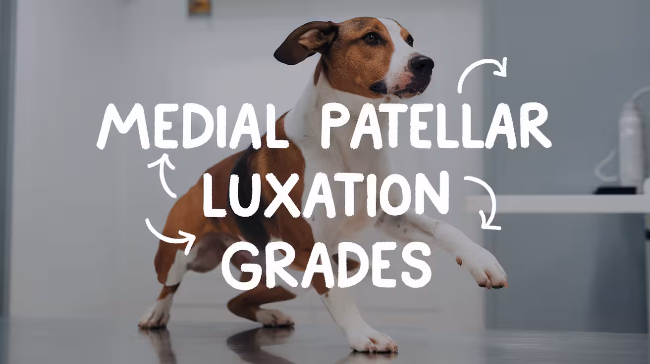
Medial Patellar Luxation Grades Explained
Clear guide to medial patellar luxation grades (I–IV) in dogs. Learn what each grade means, symptoms to watch for, and treatment options based on severity

This article is for informational purposes only and is not a substitute for professional veterinary advice. Every case is unique, so always consult your veterinarian for guidance specific to your pet.
What Is Medial Patellar Luxation (MPL)?
Medial Patellar Luxation (MPL) is a condition where a dog’s kneecap (patella) slips out of its normal position inside the femoral groove and moves toward the inside of the leg. This causes pain, limping, or skipping while walking. Over time, it can lead to joint damage or arthritis if not treated.
Commonly Affected Breeds
MPL is more common in small and toy breeds, though large breeds can also be affected.
- Pomeranians
- Yorkshire Terriers
- Chihuahuas
- Miniature and Toy Poodles
- Boston Terriers
- Pekingese
- Shih Tzus
- Some large breeds like Labrador Retrievers and Akitas
In many cases, the condition is inherited and may appear in both legs.
Why Grading Matters for Treatment
MPL is classified into four grades based on how easily the kneecap moves out of place. Grading helps the veterinarian decide the best treatment plan.
- Lower grades may not need surgery and can be managed with lifestyle changes.
- Higher grades often require surgery to correct the joint and improve function.
Understanding the grade is key to choosing the right care and predicting recovery success.
Read more:
Understanding the MPL Grading System
Grade I Medial Patellar Luxation
In Grade I MPL, the patella can be moved out of place during a physical exam but returns to its normal position on its own. Most dogs do not show obvious signs of pain or discomfort.
Typical signs:
- Intermittent “skipping” gait on one back leg
- Kneecap slips briefly, then goes back into place
- No swelling or pain when resting
- Dogs remain active and playful
Is surgery needed?
- Usually not required.
- Vets often recommend monitoring, weight control, and joint supplements.
- Surgery may be considered if the condition worsens over time.
Most dogs with Grade I live comfortably without surgery, especially if they stay active, lean, and avoid rough play. Early diagnosis helps prevent progression to higher grades.
Grade II Medial Patellar Luxation
In Grade II, the kneecap slips out more often and may stay out until it is manually repositioned. Dogs may limp or have periods of discomfort after exercise.
Common symptoms:
- More frequent skipping or limping
- Patella may remain out for several steps
- Discomfort when running or turning quickly
- Muscle loss may begin over time if not treated
When is surgery considered?
- Surgery is recommended if symptoms worsen or become regular.
- Dogs that limp often or avoid using the leg benefit from correction.
- Joint damage may occur over time if left untreated.
Some dogs with Grade II improve with conservative care, but many eventually need surgery to avoid arthritis and improve leg use.
Grade III Medial Patellar Luxation
With Grade III MPL, the kneecap is out of place most of the time but can be pushed back into the groove during an exam. Dogs often have an abnormal gait and show clear signs of discomfort.
Functional impact:
- Frequent limping or stiffness, especially after activity
- “Bunny hopping” gait or dragging one leg
- Muscle wasting on the affected leg
- Joint swelling or changes in alignment
Surgical recommendation:
- Surgery is strongly recommended to restore leg function.
- If left untreated, arthritis and permanent joint damage may occur.
- Recovery may take longer, but outcomes are usually very good.
Grade III dogs often need both soft tissue and bone procedures. Timely surgery greatly improves their quality of life and movement.
Grade IV Medial Patellar Luxation
Grade IV is the most severe form. The patella is always out of place and cannot be manually moved back into position. The leg often appears twisted or underdeveloped due to poor use.
Severe signs:
- Constant lameness or inability to use the leg
- Abnormal bone shape and knee structure
- Significant pain, joint swelling, or deformity
- Difficulty standing, sitting, or walking normally
Need for advanced correction:
- Advanced surgery is required, often involving bone realignment.
- In some cases, staged procedures are needed to fully correct the limb.
- Recovery is longer and more closely monitored.
Even though Grade IV is complex, many dogs improve with surgery. While some may still have a limp, pain is usually reduced and mobility increases. Early treatment offers the best chance for comfort and stability.
Read more:
How Vets Diagnose the Grade of MPL
Veterinarians use a mix of hands-on exams and imaging to diagnose the grade of Medial Patellar Luxation (MPL). The goal is to check how easily the kneecap moves out of place and how stable the joint is during movement.
Key Diagnostic Methods
- Physical exam: The vet checks for signs of discomfort, joint looseness, and muscle loss in the hind legs.
- Manual luxation test: While the dog is relaxed, the vet gently moves the patella to see if it slides out of place, how easily it moves, and whether it returns to the groove.
- Radiographs (X-rays): Used to check bone shape, joint alignment, and other knee problems like arthritis or bone rotation.
- Limb alignment studies: In advanced cases, special X-rays help measure bone angles and rotation, especially for surgical planning in Grade III or IV.
Accurate grading is key to choosing the right treatment. Early diagnosis allows for better planning and can prevent the condition from getting worse over time.
Read more:
Signs and Symptoms That Help Determine the Grade
MPL affects dogs differently depending on the grade. Symptoms often start mild but can worsen if the kneecap slips more often or stays out.
Common Symptoms by Grade
- Grade I: Mild, with occasional skipping or brief lameness. The dog usually walks normally between episodes.
- Grade II: More frequent skipping or limping. Some dogs start avoiding exercise or show leg stiffness after activity.
- Grade III: Constant limping or altered gait. Dogs may hop with both legs or show signs of pain during touch or movement.
- Grade IV: Severe dysfunction. Dogs often drag one leg or walk with bent knees. The limb may look twisted or underdeveloped.
Other Key Signs
- Skipping or bunny-hopping gait: Common in Grades II–III
- Bilateral signs: Both knees are often affected in small breeds
- Unilateral signs: Seen more often when injury causes MPL on one side
Observing the dog’s movement helps the vet match symptoms with the correct MPL grade and decide on treatment.
Treatment Options by Grade
Treatment for MPL depends on the grade, the dog’s age, weight, and symptoms. Lower grades may improve with non-surgical care, but higher grades usually need surgery.
Conservative Management (Grade I–II)
- Weight control
- Joint supplements
- Physical therapy
- Limiting jumping or rough play
- Pain medication, if needed
Dogs with mild signs often live comfortably without surgery if their condition stays stable.
Surgical Treatment (Grade II–IV)
When symptoms worsen or the patella stays out often, surgery is usually recommended.
- Trochlear sulcoplasty: Deepens the groove where the kneecap sits
- Tibial tuberosity transposition (TTT): Realigns the patellar tendon
- Soft tissue adjustments: Tighten or release tissues to improve tracking
Advanced Surgeries
- Needed for Grade III–IV
- May include bone cutting, plate fixation, or staged corrections
- Tailored to the dog's unique joint shape and deformity
Surgical plans vary, but the goal is always to keep the patella in place and restore comfortable movement.
Prognosis Based on Grade
The long-term outlook after MPL surgery is usually very good, especially when the condition is treated early and post-op care is followed closely.
Grade-Based Outcomes
- Grade I–II: Excellent prognosis; many dogs return to full activity and never need surgery
- Grade III: High success rates, but there is a slightly higher risk of reluxation or needing a second procedure
- Grade IV: Outcomes vary depending on joint damage. Dogs often improve, but some may keep a limp or limited motion
Reluxation Risk
- Occurs in about 10–21% of cases
- More common in Grade III and IV
- Risks decrease with proper rehab and weight control
Benefits of Early Treatment
- Helps prevent arthritis and joint damage
- Improves surgical success
- Shortens recovery time
With early diagnosis, a tailored surgical plan, and careful recovery, most dogs live active, pain-free lives after MPL correction.
Read more:
FAQs
How do vets grade a dog’s patellar luxation?
Vets grade MPL by gently moving the kneecap during a physical exam to see how easily it slips out and whether it goes back into place. They also check how the dog walks and may use X-rays to study bone shape and joint alignment. Grades range from I (mild) to IV (severe and permanent).
What is the difference between Grade I and Grade IV MPL?
Grade I MPL is the mildest form, where the kneecap only slips out during an exam and quickly returns on its own. Dogs usually show no pain or limping. Grade IV is the most severe. The kneecap is always out, cannot be pushed back in, and the leg may look twisted or underused, often needing complex surgery.
Can Grade I MPL become worse over time?
Yes, Grade I MPL can worsen if the dog gains weight, gets injured, or has weak joint support. Over time, the kneecap may start slipping more often, leading to joint wear and pain. Regular checkups, weight control, and joint care can help prevent it from progressing to higher grades.
Is surgery always needed for Grade II MPL?
Not always. Some dogs with Grade II MPL respond well to weight control, joint supplements, and restricted activity. But if the dog shows regular limping, pain, or reduced movement, surgery may be the better option. Your vet will decide based on symptoms, age, and how the joint is behaving.
What happens if MPL is left untreated?
If MPL is not treated, it can lead to chronic pain, joint damage, and arthritis. The dog may limp more often, avoid using the leg, or develop muscle loss. In higher-grade cases, untreated MPL can result in permanent joint deformity or the need for more complex surgery later.
Are some breeds more likely to have higher-grade MPL?
Yes, small breeds like Pomeranians, Chihuahuas, Yorkies, and Toy Poodles are more likely to have MPL, and some may develop higher-grade luxation due to their bone shape and genetics. However, large breeds can also be affected. Early screening in at-risk breeds helps catch problems before they get worse.

Get a Free Poster
Enhance your workspace with a high-quality radiographs reference poster, designed for veterinary professionals. This free physical poster will be shipped directly to you—just fill out the form to request your copy.

Taking Great TPLO Radiographs
Click Below to Watch Live Video Demos


We'll send you a Free Wall Poster with all the steps
Step #1
Getting Ready
Ensuring a clean surgical field starts with proper skin preparation. This video demonstrates the best practices for:
- Shaving the patient – Achieving a close, even shave while minimizing skin irritation
- The Dirty Scrub – The initial skin prep step to remove surface debris and reduce bacterial load before the sterile scrub.
Following these techniques helps reduce infection risk and improve surgical outcomes. Watch the video to see how it’s done effectively!


Step #2
Reduce Your Risks
Many surgeons are shocked to find out that their patients are not protected from biofilms and resistant bacteria when they use saline and post-op antibiotics.
That’s Where Simini Comes In.
Why leave these risks and unmanaged? Just apply Simini Protect Lavage for one minute. Biofilms and resistant bacteria can be removed, and you can reduce two significant sources of infection.
Step #3
Take the Course
Preventing surgical infections is critical for patient safety and successful outcomes. This course covers:
- Aseptic techniques – Best practices to maintain a sterile field.
- Skin prep & draping – Proper methods to minimize contamination.
- Antibiotic stewardship – When and how to use perioperative antibiotics effectively.
Stay up to date with the latest evidence-based protocols. Click the link to start learning and earn CE credits!

Things to know
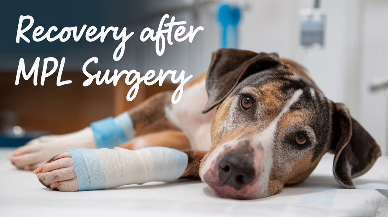
Recovery After Medial Patellar Luxation Surgery in Dogs
Discover the typical recovery timeline, care tips, and pain management after medial patellar luxation surgery to help your dog heal safely and comfortably
Typical Recovery Timeline After MPL Surgery
After medial patellar luxation surgery, your dog’s healing usually takes about 6 to 8 weeks. During this time, the knee joint repairs itself, and your dog gradually regains strength and mobility.
In the first few days, your dog may feel sore and show limited movement. Pain and swelling are normal but should improve with medication prescribed by the vet. It’s important to keep your dog calm and limit activity to help the knee heal.
Here’s what to expect during recovery:
- Weeks 1-2: Rest and restricted movement are critical. Use a leash for short bathroom breaks only. Apply cold packs if recommended by your vet to reduce swelling.
- Weeks 3-4: Your dog may start gentle walking or physical therapy exercises. Muscle strengthening begins carefully to support the knee.
- Weeks 5-6: Gradual increase in activity, including short walks and controlled play. Monitor for any signs of pain or limping.
- Week 7-8: Most dogs regain good function and strength. Your vet will check progress and may clear your dog for normal activities.
Following your vet’s recovery plan closely improves healing and helps prevent complications.
Post-Surgery Care and Activity Restrictions
After medial patellar luxation surgery, strict rest is essential to help your dog heal properly. Rest allows the repaired tissues and bones to recover without stress. Limiting movement prevents the kneecap from slipping again or causing damage during the healing process.
To keep your dog calm, restrict them to a small, quiet area like a crate or a room. Use a leash when taking them outside for bathroom breaks. Avoid letting your dog run, jump, or play freely during the first 6 to 8 weeks after surgery.
Certain activities should be avoided because they put extra pressure on the knee:
- Jumping on and off furniture or into cars
- Running or playing fetch
- Climbing stairs or steep slopes
These movements can strain the healing joint, cause pain, and slow recovery. Instead, focus on short, controlled walks as advised by your vet. Following these activity restrictions helps reduce complications and supports a smoother, faster recovery for your dog.
Managing Pain and Inflammation
Managing pain and inflammation after medial patellar luxation surgery is key to your dog’s comfort and healing. Vets commonly prescribe pain relief medications like non-steroidal anti-inflammatory drugs (NSAIDs) to reduce pain and swelling. These medicines help your dog feel more comfortable and encourage gentle movement during recovery.
Sometimes, vets may also recommend mild painkillers or muscle relaxants if the pain is more severe. It’s important to give all medications exactly as prescribed and never use human pain medicines without veterinary advice.
Cold therapy is another effective way to control swelling and reduce pain after surgery. Applying cold packs or ice wrapped in a towel to the knee for 10-15 minutes, several times a day, can help shrink blood vessels and lower inflammation. Cold therapy is most useful in the first 48-72 hours after surgery.
Supportive treatments like gentle massage or physical therapy may also be suggested by your vet to improve circulation and promote healing. Together, these methods reduce discomfort and support a smoother recovery process for your dog.
Wound Care and Monitoring
Proper wound care after medial patellar luxation surgery is crucial to avoid infection and help healing. Keeping the surgical site clean and watching for problems supports a smooth recovery.
- Check the incision daily: Look for redness, swelling, warmth, discharge, or bad smell. Mild swelling and bruising are normal, but worsening signs need a vet’s attention.
- Keep the area dry and clean: Avoid bathing your dog until the vet allows it. If cleaning is needed, gently use a mild antiseptic or saline with a clean cloth or cotton swab. Avoid harsh chemicals like alcohol.
- Prevent licking or chewing: Use an Elizabethan collar (cone) or other protective devices to stop your dog from irritating the wound. Licking can cause infection and delay healing.
- Do not touch stitches or staples: Let your vet remove them during follow-up visits, usually 10 to 14 days after surgery.
Following these steps carefully helps detect issues early and keeps the wound healthy for faster healing. Regular vet check-ups ensure the recovery is progressing well.
Physical Rehabilitation and Exercise
Physical rehabilitation plays a key role in helping your dog recover after medial patellar luxation surgery. Gentle exercises rebuild muscle strength, improve joint mobility, and support the healing knee. Without rehab, muscles can weaken and the joint may become stiff, slowing recovery.
Rehabilitation exercises usually start once the initial healing phase is over, often around 2 to 4 weeks after surgery. These exercises focus on gentle stretching, controlled leg movements, and muscle strengthening to restore normal function. A vet or veterinary physical therapist can guide you with safe, effective exercises tailored to your dog’s condition.
Gradual Reintroduction of Controlled Exercise
Gradual reintroduction of controlled exercise is essential to avoid overloading the healing knee. Short, slow walks on a leash are usually allowed first, increasing in length and intensity over several weeks. Running, jumping, and rough play should be avoided until your vet confirms the knee is strong enough.
Here’s how to safely increase activity during recovery:
- Start with brief leash walks of 5-10 minutes
- Slowly increase walk time and gentle movements
- Monitor your dog for signs of pain or limping
- Follow your vet’s advice on physical therapy sessions
Proper rehab and controlled exercise help your dog regain strength, improve joint stability, and return to normal activity safely and comfortably.
Supporting Recovery Through Diet and Environment
Dietary Considerations and Weight Management
Maintaining a healthy weight is very important during your dog’s recovery after medial patellar luxation surgery. Extra weight puts pressure on the healing knee, causing pain and slowing recovery. A balanced diet supports tissue repair and overall health.
- Control calories: Avoid high-calorie treats and table scraps that cause weight gain.
- Vet-recommended diet: Follow your vet’s advice for a diet rich in vitamins and minerals but low in excess calories.
- Gradual weight loss: If your dog is overweight, lose weight slowly under veterinary guidance to reduce joint stress.
- Nutritional support: Proper nutrients help the body heal faster and keep your dog comfortable.
Keeping your dog at a healthy weight lowers the risk of arthritis and helps the knee heal well.
Home Environment Adjustments
Making changes at home helps keep your dog safe and comfortable while recovering.
- Limit slippery floors: Use rugs or mats to prevent slips and falls.
- Block stairs and furniture: Stop your dog from jumping on or off places that strain the knee.
- Create a quiet rest area: Provide a soft, supportive bed in a calm space to encourage rest.
- Easy access: Keep food, water, and toys close so your dog doesn’t have to move too much.
These adjustments reduce injury risk and support a smooth, safe recovery.
Monitoring Healing and Follow-Up
Careful monitoring during recovery helps ensure your dog heals well and catches any problems early.
Importance of Follow-Up Veterinary Visits
Follow-up visits allow your vet to check how your dog’s knee is healing. They will assess swelling, pain levels, and joint stability. These visits help the vet adjust medications, recommend physical therapy, or suggest further treatment if needed. X-rays may be taken to see how the bones and tissues are repairing. Regular check-ups are essential for tracking progress and preventing complications, ensuring your dog recovers safely and fully.
Recognizing Possible Complications
Watch for signs like increased redness, swelling, discharge, or heat around the incision, which may indicate infection. If your dog suddenly limps more or holds the leg differently, the kneecap might have slipped again (patellar reluxation). Other signs include worsening pain, fever, or loss of appetite. Early detection of complications allows prompt veterinary care to avoid further damage and pain.
When to Contact the Vet
Contact your vet immediately if your dog shows severe limping, refuses to use the leg, or if you see swelling or discharge at the surgery site. Also, urgent care is needed if your dog shows signs of infection, such as fever or lethargy. Early communication with your vet helps manage issues quickly and supports a smoother recovery.
Owner’s Role in Successful Recovery
Your role as a dog owner is crucial for a smooth and successful recovery after medial patellar luxation surgery. Following your vet’s care instructions carefully directly affects how well and how quickly your dog heals.
- Strictly follow activity restrictions: Limiting your dog’s movement helps prevent stress on the healing knee and avoids complications.
- Administer medications as prescribed: Giving pain relief and anti-inflammatory medicines on schedule controls discomfort and swelling.
- Monitor the surgical site daily: Checking for signs of infection or other problems helps catch issues early.
- Provide a safe, comfortable environment: Making home adjustments supports rest and reduces injury risks.
- Attend all follow-up vet visits: These appointments let the vet track progress and make necessary treatment changes.
- Support rehabilitation exercises: Helping your dog with physical therapy improves strength and joint function.
Your commitment to these care steps ensures your dog stays comfortable and recovers well. Missing instructions or allowing too much activity can slow healing or cause setbacks. By staying attentive and proactive, you give your dog the best chance for a full, healthy recovery.
Recovery Variations Based on Surgery Type
Recovery after medial patellar luxation surgery can vary depending on the surgical method used. Different techniques address specific issues with the kneecap and surrounding structures, which affects the healing process and activity restrictions.
Some common surgical methods include deepening the groove where the kneecap sits (trochleoplasty), tightening or loosening tendons and ligaments around the knee, and correcting bone deformities in the thigh or shin bone. Each approach has slightly different recovery needs.
- Trochleoplasty (groove deepening): This method involves reshaping the bone groove, so healing may take longer because bone tissue needs to remodel. Strict rest is essential for 6 to 8 weeks to allow the bone to heal properly.
- Soft tissue procedures (tendon or ligament adjustments): These surgeries often involve less bone healing, so your dog might regain movement slightly faster but still requires controlled activity.
- Corrective osteotomy (bone realignment): This is a more complex procedure where bones are cut and repositioned. Recovery can be longer and may require additional pain management and physical therapy.
Your vet will explain the specific recovery plan based on the surgery type, helping you manage activity levels, pain control, and rehabilitation to ensure the best healing outcome. Understanding these differences prepares you to support your dog properly through recovery.
FAQs About Recovery After Medial Patellar Luxation Surgery
How long does recovery from MPL surgery usually take?
Recovery from medial patellar luxation surgery generally lasts between 6 and 8 weeks. During this time, your dog needs limited activity, pain management, and regular veterinary check-ups to ensure proper healing. Following your vet’s instructions helps your dog regain knee strength and mobility safely, reducing risks of complications or setbacks.
What activities should I avoid during my dog’s recovery?
Avoid activities like running, jumping, climbing stairs, or rough play during recovery. These movements put stress on the healing knee and can cause the kneecap to slip again or delay healing. Controlled, gentle walks are usually allowed under veterinary guidance. Strict activity restrictions help prevent injury and promote faster recovery.
How can I manage my dog’s pain after surgery?
Pain management includes giving prescribed medications such as NSAIDs or mild painkillers exactly as directed by your vet. Cold therapy, like applying ice packs, can help reduce swelling and discomfort. Never give human pain medicine without veterinary advice. Keeping your dog calm and comfortable is important for a smooth recovery.
When can my dog start physical therapy or exercise?
Physical therapy usually starts 2 to 4 weeks after surgery once initial healing occurs. Gentle exercises focus on improving joint mobility and rebuilding muscle strength without stressing the knee. A vet or physical therapist will guide you on safe exercises and gradually increase activity to support recovery.
How do I know if my dog’s surgical wound is healing well?
A healing wound should have minimal swelling, no redness spreading beyond the incision, and no foul-smelling discharge. Some bruising and mild swelling are normal early on. If you notice increased redness, warmth, pus, or your dog excessively licking the area, contact your vet promptly to prevent infection.
What signs mean I should call the vet during recovery?
Call your vet if your dog shows severe limping, refuses to use the leg, has swelling or discharge at the surgery site, or develops fever, lethargy, or loss of appetite. These signs may indicate infection, reluxation, or other complications requiring urgent veterinary care to protect your dog’s recovery.

Medial Patellar Luxation Treatment Without Surgery
Explore safe, effective ways to treat medial patellar luxation in dogs without surgery—home care, rehab, supplements, braces, and more
Can You Treat Medial Patellar Luxation Without Surgery?
Some dogs with MPL can be treated without surgery, but it depends on how serious the condition is.
Medial Patellar Luxation (MPL) means the kneecap (patella) slides out of its normal place inside the groove of the thigh bone. This makes the leg feel unstable and can cause pain or limping.
MPL is divided into four grades:
- Grade I: The kneecap moves out sometimes but goes back easily. Usually no pain.
- Grade II: The kneecap pops out more often and may cause mild limping.
- Grade III: The kneecap stays out most of the time but can be moved back with the hand.
- Grade IV: The kneecap is always out and can’t be moved back in.
Dogs with Grade I and some Grade II luxations can often be treated without surgery using physical therapy, weight control, and joint supplements. These help build muscle and reduce pressure on the knee.
However, Grade III and IV usually need surgery. Without it, the joint may become more damaged, and the dog may have long-term pain or difficulty walking.
Always work with your vet to decide what’s best for your dog’s specific case.
Signs Your Dog Might Benefit From Non-Surgical Treatment
Not all dogs with a loose kneecap need surgery. Some signs show your dog may do well with conservative care instead.
Dogs with mild Medial Patellar Luxation often have symptoms that come and go. One common sign is an occasional limp or a brief “skipping” step during walks. This happens when the kneecap pops out and then goes back in place. These dogs usually return to normal quickly after a few steps.
You may also hear a soft popping sound or notice the knee shift when your dog moves. If your dog rests and the signs improve, that’s another clue that non-surgical care might help.
Some symptoms that respond to:
- Rest or limited activity
- Joint supplements like glucosamine
- Gentle physical therapy
- Maintaining a healthy weight
But if the limping gets more frequent, or if your dog avoids putting weight on the leg, the condition could be getting worse. In that case, see a vet right away. Early help gives the best chance to avoid long-term joint problems.
Read more | Signs and Symptoms of Medial Patellar Luxation in Dogs
Vet Diagnosis Is Still a Must (Even for Non-Surgical Plans)
Even if your dog has mild symptoms, a vet should always confirm the diagnosis before you start treatment.
Vets use a hands-on exam to check how easily the kneecap moves out of place and which grade the MPL is. Grades I and II might not need surgery, but you need to know the grade first.
Your vet may also take X-rays to check for other problems in the knee or hip. Sometimes a dog has both MPL and hip issues, which changes the treatment plan. Without this full checkup, you might miss other problems.
Skipping the vet visit can lead to:
- Wrong treatment choices
- Delays in healing
- Worsening joint damage
- Unnecessary pain for your dog
Even if you plan to use rest, supplements, or therapy, it’s important to be sure you’re treating the right thing. A proper diagnosis helps you avoid wasting time and keeps your dog safe and comfortable. Always make the first step a vet exam.
Read more | Recovery After Medial Patellar Luxation Surgery in Dogs
Best Non-Surgical Treatments for MPL in Dogs
If your dog has a mild or moderate case of MPL, these non-surgical treatments can help manage symptoms and slow progression.
1. Weight Management to Reduce Knee Stress
Even a few extra pounds can put added pressure on your dog’s knees, especially with an unstable kneecap. Keeping your dog at a healthy weight is one of the most important steps in managing MPL.
Extra weight increases the strain on the joint and can make the patella slip out more often. Weight loss reduces inflammation and improves your dog’s ability to move comfortably.
To manage weight:
- Feed based on your vet’s calorie recommendation
- Use a body condition score chart to check for ideal shape
- Avoid table scraps and high-calorie treats
Ask your vet about the target weight for your dog’s breed and size. Track it every 2–4 weeks to stay on course. A lighter frame means less knee strain and fewer symptoms.
2. Joint Supplements for Lubrication and Repair
Joint supplements help cushion and support your dog’s knee over time. They don’t fix the luxation but can reduce pain and inflammation.
Look for products that contain:
- Glucosamine and chondroitin to support cartilage
- MSM for anti-inflammatory support
- Omega-3 fatty acids for joint lubrication
These ingredients work slowly, so it may take 4–8 weeks to see visible improvement. You might notice your dog limping less or moving more easily.
Always choose veterinary-grade supplements and follow the correct dosage for your dog’s weight. Giving more than recommended won’t help faster and can upset the stomach. If you’re unsure, ask your vet to guide you toward trusted brands.
Supplements are most effective when paired with weight control and light exercise.
Read more | How to Tell If Your Dog Has a Medially Luxating Patella
3. Controlled Exercise to Build Stability
Exercise is helpful, but it must be controlled to avoid stress on the joint. The goal is to strengthen muscles around the knee without making the luxation worse.
Focus on:
- Short, slow leash walks
- Avoiding fast turns or running
- Flat, even surfaces only
Avoid activities like:
- Stairs
- Rough play or fetch
- Jumping on furniture
Even five to ten minutes of calm walking twice a day can help. Over time, it builds leg strength and supports the kneecap’s position. Always monitor your dog after exercise for signs of soreness or increased limping. If symptoms get worse, reduce the activity and speak to your vet.
4. Strengthening Exercises You Can Try at Home
Home exercises help build muscle around the knee, improving joint stability. These are gentle and easy to do with dogs that have mild MPL.
Try these options:
- Sit-to-stand: Ask your dog to sit and then stand 5–10 times slowly
- Cavaletti poles: Use broomsticks laid low to encourage step-over motion
- Ramp or hill walking: Use a slight incline to build thigh strength
- Army crawls: Gently encourage your dog to crawl short distances (only if no pain)
Do these exercises on soft ground or carpet. Keep sessions short—5 to 10 minutes daily—and always watch your dog’s reaction. If your dog seems tired, sore, or limping more, stop the activity and check with your vet. Never force any motion.
5. Physiotherapy and Professional Rehab
Working with a certified canine rehab vet can make a big difference. Physiotherapy targets specific muscles and movements that support the knee joint.
Your rehab vet might recommend:
- Range-of-motion stretches to keep the joint flexible
- Balance exercises using wobble boards or cushions
- Laser or cold therapy to reduce pain
Sessions are usually once or twice a week at first, depending on your dog’s condition. At-home plans may be included for daily practice.
The goal is to improve strength, reduce discomfort, and keep the luxation from worsening. Ask your vet to refer you to a certified canine rehab therapist in your area.
Read more | Best Exercises and Rehab for Dogs After MPL Surgery
6. Hydrotherapy for Low-Impact Strength Building
Hydrotherapy is one of the best ways to build strength without stressing the knee. It uses water to support your dog’s body while still allowing muscle movement.
Two main methods are:
- Underwater treadmill: Your dog walks on a belt inside a water tank
- Swimming in a rehab pool: Guided by a therapist, if safe for your dog
Water reduces joint pressure by supporting weight and allowing a fuller range of motion. At the same time, resistance helps improve strength.
Hydrotherapy is ideal for dogs that can’t tolerate land exercises or have arthritis along with MPL. Most dogs enjoy the sessions and improve steadily over a few weeks. Your vet or rehab therapist will tell you how often to go—usually once or twice per week at first.
7. Pain Relief with Vet-Approved Medications
Even with mild MPL, some dogs have joint pain. Medications help manage this discomfort so your dog can stay active and heal better.
Common vet-approved options include:
- NSAIDs like Galliprant, Carprofen, or Meloxicam
- Gabapentin for nerve-related pain
- Joint-protecting injections (like Adequan)
These meds must always be prescribed by your vet, and the dose must match your dog’s weight and health condition. Never give human painkillers—many are toxic to dogs.
Pain control makes it easier for your dog to walk, rest, and take part in therapy. It also prevents muscle loss due to reduced activity.
Always watch for side effects like stomach upset or changes in appetite and report them to your vet.
Read more | Medial Patellar Luxation in Small vs. Large Dogs
8. Physical Therapy Extras That Help
Besides basic rehab, some alternative therapies can help reduce pain and improve movement. They’re not required but can support the main treatment plan.
Helpful options include:
- Massage therapy to relax tight muscles and improve circulation
- Chiropractic care, but only by trained, certified animal chiropractors
- Acupuncture or cold laser therapy for joint pain and inflammation
These options should always be done by professionals trained in canine care. They can be used alone or along with other treatments like hydrotherapy or medication.
Some dogs respond well to one method, while others need a mix. Discuss with your vet or rehab team to find what fits your dog best.
9. Using Dog Knee Braces for Extra Support
Knee braces can help dogs with Grade I or II MPL by giving extra support during movement. They don't fix the kneecap but can stop it from slipping as often.
You might consider a brace if:
- Your dog limps during walks
- You want to avoid surgery
- You’re starting physical therapy
Look for braces that offer side support and adjustable straps. Brands like Walkin’ Pets and Ortocanis make popular models. Always measure your dog’s leg carefully for a good fit.
Some dogs take time to adjust to wearing a brace. Start with short sessions indoors. Reward calm behavior and watch for rubbing or skin problems.
Use braces during walks or active periods, not all day. Ask your vet or therapist to show you how to put it on safely and check for signs of irritation.
Home Modifications That Support Recovery
Making small changes in your home can help your dog heal more comfortably and safely from MPL without surgery.
Dogs with unstable kneecaps need secure footing. Slippery floors like tile or wood can cause the knee to slide out of place. To fix this, place non-slip rugs or yoga mats in areas where your dog walks often, especially around food bowls and beds.
You should also use pet stairs or ramps to help your dog get on furniture or in and out of the car. Jumping up or down puts pressure on the knee and can worsen the condition.
Helpful home changes include:
- Block off stairs or high beds
- Avoid letting your dog jump on sofas or beds
- Use baby gates to limit movement during recovery
- Keep your dog’s essentials on one floor
These simple changes reduce strain on the joint and make your dog feel more secure. They also support the effects of weight control and therapy. Always watch how your dog moves through the house and adjust the environment as needed to prevent slips or overuse.
Read more | How to Prevent Medial Patellar Luxation in Puppies
What a Weekly Routine Might Look Like
Creating a weekly routine helps manage MPL without surgery by balancing exercise, rest, and joint support. A structured plan keeps your dog active without overloading the knee.
Here’s what a sample day might look like:
- Morning: Short 5-minute leash walk, joint supplement with breakfast
- Midday: Sit-to-stand exercise or gentle hill walking
- Evening: Massage or range-of-motion stretches, then rest
You can adjust based on your dog’s energy level and vet recommendations. For example, if using hydrotherapy or rehab, schedule it 1–2 times per week. Keep the rest of the week low-impact to avoid fatigue.
Track progress with:
- A journal of limp-free days
- Changes in mood or energy
- Photos or videos of movement
Avoid doing the same routine every single day. Dogs also need rest days with minimal activity to allow muscle recovery. Balance is key.
Your vet or canine rehab therapist can help tailor the plan further. Sticking to a schedule makes it easier to spot progress or setbacks quickly.
What to Avoid When Managing MPL Without Surgery
While there are many ways to help your dog without surgery, some mistakes can slow recovery or even make things worse.
One common issue is skipping rest days. Muscles need time to recover, especially after exercise or therapy. Too much activity without breaks can lead to swelling or pain.
Also avoid:
- Overexercising too soon – Build strength slowly to avoid injury
- Letting your dog run, jump, or climb stairs without guidance
- Using only joint supplements without a complete care plan
- Delaying vet checkups when symptoms change
Some dogs act fine even when their knee is unstable. But ignoring small changes—like more frequent limping or reduced play—can lead to worse problems later.
You don’t need to overprotect your dog, but structure and balance matter. Always adjust based on how your dog responds, and check with your vet if anything seems off.
Small daily mistakes can undo weeks of good care, so stay consistent and alert.
Read more | Grade 2 Medial Patellar Luxation: Surgery Decision Guide
How Long Does Recovery Take Without Surgery?
Recovery from MPL without surgery takes time and patience. The timeline depends on the severity of the condition, your dog’s age, and how closely you follow the care plan.
For dogs with Grade I or mild Grade II, visible improvement can start in 4 to 6 weeks. Full recovery, where symptoms are rare or gone, may take 8 to 12 weeks or longer. Some dogs need ongoing support through supplements and light exercise for life.
Signs that your dog is improving include:
- Less frequent limping or skipping steps
- Better muscle tone in the hind legs
- Increased comfort during walks
However, if the condition worsens—more frequent knee popping, longer limping episodes, or pain while resting—it’s time to re-evaluate. A follow-up vet visit can help adjust the plan or decide if surgery is now the better option.
MPL often stays manageable with consistent care, but don’t expect overnight results. Keep a record of changes to help your vet guide next steps. Recovery is a journey, and small improvements matter.
When to Reconsider Surgery
Even with the best home care, some dogs may not improve enough and will need surgery. Knowing when to shift gears can protect your dog’s comfort and long-term joint health.
You should talk to your vet about surgery if:
- Limping continues for more than 8–12 weeks
- Your dog has a Grade III or IV MPL that’s not responding
- Your dog avoids using the leg, even with treatment
Other signs to watch for include worsening pain, trouble rising, or changes in behavior like less interest in walks. These may mean that conservative treatments are no longer enough.
Quality of life is the key factor. If your dog can’t enjoy normal daily activities without pain, surgery becomes the better path.
While we all want to avoid invasive procedures, surgery often gives long-term stability and comfort for higher-grade cases. Your vet will help you weigh the risks and benefits.
It’s okay to start with non-surgical care, but be open to changing the plan if your dog isn’t getting better.
Read more | Unilateral vs. Bilateral Medial Patellar Luxation in Dogs
Final Thoughts on Avoiding Surgery for MPL
Treating Medial Patellar Luxation without surgery is possible—especially in mild cases—and often helps dogs live more comfortably when done with care.
Key strategies for success include:
- Weight control
- Joint supplements
- Structured exercise and home safety changes
- Regular vet visits and tracking symptoms
The most important part is consistency. Daily effort with even small changes builds up over time. Dogs with mild MPL can stay pain-free for years if managed properly.
However, always stay in touch with your vet. They’ll guide you when to push forward and when it’s time to consider other options like surgery.
You know your dog best, and with a clear plan, steady care, and expert guidance, many dogs can avoid surgery and still enjoy a happy, active life.
FAQs
Is it safe to avoid surgery for patellar luxation?
Yes, it’s safe in mild cases (Grade I or some Grade II) if managed correctly. Weight control, joint support, and regular vet checkups can help reduce symptoms. But if your dog’s limping worsens or pain increases, surgery may become necessary. Always follow your vet’s advice to ensure the safest outcome for your dog.
How do I know if my dog’s MPL is getting worse?
Watch for more frequent limping, longer recovery after walks, or signs of pain while resting. If your dog avoids using the leg, hesitates to climb stairs, or shows stiffness after rest, these may be signs the MPL is progressing. A worsening condition needs a vet recheck to adjust the treatment plan or consider surgery.
What supplements are best for dogs with MPL?
Good joint supplements often include glucosamine, chondroitin, MSM, and omega-3 fatty acids. These support cartilage, reduce inflammation, and improve joint function over time. Choose veterinary-grade brands and use the correct dose based on your dog’s weight. Talk to your vet before starting supplements to make sure they’re right for your dog’s condition.
Can braces fix luxating patella permanently?
No, braces don’t permanently fix the condition. They provide temporary support by helping the kneecap stay in place during movement. Braces work best in mild cases or while doing rehab. They may reduce symptoms but don’t correct bone or joint shape. For permanent correction, surgery is needed in moderate to severe cases.
How long should I try non-surgical treatment before seeing results?
Most dogs show improvement in 4 to 6 weeks with consistent care. This includes exercise, joint support, and weight control. Full benefits may take 8 to 12 weeks. If there’s no progress or symptoms worsen during that time, see your vet. They’ll help decide if surgery or a new plan is needed.
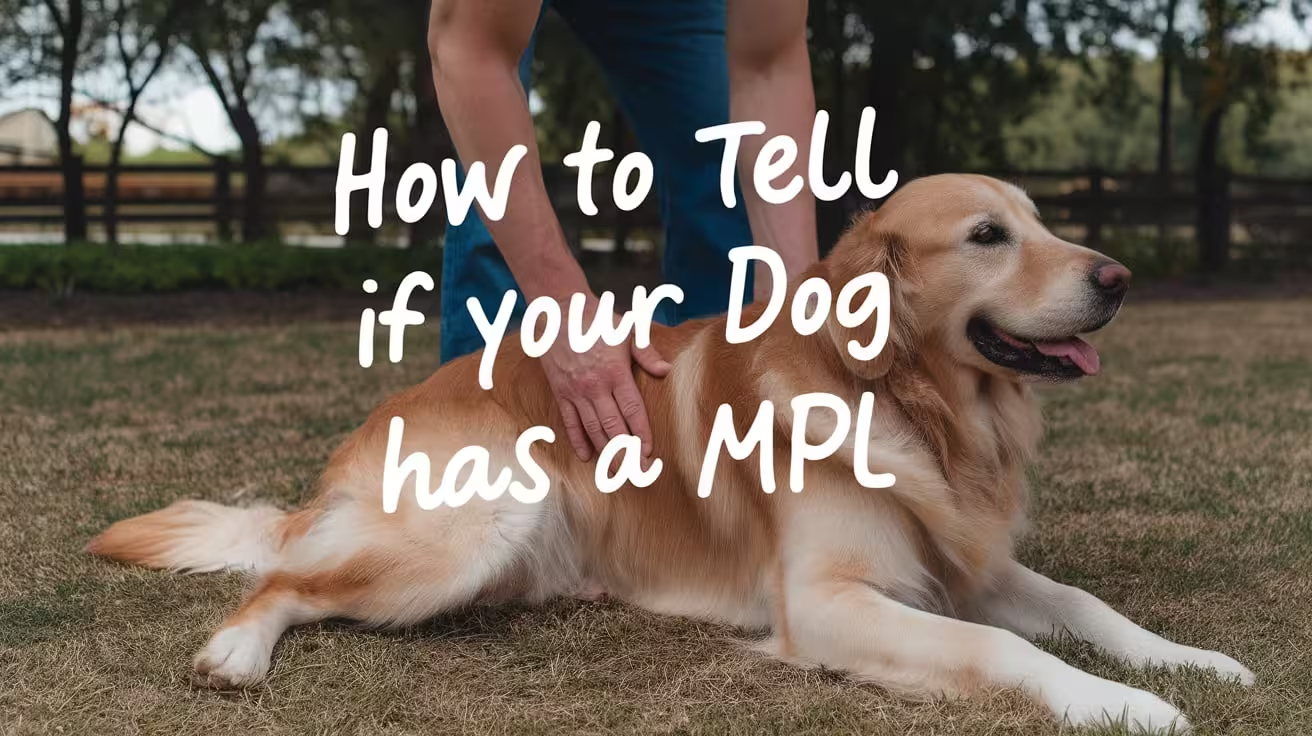
How to Tell If Your Dog Has a Medially Luxating Patella
Learn how to recognize signs of medial patellar luxation in dogs early, including limping, clicking sounds, and changes in gait for timely treatment
What Is Medial Patellar Luxation in Dogs?
Medial Patellar Luxation (MPL) is a condition where the kneecap (patella) slips out of its normal groove toward the inside of a dog’s leg. This causes the knee joint to become unstable, leading to pain, limping, and difficulty walking. MPL is especially common in small and toy dog breeds but can affect dogs of any size.
MPL matters because if left untreated, it can cause long-term joint damage, arthritis, and chronic pain. The slipping kneecap puts extra stress on the knee, making movement uncomfortable and sometimes causing your dog to avoid using the leg.
Early detection is very important for your dog’s health and comfort. Recognizing signs like limping, skipping steps, or holding up a leg allows you to seek veterinary care sooner. Early treatment can reduce pain, prevent further damage, and improve your dog’s quality of life. Keeping a close eye on your dog’s movement helps catch MPL before it worsens.
Common Signs That Your Dog May Have MPL
Here are some common signs that can help you identify if your dog may be suffering from medial patellar luxation (MPL).
1. Limping or Skipping Steps
Limping or skipping steps is one of the earliest signs your dog might have medial patellar luxation (MPL). When the kneecap slips out of place, your dog may feel pain or instability, causing uneven movement.
- Watch for your dog lifting a leg briefly while walking or running.
- Notice if your dog skips steps or hops on one leg suddenly.
- Limping can be subtle at first and may only happen after exercise or prolonged activity.
If you see your dog favoring one leg or walking unevenly, it’s a sign to check with your vet. Early limping or skipping indicates discomfort or instability in the knee, which can worsen if untreated. Tracking these patterns helps you catch MPL early and get your dog the care needed to prevent long-term damage.
2. Intermittent or Persistent Lameness
Lameness means your dog is limping or not using a leg normally. It can be intermittent (comes and goes) or persistent (constant). Both can be warning signs of MPL.
- Intermittent lameness often appears after exercise or activity when the kneecap slips out temporarily.
- Persistent lameness means ongoing pain or instability in the knee, which needs urgent attention.
- Your dog may hold up the leg or limp constantly if the condition is severe.
Recognizing when lameness changes from occasional to frequent is important. Early veterinary diagnosis can help treat mild lameness before it becomes severe. Persistent lameness might indicate worsening MPL or other complications. Watching how often your dog limps and seeking veterinary advice quickly improves treatment outcomes.
3. Abnormal Gait or Skipping Leg Movement
An abnormal gait means your dog’s walk or run looks different from normal. In dogs with MPL, this often shows as unusual skipping or jerky leg movement.
- Your dog may appear to skip or hop instead of walking smoothly.
- The affected leg might move differently, seeming stiff or shaky.
- This irregular movement happens because the kneecap slips, causing discomfort or instability.
This gait change may be subtle at first and can be mistaken for other issues. Watching carefully during walks or play helps spot unusual leg movements. Early detection allows for prompt treatment to restore normal walking patterns and reduce pain.
4. Audible Clicking or Popping Sounds
Clicking or popping sounds from your dog’s knee are signs that the kneecap is moving abnormally. These sounds happen when the patella slips out of its groove and snaps back.
- You might hear a faint “click” when your dog walks, runs, or moves the leg.
- These noises show joint instability and possible damage to soft tissues.
- Not all dogs make these sounds, but if you hear them often, it’s a sign to get a vet check.
Listening for these sounds during activity or when your dog moves the leg gently can provide clues about MPL. Early veterinary diagnosis helps prevent joint damage from frequent slipping.
5. Visible or Palpable Slipping of the Kneecap
Sometimes, you can see or feel the kneecap slipping out of place. This popping or luxation happens when the kneecap moves out of its normal groove on the thigh bone.
- Gently feel your dog’s knee when the leg is bent and straightened to check for slipping.
- You might see the kneecap visibly move or pop to the side during leg movement.
- If unsure, have a vet perform this test to avoid causing pain or injury.
Feeling or seeing the patella slip is a clear sign of MPL. If you notice this, it’s important to visit your vet for a full examination and diagnosis.
6. Holding Up the Leg or Avoiding Weight Bearing
Dogs with MPL often hold up the affected leg or avoid putting weight on it to relieve pain or discomfort.
- Your dog may lift the leg while standing or walking, especially after activity.
- Avoidance of weight bearing is a common way dogs protect an injured or painful knee.
- This behavior can be temporary or frequent depending on MPL severity.
If your dog regularly holds up a leg or refuses to walk on it, it’s a strong sign of knee pain. Early vet evaluation helps manage discomfort and improves healing.
7. Difficulty or Reluctance to Jump, Run, or Climb Stairs
Changes in your dog’s activity level, like difficulty or unwillingness to jump, run, or climb stairs, often point to knee problems like MPL.
- Your dog may avoid stairs or hesitate before jumping onto furniture or into cars.
- Running or playing less than usual can indicate discomfort during high-impact activities.
- These behavioral changes help protect the painful knee from stress.
Noticing reluctance to be active is important for early MPL detection. Discussing these changes with your vet can lead to timely diagnosis and treatment.
8. Swelling or Pain Around the Knee Joint
Swelling or pain near the knee joint may develop with MPL due to inflammation from repeated kneecap slipping.
- Look for visible swelling, warmth, or tenderness around the knee.
- Your dog might lick or chew the knee area more than usual.
- Pain signs include limping, whining, or reluctance to move.
Swelling and pain indicate irritation or early joint damage. Prompt veterinary care can reduce inflammation and prevent progression.
9. Changes in Behavior Like Reluctance to Play or Exercise
Discomfort from MPL often causes changes in your dog’s behavior, such as reduced playfulness or exercise reluctance.
- Your dog may become less active or hide more than usual.
- Decreased interest in walks, toys, or interaction can signal pain.
- These subtle mood changes are important clues to underlying knee issues.
Recognizing these behavior shifts early helps you seek veterinary care and improve your dog’s comfort and quality of life.
How MPL Symptoms Can Worsen Over Time
If medial patellar luxation (MPL) is not treated, your dog’s symptoms can get worse and cause more serious problems.
- More frequent kneecap slipping: The patella may move out of place more often, causing pain and joint instability.
- Joint damage and arthritis: Repeated slipping can wear down cartilage and bones, leading to arthritis. This causes swelling, stiffness, and long-term pain.
- Increased limping or holding up the leg: Your dog may limp more or avoid using the affected leg due to discomfort.
- Posture and gait changes: To reduce pain, your dog might change how they stand or walk, which can cause muscle loss and strain on other legs.
- Both knees affected: Sometimes MPL develops in both legs, worsening mobility and quality of life.
Monitoring your dog’s symptoms closely helps catch these changes early. Watch for increased limping, reduced activity, or changes in behavior. Early vet care can reduce pain, prevent joint damage, and improve your dog’s chances of a happy, active life.
How to Monitor Your Dog’s Mobility and Pain at Home
Monitoring your dog’s mobility and pain at home helps you track their condition and notice any worsening signs early. Regular observation lets you provide important information to your vet for better care.
- Watch your dog’s walking: Look for limping, skipping steps, or difficulty standing up. Notice if your dog favors one leg or hesitates to move.
- Observe activity levels: Pay attention to changes in how much your dog wants to play, run, or climb stairs. Reduced activity can signal pain or discomfort.
- Check for stiffness: Notice if your dog is stiff or slow to get moving after resting or sleeping.
- Look for pain behaviors: Whining, licking the knee, or sudden stops during movement may show discomfort.
- Examine posture: Watch for changes in how your dog holds their leg or stands.
Keep a simple journal to record daily observations, noting any new or worsening symptoms. Include details about when symptoms appear, their severity, and any triggers like exercise. Sharing this information with your vet helps tailor treatment and improves your dog’s care. Regular monitoring is key to managing MPL effectively.
When to Seek Veterinary Care for Suspected MPL
Knowing when to seek veterinary care for suspected medial patellar luxation (MPL) is crucial to protect your dog’s health and comfort. Early veterinary attention can prevent worsening damage and reduce pain.
- Sudden or severe limping: If your dog starts limping suddenly or cannot put weight on a leg, it needs immediate vet care.
- Persistent or worsening lameness: Continuous limping or increasing difficulty walking are signs of serious knee issues.
- Visible swelling or redness: Swelling, heat, or redness around the knee may indicate inflammation or infection.
- Audible clicking or popping: Hearing frequent clicking sounds from the knee can signal instability needing professional evaluation.
- Reluctance to move or play: A sudden decrease in activity or reluctance to jump, run, or climb stairs suggests discomfort.
Early diagnosis allows your vet to assess the severity of MPL and recommend the best treatment, whether conservative care or surgery. Prompt treatment reduces pain, slows joint damage, and improves your dog’s quality of life. Don’t wait for symptoms to worsen—early veterinary care is key to a better outcome.
FAQs About How to Tell If Your Dog Has a Medially Luxating Patella
What is medial patellar luxation in dogs?
Medial patellar luxation (MPL) occurs when the kneecap slips out of its normal position toward the inside of the leg. It causes pain, limping, and joint instability. It’s common in small breeds but can affect all dogs. Early detection is key to preventing long-term damage.
How can I spot limping caused by MPL?
Limping or skipping steps may happen suddenly or after activity. Your dog might lift or favor one leg, showing discomfort. Limping can be subtle at first, so careful observation during walks is important to catch early signs of MPL.
What does an abnormal gait look like in dogs with MPL?
An abnormal gait may appear as skipping, hopping, or uneven leg movement. The affected leg might move stiffly or jerk unexpectedly due to the kneecap slipping out of place, causing discomfort and instability while walking or running.
Why does my dog’s knee make clicking sounds?
Clicking or popping noises occur when the kneecap moves out and back into its groove. These sounds indicate joint instability and frequent slipping of the patella. Hearing this often suggests your dog should be checked by a vet.
How can I check if my dog’s kneecap is slipping?
Gently feel your dog’s knee while moving the leg to detect popping or slipping of the kneecap. Be careful to avoid causing pain. If unsure, let a vet perform the test safely for an accurate diagnosis.
When should I take my dog to the vet for suspected MPL?
Visit the vet if your dog shows limping, skipping steps, leg holding, swelling, or audible knee clicks. Early veterinary care is important to diagnose MPL, start treatment, and prevent worsening symptoms and joint damage.

Medial Patellar Luxation in Small vs. Large Dogs
Learn how medial patellar luxation affects small vs. large dogs—compare symptoms, treatment options, surgery needs, recovery, and care tips by size
What Is Medial Patellar Luxation (MPL)?
Medial Patellar Luxation (MPL) is a knee condition where the kneecap (patella) slips out of its normal groove toward the inside of the leg. This makes the joint unstable and can lead to limping, pain, or changes in how your dog walks.
MPL is different from Lateral Patellar Luxation (LPL). In MPL, the kneecap moves inward (toward the other leg). In LPL, it slips outward. MPL is much more common, especially in small dogs.
When the patella doesn’t stay in place, the knee joint becomes weak and unstable. Over time, this can cause joint damage, pain, and arthritis if not treated.
MPL is graded by severity:
- Grade I: Kneecap pops out but goes back easily
- Grade II: Slips out more often, may cause limping
- Grade III: Stays out, but can be moved back by hand
- Grade IV: Always out and can’t be returned without surgery
Understanding the grade helps guide the right treatment for your dog’s needs.
How MPL Differs Between Small and Large Dogs (Quick Comparison)
MPL appears in both small and large dogs, but it behaves differently depending on the dog’s size. Small dogs usually have it from a young age, often due to genetics. In large dogs, MPL may develop later and is sometimes linked to trauma or uneven growth. Understanding these differences helps in planning the right treatment and recovery.
Key differences include:
- Prevalence: MPL is more common in small dogs, while large dogs may have lateral luxation more often.
- Type of Luxation: Small dogs typically have medial luxation; large dogs may develop medial or lateral types.
- Common Breeds: Small breeds include Pomeranians, Chihuahuas, and Poodles. Large breeds include Labradors and German Shepherds.
- Severity and Grading: Small dogs usually have mild to moderate (Grade I–II) MPL. Large dogs often show severe grades (III–IV).
- Age of Onset: Small dogs often show signs before 1 year old. In large dogs, signs may appear later.
- Bone Deformity Likelihood: Small dogs have a higher chance of congenital bone deformities. Large dogs may develop changes over time.
- Surgical Approach: Surgery in large dogs is more complex due to size and joint stress. Small dogs often respond well to simpler procedures.
- Recovery Outlook: Small dogs recover faster and need less rehab. Large dogs may require longer recovery and stricter care.
Read more | Signs and Symptoms of Medial Patellar Luxation in Dogs
Which Breeds Are Most at Risk?
Certain dog breeds are more likely to develop Medial Patellar Luxation (MPL) due to inherited traits and body structure. Genetics play a major role, especially in small dogs, where MPL often shows up early in life. Large breeds can also be affected, though less frequently, and often with more complex presentations.
Small breeds most at risk include:
- Yorkshire Terrier
- Pomeranian
- Chihuahua
- Miniature and Toy Poodle
- Pekingese
These breeds are often born with shallow knee grooves or loose ligaments, making it easier for the kneecap to slip out of place.
Large breeds that may develop MPL include:
- Labrador Retriever
- Flat-Coated Retriever
- Great Pyrenees
- Akita
- Newfoundland
In large breeds, MPL may not appear until later in life and can be linked to trauma, poor joint alignment, or fast growth during puppyhood.
While not every dog in these breeds will get MPL, their genetic makeup increases the risk. Breeders are encouraged to screen for joint issues and avoid breeding dogs with known luxation problems.
If your dog belongs to one of these breeds, keep an eye out for early symptoms like limping or skipping steps, and have your vet assess their knees during routine checkups. Early detection gives your dog the best chance at long-term joint health.
Read more | Recovery After Medial Patellar Luxation Surgery in Dogs
Symptoms in Small vs. Large Dogs
The signs of Medial Patellar Luxation (MPL) can look different depending on your dog’s size. Small dogs often show quick, obvious signs, while large dogs may hide discomfort until it worsens.
In small dogs, the kneecap may pop out and back in during movement. This causes:
- Limping that comes and goes
- A sudden “skipping” step during walks
- Temporary leg lifting followed by normal walking
- Mild pain or licking at the knee
These signs may be brief but happen often, especially after play or getting up from rest.
In large dogs, symptoms can be harder to spot at first. Instead of skipping steps, they may show:
- Slow or stiff movements
- Reluctance to exercise or climb stairs
- Subtle lameness that worsens with activity
- Trouble rising or turning quickly
Some dogs have bilateral MPL, meaning both knees are affected. This can make them walk stiffly or show signs in both legs.
If your dog shows any of these changes, even if mild, it’s important to see your vet. Early diagnosis helps prevent long-term joint damage and allows for better treatment planning tailored to your dog’s size and condition.
Read more | How to Tell If Your Dog Has a Medially Luxating Patella
Diagnosis and Grading in Both Size Groups
Medial Patellar Luxation is diagnosed through a physical exam and imaging. Your vet will check how easily the kneecap moves out of place and assess joint function. This helps decide the severity and treatment.
The grading system is the same for all dogs:
- Grade I: Kneecap moves out easily but pops back in
- Grade II: Slips out on its own and stays briefly
- Grade III: Always out but can be moved back by hand
- Grade IV: Always out and can’t be moved back
X-rays are used to confirm joint structure and rule out other issues like hip dysplasia or trauma. This is especially important in large breeds, where symptoms may overlap with other orthopedic conditions.
Common misdiagnoses in large dogs include arthritis, hip problems, or muscle strain. That’s why a full orthopedic exam is key, not just looking at gait.
Although the grading is the same, large dogs may have more bone involvement or need more detailed imaging. Small dogs often show clearer signs during a basic exam. In all cases, grading helps guide whether surgery or conservative care is the right path.
Treatment Options: Small Dogs vs. Large Dogs
The choice between surgical and non-surgical treatment depends not just on the grade of MPL but also on your dog’s size. Small and large dogs respond differently to conservative care.
Small dogs with Grade I or II often improve with:
- Weight management
- Joint supplements
- Controlled exercise
- Bracing and physiotherapy
These treatments can stabilize the knee and reduce symptoms without surgery. Small dogs benefit from lighter body weight and lower joint stress.
Large dogs, even with Grade II, may require surgery because:
- Their size adds pressure to the joint
- Conservative care may not fully control the luxation
- Mobility problems worsen faster with weight-bearing stress
Challenges by size include:
- Harder to limit large dogs’ movement indoors
- Weight loss is slower in big breeds
- Braces are harder to fit and keep in place
For both groups, combining care—like supplements, therapy, and lifestyle changes—can reduce pain and protect the knee. But large dogs are less likely to succeed with non-surgical treatment alone. Your vet will consider all factors before creating a plan.
Read more | Medial Patellar Luxation Treatment Without Surgery
Surgery: What’s Different by Dog Size?
Surgical treatment for MPL is common and effective, but the approach depends heavily on your dog’s size and the grade of luxation.
Small dogs usually need simpler procedures such as:
- Trochlear sulcoplasty (deepening the groove for the kneecap)
- Soft tissue release or tightening
- Tibial tuberosity transposition (in more advanced cases)
These surgeries are effective in most small breeds and carry a low risk of complications when done early.
Large dogs often require more advanced techniques like:
- Corrective osteotomies (cutting and realigning the bone)
- Use of surgical implants for joint support
- More extensive soft tissue reconstruction
Key differences in large breeds:
- Higher force on the joint requires stronger repairs
- Increased anesthesia risk due to body weight
- Longer surgical time and more complex aftercare
Post-op care also varies. Small dogs may return to light activity in 4–6 weeks. Large dogs may need 8–12 weeks of structured rehab and close monitoring.
Surgical success is high in both groups, but choosing the right procedure for your dog’s size is critical for long-term joint health and mobility.
Read more | Best Exercises and Rehab for Dogs After MPL Surgery
Recovery and Prognosis: What to Expect
Recovery from MPL surgery or conservative care varies by dog size, but most dogs show excellent outcomes with proper treatment.
Small dogs often bounce back faster due to their lighter weight and easier mobility control. With routine care and basic rehab, many are back to normal within 4–6 weeks. Risks like implant failure or complications are rare if surgery is done early.
Large dogs may need:
- More time to heal
- Ongoing physical therapy
- Strict activity limits for 8–12 weeks
- Closer monitoring for complications like joint swelling or stiffness
Success rates for both groups are high—over 90% in most cases. However, complications are more common in large dogs due to size, strength, and stress on healing tissues.
Long-term, most dogs regain normal function and show no signs of lameness. Some may need continued supplements or periodic vet checkups to keep joints healthy.
Whether surgical or non-surgical, the key to a good recovery is sticking to the plan, avoiding overuse, and adjusting care based on your dog’s response. Vet-guided follow-ups improve long-term outcomes significantly.
Cost Differences in Treatment
Cost is another important factor when deciding how to treat MPL, and it varies greatly between small and large dogs.
Surgical costs for small dogs are typically lower due to:
- Smaller bone and joint size
- Simpler procedures
- Less anesthesia and material use
Most small dog MPL surgeries cost $1,500–$2,500 depending on location and hospital fees.
Large dog surgeries are more expensive due to:
- Longer surgical time
- Larger implants or plates
- Increased anesthesia needs
- More rehab or post-op care
For large breeds, costs may range from $3,000–$5,000+, especially if bone realignment or custom plates are needed.
Non-surgical costs include:
- Joint supplements ($30–$80/month)
- Rehab or hydrotherapy sessions ($50–$100/session)
- Braces ($100–$400 depending on size)
Budgeting ahead is essential. Ask your vet for a full breakdown of possible costs for both surgical and non-surgical options. Some clinics offer payment plans or referrals to orthopedic specialists with package pricing.
Choosing the right treatment should balance cost, outcome, and your dog’s long-term comfort.
Read more | How to Prevent Medial Patellar Luxation in Puppies
Managing MPL at Home: Tips by Dog Size
Managing MPL at home requires adapting your environment and care routine to suit your dog’s size. Small and large dogs need different setups for safety and support.
For small dogs:
- Use pet stairs or carry them to avoid jumping
- Place soft mats or rugs in walkways to prevent slipping
- Choose braces designed for toy or small breeds
- Encourage short leash walks to build strength
For large dogs:
- Use ramps for cars or beds
- Block access to stairs with baby gates
- Add non-slip flooring in main rooms
- Use heavy-duty braces made for large breeds
- Focus on structured, slow-paced exercises
For all dogs:
- Keep weight under control with portioned meals
- Stick to your vet’s rehab or supplement plan
- Avoid off-leash running or rough play
Customizing care by size helps reduce joint stress and supports long-term joint health. Your vet or canine therapist can give breed-specific advice to improve comfort and prevent flare-ups.
Read more | Grade 2 Medial Patellar Luxation: Surgery Decision Guide
Final Thoughts: Does Dog Size Change the MPL Plan?
Yes—dog size has a big impact on how MPL is managed. From diagnosis to treatment and recovery, small and large dogs face different challenges and require tailored plans.
Small dogs often respond well to conservative care or simple surgery. They recover quickly, and the procedures are usually less costly. Large dogs may need more complex treatment, longer rehab, and closer monitoring due to their size and weight.
Major points to remember:
- MPL is more common in small breeds
- Symptoms may be subtle in large dogs
- Treatment should match the dog’s grade and size
- Surgery is often more urgent in large breeds
- Recovery time and cost are higher in larger dogs
Early diagnosis, consistent care, and working closely with your vet give your dog the best chance for a pain-free, active life. Whether small or large, your dog can recover well with the right plan.
FAQs
Is MPL more serious in large dogs than small dogs?
Yes, MPL tends to be more serious in large dogs. Their size puts more pressure on the knee joint, which can lead to faster joint damage and a higher need for surgery. Larger dogs also face more complex surgeries and longer recovery times compared to smaller breeds with the same grade of luxation.
Can large dogs recover without surgery?
Some large dogs with mild MPL may improve with non-surgical care, but this is less common. Their heavier weight makes it harder to manage joint stress with therapy alone. Surgery is often recommended for long-term stability and comfort, especially for Grade II or higher cases in large-breed dogs.
Are certain breeds more prone to complications?
Yes, large breeds like Labrador Retrievers or Great Pyrenees are more prone to surgical complications due to their size and joint stress. Small dogs generally recover faster and with fewer issues. Dogs with severe grades or other orthopedic problems may also have higher risks during and after treatment.
Is lateral luxation more common in big dogs?
Yes, lateral patellar luxation (LPL), where the kneecap moves outward, is more common in large dogs. While MPL is still seen in big breeds, LPL tends to occur more frequently due to different bone angles and joint stresses found in larger body structures.
What is the recovery time for small vs. large dogs after surgery?
Small dogs typically recover in 4–6 weeks with light care. Large dogs may need 8–12 weeks or longer, along with structured rehab and activity limits. Recovery depends on surgery type, joint condition, and post-op care, but large dogs often take more time due to body weight and joint pressure.
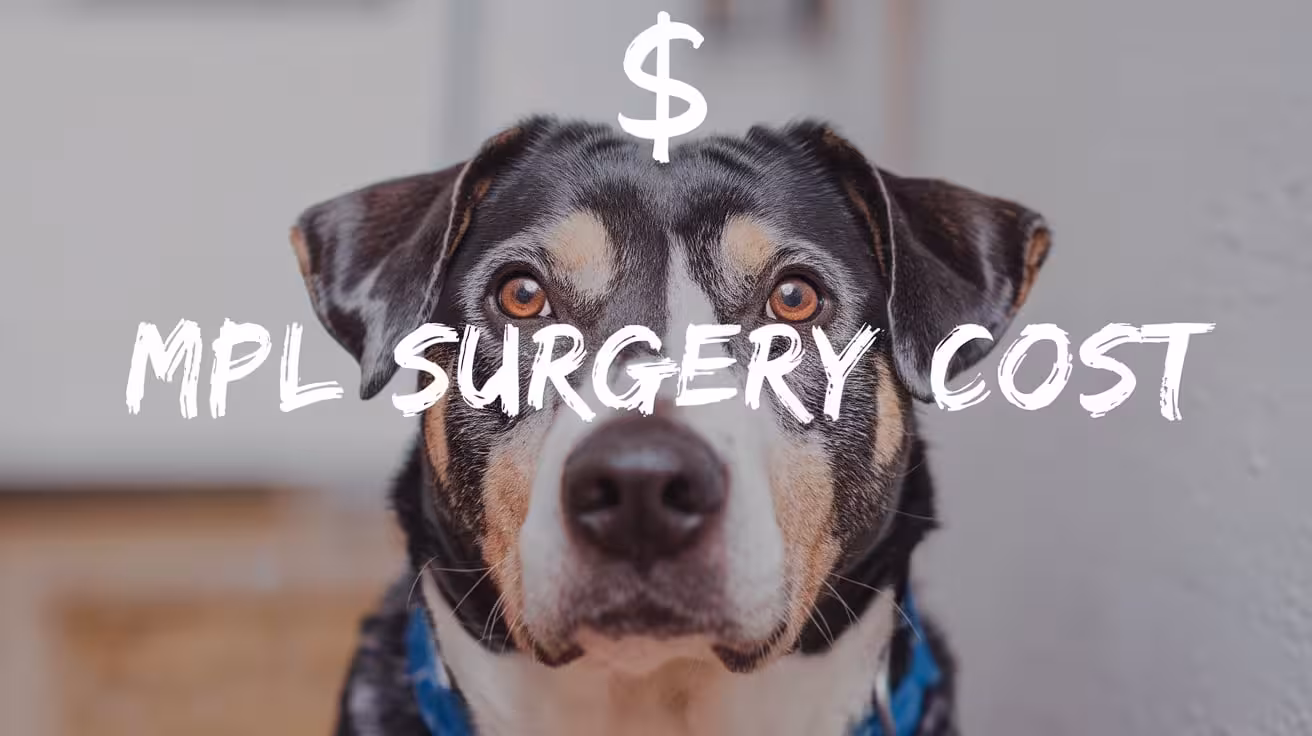
How Much Does MPL Surgery Cost for Dogs?
Learn how much MPL surgery costs for dogs, what affects pricing, and real-world estimates. Includes rehab, insurance, and cost-saving tips
Average Cost of MPL Surgery
- Common cost per knee: Most MPL surgeries range from $1,500 to $3,500 per knee in the U.S.
- Bilateral surgery (both knees): Costs can double, totaling $3,000 to $7,000, depending on complexity and clinic.
- Additional charges: Include diagnostics, post-op care, medications, and follow-up visits.
Many dog owners on forums like Reddit report paying between $2,000 and $2,800 for a single knee surgery at specialty clinics, while others paid over $5,000 for more complex bilateral cases. Prices often vary based on location, surgeon expertise, and whether orthopedic specialists are involved.
For example:
- In urban areas, surgery at a referral hospital may cost $3,500+ per knee.
- In smaller towns, general vet clinics may charge closer to $1,500–$2,000.
Always ask for a full estimate, including recovery costs. Total expenses can vary greatly, but the surgery is often worth it to restore mobility and reduce pain.
Read more:
What Affects the Cost of MPL Surgery?
- Clinic location: Urban clinics often charge more due to higher operating costs.
- Surgeon type: Board-certified orthopedic surgeons usually charge more than general vets.
- Dog-specific factors: Size, weight, and breed can affect anesthesia and recovery needs.
- Luxation grade: Higher grades (III or IV) require more complex surgery.
- Unilateral vs. bilateral: Correcting both knees doubles the cost in most cases.
The cost of MPL surgery can vary widely depending on these factors. For example, a Grade I case in a small dog might only require a basic procedure done by a general vet, costing under $2,000. But a Grade IV case in a large breed with both knees affected may need a specialist, pushing costs beyond $6,000.
These variables highlight why vets provide a cost estimate only after physical exams and imaging. Tailoring treatment to the dog’s needs ensures the best care and cost accuracy.
Additional Expenses Beyond Surgery
- Rehabilitation therapy: Sessions like physiotherapy or hydrotherapy can add $50–$150 per session.
- Medications and supplements: Pain relievers, antibiotics, and joint support can cost $100–$300 post-op.
- Follow-ups and imaging: X-rays and exams may total another $200–$500 over several visits.
While the surgery itself is a major cost, owners should prepare for extra recovery-related expenses. Post-surgery rehab is especially important for Grade III–IV MPL cases and large breed dogs, helping them regain strength and avoid complications.
Some clinics bundle post-op care in the initial estimate, while others bill separately. Ask for a breakdown so you’re not caught off guard. Keeping up with follow-ups and wound care is key to your dog’s full recovery and successful long-term outcome.
Read more:
Can Pet Insurance Help Cover the Cost?
- What’s covered: Most accident and illness plans include MPL surgery if the condition wasn’t pre-existing.
- Limits and waiting periods: Many policies exclude MPL if diagnosed or noted before the waiting period ends.
- Reimbursement tips: Choose a plan with orthopedic coverage, submit all vet records, and clarify exclusions early.
Pet insurance can be a big help in covering surgery costs, but it’s not guaranteed. If your dog had prior signs of lameness or joint issues, some insurers may deny MPL claims.
To maximize coverage, insure your dog while still young and healthy. Policies from providers like Trupanion or Healthy Paws often reimburse 70–90% of eligible costs after deductibles. This can ease the burden, especially if both knees require surgery.
Cost of MPL with Other Procedures
- TPLO + MPL correction: These dual procedures may cost $4,000 to $6,000 or more depending on the dog’s size.
- Bilateral TPLO with MPL: This complex combo can exceed $7,000–$10,000, especially in large breeds.
Some dogs need additional knee procedures alongside MPL correction. This is most common when a dog has both a luxating patella and a torn cruciate ligament, requiring TPLO (Tibial Plateau Leveling Osteotomy).
In such cases, vets may combine surgeries to reduce overall anesthesia time and recovery periods. This can save on costs in the long run but increases the upfront price. If both knees are affected, expenses rise further due to surgical time, implant hardware, and follow-up therapy.
Always ask your vet if bundled procedures are possible and what recovery support will be required.
Read more:
Final Thoughts on Affording MPL Surgery
- Plan ahead: Start budgeting once symptoms begin or diagnosis is confirmed.
- Explore financing: CareCredit and vet-specific payment plans can ease the burden.
- Look into nonprofits: Some organizations offer grants or low-cost surgery for pet owners in need.
MPL surgery can be a big financial decision, but it often brings life-changing results for your dog. With good pain control, rehab, and vet support, many dogs return to an active, pain-free life.
Talk to your vet about all your options—from staged surgery to partial treatments. The key is addressing the condition early to avoid worsening pain and more costly interventions down the line.
Read more:
FAQs
Is MPL surgery worth the cost for dogs?
Yes, for many dogs, MPL surgery greatly improves comfort, mobility, and long-term joint health. It prevents chronic pain, arthritis, and further injury like cruciate ligament tears. While the upfront cost may seem high, the benefits in quality of life and reduced future medical expenses often make it a worthwhile investment for your dog’s well-being.
Why do MPL surgery costs vary so much?
MPL surgery costs vary due to factors like the surgeon’s expertise, clinic location, and whether the surgery is for one or both knees. Dogs with higher-grade luxation or additional orthopedic problems may need more complex procedures, which cost more. Post-op care and imaging needs also influence the total cost, causing wide price ranges across clinics.
Can MPL be treated without surgery to save money?
Mild cases (Grade I or some Grade II) can sometimes be managed conservatively with weight control, joint supplements, and limited activity. However, this doesn’t fix the joint alignment. If symptoms worsen or quality of life declines, surgery becomes necessary. Non-surgical management may delay, but not always avoid, the need for future surgical correction.
What’s the cheapest way to get quality MPL surgery?
To save costs while ensuring quality, compare quotes from multiple clinics, including university vet hospitals or nonprofit organizations. Some general veterinarians with orthopedic experience may charge less than board-certified specialists. Also, ask about bundled pricing or payment plans. Just ensure the clinic follows modern surgical standards and post-op protocols.
Does MPL surgery cost more for larger dogs?
Yes, larger dogs often require more anesthesia, larger implants, and longer recovery support, which increase costs. Their surgeries may also be more complex due to weight-bearing pressure on the joints. That said, early treatment in large breeds can still prevent costlier issues like torn ligaments or advanced arthritis later in life.
Are there affordable rehab options after surgery?
Yes. While professional rehab centers offer hydrotherapy and guided exercises, many owners manage recovery with home-based rehab under veterinary guidance. Simple exercises like controlled leash walks, sit-to-stand drills, and gentle massage can be effective. Ask your vet for a customized plan, and consider occasional check-ins with a rehab specialist if needed.
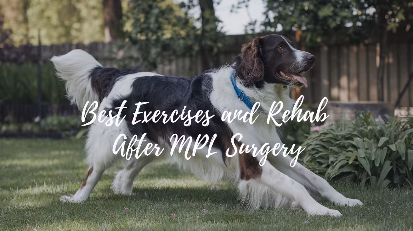
Best Exercises and Rehab for Dogs After MPL Surgery
Discover the best rehab and exercises for dogs after MPL surgery—step-by-step recovery timeline, home tips, and safe strengthening routines
Why Rehab Is Important After MPL Surgery
Rehabilitation is a key part of recovery and helps your dog heal safely and fully after MPL surgery.
After surgery, your dog’s muscles may weaken from rest, and the knee joint can become stiff. Rehab exercises help rebuild strength and improve how the leg moves. Without movement, swelling and tightness can slow healing and cause discomfort.
Proper rehab also lowers the risk of future problems, such as the kneecap slipping again or the other knee becoming injured. By keeping the joint strong and balanced, your dog is less likely to need more surgery later.
Key benefits of post-surgical rehab include:
- Helps regain muscle strength and joint mobility
- Reduces stiffness and swelling in the knee
- Prevents re-injury or surgical complications
- Supports a faster, safer return to normal activity
Rehab should start slowly and be guided by your vet or a certified canine physiotherapist to match your dog’s healing stage.
Recovery Timeline After MPL Surgery (By Weeks)
Recovery after MPL surgery usually takes 8–12 weeks or more and follows a step-by-step timeline based on healing progress.
Weeks 1–2 focus on rest and pain control. Your dog should have limited movement during this time. Short sling-supported potty walks and gentle range-of-motion exercises help prevent stiffness.
- Cold therapy to reduce swelling
- Pain medications as prescribed
- Passive range-of-motion (PROM) under vet guidance
- Short sling walks for potty breaks
- Strict rest and crate time when unsupervised
Weeks 3–6 introduce basic strength-building.
- Controlled leash walks (5–10 minutes, 2–3 times a day)
- Sit-to-stand exercises for muscle tone
- Gentle massage to improve circulation
- Hill walking (short and gradual)
Weeks 6–8 add more challenging movements.
- Longer leash walks
- Figure-eight drills for joint control
- Stair walking (only if cleared by your vet)
Weeks 8–12+ focus on returning to normal activities.
- Off-leash movement in controlled areas
- Slow trotting on soft ground
- Hydrotherapy like underwater treadmill or swimming
Every dog heals at a different pace. Always follow your vet’s guidance before moving to the next phase.
Read more | Unilateral vs. Bilateral Medial Patellar Luxation in Dogs
Best Exercises for Dogs After MPL Surgery
After MPL surgery, exercise should be safe, slow, and designed to rebuild strength without stressing the joint. Each movement should match your dog’s healing stage and be guided by your vet or rehab therapist.
Start with simple exercises like sit-to-stand (also called puppy squats) to build back leg muscles. Leash walks should be short and done on flat ground at a steady pace. As your dog progresses, you can add more advanced exercises to improve control and balance.
Effective rehab exercises include:
- Sit-to-stand: Strengthens thigh and hip muscles
- Short, controlled leash walks: 5–10 minutes, 2–3 times daily
- Figure-eight drills: Improves coordination and balance
- Hill or ramp walking: Builds rear-end strength
- Cavaletti poles: Encourages high stepping and joint control
- Stair walking: Only after vet approval, helps joint use
- Wobble board routines: Builds stability and balance
- Weight-shifting: Helps dogs learn to use the surgical leg again
Avoid rushing into too much exercise. If limping increases or your dog seems sore afterward, reduce the activity and contact your vet. Slow and steady movement is best for healing.
Read more | Grade 2 Medial Patellar Luxation: Surgery Decision Guide
Hydrotherapy and Water Exercises
Hydrotherapy is one of the best low-impact ways to help dogs recover after MPL surgery. Water supports the body and reduces joint stress while still allowing muscles to work.
Swimming is a great starting point once your vet approves. It builds strength evenly and improves joint range without the pressure of land exercise. Always supervise and use a dog life vest if needed.
Another excellent option is the underwater treadmill, where your dog walks in water at a slow, steady pace. It provides resistance to strengthen muscles and supports the joint for safer movement.
Water-based rehab includes:
- Swimming: Improves endurance and flexibility
- Underwater treadmill: Controls speed, resistance, and water level
- Warm water sessions: Loosens tight tissues and reduces pain
- Start 3–4 weeks after surgery (with vet clearance)
- Frequency: 1–2 times per week depending on progress
Hydrotherapy should always be done at a certified facility with trained staff. It’s especially helpful for large dogs, overweight pets, or those struggling with land exercises.
Read more | How to Prevent Medial Patellar Luxation in Puppies
Pain and Inflammation Management During Rehab
Keeping pain and swelling under control is critical for a successful recovery. A dog in pain won’t move normally, which slows healing and may cause stiffness or more joint damage.
Veterinarians often prescribe NSAIDs (like Carprofen or Galliprant) to manage post-op inflammation. These medications should always be given as directed and never mixed with human pain relievers.
In the first 1–2 weeks, cold packs help reduce swelling and numb the area. Later, once inflammation goes down, heat therapy can be used to loosen muscles before exercise.
Pain relief and comfort tools include:
- NSAIDs or vet-prescribed meds for daily pain control
- Cold therapy: 10–15 minutes, 2–3 times a day early on
- Heat therapy: After 2 weeks, before stretching or walking
- Massage: Eases soreness and helps circulation
- PROM exercises: Gently move the joint without weight
Never force motion or skip pain meds. If your dog is limping more, stops eating, or acts withdrawn, contact your vet. Comfort is key to keeping rehab on track.
Read more | Medial Patellar Luxation in Small vs. Large Dogs
Home Setup Tips for Safe Recovery
A safe home environment helps prevent injury and supports healing after MPL surgery. Your dog’s space should reduce slipping, prevent jumping, and make walking easy during the recovery period.
Start by using rugs or foam mats on slippery floors to give better traction. Block off stairs and furniture with baby gates or barriers to avoid unwanted movement. When walking, a towel sling under the belly can help support your dog, especially in the first two weeks.
Helpful home tips:
- Place non-slip rugs on all walking areas
- Block stairs or furniture access with gates
- Use a towel or sling during short walks for support
- Lower food portions by 25–33% to prevent weight gain
- Provide a crate or quiet space for rest when unsupervised
If your dog is very active, crate rest might be needed during early recovery to prevent injury. Keep toys and play areas calm and simple. The more you manage the environment, the easier recovery becomes for both you and your dog.
Read more | Medial Patellar Luxation Treatment Without Surgery
Mistakes to Avoid During MPL Recovery
Even with a good plan, recovery can be delayed by a few common mistakes. Avoiding these will help your dog heal faster and reduce the risk of complications.
One of the biggest mistakes is starting rehab too early. While movement is important, doing too much too soon can lead to swelling, pain, or re-injury. Skipping recheck appointments also puts your dog at risk since you may miss signs of poor healing.
Mistakes to avoid include:
- Starting rehab before your vet says it’s safe
- Skipping follow-up vet visits
- Allowing your dog to jump on furniture or run freely
- Letting them use stairs too early
- Ignoring new limping or signs of pain
Each stage of recovery has a specific goal. Trying to rush progress only increases the risk of setbacks. Stay consistent with rehab, monitor your dog’s behavior, and keep communication open with your vet. Slow progress is still progress.
Read more | How to Tell If Your Dog Has a Medially Luxating Patella
When to See the Vet Again
Follow-up visits are a critical part of MPL surgery recovery. Your vet checks how the joint is healing, adjusts your rehab plan, and helps prevent long-term issues.
Most dogs have their first recheck around 6 to 8 weeks after surgery. At this visit, your vet may perform a physical exam and possibly take follow-up X-rays to check bone healing or implant position. If needed, your vet may also adjust pain meds or rehab exercises.
Reasons to schedule follow-ups:
- Routine recheck at 6–8 weeks
- Evaluation of healing progress and joint function
- New signs of limping or swelling
- Concerns about infection, discharge, or fever
- Sudden changes in walking pattern or behavior
Don’t wait until something goes wrong. Even if things seem fine, scheduled check-ins help ensure your dog stays on track. If you notice any setback—big or small—call your vet for guidance.
Read more | Recovery After Medial Patellar Luxation Surgery in Dogs
Adjusting Rehab by Dog Type and Condition
Not all dogs recover the same after MPL surgery. Breed, age, body weight, and any other health conditions all affect how rehab should be planned.
Small dogs often heal faster and can do simple exercises at home. Large dogs may need professional therapy, especially to manage weight and avoid overloading the knee. Senior or overweight dogs usually need more time and support.
Rehab adjustments may include:
- Slower pace for large, heavy breeds
- Shorter sessions for older dogs with less endurance
- Extra care for dogs with previous ACL (CrCL) injuries
- Breeds like Bulldogs may need gentler activities
- Smaller dogs can often do balance work earlier
Your vet or rehab therapist will consider these factors when building a plan. What works for one dog may not work for another, even with the same surgery. Customized care ensures your dog’s recovery is safe and effective.
Sample Daily Rehab Routine (Week 3–6 Example)
A structured daily routine helps build strength without overloading the healing knee. This sample plan works well for many dogs in the week 3 to 6 recovery stage.
Morning Routine:
- 5–10 minutes of PROM (passive range of motion) exercises
- Short leash walk on flat ground (5 minutes)
Midday Routine:
- Gentle massage or cold compress if swelling is present
- Quiet rest in a crate or bed with limited movement
Evening Routine:
- Sit-to-stand exercises (5–10 reps)
- Short incline walk (small hill or ramp)
Always include rest periods between sessions to avoid fatigue. Limit total daily exercise time and adjust based on how your dog feels. If you notice limping after an activity, reduce intensity and ask your vet for guidance.
Tracking your dog’s progress each day can also help you spot improvements or problems early.
Read more | Signs and Symptoms of Medial Patellar Luxation in Dogs
Final Thoughts on Safe Rehab After MPL Surgery
Rehab is just as important as the surgery itself when it comes to helping your dog recover fully. Success depends on consistency, patience, and careful planning.
Stick to the timeline, don’t rush progress, and always monitor your dog’s comfort. Follow-up appointments, rest days, and gradual activity are all part of a complete recovery.
Keep in mind:
- Consistency is key—do a little each day
- Avoid pushing too hard, too fast
- Watch for signs of pain or limping
- Work with your vet or rehab therapist throughout
Every dog heals at a different pace. With the right support and a structured rehab plan, your dog can return to a happy, active life after MPL surgery.
FAQs
When can my dog start walking after MPL surgery?
Most dogs can begin short, controlled leash walks within 7–10 days after surgery, but only with vet approval. These walks are usually just for potty breaks at first. Gradual increase in walking time and difficulty should follow a structured rehab plan to avoid stressing the joint too soon.
How long does rehab take after MPL surgery?
Rehab usually lasts 8 to 12 weeks, depending on your dog’s size, age, and surgical grade. Some dogs need longer, especially large breeds or those with other joint issues. Progress is gradual, moving from rest to full activity in phases. Your vet will guide each stage to ensure safe, steady healing.
Are home exercises safe or should I visit a rehab center?
Home exercises can be very effective when done correctly and under veterinary guidance. Sit-to-stands, leash walks, and massage are helpful at home. However, rehab centers offer tools like underwater treadmills and trained therapists. For large dogs or complex cases, combining both home care and professional rehab works best.
Can dogs climb stairs again after recovery?
Yes, most dogs can use stairs again after full recovery, usually by week 8 to 12, if cleared by the vet. Stair climbing helps rebuild strength, but should be introduced gradually. Always start with supervision, and use gates early in recovery to prevent accidental stair use.
Is swimming safe after MPL surgery?
Yes, swimming is safe and highly recommended after MPL surgery—but only with vet approval. It usually starts around week 4 to 6. Swimming builds strength without stressing joints. Start slow in calm water, always supervised, and avoid slippery entry points. Never force your dog to swim if they’re uncomfortable.
Success Rates for Medial Patellar Luxation Surgery
Explore success rates for medial patellar luxation surgery by grade, including complications, recovery outcomes, and key factors that affect surgical success
What Is Medial Patellar Luxation (MPL)?
Medial Patellar Luxation (MPL) is a knee condition in dogs where the kneecap (patella) slips out of its normal groove on the femur. Instead of staying in place, it moves toward the inside (medial side) of the leg. This can cause pain, limping, or long-term joint damage if not treated.
Grades of MPL
- Grade I: Patella slips out but returns on its own
- Grade II: Patella comes out easily and may stay out sometimes
- Grade III: Patella is out most of the time but can be pushed back in
- Grade IV: Patella is always out and cannot be returned manually
Common Breeds and Risk Factors
- Small breeds like Pomeranians, Chihuahuas, Poodles, and Yorkies
- Genetic traits, shallow femoral grooves, or injury increase the risk
What Does MPL Surgery Involve?
Medial Patellar Luxation (MPL) surgery is done to correct the position of a dog's kneecap. When the patella keeps slipping out of place, it causes pain and changes how your dog walks. Surgery helps keep the kneecap stable and prevents long-term damage to the joint.
Goals of the Procedure
- Keep the patella inside its groove during movement
- Restore normal leg alignment and function
- Reduce pain, limping, and joint wear
- Prevent arthritis and other joint issues in the future
Common Surgical Techniques
Veterinary surgeons choose one or more techniques based on the dog’s grade of MPL and joint shape:
- Trochleoplasty: Deepens the femoral groove so the kneecap stays in place
- Tibial Tuberosity Transposition (TTT): Realigns the patellar tendon by moving a small bone piece
- Lateral Imbrication: Tightens tissue on the outer side of the joint to support the kneecap
- Medial Release: Loosens tight inner tissues that pull the patella out of place
Recovery Timeline in Brief
- Dogs can bear weight in a few days
- Activity must be limited for 6–8 weeks
- Full healing takes about 12–16 weeks, including rehab and follow-ups
Read more:
Success Rates by MPL Grade
Grade I–II: Highest Success (Up to 100%)
Surgery for Grade I and II MPL cases has the best results. These are mild forms where the kneecap may slip occasionally (Grade I) or more often but still return on its own or with help (Grade II). In both cases, joint anatomy is mostly normal.
- Success rate: Up to 100%
- Common procedures: Soft tissue tightening, medial release, trochleoplasty
- Complication rate: Very low
- Recovery time: 8–12 weeks for full return to normal function
Most dogs regain full use of the leg after healing. They walk and run normally without pain. Surgery at this stage also helps prevent arthritis or worsening joint issues in the future. In many Grade I cases, surgery isn’t even needed unless symptoms worsen.
Follow-up care includes rest, anti-inflammatory meds, and light rehab. These dogs typically do not need revision surgery, and the long-term outcome is excellent. Early treatment means faster recovery and fewer risks overall.
Grade III: High Success (90–100%) but Higher Recurrence
Grade III MPL is more advanced. The patella stays out of place most of the time but can be manually repositioned. Dogs often show more lameness, and their joint structure may need stronger correction.
- Success rate: Around 90–100%
- Common procedures: Trochleoplasty, TTT, lateral imbrication, medial release
- Complication risk: Moderate, especially reluxation
- Recovery time: 10–14 weeks, including rehab and checkups
Surgery often involves both bone and soft tissue work. In some dogs, the joint may still loosen over time, leading to partial luxation again. This is more likely if recovery is rushed or if the dog is very active during healing.
While most dogs return to near-normal movement, some may show minor stiffness or a mild limp, especially during exercise. A small number of dogs may need revision surgery later in life.
With proper surgical planning, weight control, and good rehab, most Grade III cases have strong outcomes and can return to a happy, active lifestyle.
Read more:
Grade IV: Variable Success (64–93%)
Grade IV MPL is the most severe form. The kneecap is always out of place and cannot be pushed back manually. Dogs often have serious skeletal changes, like rotated tibias or shallow grooves, which require more complex surgery.
- Success rate: Ranges from 64% to 93%
- Common procedures: TTT, deep trochleoplasty, rotational correction, staged surgeries
- Complication risk: High, especially reluxation or implant failure
- Recovery time: 12–16 weeks or longer, depending on case complexity
These surgeries are more challenging and may involve multiple steps. In some cases, staged correction is needed to slowly fix the bone alignment over time. Healing is slower, and strict rest is very important to avoid complications.
Some dogs improve but may not regain full, normal motion. A mild limp or stiffness can remain, especially in older dogs or those with long-standing luxation. However, pain relief and improved comfort are still strong outcomes.
With skilled surgery, proper follow-up, and careful rehab, many Grade IV dogs live more comfortable, mobile lives—even if their function isn't perfect.
Complication and Recurrence Rates
While most dogs recover well after Medial Patellar Luxation (MPL) surgery, there are some risks to be aware of. The chance of complications or the kneecap slipping again depends on the grade of MPL, the dog’s age, the surgeon’s technique, and how well post-surgery care is followed.
Average Rates
- Recurrence (reluxation) rate: 10–21%
- Major complication rate: 16–24%
These numbers are higher in dogs with Grade III and IV luxation, where the joint is more unstable or the bone structure is more deformed.
Common Complications
- Swelling or seroma: Fluid may collect near the incision site
- Implant issues: Pins or wires used in surgery may move or loosen
- Infection: Can happen at the wound site if not kept clean
- Joint stiffness: Especially if rest is not followed properly
- Recurrence: The patella may slip out again if healing is poor
Rare but Serious Outcomes
- Nerve damage or long-term lameness
- Poor bone healing in very active or older dogs
- Amputation (very rare): Only needed if the leg becomes non-functional due to severe infection or failure
Proper rest, weight control, and follow-up care reduce most risks.
Read more:
Factors That Affect Surgical Success
Not all MPL surgeries have the same outcome. Several key factors can affect how well a dog recovers and how stable the knee stays after surgery.
Key Factors
- Age of the dog: Younger dogs tend to heal faster and adapt better. Older dogs may have slower recovery and higher arthritis risk.
- Weight/body condition: Overweight dogs have more pressure on their joints, which increases the risk of complications and slower healing.
- Other joint issues: Dogs with a torn cranial cruciate ligament (CCL) or hip problems may need more complex surgery.
- Breed-related anatomy: Some breeds have shallow grooves or bone rotation, making surgery more difficult.
- Unilateral vs bilateral surgery: Dogs with both knees affected (bilateral MPL) may take longer to recover, especially if both surgeries are done together.
Each of these factors plays a role in the dog’s outcome. A well-planned surgery combined with personalized care improves the success rate, even in more complex cases. Pre-surgical exams and imaging help identify these factors early so the treatment can be tailored to each dog’s needs.
Long-Term Outcomes and Owner Satisfaction
Most dogs that undergo MPL surgery enjoy long-lasting relief from pain and improved movement. Long-term success depends on the surgery quality and how well the dog is cared for afterward.
What Owners Can Expect Long-Term
- Pain control: Most dogs are more comfortable and pain-free after healing. Long-term pain is rare if the joint is stable.
- Activity levels: Many dogs return to normal activity—walking, running, and playing—within 3–4 months.
- Satisfaction rates: Over 90% of owners report high satisfaction with the surgery, especially in Grades I–III.
Some dogs may still show mild stiffness or avoid certain movements, especially if they had a high-grade luxation or other joint conditions. Regular exercise, weight control, and avoiding intense jumping or rough play help preserve joint health over time.
Overall, MPL surgery has a strong track record of improving quality of life. Dogs move more freely, show less pain, and become more active after recovery. When paired with proper follow-up care, most dogs remain stable for many years after surgery.
Read more | Signs and Symptoms of Medial Patellar Luxation in Dogs
How to Maximize the Chances of a Successful Outcome
Good surgical technique is just one part of MPL recovery. What happens after surgery is just as important for a full and lasting recovery.
Key Steps for Better Results
- Post-op care: Strict crate rest for 6–8 weeks is critical. It prevents implant movement and allows the joint to heal properly.
- Rehabilitation: Gentle rehab like controlled leash walks, underwater treadmill, or physiotherapy helps regain strength and range of motion.
- Follow-ups: Regular vet checkups ensure healing is on track. X-rays may be needed to confirm bone healing and implant position.
Owners play a big role in the recovery process. Keeping the dog calm, using medications as prescribed, and following rehab steps all support better outcomes. It’s also important to avoid running, jumping, or stairs until your vet gives the all-clear.
Feeding a balanced diet and keeping your dog at a healthy weight also reduces joint stress. With the right support at home and guidance from your vet, your dog has the best chance to heal fully and return to an active, pain-free life.
Conclusion
Medial Patellar Luxation (MPL) surgery is a highly effective treatment for dogs with kneecap instability. With proper surgical planning and careful post-op care, most dogs return to normal or near-normal function. Success rates are especially high in Grade I–III cases, and even dogs with Grade IV can show major improvement.
While complications are possible, they are often manageable with early attention. Owners who follow vet instructions—especially crate rest and rehab—help their dogs heal faster and avoid reluxation. With patience and proper care, most dogs enjoy a more active, pain-free life after surgery.
FAQs
What is the average success rate for MPL surgery in dogs?
The success rate for MPL surgery is high. For Grade I–III cases, it ranges from 90% to 100%. Grade IV cases have a slightly lower success rate, between 64% and 93%, due to complex joint changes. Most dogs regain good function and live comfortably with proper care and follow-up.
Does the grade of MPL affect the surgery results?
Yes, the grade of MPL directly affects the surgery outcome. Lower grades (I–II) have simpler issues and near-perfect success. Grade III surgeries are more complex but still have high success. Grade IV often needs advanced correction and has a higher risk of complications, though many dogs still improve significantly.
What are the chances of MPL coming back after surgery?
The chance of the kneecap slipping again, called reluxation, is around 10–21%. This depends on the grade of MPL, the dog’s activity level, and how closely post-op care is followed. Proper rest, weight control, and vet follow-ups can lower this risk and help keep the knee stable.
How long does it take for a dog to recover from MPL surgery?
Most dogs begin walking within a few days but need 6–8 weeks of crate rest. Full recovery can take 3–4 months, especially for higher-grade surgeries. During this time, rehab and regular checkups help rebuild strength and ensure healing is on track.
Can my dog walk normally after MPL surgery?
Yes, most dogs can walk and even run normally after MPL surgery. Once healing is complete, they often return to normal activity levels. Minor stiffness or a limp may remain in rare cases, especially in Grade IV or older dogs, but pain and lameness usually improve.
Is MPL surgery safe for older or overweight dogs?
MPL surgery can be done safely in older or overweight dogs, but the risks are slightly higher. These dogs may heal slower or face more stress on the joint. With proper planning, pain management, and weight control, they can still have excellent results and better comfort long-term.
Medial Patellar Luxation Treatment Without Surgery
Explore safe, effective ways to treat medial patellar luxation in dogs without surgery—home care, rehab, supplements, braces, and more
Can You Treat Medial Patellar Luxation Without Surgery?
Some dogs with MPL can be treated without surgery, but it depends on how serious the condition is.
Medial Patellar Luxation (MPL) means the kneecap (patella) slides out of its normal place inside the groove of the thigh bone. This makes the leg feel unstable and can cause pain or limping.
MPL is divided into four grades:
- Grade I: The kneecap moves out sometimes but goes back easily. Usually no pain.
- Grade II: The kneecap pops out more often and may cause mild limping.
- Grade III: The kneecap stays out most of the time but can be moved back with the hand.
- Grade IV: The kneecap is always out and can’t be moved back in.
Dogs with Grade I and some Grade II luxations can often be treated without surgery using physical therapy, weight control, and joint supplements. These help build muscle and reduce pressure on the knee.
However, Grade III and IV usually need surgery. Without it, the joint may become more damaged, and the dog may have long-term pain or difficulty walking.
Always work with your vet to decide what’s best for your dog’s specific case.
Signs Your Dog Might Benefit From Non-Surgical Treatment
Not all dogs with a loose kneecap need surgery. Some signs show your dog may do well with conservative care instead.
Dogs with mild Medial Patellar Luxation often have symptoms that come and go. One common sign is an occasional limp or a brief “skipping” step during walks. This happens when the kneecap pops out and then goes back in place. These dogs usually return to normal quickly after a few steps.
You may also hear a soft popping sound or notice the knee shift when your dog moves. If your dog rests and the signs improve, that’s another clue that non-surgical care might help.
Some symptoms that respond to:
- Rest or limited activity
- Joint supplements like glucosamine
- Gentle physical therapy
- Maintaining a healthy weight
But if the limping gets more frequent, or if your dog avoids putting weight on the leg, the condition could be getting worse. In that case, see a vet right away. Early help gives the best chance to avoid long-term joint problems.
Read more | Signs and Symptoms of Medial Patellar Luxation in Dogs
Vet Diagnosis Is Still a Must (Even for Non-Surgical Plans)
Even if your dog has mild symptoms, a vet should always confirm the diagnosis before you start treatment.
Vets use a hands-on exam to check how easily the kneecap moves out of place and which grade the MPL is. Grades I and II might not need surgery, but you need to know the grade first.
Your vet may also take X-rays to check for other problems in the knee or hip. Sometimes a dog has both MPL and hip issues, which changes the treatment plan. Without this full checkup, you might miss other problems.
Skipping the vet visit can lead to:
- Wrong treatment choices
- Delays in healing
- Worsening joint damage
- Unnecessary pain for your dog
Even if you plan to use rest, supplements, or therapy, it’s important to be sure you’re treating the right thing. A proper diagnosis helps you avoid wasting time and keeps your dog safe and comfortable. Always make the first step a vet exam.
Read more | Recovery After Medial Patellar Luxation Surgery in Dogs
Best Non-Surgical Treatments for MPL in Dogs
If your dog has a mild or moderate case of MPL, these non-surgical treatments can help manage symptoms and slow progression.
1. Weight Management to Reduce Knee Stress
Even a few extra pounds can put added pressure on your dog’s knees, especially with an unstable kneecap. Keeping your dog at a healthy weight is one of the most important steps in managing MPL.
Extra weight increases the strain on the joint and can make the patella slip out more often. Weight loss reduces inflammation and improves your dog’s ability to move comfortably.
To manage weight:
- Feed based on your vet’s calorie recommendation
- Use a body condition score chart to check for ideal shape
- Avoid table scraps and high-calorie treats
Ask your vet about the target weight for your dog’s breed and size. Track it every 2–4 weeks to stay on course. A lighter frame means less knee strain and fewer symptoms.
2. Joint Supplements for Lubrication and Repair
Joint supplements help cushion and support your dog’s knee over time. They don’t fix the luxation but can reduce pain and inflammation.
Look for products that contain:
- Glucosamine and chondroitin to support cartilage
- MSM for anti-inflammatory support
- Omega-3 fatty acids for joint lubrication
These ingredients work slowly, so it may take 4–8 weeks to see visible improvement. You might notice your dog limping less or moving more easily.
Always choose veterinary-grade supplements and follow the correct dosage for your dog’s weight. Giving more than recommended won’t help faster and can upset the stomach. If you’re unsure, ask your vet to guide you toward trusted brands.
Supplements are most effective when paired with weight control and light exercise.
Read more | How to Tell If Your Dog Has a Medially Luxating Patella
3. Controlled Exercise to Build Stability
Exercise is helpful, but it must be controlled to avoid stress on the joint. The goal is to strengthen muscles around the knee without making the luxation worse.
Focus on:
- Short, slow leash walks
- Avoiding fast turns or running
- Flat, even surfaces only
Avoid activities like:
- Stairs
- Rough play or fetch
- Jumping on furniture
Even five to ten minutes of calm walking twice a day can help. Over time, it builds leg strength and supports the kneecap’s position. Always monitor your dog after exercise for signs of soreness or increased limping. If symptoms get worse, reduce the activity and speak to your vet.
4. Strengthening Exercises You Can Try at Home
Home exercises help build muscle around the knee, improving joint stability. These are gentle and easy to do with dogs that have mild MPL.
Try these options:
- Sit-to-stand: Ask your dog to sit and then stand 5–10 times slowly
- Cavaletti poles: Use broomsticks laid low to encourage step-over motion
- Ramp or hill walking: Use a slight incline to build thigh strength
- Army crawls: Gently encourage your dog to crawl short distances (only if no pain)
Do these exercises on soft ground or carpet. Keep sessions short—5 to 10 minutes daily—and always watch your dog’s reaction. If your dog seems tired, sore, or limping more, stop the activity and check with your vet. Never force any motion.
5. Physiotherapy and Professional Rehab
Working with a certified canine rehab vet can make a big difference. Physiotherapy targets specific muscles and movements that support the knee joint.
Your rehab vet might recommend:
- Range-of-motion stretches to keep the joint flexible
- Balance exercises using wobble boards or cushions
- Laser or cold therapy to reduce pain
Sessions are usually once or twice a week at first, depending on your dog’s condition. At-home plans may be included for daily practice.
The goal is to improve strength, reduce discomfort, and keep the luxation from worsening. Ask your vet to refer you to a certified canine rehab therapist in your area.
Read more | Best Exercises and Rehab for Dogs After MPL Surgery
6. Hydrotherapy for Low-Impact Strength Building
Hydrotherapy is one of the best ways to build strength without stressing the knee. It uses water to support your dog’s body while still allowing muscle movement.
Two main methods are:
- Underwater treadmill: Your dog walks on a belt inside a water tank
- Swimming in a rehab pool: Guided by a therapist, if safe for your dog
Water reduces joint pressure by supporting weight and allowing a fuller range of motion. At the same time, resistance helps improve strength.
Hydrotherapy is ideal for dogs that can’t tolerate land exercises or have arthritis along with MPL. Most dogs enjoy the sessions and improve steadily over a few weeks. Your vet or rehab therapist will tell you how often to go—usually once or twice per week at first.
7. Pain Relief with Vet-Approved Medications
Even with mild MPL, some dogs have joint pain. Medications help manage this discomfort so your dog can stay active and heal better.
Common vet-approved options include:
- NSAIDs like Galliprant, Carprofen, or Meloxicam
- Gabapentin for nerve-related pain
- Joint-protecting injections (like Adequan)
These meds must always be prescribed by your vet, and the dose must match your dog’s weight and health condition. Never give human painkillers—many are toxic to dogs.
Pain control makes it easier for your dog to walk, rest, and take part in therapy. It also prevents muscle loss due to reduced activity.
Always watch for side effects like stomach upset or changes in appetite and report them to your vet.
Read more | Medial Patellar Luxation in Small vs. Large Dogs
8. Physical Therapy Extras That Help
Besides basic rehab, some alternative therapies can help reduce pain and improve movement. They’re not required but can support the main treatment plan.
Helpful options include:
- Massage therapy to relax tight muscles and improve circulation
- Chiropractic care, but only by trained, certified animal chiropractors
- Acupuncture or cold laser therapy for joint pain and inflammation
These options should always be done by professionals trained in canine care. They can be used alone or along with other treatments like hydrotherapy or medication.
Some dogs respond well to one method, while others need a mix. Discuss with your vet or rehab team to find what fits your dog best.
9. Using Dog Knee Braces for Extra Support
Knee braces can help dogs with Grade I or II MPL by giving extra support during movement. They don't fix the kneecap but can stop it from slipping as often.
You might consider a brace if:
- Your dog limps during walks
- You want to avoid surgery
- You’re starting physical therapy
Look for braces that offer side support and adjustable straps. Brands like Walkin’ Pets and Ortocanis make popular models. Always measure your dog’s leg carefully for a good fit.
Some dogs take time to adjust to wearing a brace. Start with short sessions indoors. Reward calm behavior and watch for rubbing or skin problems.
Use braces during walks or active periods, not all day. Ask your vet or therapist to show you how to put it on safely and check for signs of irritation.
Home Modifications That Support Recovery
Making small changes in your home can help your dog heal more comfortably and safely from MPL without surgery.
Dogs with unstable kneecaps need secure footing. Slippery floors like tile or wood can cause the knee to slide out of place. To fix this, place non-slip rugs or yoga mats in areas where your dog walks often, especially around food bowls and beds.
You should also use pet stairs or ramps to help your dog get on furniture or in and out of the car. Jumping up or down puts pressure on the knee and can worsen the condition.
Helpful home changes include:
- Block off stairs or high beds
- Avoid letting your dog jump on sofas or beds
- Use baby gates to limit movement during recovery
- Keep your dog’s essentials on one floor
These simple changes reduce strain on the joint and make your dog feel more secure. They also support the effects of weight control and therapy. Always watch how your dog moves through the house and adjust the environment as needed to prevent slips or overuse.
Read more | How to Prevent Medial Patellar Luxation in Puppies
What a Weekly Routine Might Look Like
Creating a weekly routine helps manage MPL without surgery by balancing exercise, rest, and joint support. A structured plan keeps your dog active without overloading the knee.
Here’s what a sample day might look like:
- Morning: Short 5-minute leash walk, joint supplement with breakfast
- Midday: Sit-to-stand exercise or gentle hill walking
- Evening: Massage or range-of-motion stretches, then rest
You can adjust based on your dog’s energy level and vet recommendations. For example, if using hydrotherapy or rehab, schedule it 1–2 times per week. Keep the rest of the week low-impact to avoid fatigue.
Track progress with:
- A journal of limp-free days
- Changes in mood or energy
- Photos or videos of movement
Avoid doing the same routine every single day. Dogs also need rest days with minimal activity to allow muscle recovery. Balance is key.
Your vet or canine rehab therapist can help tailor the plan further. Sticking to a schedule makes it easier to spot progress or setbacks quickly.
What to Avoid When Managing MPL Without Surgery
While there are many ways to help your dog without surgery, some mistakes can slow recovery or even make things worse.
One common issue is skipping rest days. Muscles need time to recover, especially after exercise or therapy. Too much activity without breaks can lead to swelling or pain.
Also avoid:
- Overexercising too soon – Build strength slowly to avoid injury
- Letting your dog run, jump, or climb stairs without guidance
- Using only joint supplements without a complete care plan
- Delaying vet checkups when symptoms change
Some dogs act fine even when their knee is unstable. But ignoring small changes—like more frequent limping or reduced play—can lead to worse problems later.
You don’t need to overprotect your dog, but structure and balance matter. Always adjust based on how your dog responds, and check with your vet if anything seems off.
Small daily mistakes can undo weeks of good care, so stay consistent and alert.
Read more | Grade 2 Medial Patellar Luxation: Surgery Decision Guide
How Long Does Recovery Take Without Surgery?
Recovery from MPL without surgery takes time and patience. The timeline depends on the severity of the condition, your dog’s age, and how closely you follow the care plan.
For dogs with Grade I or mild Grade II, visible improvement can start in 4 to 6 weeks. Full recovery, where symptoms are rare or gone, may take 8 to 12 weeks or longer. Some dogs need ongoing support through supplements and light exercise for life.
Signs that your dog is improving include:
- Less frequent limping or skipping steps
- Better muscle tone in the hind legs
- Increased comfort during walks
However, if the condition worsens—more frequent knee popping, longer limping episodes, or pain while resting—it’s time to re-evaluate. A follow-up vet visit can help adjust the plan or decide if surgery is now the better option.
MPL often stays manageable with consistent care, but don’t expect overnight results. Keep a record of changes to help your vet guide next steps. Recovery is a journey, and small improvements matter.
When to Reconsider Surgery
Even with the best home care, some dogs may not improve enough and will need surgery. Knowing when to shift gears can protect your dog’s comfort and long-term joint health.
You should talk to your vet about surgery if:
- Limping continues for more than 8–12 weeks
- Your dog has a Grade III or IV MPL that’s not responding
- Your dog avoids using the leg, even with treatment
Other signs to watch for include worsening pain, trouble rising, or changes in behavior like less interest in walks. These may mean that conservative treatments are no longer enough.
Quality of life is the key factor. If your dog can’t enjoy normal daily activities without pain, surgery becomes the better path.
While we all want to avoid invasive procedures, surgery often gives long-term stability and comfort for higher-grade cases. Your vet will help you weigh the risks and benefits.
It’s okay to start with non-surgical care, but be open to changing the plan if your dog isn’t getting better.
Read more | Unilateral vs. Bilateral Medial Patellar Luxation in Dogs
Final Thoughts on Avoiding Surgery for MPL
Treating Medial Patellar Luxation without surgery is possible—especially in mild cases—and often helps dogs live more comfortably when done with care.
Key strategies for success include:
- Weight control
- Joint supplements
- Structured exercise and home safety changes
- Regular vet visits and tracking symptoms
The most important part is consistency. Daily effort with even small changes builds up over time. Dogs with mild MPL can stay pain-free for years if managed properly.
However, always stay in touch with your vet. They’ll guide you when to push forward and when it’s time to consider other options like surgery.
You know your dog best, and with a clear plan, steady care, and expert guidance, many dogs can avoid surgery and still enjoy a happy, active life.
FAQs
Is it safe to avoid surgery for patellar luxation?
Yes, it’s safe in mild cases (Grade I or some Grade II) if managed correctly. Weight control, joint support, and regular vet checkups can help reduce symptoms. But if your dog’s limping worsens or pain increases, surgery may become necessary. Always follow your vet’s advice to ensure the safest outcome for your dog.
How do I know if my dog’s MPL is getting worse?
Watch for more frequent limping, longer recovery after walks, or signs of pain while resting. If your dog avoids using the leg, hesitates to climb stairs, or shows stiffness after rest, these may be signs the MPL is progressing. A worsening condition needs a vet recheck to adjust the treatment plan or consider surgery.
What supplements are best for dogs with MPL?
Good joint supplements often include glucosamine, chondroitin, MSM, and omega-3 fatty acids. These support cartilage, reduce inflammation, and improve joint function over time. Choose veterinary-grade brands and use the correct dose based on your dog’s weight. Talk to your vet before starting supplements to make sure they’re right for your dog’s condition.
Can braces fix luxating patella permanently?
No, braces don’t permanently fix the condition. They provide temporary support by helping the kneecap stay in place during movement. Braces work best in mild cases or while doing rehab. They may reduce symptoms but don’t correct bone or joint shape. For permanent correction, surgery is needed in moderate to severe cases.
How long should I try non-surgical treatment before seeing results?
Most dogs show improvement in 4 to 6 weeks with consistent care. This includes exercise, joint support, and weight control. Full benefits may take 8 to 12 weeks. If there’s no progress or symptoms worsen during that time, see your vet. They’ll help decide if surgery or a new plan is needed.
Can Medial Patellar Luxation Be Prevented in Puppies?
Learn how to reduce the risk of medial patellar luxation in puppies with early care tips, safe exercise, diet, and vet-guided joint support
What Is Medial Patellar Luxation in Puppies?
Medial Patellar Luxation (MPL) is a common knee problem in puppies, where the kneecap doesn’t stay in the right place.
In a healthy knee, the patella (kneecap) sits in a groove at the end of the femur and slides smoothly as the leg moves. In puppies with MPL, the kneecap slips inward—toward the other leg—causing an unstable joint. This can lead to limping, skipping steps, or trouble walking as the puppy grows.
MPL can be congenital, meaning the puppy is born with a shallow groove or weak ligaments that allow the kneecap to move out of place. It can also be acquired, which means it develops later due to injury, poor nutrition, or uneven bone growth during puppyhood.
Some breeds are more prone to MPL due to their body structure and genetics. These include:
- Yorkshire Terrier
- Pomeranian
- Chihuahua
- Poodle
- Boston Terrier
While not all puppies will show signs early, it’s important for owners of at-risk breeds to monitor for any leg issues and begin prevention early. Early care can reduce long-term problems.
Is Medial Patellar Luxation Preventable?
MPL is mostly genetic, but there are ways to reduce the risk and support healthy joint development in puppies.
Most cases of MPL are hereditary, especially in small breeds. Puppies can be born with shallow grooves in the knee or loose ligaments, which cause the kneecap to slip. While there's no guaranteed way to prevent it, smart early care can delay or reduce the severity of symptoms.
Ways to lower MPL risk include:
- Avoid breeding dogs with a known history of MPL
- Choose responsible breeders who screen for orthopedic issues
- Support joint health through proper weight, nutrition, and exercise
- Detect early signs like “skipping” steps or limping during puppy exams
Starting joint-friendly habits early helps protect the knees during growth. Supplements and low-impact activity don’t stop MPL from forming, but they can improve joint strength and comfort.
If your puppy is in a high-risk breed, talk to your vet about a prevention plan. While MPL may still develop, you can reduce pain, improve function, and possibly avoid surgery later in life with early care and close monitoring.
Read more | Unilateral vs. Bilateral Medial Patellar Luxation in Dogs
How to Identify Puppies at Higher Risk
Some puppies are born more likely to develop MPL based on their breed and family background. Spotting risk early can help you act before the condition worsens.
High-risk breeds often include small or toy dogs. Their bone structure and genetics make MPL more common.
Breeds at higher risk:
- Yorkshire Terrier
- Chihuahua
- Pomeranian
- French Bulldog
- Poodle
Family history also matters. If a parent or sibling had MPL, the puppy has a higher chance of getting it. Even if they look fine now, underlying joint issues could appear as they grow.
Signs of early MPL include:
- Occasional “skipping” steps
- Limping that comes and goes
- Back leg held up for a few strides
- Reluctance to play or jump
Ask your vet during puppy checkups:
- Are the kneecaps tracking normally?
- Is there any looseness or instability in the joints?
- Should we consider early joint support or supplements?
The earlier MPL is identified, the easier it is to manage. Don’t wait until limping becomes regular—track movement and behavior from the start.
Read more | Grade 2 Medial Patellar Luxation: Surgery Decision Guide
Joint Health Tips to Lower Risk in Puppies
Simple changes in daily care can help reduce joint stress as your puppy grows. These habits won’t prevent MPL completely but can lower the chances of joint damage or worsening symptoms.
Maintain a Healthy Weight During Growth
Keeping your puppy at a healthy weight is one of the best ways to protect their knees.
- Avoid overfeeding or too many treats
- Watch for rapid weight gain in the first year
- Monitor weight monthly, especially in small breeds
Extra weight adds stress to developing joints and increases the risk of instability.
Use Low-Impact Exercise to Build Muscle
Gentle movement builds strength without causing joint strain.
- Short daily leash walks on even surfaces
- No fetch, running, or jumping games
- Safe play on soft grass or padded floors
This keeps the leg muscles strong while protecting the kneecap.
Avoid High-Risk Movements and Environments
Jumping or slipping increases injury risk during growth.
- Don’t let puppies use stairs or jump from furniture
- Use ramps or steps for couches and beds
- Cover floors with non-slip rugs or foam mats
Start Joint-Friendly Supplements Early (If Vet Approves)
Talk to your vet before starting anything, but early support may help.
- Glucosamine and chondroitin
- Omega-3 fatty acids for joint lubrication
- Vitamin C and manganese-rich diets to support collagen
Each small step adds protection as the puppy grows.
Read more | Medial Patellar Luxation in Small vs. Large Dogs
Puppy Growth and Bone Development: What Matters Most
Puppies grow rapidly in their first year, and how their bones form during this stage can affect joint health for life. Balanced nutrition and proper rest play a huge role in preventing joint stress.
One key factor is calcium and phosphorus balance. Too much calcium can cause uneven bone growth or joint issues. That’s why you should always feed vet-recommended puppy food, not adult food or homemade diets without guidance.
Important tips for healthy bone development:
- Choose a complete and balanced puppy formula based on breed size
- Avoid adding extra calcium or supplements unless approved
- Don’t let your puppy over-exercise during growth spurts
- Provide crate or pen rest periods during the day to avoid fatigue
- Monitor height and weight with regular vet visits
For large or small breeds, each bone growth stage must be supported. Sudden changes in weight or intense exercise can affect the knee structure. Puppy bones are soft and flexible—good habits during this stage create stronger joints for the future.
Read more | Best Exercises and Rehab for Dogs After MPL Surgery
Sample Prevention Plan for Puppy Owners
Building joint-friendly habits into daily life can make a big difference, especially if your puppy is in a high-risk breed. A prevention plan doesn’t need to be complex—it just needs to be consistent.
Vet care routine:
- Full orthopedic exam at each puppy visit
- Ask about kneecap tracking and joint health
- Discuss early signs like skipping or leg favoring
Daily activity:
- Controlled leash walks on grass or carpet
- Avoid rough play with larger dogs
- Play with puzzle toys or soft tug games instead of fetch
Feeding tips:
- Use puppy food with proper calcium-phosphorus ratio
- Keep portions small and frequent
- Monitor body condition weekly (you should feel ribs but not see them)
Home setup:
- Place rugs on tile or wood floors
- Use ramps or steps for furniture
- Block off stairs with baby gates
Starting these practices early supports strong joint development and reduces the chance of painful knee problems later in life.
Read more | Medial Patellar Luxation Treatment Without Surgery
Myths About MPL Prevention You Should Know
Many well-meaning owners believe myths that could delay proper care for MPL. Knowing the facts helps you make better choices for your puppy’s joint health.
Myth 1: “Supplements will prevent MPL.”
Supplements can support joint health, but they don’t prevent misaligned bones or hereditary luxation.
Myth 2: “If there’s no limp, there’s no problem.”
Early MPL signs may be subtle or come and go. Skipping steps or brief lameness may be the first signs.
Myth 3: “Muscle strength alone can fix it.”
Strong muscles help stabilize the joint, but they can’t correct bone structure problems if the kneecap doesn’t track properly.
Myth 4: “All limping is due to injury.”
Puppies with recurring limping or skipping should always be checked by a vet, even if it seems mild.
Avoiding these myths helps you take early action when needed. Prevention isn’t about guessing—it’s about watching closely and supporting healthy growth.
Read more:
Final Thoughts on Preventing MPL in Puppies
You can’t completely prevent Medial Patellar Luxation, but you can lower the risk and reduce long-term joint problems by making smart choices early in your puppy’s life.
Dogs with strong muscles, healthy joints, and good weight control are less likely to develop painful symptoms, even if they carry a genetic risk.
Key takeaways:
- You can’t control genetics, but you can control environment and care
- Early joint support leads to better long-term outcomes
- Regular vet exams help spot MPL early
- Focus on safe play, joint-friendly diets, and avoiding high-impact activity
Prevention is about setting up your puppy for a healthy future. Every small step—from soft flooring to gentle exercise—adds up. With close vet guidance and a bit of planning, your puppy can grow into a strong, active dog with minimal joint issues.
FAQs
Can you stop MPL from developing in puppies?
You can’t fully stop MPL from developing, especially if it’s genetic. However, you can reduce the risk of symptoms by managing your puppy’s weight, providing joint-friendly exercise, and avoiding high-impact activity. Early vet care and a proper home environment also help minimize joint stress during growth.
What breeds are most prone to MPL in puppies?
Small and toy breeds are most commonly affected by MPL. This includes Yorkshire Terriers, Chihuahuas, Pomeranians, Toy Poodles, and French Bulldogs. These breeds often have shallow knee grooves and soft tissue looseness from birth, increasing the chance of the kneecap slipping out of place as they grow.
Do joint supplements work for prevention?
Joint supplements don’t prevent MPL, but they can support healthy cartilage and reduce inflammation. Ingredients like glucosamine, chondroitin, and omega-3s may help protect the joint as the puppy grows. Always ask your vet before starting supplements, especially in very young or small-breed puppies.
Is exercise safe for puppies with suspected MPL?
Yes, but only low-impact, vet-approved exercise is safe. Avoid running, fetch, or jumping. Instead, stick to short, slow leash walks on soft ground. If your puppy shows signs like skipping steps or limping, limit activity and consult your vet before continuing any movement routine.
When should a vet examine patella alignment in puppies?
A vet should check your puppy’s kneecaps during every routine exam, starting as early as 8 weeks. Early detection of loose or misaligned patellae helps guide care. If your puppy is in a high-risk breed or showing unusual movement, ask your vet to assess patellar tracking closely.
Bilateral MPL in Dogs: What You Need to Know
Learn what bilateral MPL in dogs means, how it’s diagnosed, treated, and what recovery looks like when both knees are affected by luxating patella
What Is a Luxating Patella in Dogs?
A luxating patella means the dog’s kneecap (patella) slips out of its normal groove in the thigh bone. Instead of gliding smoothly when the knee bends, the patella pops to the side. This causes pain, limping, or skipping during movement.
Medial patellar luxation (MPL) is the most common type, where the kneecap shifts inward toward the other leg. It’s more frequent than lateral luxation, which moves outward and is usually seen in larger breeds.
In bilateral MPL, both knees are affected. Dogs may show lameness in both hind limbs, bunny-hopping, or trouble rising and walking. Symptoms can appear on one side first but usually involve both knees over time. Bilateral cases can range from mild to severe and may require surgery to correct pain, improve limb use, and prevent long-term joint damage.
Why Do Some Dogs Get MPL in Both Knees?
Bilateral medial patellar luxation (MPL) often develops due to genetic and anatomical reasons. Some dogs are born with shallow grooves in their femur bones, which don’t hold the kneecap in place well. Over time, this leads to luxation—often in both legs.
Small breeds are more prone to bilateral MPL. Dogs like Pomeranians, Yorkshire Terriers, Chihuahuas, and Toy Poodles are commonly affected. These breeds tend to have inherited bone structures that don’t properly support the patella during movement.
Another major factor is limb alignment. If a dog’s legs are naturally bowed or rotated, it puts abnormal pressure on both knees. This uneven force allows the patella to move out of place in both limbs. Bone deformities in the femur or tibia, or tight soft tissues, also contribute.
Because these traits are often present in both hind limbs, it’s not unusual for both knees to develop MPL. In many cases, one side starts showing symptoms first, but the second knee usually follows. Early diagnosis helps prevent worsening on both sides.
Signs of Bilateral MPL You Should Watch For
When both knees are affected by MPL, the symptoms are often more noticeable and can affect your dog’s daily life. One of the most common signs is a skipping gait—your dog may hop with both back legs off the ground, especially during fast movement. This happens when both kneecaps slip out of place.
Other warning signs include:
- Limping or stiffness in both hind legs
- Bunny-hopping while walking or running
- Difficulty getting up from rest or lying down
- Reluctance to climb stairs or jump onto furniture
- Occasional yelps when moving awkwardly
Some dogs may try to avoid using their hind legs altogether or shift more weight to their front limbs. This can lead to muscle loss in the back legs and an arched posture over time.
Because bilateral MPL affects both knees, these signs can be harder to spot than a limp on one side. Owners might think their dog is just lazy or tired, especially in small breeds that don’t show pain clearly. But early signs like bunny-hopping or hesitation with movement usually indicate both knees are involved. Timely vet evaluation can confirm the condition and guide treatment before it worsens.
Read more:
- MPL Surgery Cost for Dogs
- Is Surgery Necessary for Medial Patellar Luxation in Dogs?
- Medial Patellar Luxation Grades Explained
- Success Rates of Medial Patellar Luxation Surgery
How Vets Grade Bilateral MPL
Veterinarians use a grading system from 1 to 4 to assess the severity of patellar luxation in each knee. In bilateral MPL, both knees are evaluated separately, but treatment planning considers the overall effect on the dog.
- Grade 1: The kneecap moves out of place with pressure but returns on its own. Dogs often show no signs.
- Grade 2: The patella slips out more easily and may stay luxated briefly. Dogs may limp or skip occasionally.
- Grade 3: The kneecap is out most of the time but can be pushed back manually. Dogs often have persistent limping.
- Grade 4: The patella is permanently luxated and cannot be moved back. This causes severe lameness and structural changes.
Each grade reflects how unstable the joint is. Dogs with different grades on each leg may use one leg more than the other, but bilateral issues still impact both limbs over time. Grading helps determine whether surgery is needed and which leg to operate on first. It also guides rehab and long-term care planning.
Read more:
How Bilateral MPL Is Diagnosed
Diagnosing bilateral medial patellar luxation (MPL) begins with a detailed physical exam. During the exam, the vet palpates both knees to feel how easily the kneecaps move out of place. The dog may be gently walked or trotted so the vet can observe signs like limping, skipping, or uneven gait.
Radiographs (X-rays) are commonly used to confirm the diagnosis. They show the alignment of the kneecap, femur, and tibia. In more complex or severe cases, especially with suspected bone deformities, CT scans may be recommended for a more detailed view of joint anatomy. Both knees are evaluated, even if symptoms appear worse on one side.
Early and accurate detection is key to successful treatment. Identifying the grade of luxation in each knee helps guide the right plan—whether that means monitoring, conservative care, or surgery. Since bilateral MPL can worsen over time, even mild cases should be regularly checked. Timely diagnosis helps prevent pain, joint damage, and complications in both knees, especially in young or small-breed dogs.
Treatment Options: Conservative vs Surgical
Not all cases of bilateral MPL require surgery. Mild or early-stage luxation (Grade 1 or 2) can often be managed conservatively—especially if the dog is not in pain and stays active.
Conservative treatment includes:
- Weight management: Reducing excess weight lowers stress on the joints
- Joint supplements: Products containing glucosamine and chondroitin may support cartilage health
- Controlled activity: Limiting jumping and fast running helps avoid injury
- Physical therapy: Light exercises can help build muscle and support knee stability
This approach works best for smaller dogs with low-grade luxation who aren’t showing consistent lameness. However, if symptoms worsen or if the patella dislocates frequently, surgery becomes necessary.
Vets typically recommend surgery for Grade 3 and 4 luxations or if both knees are causing discomfort and mobility issues. The decision depends on the dog’s quality of life, level of pain, and risk of long-term joint damage if left untreated.
Surgical Options for Bilateral MPL
When surgery is needed for bilateral MPL, the goal is to restore normal patella alignment and joint function. Surgeons may use one or more of these procedures:
- Trochleoplasty: Deepening the groove in the femur to hold the patella in place
- Tibial tuberosity transposition (TTT): Repositioning the bony attachment of the patellar tendon
- Soft tissue balancing: Tightening or releasing tissues to improve kneecap tracking
Some dogs may also need bone reshaping or correction if limb deformities contribute to the luxation. Each knee is assessed individually, and the surgical plan is tailored to the severity on each side.
For bilateral cases, vets may stage the surgeries—correcting one knee first, then the second after initial recovery. This reduces stress during rehab and allows the dog to use one stable leg. In select cases, both knees may be operated on at once, but only if the dog’s health and recovery environment are well controlled. The decision depends on age, weight, support at home, and overall health.
Read more:
What to Expect After Bilateral Surgery
Recovery from bilateral MPL surgery requires patience and planning. Whether surgeries are done together or separately, dogs need strict rest and restricted movement for the first 4 to 6 weeks. Crate rest, short leash walks, and avoiding stairs or furniture are essential.
- Timeline: Weight-bearing usually starts within 1–2 weeks. Stronger walking returns by 4–6 weeks, and full recovery may take 3–4 months per leg.
- Post-op care: Pain medication, anti-inflammatories, and ice therapy help in the early days.
- Physical therapy: Gentle exercises and hydrotherapy build strength, restore motion, and prevent stiffness.
Follow-up visits are important to monitor progress, remove sutures, and assess healing through exams or X-rays. If only one leg is repaired first, the second surgery is often scheduled 6–8 weeks later, depending on the dog’s progress. Owners should be prepared for close monitoring and regular vet guidance during recovery.
Long-Term Outlook for Dogs With Bilateral MPL
With proper treatment, most dogs with bilateral MPL do very well long-term. The prognosis depends on the grade of luxation and treatment method. Dogs with mild cases managed conservatively can live comfortably with lifestyle changes and monitoring.
For those who undergo surgery:
- Over 90% return to normal or near-normal activity, especially small to medium-sized breeds
- Many regain the ability to walk, run, and play without pain
- Regular rehab and weight control support long-term success
However, untreated or poorly managed bilateral MPL can lead to complications. Chronic luxation causes cartilage wear, arthritis, and even rupture of the cranial cruciate ligament (CCL). These issues make future treatment more complex and reduce quality of life.
Timely surgery, rehab, and follow-up care give the best outcomes. With the right plan, most dogs can enjoy a happy, active life—even after dealing with MPL in both knees.
Read more:
FAQs
Is it common for dogs to have MPL in both legs?
Yes, it’s fairly common for dogs—especially small breeds—to develop medial patellar luxation (MPL) in both knees. Genetic factors and limb alignment often affect both legs equally. Even if signs appear on one side first, the second knee may develop symptoms later. Bilateral cases are more likely in breeds like Pomeranians, Yorkies, and Toy Poodles.
Can my dog walk normally after bilateral MPL surgery?
Most dogs regain normal or near-normal walking after bilateral MPL surgery, especially with good post-op care and physical therapy. Some dogs may have a mild limp during recovery, but this usually improves with time. Once healing is complete, many dogs return to regular activity, including running and playing, without ongoing pain.
How long is recovery if both knees are treated?
If both knees are treated (either together or one at a time), full recovery can take 4 to 6 months. Initial weight-bearing starts within 1–2 weeks. Dogs typically walk better by 4–6 weeks per leg. Physical therapy and vet follow-ups help track healing and speed up muscle strength return.
Should both knees be operated at the same time?
In some cases, yes—both knees can be operated on at the same time, especially in smaller, lighter dogs with strong home care support. However, most vets prefer staging the surgeries, treating one knee first and the other after recovery. This reduces stress and makes rehab easier for the dog and caregiver.
Is surgery always needed for bilateral MPL?
No, not always. Mild cases (Grade 1 or 2) in both knees may be managed with weight control, supplements, and activity changes. But if your dog shows frequent limping, pain, or reduced mobility, surgery becomes necessary. Your vet will grade both knees and recommend the best course based on your dog’s comfort and function.
What happens if I don’t treat MPL in both knees?
Untreated bilateral MPL can lead to chronic pain, joint damage, and arthritis. Over time, the abnormal movement wears down cartilage, affects muscle balance, and increases the risk of ligament injuries like cruciate tears. Delaying treatment can make surgery more complex and limit long-term success. Early intervention offers the best outcome.
Is Surgery Necessary for Medial Patellar Luxation in Dogs?
Learn when surgery is necessary for medial patellar luxation in dogs, what to expect, and when non-surgical treatment may be enough
What Is Medial Patellar Luxation (MPL)?
Medial Patellar Luxation (MPL) is a condition where a dog’s kneecap (patella) slips out of its normal groove and moves toward the inside of the leg. This misalignment causes the knee joint to become unstable and painful.
- Knee and mobility impact: When the patella moves out of place, it interferes with the smooth motion of the leg. This makes walking, running, and standing more difficult. Over time, the joint may develop inflammation, arthritis, or muscle loss if left untreated.
- Signs and symptoms: Most owners notice limping, skipping steps, or the dog holding one leg up. Some dogs may show a hopping gait or reluctance to run, climb stairs, or jump. The signs may come and go in mild cases or become more constant as the condition worsens.
Recognizing these symptoms early helps ensure your dog receives proper care before long-term damage sets in. A veterinary exam is needed to confirm MPL and determine its severity.
Understanding MPL Grades and Why They Matter
Veterinarians classify Medial Patellar Luxation (MPL) into four grades based on how easily the kneecap moves out of place and how often it stays there. These grades help guide treatment decisions and predict outcomes.
- Grade I: The kneecap can be manually moved out of place but returns on its own. Dogs may not show clear symptoms and often walk normally.
- Grade II: The patella dislocates on its own but can return manually or with movement. Dogs may show occasional limping or skipping.
- Grade III: The kneecap is out most of the time but can be pushed back into place. Dogs often have lameness or abnormal gait.
- Grade IV: The kneecap is permanently out of place and cannot be returned. Dogs have trouble walking and may show pain or severe limb deformity.
Grades I and some Grade II cases may be managed without surgery. Grades III and IV almost always require surgical correction to prevent long-term joint damage and restore function.
Read more:
Can Mild Cases Be Managed Without Surgery?
Yes, surgery is not always necessary for every case of Medial Patellar Luxation. Mild cases—especially Grade I and some Grade II—can often be managed successfully with conservative treatment, especially when symptoms are minimal.
- Good candidates for non-surgical care: Small breed dogs under 20–25 pounds, dogs with occasional limping but no consistent pain, or older pets with lower activity levels.
- When surgery may not be needed: If your dog moves normally most of the time, does not show signs of chronic pain, and maintains good muscle strength in the leg.
- Monitoring is key: Dogs with mild MPL should be checked regularly to make sure the condition isn’t getting worse.
Managing mild MPL early can delay or even avoid the need for surgery. However, owners must stay alert for signs of progression, such as frequent limping or reduced activity. Your veterinarian can help determine if surgery is needed later based on your dog’s symptoms and lifestyle.
Non-Surgical Management Options
For dogs with mild MPL or those not ready for surgery, there are several non-surgical options that help reduce pain and support joint function.
- Weight control: Keeping your dog at a healthy weight reduces stress on the knees and slows joint wear.
- Exercise restrictions: Avoid jumping, fast running, or slippery surfaces. Controlled walks and low-impact movement are safer.
- Joint supplements: Products containing glucosamine, chondroitin, or omega-3 fatty acids support cartilage health and reduce inflammation.
- Anti-inflammatory medications: Vets may prescribe NSAIDs to relieve pain and improve movement in flare-up periods.
- Physical therapy: Gentle stretching, strength-building exercises, or laser therapy help maintain muscle tone and balance.
- Hydrotherapy: Swimming or underwater treadmill therapy allows your dog to exercise without putting weight on the joint.
These approaches do not cure MPL, but they often keep symptoms under control, especially in mild or early-stage cases. Regular follow-ups are important to monitor changes in mobility or comfort.
Read more:
When Surgery Becomes Necessary
Surgery becomes necessary when non-surgical treatments no longer manage symptoms or when the patellar luxation is more severe (Grades III–IV). Early surgery can prevent joint damage, chronic pain, and loss of mobility.
- Signs conservative care isn’t working: Frequent limping, pain during movement, decreased activity, or worsening gait even with rest and medications.
- When surgery is recommended right away: Grade III or IV MPL, kneecap always dislocated, clear signs of pain, or limb deformity. Puppies with severe signs may need early correction to avoid growth issues.
- Risks of delaying surgery: Untreated MPL can lead to arthritis, muscle loss, and worsening joint misalignment. Long-term damage may make surgery and recovery more difficult later.
If your dog struggles to walk or play comfortably, it’s time to discuss surgery. Procedures are generally safe and have a high success rate, especially when done early. Your vet will guide you through the options based on your dog’s condition, age, and lifestyle.
What Happens If MPL Is Left Untreated?
Leaving Medial Patellar Luxation (MPL) untreated can lead to long-term problems in the knee joint. Even if symptoms seem mild at first, the condition often worsens over time.
- Joint degeneration and arthritis: Repeated luxation wears down cartilage, leading to joint inflammation and permanent damage.
- Pain and worsening lameness: As the kneecap slips more often, your dog may limp more, avoid activity, or show signs of constant discomfort.
- Cranial cruciate ligament (CCL) tears: Long-term instability puts added strain on other structures, especially the CCL, increasing the risk of rupture and more serious surgery.
Delaying treatment can turn a manageable condition into a much more complex problem. While mild cases can be monitored, dogs with moderate or severe MPL often benefit from early surgical correction to avoid joint breakdown and pain. Regular vet check-ups help track changes and guide timely intervention.
Read more:
Surgical Treatment Options for MPL
Several surgical options are available to correct Medial Patellar Luxation, depending on the grade of the condition and your dog’s unique anatomy.
- Trochlear sulcoplasty: The surgeon deepens the groove where the kneecap sits, helping it stay in place during movement.
- Tibial tuberosity transposition (TTT): This procedure moves the bony attachment of the patellar tendon to better align the kneecap with the groove.
- Soft tissue techniques: Tight or loose tissues around the knee are either released or tightened to improve stability.
- Bone corrections (osteotomy): In severe cases with limb deformities, cutting and realigning bones is needed to restore proper function.
These procedures are often combined during surgery to give the best results. The goal is to keep the kneecap stable, reduce pain, and prevent further joint damage. Your vet or surgeon will select the right combination based on X-rays, gait analysis, and physical exam findings.
Recovery and Long-Term Outlook After Surgery
Recovery after MPL surgery involves several weeks of rest, careful monitoring, and gradual return to activity. Most dogs do very well when owners follow post-operative care closely.
- Initial recovery: The first 2 weeks require crate rest and strict activity restriction. Pain medications and anti-inflammatories are typically prescribed.
- Gradual rehab: Controlled leash walks and home exercises start in weeks 3–6. Jumping, running, and stairs are limited until the vet gives clearance.
- Physical therapy: Many dogs benefit from structured rehab, including underwater treadmill, stretching, and muscle-strengthening to rebuild leg function.
Most dogs return to normal or near-normal activity within 8–12 weeks. Long-term outcomes are excellent for most cases, especially if the surgery is done before severe joint damage sets in. With successful surgery and proper care, your dog can enjoy a pain-free, active life again.
Does Breed or Size Affect Surgical Decisions?
Yes, breed and size play a major role in how veterinarians approach MPL treatment. While MPL is most common in small breeds, it can affect larger dogs too.
- Smaller breeds: Dogs like Pomeranians, Chihuahuas, and Yorkies commonly develop MPL and often respond very well to surgical correction.
- Large breeds: Surgery can be more complex due to body weight and bone structure. Outcomes are still good but may require more rehab and monitoring.
- Age and activity level: Younger dogs recover faster and may benefit more from early surgery. Active dogs are often treated sooner to prevent injury to other joints.
Your vet will consider breed, weight, limb alignment, and lifestyle when recommending surgery. Even large dogs can do well with the right surgical plan and consistent follow-up care.
Is Surgery Worth It? Cost vs Outcome
For many dogs, MPL surgery is a long-term investment in comfort and mobility. While the upfront cost can be significant, the benefits are often life-changing.
- Typical cost: MPL surgery ranges from $1,500 to $4,000 per knee depending on location, surgeon experience, and complexity.
- Early intervention saves money: Addressing MPL before it worsens reduces the chance of arthritis or ligament injuries, which require more costly treatment.
- Satisfaction and results: Most owners report high satisfaction. Dogs often return to walking, running, and playing with no pain and minimal limp.
While not every case requires surgery, it’s highly effective in moderate to severe cases or when quality of life is reduced. Discussing options with your vet can help you make a confident decision based on your dog’s needs and your budget.
Read more:
FAQs
How do I know if my dog needs surgery for MPL?
Your vet will assess the severity (Grade I–IV), signs like limping, pain, or kneecap dislocation, and how much it affects your dog’s daily life. Surgery is usually needed for Grade III or IV, or if conservative care fails. A full exam, gait observation, and X-rays help decide if surgery is necessary.
Is surgery the only option for a Grade II luxating patella?
Not always. Some Grade II cases can be managed with rest, weight control, joint supplements, and physical therapy. But if your dog shows worsening pain or lameness, or the condition doesn't improve, surgery may still be recommended. Your vet will guide the best option based on how your dog responds.
Can a dog live a normal life without MPL surgery?
Mild cases (like Grade I) often don’t need surgery and dogs can live comfortably with conservative care. But moderate or severe cases may get worse without surgery, leading to pain, arthritis, or ligament injuries. A dog’s quality of life depends on how well symptoms are managed over time.
How successful is MPL surgery in dogs?
MPL surgery has a high success rate, especially when done early. Most dogs recover well, with reduced pain and improved mobility. Success depends on the dog’s size, the grade of luxation, and how well post-surgical rehab is followed. Reluxation or complications are rare but possible if care is inconsistent.
How much does MPL surgery cost on average?
The cost of MPL surgery typically ranges from $1,500 to $4,000 per knee, depending on your location, surgeon, and whether one or both knees are affected. Additional costs may include diagnostics, follow-up visits, and physical therapy. Some clinics offer financing or package plans to help manage expenses.
Is MPL surgery more risky in large breed dogs?
Surgery in large breeds may involve more challenges due to body weight and joint structure. While the risk of complications is slightly higher, outcomes are still good with proper surgical technique and rehab. Large dogs may need longer recovery and physical therapy, but many return to normal activity.
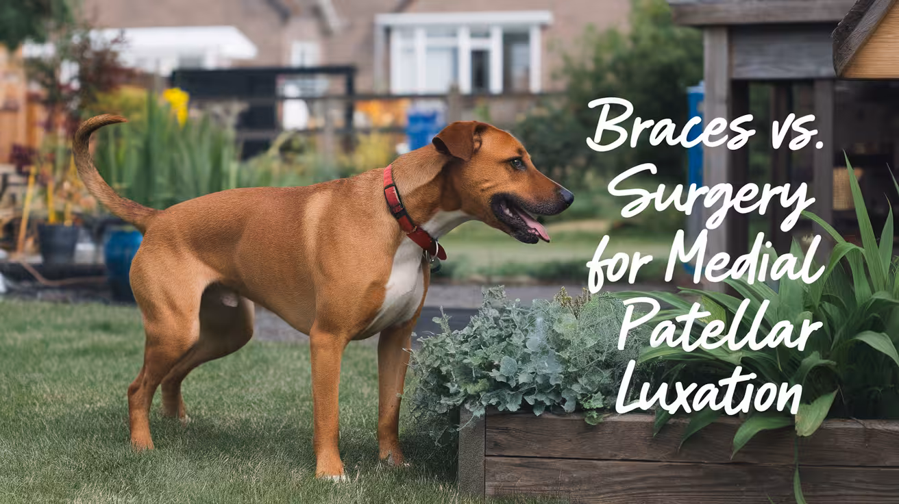
Braces vs. Surgery for Medial Patellar Luxation
Compare braces vs. surgery for medial patellar luxation in dogs. Learn what’s best based on severity, cost, recovery, and long-term outcomes
What Is Medial Patellar Luxation (MPL)?
Medial Patellar Luxation (MPL) is a condition where a dog’s kneecap (patella) slips out of its normal groove in the femur and moves toward the inside of the leg. It’s one of the most common orthopedic problems in small breeds but can affect dogs of any size.
Dogs with MPL may show signs like skipping, limping, or bunny-hopping in the hind leg. Some dogs quickly return to normal movement, while others develop more frequent or painful episodes. The condition is typically graded on a scale of 1 to 4, with Grade 1 being mild (occasional luxation with no pain) and Grade 4 being severe (permanent dislocation, pain, and limited function).
Over time, untreated MPL can lead to joint wear, arthritis, or ligament damage. Early diagnosis and treatment—ranging from conservative care to surgery—help improve outcomes and reduce long-term complications.
What Are Braces for Patellar Luxation?
Braces for patellar luxation are orthopedic devices designed to stabilize the dog’s knee and reduce kneecap movement. These braces help support soft tissues around the joint, improving alignment and reducing pain during activity.
Most braces are made from soft, flexible neoprene or rigid molded supports, depending on the severity of luxation. They typically wrap around the leg with adjustable straps to hold the patella in place during walking or light exercise. Some designs have hinges to allow controlled motion while limiting side-to-side movement.
Vets may recommend braces in mild or moderate cases (Grades 1–2), especially when surgery isn’t immediately needed. They are also used during rehabilitation after surgery or for senior dogs who aren’t good surgical candidates. While braces don’t fix the structural issue, they can reduce symptoms, improve comfort, and delay the need for surgery when used properly with weight management and activity control.
How Effective Are Braces for MPL?
Braces can help manage mild to moderate medial patellar luxation (MPL), but their effectiveness has limits. Many dogs show reduced limping, improved stability, and increased comfort when wearing a brace—especially in Grade 1 or 2 cases. Real-world results vary, though, depending on brace fit, compliance, and the dog’s activity level.
While braces may offer short-term relief, they do not correct the anatomical problem. As a result, recurrence of symptoms is common once the brace is removed or not worn consistently. Long-term use may also lead to muscle atrophy, since the brace reduces workload on the leg. This can make the knee even less stable over time.
Another drawback is limited range of motion, especially with rigid braces. Some dogs tolerate this well; others may resist wearing the brace entirely. Braces can be helpful as part of a non-surgical management plan—including weight control, activity restriction, and physical therapy—but they are not a cure. For lasting results, especially in progressing or high-grade cases, surgery remains the more effective option.
What Does Surgery Involve for MPL?
Surgery for medial patellar luxation (MPL) aims to permanently correct the kneecap’s position and restore stable movement. The procedure your dog receives depends on the severity and any underlying bone deformities.
Common surgical techniques include:
- Trochlear sulcoplasty: Deepening the groove in the femur so the patella fits securely
- Tibial tuberosity transposition (TTT): Shifting the attachment point of the patellar tendon to realign pull
- Soft tissue balancing: Tightening or loosening tendons and ligaments around the joint
These procedures may be used alone or combined based on the individual case. Surgery directly addresses the mechanical cause of luxation, offering a long-term solution rather than temporary support.
The typical recovery time ranges from 8 to 12 weeks, depending on age, grade, and whether one or both legs are treated. Dogs usually start gentle weight-bearing within days, followed by structured rehab to rebuild muscle. Post-op care includes crate rest, leash walks, pain meds, and follow-up exams to monitor healing. With proper care, most dogs regain stable, comfortable movement and significantly reduced risk of recurrence.
Braces vs. Surgery: Side-by-Side Comparison
When comparing braces vs. surgery for MPL, it’s important to consider effectiveness, severity, and long-term results.
- Effectiveness in mild cases (Grade 1–2): Braces can help reduce symptoms and delay surgery. They support the joint during movement but don’t stop progression. Surgery is not always needed in early cases if the dog remains stable and pain-free.
- Effectiveness in severe cases (Grade 3–4): Surgery is the clear choice. Braces won’t correct permanent dislocation or severe pain.
- Recurrence rates: Braced dogs often relapse once the brace is removed or becomes less effective. Surgery has a much lower risk of recurrence, especially with proper rehab.
Surgery provides better long-term joint function and prevents secondary issues like cartilage damage or cruciate ligament tears. Braces are non-invasive and less costly but do not improve joint structure. For dogs needing full mobility and comfort, especially active breeds or working dogs, surgery remains the gold standard. Braces may still have value in select cases, but they are typically part of a conservative management plan—not a substitute for correction.
Read more:
Which Dogs Are Better Suited for Braces?
Braces can be a good option for certain dogs with mild or early-stage MPL (Grades 1–2) who don’t yet show signs of ongoing pain or instability. These cases benefit most when symptoms are intermittent and managed with lifestyle adjustments.
Braces are especially useful for:
- Older dogs who are not good surgical candidates due to age or underlying health issues
- Dogs with heart, kidney, or anesthetic risks that make surgery unsafe
- Dogs with low to moderate activity levels, especially indoor pets
- Lightweight or small breeds, where joint forces are lower
Puppies with mild signs may also be monitored with bracing while they finish growing. In these cases, vets aim to slow progression and maintain joint comfort until skeletal maturity.
However, even when braces are used, they work best as part of a full plan that includes physical therapy, weight management, and activity restrictions. Bracing alone rarely prevents worsening in moderate to severe cases. Regular vet follow-ups are important to assess progression and adjust treatment if needed.
When Is Surgery the Better Option?
Surgery becomes the better choice when a dog shows frequent or severe kneecap dislocation, or when bracing and conservative care fail to control symptoms.
Surgical correction is typically recommended when:
- Grade 3 or 4 MPL is diagnosed
- The kneecap is permanently out of place or luxates daily
- The dog shows significant lameness or pain
- The condition affects both knees, causing difficulty walking
- The dog is active, athletic, or working, needing full joint function
In these cases, delaying surgery may lead to joint damage, arthritis, or ligament tears like cranial cruciate ligament (CCL) injuries. These complications make recovery harder and more expensive later.
Surgery restores alignment and reduces stress on surrounding tissues. It gives dogs the best chance for long-term comfort and stable use of the leg. Dogs that have already tried bracing without improvement are often excellent surgical candidates. A full evaluation—including X-rays and orthopedic exam—helps your vet determine if surgery is the most effective and appropriate next step.
Read more:
Cost and Recovery: What to Expect
Bracing is often more affordable upfront, but results vary. A high-quality dog knee brace typically costs $150–$500, depending on size and customization. There may also be costs for physical therapy, vet rechecks, and replacing worn-out braces. Some dogs need multiple fittings before finding the right support.
Surgery for MPL costs between $1,500 and $3,500 per knee, depending on the severity and region. This includes anesthesia, surgical procedure, hospital stay, pain meds, and post-op follow-ups. If both knees are treated, the cost doubles—though some clinics offer discounts for staged procedures.
Recovery time for bracing is shorter, often just a few days of adjustment. However, long-term use can cause issues like muscle loss or skin irritation. Surgery requires a longer healing period—typically 8–12 weeks—with crate rest, leash walks, and physical therapy.
Ultimately, surgery provides more durable results. Bracing may cost less at first but could lead to added expenses if symptoms worsen or additional care is needed later.
Final Decision: What’s Best for Your Dog?
Choosing between bracing and surgery depends on your dog’s MPL grade, age, activity level, and overall health. There’s no one-size-fits-all answer, but understanding the benefits and limits of each option will help you make the right decision.
- Braces are best for mild cases, older dogs, or when surgery isn’t possible due to medical or financial reasons.
- Surgery is ideal for moderate to severe luxation, active dogs, or when conservative care fails.
Talk to your vet or a board-certified surgeon about:
- How advanced is the luxation?
- Is your dog in pain or losing muscle?
- What are the risks of delaying treatment?
- What’s your budget and capacity for post-op care?
Together, you’ll weigh cost, comfort, and quality of life. With the right plan—surgical or non-surgical—most dogs with MPL can return to a happy, mobile life.
Read more:
FAQs
Can a brace cure medial patellar luxation?
No, a brace cannot cure medial patellar luxation (MPL). Braces help manage symptoms by stabilizing the knee and reducing pain, especially in mild cases. However, they do not correct the underlying bone or soft tissue issues causing the kneecap to dislocate. Braces are a supportive option, not a permanent fix. Surgery is the only way to structurally realign the joint and prevent long-term damage.
How long does it take for a brace to show results?
Most dogs show some improvement within 1 to 2 weeks of consistent brace use. You may notice reduced limping, fewer skipping steps, and more comfort during walks. However, results depend on brace fit, usage consistency, and the severity of the luxation. Keep in mind, braces work best when combined with weight control and limited activity, especially in Grade 1 or 2 cases.
Is surgery risky for older dogs with MPL?
Surgery in older dogs does carry more risk, but many senior dogs still recover well with proper screening and care. Vets will evaluate your dog’s heart, kidney, and overall health before surgery. An experienced surgical team can manage anesthesia safely. If your older dog is in chronic pain or losing mobility, surgery may greatly improve comfort and quality of life.
Can a dog go from brace to surgery later?
Yes, many dogs start with bracing and move to surgery later if symptoms worsen or braces stop working. This approach can buy time, especially in mild cases or when owners need to prepare for surgery costs. Your vet will monitor progression and recommend surgery when conservative care no longer provides relief. Early planning helps ensure a smooth transition if surgery becomes necessary.
What if I can’t afford MPL surgery?
If surgery isn’t affordable right now, bracing, physical therapy, and lifestyle changes can help manage symptoms in the short term. Focus on maintaining a healthy weight, avoiding stairs or jumping, and using joint supplements. Ask your vet about payment plans or surgical assistance programs. In some cases, non-profit or teaching hospitals may offer lower-cost surgery options. Prioritizing comfort and regular checkups is key.

