
Protecting
Pets, People & Planet
Join a group of veterinarians leveraging the latest technologies to deliver excellent care to their patients while being a responsible and positive force for their local and global communities.
100% secure. We do not share your information
Recent Articles
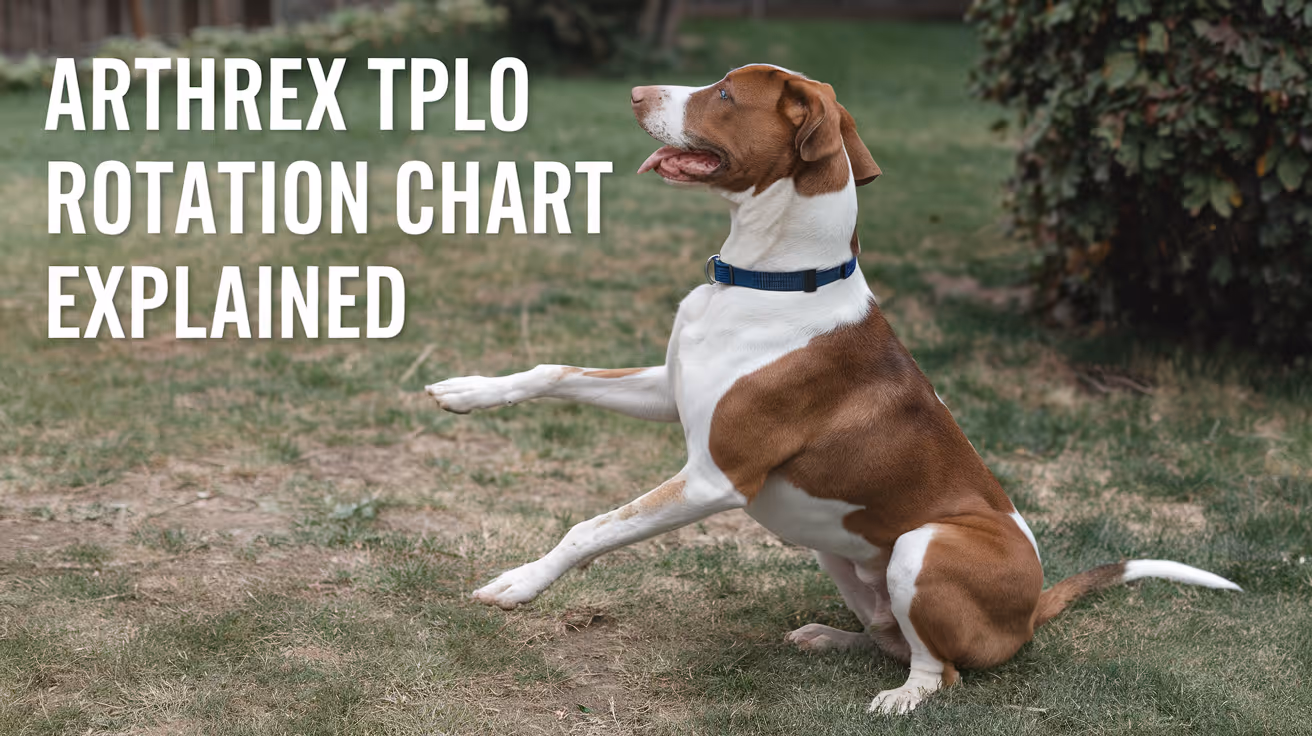
Arthrex TPLO Rotation Chart Explained
Learn how to read and use the Arthrex TPLO rotation chart for precise tibial plateau leveling osteotomy surgery in dogs.
Performing a tibial plateau leveling osteotomy (TPLO) is a common surgical procedure to treat cranial cruciate ligament injuries in dogs. The Arthrex TPLO rotation chart is a vital tool that helps veterinary surgeons determine the exact amount of tibial rotation needed during surgery. Understanding this chart ensures accurate bone alignment and improves surgical outcomes.
This article explains the Arthrex TPLO rotation chart in detail. You will learn how to interpret the chart, why it matters for your pet’s surgery, and how it guides surgeons in achieving the correct tibial plateau angle. This knowledge helps pet owners understand the surgical process better and supports informed discussions with your veterinary surgeon.
What is the Arthrex TPLO rotation chart?
The Arthrex TPLO rotation chart is a reference guide used during TPLO surgery. It shows the degrees of rotation needed to achieve a target tibial plateau angle based on the preoperative measurement. This chart helps surgeons plan and execute the bone cut and rotation precisely.
Using this chart reduces guesswork and improves the accuracy of the surgical correction. It is designed specifically for the Arthrex TPLO surgical system, which includes specialized plates and instruments.
- Rotation guidance: The chart provides exact degrees of tibial rotation required to reach the desired postoperative tibial plateau angle, ensuring surgical precision.
- Preoperative planning: It uses the measured preoperative tibial plateau angle to determine how much rotation is necessary during surgery.
- Standardization tool: The chart standardizes the surgical approach, reducing variability between different surgeons and cases.
- Integration with Arthrex system: It is designed to work with Arthrex-specific implants and instruments for seamless surgical workflow.
Understanding this chart is essential for surgeons performing TPLO with Arthrex equipment and benefits pet owners by improving surgical success rates.
How does the Arthrex TPLO rotation chart improve surgical accuracy?
Accurate rotation of the tibial plateau is critical to restore normal joint mechanics after TPLO surgery. The rotation chart helps surgeons avoid under- or over-rotation, which can lead to poor outcomes or complications.
By providing a clear reference, the chart minimizes errors during the procedure. It also supports consistent results across different patients and surgeons.
- Precise angle correction: The chart ensures the tibial plateau angle is corrected to the target, improving joint stability post-surgery.
- Reduces complications: Proper rotation lowers risks of implant failure, arthritis progression, and abnormal gait after surgery.
- Improves recovery: Accurate alignment supports better healing and faster return to normal activity for dogs.
- Supports training: The chart helps less experienced surgeons perform TPLO with confidence and accuracy.
Using the Arthrex TPLO rotation chart is a key step in achieving the best surgical outcomes for dogs undergoing cruciate ligament repair.
What measurements are needed before using the Arthrex TPLO rotation chart?
Before referencing the rotation chart, the surgeon must measure the tibial plateau angle on preoperative radiographs. This angle indicates how sloped the tibial plateau is, which influences the degree of rotation needed.
Accurate measurement is essential for the chart to provide correct rotation values. The process involves specific radiographic positioning and angle calculation techniques.
- Radiographic positioning: Proper lateral X-rays of the stifle joint are required to visualize the tibial plateau accurately.
- Angle measurement: The tibial plateau angle is measured using anatomical landmarks on the radiograph, typically with digital tools.
- Recording values: The measured angle is noted and used as the input for the rotation chart to find the corresponding rotation degree.
- Repeatability: Consistent measurement technique ensures reliable data for surgical planning and chart use.
Accurate preoperative measurements are the foundation for effective use of the Arthrex TPLO rotation chart during surgery.
How do surgeons use the Arthrex TPLO rotation chart during surgery?
During TPLO surgery, the surgeon references the rotation chart after measuring the tibial plateau angle. The chart indicates how many degrees to rotate the tibial segment after the osteotomy cut.
This rotation changes the slope of the tibial plateau to a safer angle, reducing strain on the cruciate ligament. The chart guides the surgeon to achieve this precisely.
- Osteotomy planning: The surgeon plans the bone cut location and angle based on the chart’s rotation recommendations.
- Rotation execution: After cutting the tibia, the surgeon rotates the bone segment by the degree indicated on the chart.
- Verification: Intraoperative imaging or jigs may be used to confirm the rotation matches the chart’s guidance.
- Plate fixation: The Arthrex TPLO plate is applied to stabilize the rotated tibia, maintaining the corrected angle during healing.
Following the rotation chart during surgery helps ensure the tibial plateau angle is corrected accurately and consistently.
What are the benefits of using the Arthrex TPLO rotation chart for pet owners?
For pet owners, the Arthrex TPLO rotation chart contributes to safer surgeries and better recovery for dogs with cruciate ligament injuries. It supports precise surgical correction, which improves joint function.
Understanding the chart can help owners feel more confident about the procedure and its outcomes.
- Improved surgical outcomes: Accurate rotation reduces complications and improves joint stability after surgery.
- Faster recovery: Correct alignment supports quicker healing and return to normal activity for pets.
- Reduced arthritis risk: Proper tibial plateau angle correction lowers the chance of arthritis development later.
- Enhanced surgeon confidence: The chart helps surgeons perform the procedure with precision, benefiting your pet’s health.
Knowing that the surgeon uses tools like the Arthrex TPLO rotation chart can reassure owners about the quality of care their pet receives.
Are there limitations or challenges with the Arthrex TPLO rotation chart?
While the Arthrex TPLO rotation chart is a valuable tool, it has some limitations. Surgeons must combine chart data with clinical judgment and experience for best results.
Variations in anatomy or measurement errors can affect the chart’s accuracy. Understanding these challenges helps set realistic expectations.
- Measurement variability: Inaccurate preoperative angle measurement can lead to incorrect rotation recommendations.
- Anatomical differences: Individual dog anatomy may require adjustments beyond the chart’s standardized values.
- Technical skill required: Proper use of the chart depends on surgeon experience and surgical technique.
- Not a standalone tool: The chart should be used alongside other surgical planning methods and intraoperative assessments.
Awareness of these limitations ensures the Arthrex TPLO rotation chart is used effectively as part of comprehensive surgical care.
Conclusion
The Arthrex TPLO rotation chart is an essential tool for veterinary surgeons performing tibial plateau leveling osteotomy in dogs. It provides clear guidance on the degree of tibial rotation needed to correct the tibial plateau angle accurately.
By understanding and using this chart, surgeons can improve surgical precision, reduce complications, and support better recovery for pets. Pet owners benefit from knowing their dog’s surgery is planned with advanced, reliable tools like the Arthrex TPLO rotation chart.
What is the Arthrex TPLO rotation chart used for?
The Arthrex TPLO rotation chart is used to determine the exact degrees of tibial rotation needed during TPLO surgery to achieve the desired tibial plateau angle correction.
How do you measure the tibial plateau angle before surgery?
The tibial plateau angle is measured on a properly positioned lateral radiograph of the stifle using anatomical landmarks and digital tools to ensure accuracy.
Can the Arthrex TPLO rotation chart prevent surgical complications?
Yes, by guiding precise tibial rotation, the chart helps reduce risks like implant failure and arthritis progression after TPLO surgery.
Is the Arthrex TPLO rotation chart suitable for all dog breeds?
The chart is designed for general use but surgeons may adjust rotation based on individual anatomical differences in certain breeds.
Do surgeons use imaging during TPLO surgery with the rotation chart?
Yes, intraoperative imaging or jigs are often used to verify that the tibial rotation matches the chart’s recommendations for accuracy.
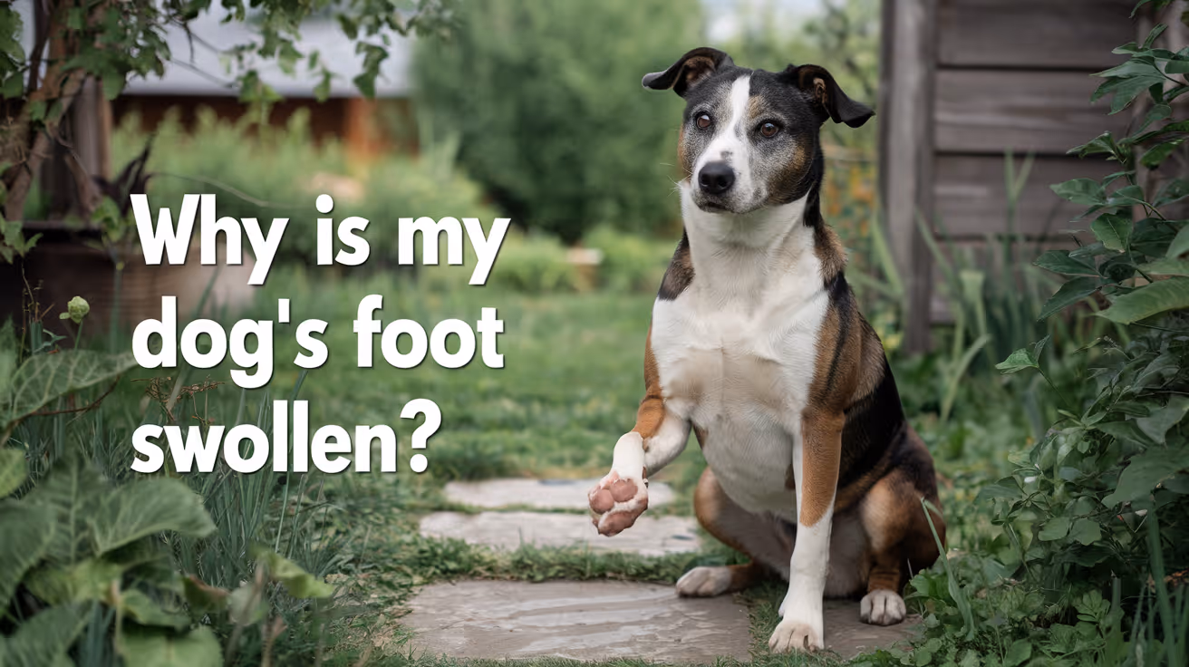
Why Is My Dog's Foot Swollen?
Discover why your dog's foot is swollen, common causes, treatments, and when to see a vet for proper care.
Seeing your dog's foot swollen can be worrying. Swelling in a dog's foot can happen for many reasons, from injuries to infections. Understanding why this happens helps you act quickly and keep your dog comfortable.
This article explains common causes of swollen dog feet, how to spot serious problems, and what treatments work best. You will learn when to treat at home and when to visit a vet for urgent care.
What Causes Swelling in a Dog's Foot?
Swelling in a dog's foot can come from many different problems. It often shows as puffiness, redness, or heat in the paw area. Knowing the cause helps you decide the right care.
Common causes include injuries, infections, allergies, and insect bites. Each cause needs a different approach to treatment.
- Injury or trauma: A cut, sprain, or broken bone can cause swelling due to inflammation and fluid buildup in the foot tissues.
- Infections: Bacterial or fungal infections can cause swelling, redness, and pain, often needing antibiotics or antifungal treatment.
- Allergic reactions: Allergies to plants, chemicals, or insect stings can cause sudden swelling and itching in the foot.
- Foreign objects: Thorns, splinters, or glass stuck in the paw can cause swelling and discomfort until removed.
Identifying the cause early helps prevent complications and speeds healing.
How Can I Tell If My Dog's Foot Swelling Is Serious?
Not all swelling is an emergency, but some signs mean you should see a vet quickly. Serious swelling can affect your dog's ability to walk or cause severe pain.
Look for symptoms like severe limping, open wounds, or signs of infection. These require prompt veterinary care.
- Severe limping or inability to walk: Indicates pain or serious injury needing urgent veterinary evaluation.
- Open wounds or bleeding: Risk of infection and need for cleaning and possibly stitches.
- Fever or lethargy: Signs that infection may have spread and requires medical treatment.
- Rapidly increasing swelling: Could signal an allergic reaction or deep infection needing emergency care.
When in doubt, it is safer to consult your vet to avoid worsening problems.
What Home Treatments Can Help a Swollen Dog Foot?
For mild swelling without serious signs, you can try some home care steps. These help reduce swelling and keep your dog comfortable.
Always watch your dog closely and stop home treatment if symptoms worsen or do not improve.
- Rest and limit activity: Keep your dog from running or jumping to reduce stress on the swollen foot.
- Cold compress application: Apply a cold pack wrapped in cloth for 10-15 minutes to reduce swelling and pain.
- Clean the paw gently: Use warm water to clean dirt or debris, especially if there are small cuts or irritations.
- Prevent licking or chewing: Use an Elizabethan collar if your dog tries to lick or bite the swollen area, which can worsen irritation.
These steps can help minor swelling but do not replace veterinary care for serious cases.
When Should I Take My Dog to the Vet for a Swollen Foot?
Knowing when to seek professional help is important. Some swelling needs medical treatment to avoid complications.
If your dog's swelling is severe, painful, or lasts more than a day or two, a vet visit is necessary. Early treatment can prevent infections or permanent damage.
- Persistent swelling over 48 hours: Indicates that the problem may not resolve without medical intervention.
- Signs of infection: Pus, foul odor, or heat around the swollen area require antibiotics or cleaning by a vet.
- Suspected broken bone or sprain: Needs X-rays and pain management from a veterinary professional.
- Severe allergic reactions: Swelling with difficulty breathing or collapse needs emergency veterinary care immediately.
Your vet can diagnose the cause and recommend the right treatment plan for your dog's recovery.
How Do Vets Diagnose the Cause of a Swollen Dog Foot?
Veterinarians use several methods to find the cause of swelling. A correct diagnosis is key to effective treatment.
They will examine your dog’s foot carefully and may use tests to look deeper into the problem.
- Physical examination: Checking for wounds, foreign objects, and signs of pain or infection in the foot.
- X-rays: Used to detect fractures, bone infections, or foreign bodies inside the paw.
- Skin scrapings or cultures: To identify infections caused by bacteria, fungi, or parasites.
- Blood tests: To check for systemic infection or allergic reactions affecting the swelling.
These tools help your vet create a treatment plan tailored to your dog's needs.
What Treatments Do Vets Use for Swollen Dog Feet?
Treatment depends on the cause of the swelling. Your vet may use medications, procedures, or supportive care to help your dog heal.
Some treatments can be done at home under vet guidance, while others require clinic visits.
- Antibiotics or antifungals: Prescribed to treat infections causing swelling and prevent spread.
- Anti-inflammatory drugs: Help reduce pain and swelling, improving your dog's comfort.
- Wound care and bandaging: Cleaning and protecting open wounds to promote healing and prevent infection.
- Surgery: May be needed to remove foreign objects or repair fractures causing swelling.
Follow your vet’s instructions carefully to ensure the best outcome for your dog.
How Can I Prevent My Dog's Foot from Swelling?
Preventing foot swelling involves protecting your dog from injuries and infections. Regular care and attention can reduce risks.
Simple habits help keep your dog's paws healthy and avoid painful swelling episodes.
- Regular paw inspections: Check your dog's feet daily for cuts, thorns, or swelling to catch problems early.
- Keep nails trimmed: Prevents nails from breaking or causing injury to the foot pads.
- Avoid walking on rough surfaces: Protect paws from sharp objects or hot pavement that can cause injuries.
- Use protective booties: Especially in harsh weather or rough terrain to shield paws from damage.
Good paw care supports your dog’s overall health and comfort.
Conclusion
Swelling in your dog's foot can have many causes, from minor injuries to serious infections. Understanding why your dog's foot is swollen helps you provide the right care quickly.
Always watch for signs of pain, infection, or worsening symptoms. When in doubt, seek veterinary advice to protect your dog's health and comfort. Early treatment can prevent complications and get your dog back on their feet faster.
Why is my dog's foot swollen after walking?
Your dog's foot may swell after walking due to minor injuries, irritation from rough surfaces, or allergic reactions. Rest and paw care usually help reduce swelling quickly.
Can a swollen dog foot heal without a vet?
Mild swelling from minor injuries or irritations can heal at home with rest and care. However, persistent or severe swelling needs veterinary evaluation to avoid complications.
How long does it take for a dog's swollen foot to go down?
Swelling may reduce within a few days with proper care. If swelling lasts more than 48 hours or worsens, consult a vet for treatment.
Is a swollen dog foot painful?
Yes, swelling often causes pain and discomfort. Your dog may limp, lick, or avoid putting weight on the swollen foot.
Can allergies cause a dog's foot to swell?
Yes, allergies to insect bites, plants, or chemicals can cause sudden swelling and itching in a dog's foot, sometimes requiring veterinary treatment.
All Articles
Choosing Suture Material for Dog Surgery
Learn how to choose the best suture material for dog surgery with expert tips on types, uses, and care for optimal healing.
Choosing the right suture material for dog surgery is a common challenge for pet owners and veterinarians. The choice affects healing, infection risk, and comfort for your dog. Understanding the options can help you make informed decisions for your pet's care.
This article explains the types of suture materials, their uses, and factors to consider when selecting sutures for dog surgery. You will learn how to support your dog's recovery with the best choices in suturing.
What types of suture materials are used in dog surgery?
There are many suture materials available for dog surgery. They differ in composition, absorbability, and strength. Knowing the main types helps you understand why a vet chooses one over another.
- Absorbable sutures: These sutures dissolve in the body over time, reducing the need for removal and minimizing stress for your dog after surgery.
- Non-absorbable sutures: Made from materials that do not dissolve, these sutures require removal but provide long-term wound support when needed.
- Natural sutures: Derived from animal or plant fibers, they are less commonly used due to higher tissue reaction risks compared to synthetic options.
- Synthetic sutures: Manufactured from man-made materials, they cause less inflammation and have predictable absorption rates, making them popular in veterinary surgery.
Understanding these types helps you discuss options with your vet and know what to expect during your dog's surgery and recovery.
How does absorbable suture material benefit dog surgery?
Absorbable sutures are designed to break down safely inside your dog's body. They are often used for internal tissues or when suture removal would be difficult or stressful.
- Reduced stress: Absorbable sutures eliminate the need for a second visit to remove stitches, reducing anxiety for both dog and owner.
- Internal use: They are ideal for closing internal tissues that heal beneath the skin, supporting healing without external removal.
- Variable absorption: Different materials absorb at different rates, allowing vets to match suture life to tissue healing time.
- Lower infection risk: Absorbable sutures reduce the chance of infection by avoiding prolonged foreign material presence on the skin surface.
Choosing absorbable sutures can improve your dog's comfort and healing, especially for surgeries involving deep tissue layers.
When are non-absorbable sutures preferred in dog surgery?
Non-absorbable sutures stay in the body until removed. They are used when long-term support is needed or for skin closures where easy removal is possible.
- Strong wound support: Non-absorbable sutures provide durable strength for wounds that require extended healing time.
- Skin closure: These sutures are often placed on the skin surface for easy removal once healing is sufficient.
- Minimal tissue reaction: Many non-absorbable sutures cause little inflammation, reducing complications during healing.
- Visible monitoring: Since they remain on the skin, vets can monitor the wound closely and remove sutures at the right time.
Non-absorbable sutures are useful when precise control over wound closure and removal timing is important for your dog's recovery.
What factors influence suture material choice for dog surgery?
Choosing suture material depends on many factors related to your dog's health, the surgery type, and healing needs. Vets consider these carefully to optimize outcomes.
- Wound location: Areas with high movement or tension may require stronger or non-absorbable sutures for better support.
- Tissue type: Different tissues heal at different rates, so suture absorption time must match the healing process.
- Infection risk: Some sutures resist bacteria better, important for contaminated or high-risk wounds.
- Dog's size and activity: Larger or more active dogs may need stronger sutures to prevent wound reopening during healing.
Discussing these factors with your vet helps ensure the suture material chosen suits your dog's specific surgery and recovery needs.
How do suture sizes and needle types affect dog surgery outcomes?
Suture size and needle shape impact how well the wound heals and how much tissue damage occurs during stitching. These details matter for your dog's comfort and healing speed.
- Suture size: Smaller sizes cause less tissue trauma but may be weaker; larger sizes provide strength but can increase inflammation.
- Needle shape: Curved needles allow precise placement in tight spaces, reducing tissue damage during suturing.
- Needle point: Cutting needles penetrate tough skin easily, while taper needles are better for soft tissues to minimize trauma.
- Matching tissue: Proper needle and suture size matching the tissue type promotes faster healing and reduces complications.
Vets select the right combination to balance strength and healing, ensuring your dog's surgery is as safe and comfortable as possible.
What care is needed for sutures after dog surgery?
Proper care of sutures after surgery is essential to prevent infection and support healing. You play a key role in monitoring and protecting your dog's wound.
- Keep clean and dry: Avoid wetting the sutures to reduce infection risk and help the wound heal properly.
- Prevent licking: Use an Elizabethan collar or other methods to stop your dog from licking or biting the sutures.
- Watch for signs: Look for redness, swelling, discharge, or discomfort that may indicate infection or complications.
- Follow vet instructions: Attend follow-up visits and remove non-absorbable sutures as directed to ensure proper healing.
Following these care steps helps your dog recover quickly and reduces the chance of problems with the sutures or wound.
How does suture material choice affect dog surgery costs?
Suture materials vary in price, which can influence the overall cost of your dog's surgery. Understanding this helps you prepare financially and discuss options with your vet.
- Material cost differences: Synthetic and specialized sutures often cost more than natural or basic types due to manufacturing and performance benefits.
- Absorbable vs non-absorbable: Absorbable sutures may reduce follow-up costs by eliminating removal visits, balancing initial expenses.
- Surgery complexity: More complex surgeries needing stronger or multiple suture types can increase material costs.
- Long-term benefits: Investing in quality sutures may reduce complications and additional treatments, saving money over time.
Discussing suture options and costs with your vet ensures you choose the best material for your dog's health and your budget.
Conclusion
Choosing the right suture material for dog surgery is vital for your pet's healing and comfort. Different types, sizes, and needle options suit various surgeries and tissue needs.
Understanding absorbable and non-absorbable sutures, care requirements, and cost implications empowers you to support your dog's recovery effectively. Always consult your vet to select the best sutures for your dog's specific surgery.
What is the difference between absorbable and non-absorbable sutures?
Absorbable sutures dissolve inside the body over time, eliminating removal, while non-absorbable sutures remain until removed by a vet, providing longer wound support.
Can suture material cause allergic reactions in dogs?
Some dogs may react to certain suture materials, especially natural ones, causing inflammation or irritation; synthetic sutures usually cause fewer allergic responses.
How long do absorbable sutures take to dissolve in dogs?
Absorbable sutures typically dissolve within 10 to 60 days, depending on the material and tissue type, matching the healing process of the wound.
When should non-absorbable sutures be removed after dog surgery?
Non-absorbable sutures are usually removed 10 to 14 days after surgery, once the wound has healed enough to stay closed without support.
Is it safe to bathe a dog with sutures?
Bathing should be avoided until sutures are removed or fully absorbed to prevent infection and wound opening; always follow your vet's advice on wound care.

Surface Disinfection Protocols in Veterinary Hospitals
Learn effective surface disinfection protocols in veterinary hospitals to prevent infections and ensure pet safety.
Surface disinfection in veterinary hospitals is crucial to prevent the spread of infections among animals and staff. Contaminated surfaces can harbor harmful pathogens, leading to disease outbreaks. Understanding proper disinfection protocols helps maintain a safe environment for pets and veterinary workers.
This article explains the best surface disinfection practices in veterinary hospitals. You will learn about effective disinfectants, cleaning steps, and how to implement protocols that reduce infection risks in clinical settings.
What are the key steps in surface disinfection in veterinary hospitals?
Surface disinfection involves cleaning and applying disinfectants to kill germs. Proper steps ensure that pathogens are removed and surfaces are safe for use. Each step plays a role in reducing contamination and preventing disease transmission.
Following a structured cleaning routine is essential for effective disinfection. Skipping steps or using incorrect methods can leave harmful microbes behind.
- Initial cleaning: Remove visible dirt and organic matter using detergent and water before disinfecting surfaces to allow disinfectants to work effectively.
- Choosing disinfectants: Select disinfectants approved for veterinary use that are effective against common pathogens like bacteria, viruses, and fungi.
- Contact time: Allow disinfectants to remain on surfaces for the recommended duration to ensure complete pathogen kill.
- Proper application: Use appropriate tools such as wipes, sprays, or mops to evenly apply disinfectants without cross-contaminating other areas.
Adhering to these steps helps maintain a hygienic hospital environment and protects animal patients from infections.
Which disinfectants are best for veterinary hospital surfaces?
Choosing the right disinfectant is vital for effective surface cleaning. Veterinary hospitals face a variety of pathogens, so disinfectants must cover a broad spectrum of microbes.
Disinfectants vary in their chemical composition, safety, and effectiveness. Selecting the right one depends on the surface type and pathogens present.
- Quaternary ammonium compounds: These are widely used for their broad antimicrobial activity and low toxicity, suitable for many surfaces.
- Chlorine-based disinfectants: Effective against viruses and bacteria but can be corrosive and irritate skin, requiring careful handling.
- Accelerated hydrogen peroxide: Offers fast action and breaks down into safe byproducts, making it environmentally friendly and effective.
- Phenolic disinfectants: Useful against a wide range of pathogens but may be toxic to cats, so use with caution in mixed-species hospitals.
Consult product labels and veterinary guidelines to select disinfectants that balance efficacy and safety for your hospital.
How often should surfaces be disinfected in veterinary hospitals?
The frequency of surface disinfection depends on the area’s use and contamination risk. High-touch and high-risk zones require more frequent cleaning to prevent pathogen buildup.
Routine disinfection schedules help maintain continuous infection control and reduce disease transmission among patients and staff.
- High-touch areas: Disinfect surfaces like door handles, exam tables, and computer keyboards multiple times daily to reduce contamination risk.
- Isolation rooms: Clean and disinfect after each patient use to prevent cross-infection between animals with contagious diseases.
- General patient areas: Perform daily disinfection to maintain overall hygiene and reduce microbial load.
- Operating rooms: Clean and disinfect before and after every surgical procedure to ensure a sterile environment.
Adjust cleaning frequency based on hospital traffic, outbreak situations, and veterinary infection control policies.
What equipment and tools are needed for effective surface disinfection?
Using the right equipment ensures disinfectants are applied correctly and surfaces are thoroughly cleaned. Proper tools also prevent cross-contamination during cleaning.
Investing in suitable cleaning supplies improves efficiency and helps staff follow protocols consistently.
- Disposable wipes: Convenient for quick disinfection of small surfaces and reduce risk of spreading pathogens compared to reusable cloths.
- Mops with detachable heads: Allow thorough cleaning of floors and easy replacement to avoid contamination buildup.
- Spray bottles: Facilitate even application of liquid disinfectants on various surfaces without waste.
- Protective gear: Gloves and masks protect staff from chemical exposure and infectious agents during cleaning tasks.
Regularly inspect and replace cleaning tools to maintain their effectiveness and hygiene.
How can veterinary hospitals train staff on surface disinfection protocols?
Staff training is essential to ensure consistent and effective surface disinfection. Proper education reduces errors and improves compliance with infection control standards.
Training programs should be clear, practical, and regularly updated to reflect current best practices and new products.
- Hands-on demonstrations: Show staff how to clean and disinfect surfaces step-by-step to build confidence and skill.
- Written protocols: Provide easy-to-understand guidelines and checklists for reference during daily tasks.
- Regular refresher courses: Reinforce knowledge and update staff on changes in protocols or disinfectant products.
- Monitoring and feedback: Observe cleaning practices and offer constructive feedback to improve adherence to protocols.
Engaging staff in infection control fosters a culture of safety and responsibility within the hospital.
What challenges affect surface disinfection in veterinary hospitals?
Several challenges can hinder effective surface disinfection in veterinary hospitals. Identifying and addressing these issues improves infection control outcomes.
Awareness of common obstacles helps hospitals develop strategies to overcome them and maintain high hygiene standards.
- High patient turnover: Rapid movement of animals can limit time available for thorough cleaning between patients.
- Surface material limitations: Some disinfectants may damage sensitive surfaces, restricting product choices and cleaning frequency.
- Staff compliance: Inconsistent adherence to protocols due to workload or lack of training can reduce disinfection effectiveness.
- Pathogen resistance: Some microbes develop tolerance to disinfectants, requiring rotation of products and updated protocols.
Addressing these challenges requires planning, education, and investment in appropriate resources.
How do surface disinfection protocols impact infection control in veterinary hospitals?
Effective surface disinfection protocols are a cornerstone of infection control in veterinary hospitals. They reduce the risk of disease spread and protect animal and human health.
Consistent disinfection practices contribute to safer clinical environments and better patient outcomes.
- Reduced pathogen transmission: Proper disinfection lowers the chance of infections spreading between animals and staff.
- Improved patient recovery: Cleaner environments decrease secondary infections, supporting faster healing and less complications.
- Enhanced staff safety: Minimizing exposure to infectious agents protects veterinary workers from zoonotic diseases.
- Compliance with regulations: Following protocols meets legal and accreditation standards for veterinary practice hygiene.
Investing in surface disinfection protocols ultimately benefits the entire veterinary hospital community.
Conclusion
Surface disinfection protocols in veterinary hospitals are essential for preventing infections and maintaining a safe environment. Proper cleaning steps, disinfectant selection, and staff training all contribute to effective infection control.
By understanding and applying these protocols, veterinary hospitals can protect their patients and staff from harmful pathogens. Consistent disinfection practices support healthier outcomes and uphold high standards of veterinary care.
What surfaces require the most frequent disinfection in veterinary hospitals?
High-touch surfaces like exam tables, door handles, and computer keyboards require frequent disinfection to prevent pathogen spread among animals and staff.
Can disinfectants harm animals if not used properly?
Yes, some disinfectants can be toxic if residues remain on surfaces or if used incorrectly. Following label instructions and rinsing surfaces when needed reduces risks.
How long should disinfectants stay on surfaces to be effective?
Disinfectants should remain wet on surfaces for the contact time specified by the manufacturer, usually between 1 to 10 minutes, to kill pathogens effectively.
Is it necessary to clean surfaces before disinfecting?
Yes, cleaning removes dirt and organic material that can block disinfectants, ensuring they work properly to kill germs on surfaces.
What role do veterinary staff play in surface disinfection?
Staff are responsible for following protocols, applying disinfectants correctly, and maintaining hygiene standards to prevent infections in the hospital.

Auditing Asepsis Compliance in Veterinary Clinics
Learn how to audit asepsis compliance in veterinary clinics to ensure infection control and patient safety effectively.
Asepsis is critical in veterinary clinics to prevent infections during surgeries and treatments. Auditing asepsis compliance helps clinics maintain high standards and protect animal health. Understanding how to perform these audits ensures safer care for pets.
This article explains what auditing asepsis compliance means, why it matters, and how veterinary clinics can implement effective auditing processes. You will learn practical steps to check aseptic techniques and improve clinic hygiene.
What is auditing asepsis compliance in veterinary clinics?
Auditing asepsis compliance means systematically checking if veterinary staff follow infection control rules. It involves observing procedures, reviewing records, and identifying risks that could cause contamination.
This process helps clinics find gaps in their aseptic practices and take action to improve safety. Auditing is a key part of quality control in veterinary medicine.
- Definition of auditing: A structured review of aseptic procedures to ensure infection prevention standards are met consistently in clinical settings.
- Purpose of auditing: To identify weaknesses in asepsis practices that could lead to infections in animals or staff.
- Scope of auditing: Includes surgical prep, instrument sterilization, hand hygiene, and environmental cleanliness.
- Frequency of audits: Regular audits, such as monthly or quarterly, help maintain ongoing compliance and catch issues early.
By understanding auditing, clinics can create safer environments and reduce infection risks during veterinary care.
Why is asepsis compliance important in veterinary clinics?
Asepsis prevents harmful microbes from entering wounds or sterile areas. In veterinary clinics, poor asepsis can cause infections that harm pets and increase treatment costs.
Maintaining asepsis protects animals, staff, and clients. Compliance reduces disease spread and improves surgical outcomes.
- Infection prevention: Proper asepsis stops bacteria and viruses from causing surgical site infections in animals.
- Animal welfare: Clean techniques reduce pain, complications, and recovery time for pets undergoing procedures.
- Staff safety: Following aseptic protocols lowers the risk of zoonotic infections for veterinary workers.
- Clinic reputation: High asepsis standards build client trust and demonstrate professional care quality.
Strong asepsis compliance is essential for safe veterinary practice and positive patient results.
How do you conduct an asepsis compliance audit?
Conducting an audit involves planning, observation, data collection, and feedback. Auditors review clinical areas and staff behavior to measure adherence to aseptic protocols.
Clear checklists and criteria guide the audit process. Results help clinics improve infection control.
- Preparation phase: Define audit goals, select audit team, and prepare checklists based on veterinary asepsis standards.
- Observation phase: Watch surgeries, cleaning, and sterilization procedures to see if protocols are followed correctly.
- Documentation review: Check sterilization logs, cleaning schedules, and training records for completeness and accuracy.
- Feedback and action: Share audit findings with staff and create plans to address any compliance gaps found.
Regular audits encourage continuous improvement in aseptic practices.
What key areas should an asepsis audit cover?
An effective audit covers all points where contamination could occur. This includes staff hygiene, equipment sterilization, and environmental cleaning.
Focusing on these areas ensures a comprehensive review of asepsis compliance.
- Hand hygiene: Assess if staff wash and sanitize hands properly before and after procedures.
- Instrument sterilization: Verify that surgical tools are cleaned, packaged, and sterilized according to protocols.
- Operating room cleanliness: Check if surfaces and floors are disinfected regularly and maintained sterile.
- Personal protective equipment (PPE): Ensure staff use gloves, gowns, and masks correctly during procedures.
Covering these areas helps prevent infection risks in veterinary clinics.
How can veterinary clinics improve asepsis compliance after an audit?
After identifying issues, clinics should implement targeted improvements. Training, policy updates, and monitoring help raise asepsis standards.
Continuous education and leadership support are key to lasting compliance.
- Staff training: Provide regular education on aseptic techniques and infection control best practices.
- Policy revision: Update clinic protocols to address audit findings and clarify expectations.
- Monitoring systems: Use checklists and spot checks to track ongoing compliance and correct lapses quickly.
- Leadership involvement: Engage clinic managers to promote a culture of safety and accountability.
These steps help embed asepsis as a priority in daily veterinary work.
What challenges affect asepsis compliance in veterinary clinics?
Several factors can make maintaining asepsis difficult. Recognizing these challenges helps clinics find practical solutions.
Addressing barriers improves audit outcomes and patient safety.
- Resource limitations: Lack of sterilization equipment or cleaning supplies can hinder proper asepsis.
- Staff workload: Busy schedules may lead to shortcuts or missed steps in infection control.
- Training gaps: Inadequate knowledge about aseptic techniques reduces compliance quality.
- Resistance to change: Some staff may resist new protocols or audits, affecting adherence.
Understanding challenges enables clinics to tailor interventions and support staff better.
What tools and checklists are used in asepsis audits?
Standardized tools help auditors evaluate asepsis objectively. Checklists list specific criteria to observe and score during audits.
Using these tools ensures consistency and thoroughness in compliance assessments.
- Hand hygiene checklist: Details steps and timing for proper handwashing and sanitizing techniques.
- Sterilization audit form: Records instrument cleaning, packaging, sterilizer function, and storage conditions.
- Environmental cleaning checklist: Covers frequency and methods for disinfecting surfaces and floors.
- PPE compliance form: Tracks correct use and disposal of gloves, gowns, masks, and caps.
These tools guide auditors and provide clear feedback for improvement.
Conclusion
Auditing asepsis compliance in veterinary clinics is vital for preventing infections and ensuring safe animal care. It helps identify weaknesses and promotes continuous improvement in hygiene practices.
By regularly auditing and addressing gaps, veterinary clinics protect pets, staff, and clients. Implementing structured audits with clear tools supports a culture of safety and high-quality veterinary medicine.
FAQs
How often should veterinary clinics perform asepsis audits?
Clinics should conduct asepsis audits at least quarterly, but monthly audits provide better ongoing monitoring and quicker identification of issues.
Who should conduct asepsis compliance audits?
Audits are best done by trained infection control officers or veterinary staff not directly involved in daily procedures to ensure objective assessments.
What are common signs of poor asepsis in clinics?
Signs include inconsistent hand hygiene, improper sterilization, dirty surgical areas, and incorrect use of personal protective equipment.
Can audit results improve surgical outcomes?
Yes, identifying and correcting asepsis lapses reduces infection rates and leads to faster recovery and better surgical success.
Are there digital tools for asepsis auditing?
Yes, some clinics use mobile apps and software to record audit data, track compliance trends, and generate reports for easier management.
Dental Surgical Asepsis in Dogs: Best Practices
Learn essential steps and best practices for dental surgical asepsis in dogs to ensure safe and effective oral surgeries.
Dental surgical asepsis in dogs is crucial to prevent infections during oral surgeries. Proper aseptic techniques protect your dog from complications and promote faster healing. Understanding these methods helps you ensure your pet’s safety during dental procedures.
This article explains what dental surgical asepsis involves, why it matters, and how veterinarians maintain a sterile environment. You will learn key steps to prepare, perform, and follow up on dental surgeries safely for your dog.
What is dental surgical asepsis in dogs?
Dental surgical asepsis refers to the practices used to keep the surgical area free from harmful bacteria and contaminants during oral surgery. It involves cleaning, sterilizing, and protecting the surgical site to prevent infection.
Maintaining asepsis is critical because the mouth naturally contains many bacteria. Without proper aseptic techniques, these bacteria can enter surgical wounds and cause serious infections.
- Definition of asepsis: It means eliminating or controlling microorganisms to prevent infection during surgery.
- Importance in dentistry: The mouth has many bacteria, so extra care is needed to keep the surgical site clean.
- Goal of asepsis: To protect the dog’s tissues from contamination and promote healing.
- Common procedures: Tooth extractions, gum surgeries, and oral tumor removals require strict aseptic methods.
Understanding dental surgical asepsis helps you appreciate why your veterinarian follows strict protocols during your dog’s dental surgery.
Why is dental surgical asepsis important for dogs?
Dental surgical asepsis is vital because it reduces the risk of infection, which can cause pain, delayed healing, or more serious health problems. Dogs are prone to oral infections due to the bacteria in their mouths.
Infections can spread beyond the mouth and affect other organs if not controlled. Proper aseptic techniques protect your dog’s overall health and improve surgical outcomes.
- Prevents infections: Asepsis stops bacteria from entering surgical wounds and causing infections.
- Improves healing: Clean surgical sites heal faster and with fewer complications.
- Reduces pain: Avoiding infection limits inflammation and discomfort after surgery.
- Protects overall health: Prevents bacteria from spreading to other parts of the body through the bloodstream.
Ensuring asepsis during dental surgery is one of the best ways to keep your dog safe and comfortable during recovery.
How do veterinarians prepare for dental surgical asepsis?
Veterinarians prepare for dental surgical asepsis by carefully cleaning and sterilizing all instruments and the surgical area. They also prepare the dog by cleaning the mouth and using antiseptics.
Preparation includes setting up a sterile environment and using personal protective equipment to avoid contamination.
- Instrument sterilization: All dental tools are cleaned and sterilized using autoclaves before surgery.
- Mouth cleaning: The dog’s oral cavity is cleaned to reduce bacteria before the procedure.
- Use of antiseptics: Antiseptic solutions are applied to the surgical site to kill surface bacteria.
- Protective gear: Veterinarians wear gloves, masks, and gowns to maintain a sterile field.
Proper preparation is essential to minimize infection risks and ensure a smooth dental surgery for your dog.
What are the key steps during dental surgical asepsis in dogs?
During dental surgery, maintaining asepsis involves several key steps. These include controlling the environment, handling instruments properly, and protecting the surgical site.
Each step helps prevent contamination and keeps the surgery safe for your dog.
- Surgical site draping: Sterile drapes cover areas around the mouth to isolate the surgical field.
- Instrument handling: Tools are handled only by sterile gloves to avoid contamination.
- Minimal exposure: The surgical site is exposed only as much as needed to reduce infection risk.
- Continuous monitoring: The surgical team watches for any breaks in asepsis and corrects them immediately.
Following these steps helps maintain a clean surgical environment and protects your dog from infection.
How is post-operative care linked to dental surgical asepsis?
Post-operative care is closely linked to dental surgical asepsis because it continues to prevent infection after surgery. Proper care supports healing and reduces complications.
Owners must follow veterinary instructions carefully to maintain cleanliness and monitor for signs of infection.
- Wound cleaning: Keeping the surgical site clean prevents bacteria from causing infections.
- Medication use: Antibiotics and pain relief help control infection and discomfort.
- Diet adjustments: Soft foods reduce irritation to the surgical site during healing.
- Monitoring signs: Watching for swelling, discharge, or bad odor helps detect infections early.
Good post-operative care complements surgical asepsis and ensures your dog recovers safely and comfortably.
What are common challenges in dental surgical asepsis for dogs?
Dental surgical asepsis in dogs faces challenges due to the mouth’s natural bacteria, the dog’s behavior, and the complexity of oral surgeries. These factors make maintaining sterility difficult.
Veterinarians use specialized techniques and equipment to overcome these challenges and protect your dog.
- High bacterial load: The mouth contains many bacteria, increasing infection risk during surgery.
- Animal movement: Dogs may move unexpectedly, risking contamination during procedures.
- Complex anatomy: The mouth has many small spaces that are hard to keep sterile.
- Equipment limitations: Some dental tools are difficult to sterilize completely due to their design.
Awareness of these challenges helps veterinarians plan and execute dental surgeries with the best aseptic practices.
How can pet owners support dental surgical asepsis in dogs?
Pet owners play a key role in supporting dental surgical asepsis by following pre- and post-surgery instructions carefully. This cooperation helps prevent infections and promotes healing.
Owners should maintain good oral hygiene and attend all veterinary appointments for the best outcomes.
- Follow fasting rules: Avoid feeding your dog before surgery as instructed to reduce anesthesia risks.
- Maintain oral hygiene: Regular brushing before surgery lowers bacteria levels in the mouth.
- Administer medications: Give prescribed antibiotics and painkillers exactly as directed.
- Attend follow-ups: Regular check-ups allow the vet to monitor healing and catch infections early.
By supporting aseptic practices, owners help ensure their dog’s dental surgery is safe and successful.
Conclusion
Dental surgical asepsis in dogs is essential to prevent infections and ensure smooth recovery after oral surgeries. It involves careful preparation, sterile techniques during surgery, and diligent post-operative care.
Understanding and supporting these aseptic practices helps protect your dog’s health and comfort. Always follow your veterinarian’s instructions closely to maintain dental surgical asepsis and promote healing.
What is the difference between asepsis and antisepsis in dental surgery?
Asepsis means preventing any bacteria from contaminating the surgical area, while antisepsis involves using chemicals to kill bacteria on tissues or surfaces before surgery.
How long does it take for a dog to heal after dental surgery?
Healing usually takes 10 to 14 days, but it depends on the procedure and the dog’s health. Proper asepsis and care speed up recovery.
Can dental surgical asepsis prevent all infections in dogs?
While asepsis greatly reduces infection risk, it cannot guarantee complete prevention. Post-operative care and monitoring are also important to avoid infections.
Are there risks if dental surgical asepsis is not followed?
Yes, ignoring aseptic techniques can lead to infections, delayed healing, pain, and more serious health problems for your dog.
Should I brush my dog’s teeth before dental surgery?
Yes, gentle brushing before surgery helps reduce bacteria in the mouth, supporting better aseptic conditions during the procedure.
Maintaining a Sterile Field in Veterinary Surgery
Learn how to maintain a sterile field in veterinary surgery to prevent infections and ensure pet safety during operations.
Maintaining a sterile field in veterinary surgery is critical to prevent infections and ensure successful outcomes for your pet's health. A sterile field is a clean, controlled area free from harmful microorganisms during surgical procedures. Without proper sterile techniques, pets face higher risks of complications and delayed healing.
This article explains how veterinary teams create and maintain sterile fields, why it matters, and what pet owners should know. You will learn the essential steps, common challenges, and best practices used daily in veterinary clinics to keep surgeries safe and effective.
What is a sterile field in veterinary surgery?
A sterile field is a designated area prepared to be free of all microorganisms during surgery. It includes surgical instruments, drapes, gloves, and the patient's skin area that must remain uncontaminated. This controlled environment helps prevent infections and promotes healing.
Veterinary teams carefully set up and monitor the sterile field throughout the procedure. Any breach can introduce bacteria or fungi, leading to serious complications for the animal.
- Definition and purpose: A sterile field is a clean zone that protects the surgical site from germs, reducing infection risk and improving recovery.
- Components included: Surgical drapes, sterile gloves, instruments, gowns, and the prepared skin area form the sterile field.
- Importance in surgery: Maintaining sterility prevents contamination that can cause wound infections or systemic illness in pets.
- Role of veterinary staff: Surgeons, nurses, and assistants work together to create and preserve the sterile environment during operations.
Understanding the sterile field helps you appreciate the care taken during your pet's surgery to keep them safe and healthy.
How do veterinary teams prepare a sterile field?
Preparing a sterile field involves several detailed steps before surgery begins. The goal is to eliminate microorganisms from the surgical area and tools. This preparation requires strict protocols and teamwork.
Each step reduces the chance of contamination and ensures the highest level of cleanliness for the procedure.
- Skin preparation: The pet's surgical site is shaved, cleaned with antiseptic solutions, and disinfected to remove bacteria.
- Instrument sterilization: Surgical tools are sterilized using autoclaves or chemical methods to kill all microorganisms.
- Use of sterile drapes: Sterile drapes cover the pet and surrounding areas, isolating the surgical site from contaminants.
- Personnel preparation: Surgical staff scrub their hands, wear sterile gloves, gowns, masks, and caps to maintain cleanliness.
These steps create a controlled environment that minimizes infection risks and supports safe surgery.
What are the common challenges in maintaining sterility during surgery?
Despite careful preparation, maintaining a sterile field can be challenging. Veterinary teams must stay vigilant to avoid accidental contamination during surgery. Understanding these challenges helps explain the strict rules followed in the operating room.
Addressing these issues promptly is essential to protect your pet's health.
- Accidental touch contamination: Touching non-sterile surfaces or objects can introduce germs to gloves or instruments.
- Movement and airflow: Excessive movement or improper airflow in the operating room can carry contaminants into the sterile field.
- Equipment failure: Sterilization equipment malfunction can result in non-sterile instruments being used unknowingly.
- Human error: Mistakes like improper glove removal or incorrect draping can break sterility and increase infection risk.
Veterinary teams train extensively to recognize and prevent these issues, ensuring the sterile field remains intact.
How do veterinary surgeons maintain sterility during surgery?
During surgery, veterinary surgeons follow strict protocols to keep the sterile field uncontaminated. They use specific techniques and teamwork to avoid introducing bacteria or other pathogens.
Maintaining sterility requires constant attention and careful handling of instruments and tissues.
- Proper glove use: Surgeons change gloves if contamination occurs and avoid touching non-sterile surfaces.
- Instrument handling: Sterile instruments are passed carefully to avoid contact with non-sterile areas.
- Minimal exposure: The surgical site is exposed only as much as necessary to reduce contamination risk.
- Communication: Team members communicate clearly to manage equipment and maintain sterility throughout the procedure.
These practices help ensure the surgery proceeds safely without introducing infections.
What role does the veterinary nurse play in sterile field maintenance?
Veterinary nurses are essential in setting up and maintaining the sterile field. They assist the surgeon and monitor the environment to prevent contamination. Their role requires knowledge, skill, and vigilance.
Nurses act as the sterile field's guardians, ensuring all protocols are followed correctly.
- Preparation assistance: Nurses prepare instruments, drapes, and supplies in a sterile manner before surgery.
- Monitoring sterility: They watch for any breaks in sterility and alert the team immediately if contamination occurs.
- Instrument management: Nurses handle and pass instruments to the surgeon without compromising sterility.
- Postoperative care: They help clean and sterilize equipment after surgery to prepare for future procedures.
Veterinary nurses’ attention to detail helps maintain a safe surgical environment for every pet.
How can pet owners support sterile field practices?
Pet owners play a role in supporting sterile field maintenance by following pre- and post-surgery instructions carefully. Their cooperation helps reduce infection risks and promotes healing.
Understanding the importance of sterility can improve outcomes and reduce complications.
- Pre-surgery hygiene: Follow instructions to keep your pet clean and avoid feeding before surgery as advised.
- Environment control: Keep your pet in a clean, calm area before and after surgery to reduce exposure to germs.
- Follow medication guidelines: Administer prescribed antibiotics or medications exactly as directed to prevent infections.
- Observe surgical site: Monitor the wound for redness, swelling, or discharge and report concerns to your veterinarian promptly.
By cooperating with veterinary advice, you help maintain the sterile field's benefits beyond the operating room.
What are the consequences of breaking the sterile field?
Breaking the sterile field can lead to serious complications for your pet. Contamination introduces bacteria or fungi that may cause infections, delayed healing, or systemic illness.
Understanding these risks highlights why sterile field protocols are so strict in veterinary surgery.
- Increased infection risk: Contamination can cause surgical site infections requiring additional treatment or surgery.
- Delayed recovery: Infections slow healing and can prolong your pet’s discomfort and hospitalization.
- Higher costs: Treating infections or complications adds to medical expenses and care time.
- Potential for systemic illness: Severe infections can spread through the bloodstream, threatening your pet’s life.
Strict sterile field maintenance protects your pet’s health and helps ensure the best surgical outcomes.
Conclusion
Maintaining a sterile field in veterinary surgery is vital to prevent infections and promote healing. Veterinary teams use detailed preparation, strict protocols, and teamwork to keep the surgical environment clean and safe for your pet.
As a pet owner, understanding these practices helps you appreciate the care taken during surgery and how you can support your pet’s recovery. Keeping the sterile field intact protects your pet’s health and leads to better surgical success.
FAQs
How long does it take to prepare a sterile field before surgery?
Preparing a sterile field typically takes 30 to 60 minutes, including skin cleaning, instrument sterilization, and draping. The time varies depending on the surgery complexity.
Can a sterile field be re-established if contamination occurs?
Yes, if contamination happens, the team can re-sterilize instruments or change gloves and drapes to restore sterility and continue surgery safely.
Are sterile fields used in all types of veterinary surgeries?
Most surgeries require a sterile field, especially invasive procedures. Minor treatments may not need full sterile precautions but still follow cleanliness standards.
What signs indicate a surgical site infection after surgery?
Signs include redness, swelling, heat, pain, discharge, or fever. Contact your veterinarian immediately if you notice these symptoms.
Do pet owners need to wear protective clothing during surgery?
Pet owners usually do not enter the operating room. Only surgical staff wear protective clothing to maintain sterility during procedures.

Medical vs Surgical Asepsis in Veterinary Practice
Explore the differences between medical and surgical asepsis in veterinary practice and learn how each protects your pet from infections.
In veterinary practice, preventing infections is crucial for the health and safety of animals. Medical and surgical asepsis are two key methods used to reduce infection risks. Understanding these methods helps ensure your pet receives the best care possible.
This article explains the differences between medical and surgical asepsis in veterinary settings. You will learn how each technique works, when they are used, and why they are important for your pet’s health.
What is medical asepsis in veterinary practice?
Medical asepsis refers to practices that reduce the number and spread of microorganisms. It is commonly called "clean technique" and is used in routine veterinary care to prevent infection.
These practices help keep the environment, instruments, and staff clean to protect animals from germs. Medical asepsis is essential for everyday procedures like wound care and injections.
- Hand hygiene: Washing hands thoroughly before and after contact with animals reduces the chance of spreading germs.
- Use of gloves: Wearing disposable gloves during exams or treatments prevents direct contact with infectious materials.
- Cleaning surfaces: Regularly disinfecting exam tables and equipment lowers microbial contamination.
- Proper waste disposal: Safely discarding used materials like needles and bandages prevents environmental contamination.
Medical asepsis focuses on controlling infection in non-surgical settings. It is the foundation of infection control in veterinary clinics.
What is surgical asepsis in veterinary practice?
Surgical asepsis, also called "sterile technique," aims to eliminate all microorganisms from surgical areas. It is used during invasive procedures to create a sterile field and prevent infections.
This method requires strict protocols to keep instruments, surgical sites, and personnel free from germs. Surgical asepsis is critical for operations like spays, neuters, and tumor removals.
- Sterilization of instruments: Using autoclaves or chemical sterilants ensures surgical tools are free of all microbes.
- Sterile gloves and gowns: Surgeons wear sterile clothing to avoid contaminating the surgical field.
- Preparing the surgical site: Shaving and disinfecting the animal’s skin removes bacteria before incision.
- Maintaining a sterile field: Only sterile items and personnel can touch the surgical area during the procedure.
Surgical asepsis is more rigorous than medical asepsis because it prevents infections during procedures that expose internal tissues.
How do medical and surgical asepsis differ in veterinary care?
Medical and surgical asepsis differ mainly in their level of cleanliness and application. Medical asepsis reduces germs, while surgical asepsis aims to eliminate them completely.
Understanding these differences helps veterinary teams choose the right method for each situation to protect animal health.
- Purpose: Medical asepsis controls infection in routine care; surgical asepsis prevents infection during invasive surgery.
- Cleanliness level: Medical asepsis is "clean" but not sterile; surgical asepsis requires complete sterility.
- Techniques used: Medical asepsis involves handwashing and disinfection; surgical asepsis requires sterilization and sterile barriers.
- Risk level: Surgical asepsis is needed when infection risk is high due to exposure of internal tissues.
Both methods are essential but serve different roles in veterinary infection control.
When should veterinary professionals use medical asepsis?
Medical asepsis is used in many common veterinary tasks that do not involve surgery. It helps reduce infection risks during routine care and minor procedures.
Veterinary staff apply medical asepsis whenever they handle animals or equipment to maintain a clean environment.
- Physical exams: Cleaning hands and surfaces before examining animals prevents germ spread.
- Wound care: Using gloves and disinfectants reduces infection risk when treating cuts or abrasions.
- Administering injections: Sterile needles and gloves protect against contamination during vaccinations or medications.
- Handling bodily fluids: Proper disposal and protective gear prevent transmission of infectious agents.
Medical asepsis is the standard for everyday veterinary care to keep animals safe from infections.
When is surgical asepsis necessary in veterinary practice?
Surgical asepsis is required during procedures that break the skin or enter sterile body areas. It prevents serious infections that can occur after surgery.
Veterinary surgeons follow strict sterile protocols to protect animals during operations.
- Spay and neuter surgeries: Maintaining sterility prevents post-operative infections in reproductive organs.
- Orthopedic surgeries: Sterile technique protects bones and joints from contamination.
- Dental surgeries: Surgical asepsis reduces infection risk when working inside the mouth.
- Emergency surgeries: Sterile procedures are critical when treating trauma or internal injuries.
Surgical asepsis is vital for any invasive procedure to ensure safe healing and recovery.
How can pet owners support asepsis during veterinary visits?
Pet owners play a role in infection prevention by following veterinary advice and maintaining hygiene at home. This helps support medical and surgical asepsis efforts.
Simple steps can reduce infection risks before and after veterinary care.
- Keep pets clean: Regular grooming reduces dirt and bacteria on the animal’s skin and coat.
- Follow wound care instructions: Proper cleaning and bandage changes prevent infections after treatment.
- Attend follow-up visits: Monitoring healing helps detect and address infections early.
- Communicate health changes: Informing the vet about any signs of illness supports timely care.
Working with your veterinary team helps protect your pet from infections through effective asepsis.
What are the risks of poor asepsis in veterinary practice?
Poor asepsis can lead to infections that cause pain, delayed healing, or serious illness in animals. It also increases treatment costs and recovery times.
Understanding these risks highlights why strict aseptic techniques are essential in veterinary care.
- Increased infections: Contaminated instruments or environments can introduce harmful bacteria to wounds or surgical sites.
- Delayed healing: Infections slow down recovery and may require additional treatments or surgeries.
- Spread of disease: Poor asepsis can transmit contagious diseases between animals or humans.
- Higher costs: Treating infections often involves more medications, longer hospital stays, and extra care.
Maintaining proper medical and surgical asepsis protects animal health and improves treatment outcomes.
Conclusion
Medical and surgical asepsis are vital practices in veterinary medicine to prevent infections. Medical asepsis focuses on reducing germs during routine care, while surgical asepsis aims for complete sterility during invasive procedures.
Both methods protect your pet’s health by minimizing infection risks. Understanding their differences helps you appreciate the care your pet receives and supports better outcomes during veterinary visits.
What is the main difference between medical and surgical asepsis?
Medical asepsis reduces germs to prevent infection, while surgical asepsis eliminates all microorganisms to maintain sterility during surgery.
When is surgical asepsis used in veterinary practice?
Surgical asepsis is used during invasive procedures like spays, neuters, and emergency surgeries to prevent infections in sterile body areas.
How does medical asepsis protect pets during routine care?
Medical asepsis uses hand hygiene, gloves, and surface cleaning to reduce germs and prevent infections during exams and minor treatments.
Can pet owners help maintain asepsis after veterinary visits?
Yes, by following wound care instructions, keeping pets clean, and attending follow-ups, owners support infection prevention at home.
What risks arise from poor asepsis in veterinary clinics?
Poor asepsis can cause infections, delayed healing, disease spread, and increased treatment costs, harming animal health and recovery.
Monofilament vs Multifilament Sutures in Dogs and Cats
Compare monofilament and multifilament sutures for dogs and cats, covering uses, benefits, risks, and care tips for pet owners.
Sutures are essential tools in veterinary surgery, especially for dogs and cats. Choosing the right suture type can affect healing, infection risk, and comfort. Monofilament and multifilament sutures are the two main categories used in small animal medicine.
This article explains the differences between monofilament and multifilament sutures in dogs and cats. You will learn their characteristics, advantages, disadvantages, and when to use each type. Understanding these sutures helps you better care for your pet after surgery.
What are monofilament sutures in dogs and cats?
Monofilament sutures consist of a single smooth strand. They are commonly used in veterinary surgeries for dogs and cats because they cause less tissue trauma. Their smooth surface reduces bacterial colonization, lowering infection risk.
These sutures are less flexible but glide easily through tissue. They are often made from materials like nylon, polypropylene, or polydioxanone (PDS). Monofilament sutures are popular for skin closure and internal tissues.
- Single strand design: Monofilament sutures have one smooth thread, which reduces tissue drag and irritation during placement and healing.
- Lower infection risk: Their smooth surface limits bacteria from hiding in suture fibers, decreasing chances of wound infection in pets.
- Less knot security: Knots may slip more easily, so surgeons must tie more secure knots or use additional throws.
- Good tensile strength: They maintain strength well over time, supporting wound edges during healing in dogs and cats.
Monofilament sutures are ideal when minimizing infection risk is a priority. They work well for skin, fascia, and subcutaneous tissues in dogs and cats.
What are multifilament sutures in dogs and cats?
Multifilament sutures are made of several smaller threads twisted or braided together. This structure provides flexibility and excellent knot security. However, the braided surface can harbor bacteria, increasing infection risk.
Common materials include braided polyester, silk, and coated polyglactin (Vicryl). Multifilament sutures are often used for internal tissues where strong knots and flexibility are needed.
- Braided structure: Multiple fibers twisted together increase flexibility and allow easier handling during surgery.
- Better knot security: Braided sutures hold knots firmly, reducing risk of loosening in active pets like dogs and cats.
- Higher infection risk: Braids can trap bacteria and debris, increasing chances of wound infection if not managed carefully.
- Good tissue support: They provide strong wound closure, especially in tissues under tension or movement.
Multifilament sutures are preferred for tissues needing strong, secure closure but require careful monitoring for infection signs in dogs and cats.
How do monofilament and multifilament sutures differ in infection risk?
Infection risk is a key factor when choosing sutures for dogs and cats. Monofilament sutures have a smooth surface that resists bacterial colonization. Multifilament sutures have a braided surface that can trap bacteria.
Proper surgical technique and wound care reduce infection risk regardless of suture type. However, monofilament sutures are generally safer in contaminated wounds or high-risk patients.
- Smoother surface advantage: Monofilament sutures reduce bacterial adhesion, lowering infection chances in surgical wounds.
- Bacterial trapping risk: Multifilament sutures’ braided fibers can harbor bacteria, increasing infection risk if not cleaned well.
- Wound environment impact: Contaminated or dirty wounds benefit from monofilament sutures to minimize infection in dogs and cats.
- Postoperative care importance: Proper cleaning and monitoring reduce infection risk for both suture types after surgery.
Choosing monofilament sutures for infected or dirty wounds is safer, but multifilament sutures can be used with strict hygiene and care.
When should vets choose monofilament sutures for dogs and cats?
Monofilament sutures are best when infection risk must be minimized or when smooth tissue passage is needed. They are commonly used for skin closure and subcutaneous tissues in dogs and cats.
These sutures are also preferred in patients with compromised immune systems or wounds exposed to dirt. Their lower tissue reaction helps promote faster healing.
- Skin closure preference: Monofilament sutures cause less irritation and reduce scarring when closing skin in dogs and cats.
- Contaminated wounds: They are safer for wounds exposed to bacteria or dirt, lowering infection risk.
- Immune-compromised pets: Dogs and cats with weak immunity benefit from monofilament sutures to avoid complications.
- Minimal tissue reaction: These sutures cause less inflammation, promoting smoother healing in delicate tissues.
Vets often select monofilament sutures for external wounds and clean surgical sites in dogs and cats to optimize healing and reduce complications.
When are multifilament sutures better for dogs and cats?
Multifilament sutures are ideal when strong knot security and flexibility are needed. They work well for internal tissues under tension or movement, such as muscles and ligaments in dogs and cats.
These sutures are also chosen when surgeons need easier handling and better control during suturing. However, infection risk must be managed carefully.
- Strong knot holding: Multifilament sutures maintain secure knots in tissues that move or stretch frequently in dogs and cats.
- Flexible handling: Their braided design allows surgeons to manipulate sutures easily during complex procedures.
- Internal tissue use: Preferred for muscles, tendons, and ligaments requiring durable closure in active pets.
- Careful infection control: Use requires strict aseptic technique and monitoring to prevent bacterial complications.
Multifilament sutures provide excellent support for internal repairs but need careful postoperative care to avoid infection in dogs and cats.
How should pet owners care for sutures after surgery?
Proper suture care is crucial for healing and preventing complications in dogs and cats. Owners should follow veterinary instructions closely to protect the surgical site.
Both monofilament and multifilament sutures require monitoring for signs of infection, swelling, or discomfort. Keeping the area clean and dry helps ensure good healing.
- Keep area clean: Gently clean around the suture site as directed to prevent dirt and bacteria buildup in dogs and cats.
- Prevent licking: Use an Elizabethan collar or other barriers to stop pets from licking or chewing sutures.
- Watch for infection: Look for redness, swelling, discharge, or odor and contact your vet if these signs appear.
- Follow vet advice: Administer prescribed medications and attend follow-up visits for suture removal or wound checks.
Good suture care helps your pet heal faster and reduces the risk of complications after surgery.
What are the pros and cons of monofilament vs multifilament sutures?
Both suture types have advantages and disadvantages depending on the surgical situation in dogs and cats. Understanding these helps vets and owners make informed choices.
Monofilament sutures reduce infection risk but have less knot security. Multifilament sutures offer strong knots but higher infection risk. The choice depends on wound type, location, and pet health.
- Monofilament pros: Lower infection risk, less tissue trauma, and minimal inflammation support faster healing in dogs and cats.
- Monofilament cons: Knots may slip more easily, requiring extra care during suturing to maintain wound closure.
- Multifilament pros: Excellent knot security and flexibility make them ideal for internal tissues under tension.
- Multifilament cons: Higher infection risk due to braided fibers requires careful wound management and hygiene.
Choosing the right suture balances these factors to optimize healing and reduce complications in dogs and cats.
Conclusion
Monofilament and multifilament sutures each have unique benefits and drawbacks for dogs and cats. Monofilament sutures reduce infection risk and cause less tissue irritation, making them ideal for skin and contaminated wounds.
Multifilament sutures provide strong knot security and flexibility, suitable for internal tissues under stress. Proper suture care and veterinary guidance ensure the best healing outcomes for your pet after surgery.
What is the difference between monofilament and multifilament sutures?
Monofilament sutures are single smooth strands, while multifilament sutures are braided or twisted fibers. This affects flexibility, knot security, and infection risk in dogs and cats.
Are monofilament sutures better for infected wounds in pets?
Yes, monofilament sutures reduce bacterial colonization, making them safer for contaminated or infected wounds in dogs and cats.
Can multifilament sutures cause more infections in dogs and cats?
Multifilament sutures can trap bacteria in their braided fibers, increasing infection risk if wound care is not properly managed.
How long do sutures stay in dogs and cats after surgery?
Sutures typically stay in place for 10 to 14 days, depending on the surgery type and tissue involved, before removal by a veterinarian.
What should I do if my pet’s sutures look red or swollen?
Contact your veterinarian immediately if you notice redness, swelling, discharge, or your pet shows pain, as these may be signs of infection.
Preventing Post-Operative Infections in Dogs
Learn effective ways to prevent post-operative infections in dogs with expert tips on care, hygiene, and monitoring after surgery.
Post-operative infections in dogs are a common concern for pet owners after surgery. These infections can cause pain, delay healing, and sometimes lead to serious complications if not managed properly. Understanding how to prevent these infections is crucial for your dog's recovery and overall health.
This article explains the key steps you can take to reduce the risk of infections after your dog’s surgery. You will learn about wound care, hygiene, signs to watch for, and when to contact your veterinarian for help.
What causes post-operative infections in dogs?
Post-operative infections occur when bacteria or other microorganisms invade the surgical site. These infections can develop from contamination during surgery or from poor wound care afterward. Knowing the causes helps you take the right precautions.
- Surgical contamination: Bacteria may enter the wound during surgery if sterile techniques are not strictly followed, increasing infection risk.
- Environmental exposure: Open wounds exposed to dirt, dust, or unclean surfaces can become infected after surgery.
- Poor hygiene: Lack of proper cleaning around the surgical site can allow bacteria to multiply and cause infection.
- Underlying health issues: Dogs with weakened immune systems or chronic diseases are more prone to infections after surgery.
Understanding these causes helps you focus on preventing contamination and supporting your dog’s immune system during recovery.
How should I care for my dog’s surgical wound?
Proper wound care is essential to prevent infections. You need to keep the area clean, dry, and protected from your dog’s licking or scratching. Following your veterinarian’s instructions is critical for successful healing.
- Keep wound clean: Gently clean the area with prescribed solutions or wipes to remove dirt without damaging healing tissue.
- Prevent licking: Use an Elizabethan collar or other devices to stop your dog from licking or biting the wound, which can introduce bacteria.
- Change bandages: Replace dressings as directed by your vet to maintain a sterile environment around the wound.
- Monitor for swelling: Watch for redness, swelling, or discharge that may indicate infection and require veterinary attention.
Consistent wound care reduces infection risk and promotes faster healing after surgery.
What hygiene practices help reduce infection risk?
Maintaining good hygiene around your dog and their environment is key to preventing infections. Cleanliness helps limit bacterial growth and keeps the surgical site safe during recovery.
- Wash hands: Always wash your hands before and after touching the wound to avoid transferring germs.
- Clean bedding: Change and wash your dog’s bedding regularly to reduce bacteria and dirt near the surgical site.
- Limit outdoor exposure: Keep your dog indoors or in a clean area to prevent contact with contaminants during healing.
- Disinfect surfaces: Clean floors and surfaces your dog contacts with pet-safe disinfectants to minimize infection risk.
Good hygiene habits create a safer healing environment and support your dog’s recovery process.
When should I give antibiotics to my dog after surgery?
Antibiotics can help prevent or treat infections but should only be used under veterinary guidance. Overuse or misuse can cause resistance and other health problems.
- Follow vet prescription: Give antibiotics exactly as prescribed, completing the full course even if your dog seems better.
- Do not self-medicate: Never give antibiotics without veterinary approval, as incorrect use can worsen infections.
- Watch for side effects: Monitor your dog for vomiting, diarrhea, or allergic reactions during antibiotic treatment.
- Use antibiotics preventively only if advised: Some surgeries require antibiotics to prevent infection, but only your vet can decide.
Proper antibiotic use supports healing and reduces the chance of resistant infections.
What signs indicate a post-operative infection in dogs?
Recognizing infection signs early allows prompt treatment and prevents complications. You should regularly check your dog’s surgical site and overall condition during recovery.
- Redness and swelling: Increased redness or swelling around the wound can signal infection.
- Discharge or pus: Yellow, green, or foul-smelling discharge from the wound is a common infection sign.
- Fever and lethargy: Your dog may feel tired, have a fever, or lose appetite if infected.
- Excessive pain: Increased pain or sensitivity at the surgical site may indicate infection or other problems.
If you notice any of these signs, contact your veterinarian immediately for evaluation and treatment.
How can I support my dog’s immune system after surgery?
A strong immune system helps your dog fight infections and heal faster. You can support immunity through nutrition, rest, and stress reduction during recovery.
- Balanced diet: Feed high-quality food rich in vitamins and minerals to boost immune function and tissue repair.
- Proper rest: Ensure your dog has a quiet, comfortable place to rest and recover without stress or excessive activity.
- Hydration: Keep fresh water available to maintain hydration, which is important for immune health.
- Follow-up care: Attend all veterinary check-ups to monitor healing and address any health issues promptly.
Supporting your dog’s immune system helps reduce infection risk and promotes a smooth recovery.
What role does the veterinarian play in preventing infections?
Your veterinarian is essential in preventing post-operative infections. They provide surgical expertise, guidance on care, and early intervention if problems arise.
- Sterile surgery: Vets use strict sterile techniques during surgery to minimize infection risk.
- Post-op instructions: They give detailed care instructions tailored to your dog’s surgery and health status.
- Monitoring healing: Regular check-ups allow vets to detect early signs of infection and treat them promptly.
- Prescribing medications: Vets decide when antibiotics or pain relief are needed to support recovery safely.
Working closely with your veterinarian ensures the best outcome and reduces infection chances after surgery.
Conclusion
Preventing post-operative infections in dogs requires careful attention to wound care, hygiene, and monitoring. By following your veterinarian’s advice and maintaining a clean environment, you can help your dog heal safely and comfortably.
Early detection of infection signs and proper antibiotic use are also vital. Supporting your dog’s immune system with good nutrition and rest further reduces infection risks. Taking these steps ensures your dog recovers well and stays healthy after surgery.
What is the typical recovery time after dog surgery?
Recovery time varies by surgery type but usually ranges from 10 to 14 days. Your veterinarian will provide specific timelines based on your dog’s procedure and health.
Can I bathe my dog after surgery?
Avoid bathing your dog until the surgical wound is fully healed and your vet approves. Moisture can increase infection risk during early healing.
How do I know if my dog is in pain after surgery?
Signs of pain include whining, restlessness, licking the wound, and reluctance to move. Contact your vet if you suspect your dog is uncomfortable.
Are there natural remedies to prevent infections?
Natural remedies are not a substitute for veterinary care. Always consult your vet before using supplements or treatments to prevent infections.
What should I do if my dog removes the stitches?
If your dog removes stitches, contact your veterinarian immediately. They may need to replace them or provide alternative wound care to prevent infection.

Walking Sutures in Large Breed Dogs
Learn about walking sutures in large breed dogs, their benefits, techniques, and care tips for optimal healing after surgery.
What are walking sutures in large breed dogs?
Walking sutures are a special stitching technique used in surgery to help close wounds in large breed dogs. They allow the skin to stretch and move without putting too much pressure on the wound edges. This method helps prevent the sutures from cutting into the skin, which is important for dogs with thick or loose skin.
These sutures act like small steps that gradually bring the wound edges together. This technique is especially useful in large breed dogs because their skin can be heavy and prone to tension after surgery.
- Definition and purpose: Walking sutures are stitches placed to reduce tension on wound edges, promoting better healing and reducing the risk of suture tearing in large dogs.
- Skin movement allowance: They allow slight movement between stitches, which helps prevent skin damage caused by tight sutures pulling on thick skin.
- Use in large breeds: Large breed dogs often have more skin tension, making walking sutures ideal to support wound closure safely.
- Healing improvement: By distributing tension evenly, walking sutures help wounds heal faster and with fewer complications.
Understanding walking sutures helps pet owners appreciate why surgeons choose this method for large dogs. It ensures the wound stays closed properly while minimizing discomfort and risk of wound breakdown.
How do walking sutures benefit large breed dogs after surgery?
Walking sutures provide several advantages for large breed dogs recovering from surgery. Their unique design helps manage the tension that naturally occurs on the skin after large wounds or incisions are closed. This reduces complications and supports a smoother healing process.
These benefits are especially important because large dogs have more skin weight and movement, which can stress surgical wounds.
- Reduced skin tension: Walking sutures lower the pull on wound edges, preventing suture tearing and skin damage in heavy or loose skin.
- Improved wound stability: They keep the wound securely closed while allowing some flexibility, which helps avoid wound reopening during normal movement.
- Lower infection risk: By preventing suture cutting and skin damage, walking sutures reduce the chance of infection and inflammation.
- Enhanced cosmetic results: These sutures promote even healing, which can lead to less scarring and better appearance after recovery.
Overall, walking sutures help large breed dogs heal more comfortably and safely after surgery, reducing the risk of complications that can delay recovery.
What materials are used for walking sutures in large breed dogs?
The choice of suture material is important for walking sutures to work effectively. Surgeons select materials that provide strength, flexibility, and minimal tissue reaction. The right suture helps maintain wound closure while allowing skin movement.
Different suture types have specific benefits depending on the wound and dog’s skin condition.
- Monofilament sutures: These single-strand sutures cause less tissue irritation and reduce infection risk, making them suitable for walking sutures.
- Absorbable materials: Absorbable sutures dissolve over time, eliminating the need for removal and reducing stress for the dog.
- Non-absorbable options: Sometimes non-absorbable sutures are used when longer wound support is needed, but they require removal later.
- Suture size selection: Larger suture sizes provide more strength for thick skin, which is common in large breed dogs.
Choosing the right suture material helps ensure walking sutures perform well and support the healing process without causing extra irritation or complications.
How are walking sutures placed in large breed dogs?
Placing walking sutures requires skill and understanding of skin anatomy. The surgeon inserts the stitches in a way that the skin is gently pulled together in small steps. This technique reduces tension and allows the skin to move slightly between stitches.
The process involves careful spacing and depth to achieve the best wound closure without damaging the skin.
- Suture spacing: Sutures are placed at regular intervals to evenly distribute tension along the wound edges.
- Depth control: The needle passes through the skin and underlying tissue layers to provide strong support without causing tissue damage.
- Gentle tension: Each stitch is tightened just enough to bring the skin edges closer without cutting into the skin.
- Stepwise closure: The technique mimics small steps, allowing gradual wound edge approximation and reducing stress on any single point.
Proper placement of walking sutures is crucial for large breed dogs to avoid complications and promote fast healing. Surgeons carefully plan each stitch to balance support and flexibility.
What are the common complications of walking sutures in large breed dogs?
While walking sutures offer many benefits, some complications can occur if the technique or care is not optimal. Recognizing these issues early helps pet owners seek timely veterinary care to prevent worsening problems.
Large breed dogs may be more prone to certain complications due to their size and skin characteristics.
- Suture loosening: If sutures are too loose, the wound may open, delaying healing and increasing infection risk.
- Suture cutting: Excessive tension or improper placement can cause sutures to cut into the skin, leading to tissue damage.
- Infection risk: Poor wound hygiene or suture reactions can cause infection, requiring veterinary treatment.
- Wound dehiscence: This is the reopening of the wound, which can happen if sutures fail or the dog is too active during healing.
Proper surgical technique and post-operative care reduce these risks. Owners should monitor the wound closely and follow veterinary instructions to support healing.
How should you care for walking sutures in large breed dogs at home?
After surgery, caring for walking sutures is essential to ensure the wound heals well. Owners must keep the area clean, prevent the dog from licking or biting the sutures, and watch for signs of problems.
Following veterinary advice carefully helps avoid complications and speeds recovery.
- Keep the wound clean: Gently clean the area as directed by your vet to prevent infection and remove debris.
- Prevent licking or chewing: Use an Elizabethan collar or other devices to stop the dog from disturbing the sutures.
- Limit activity: Restrict running, jumping, and rough play to avoid stress on the wound and sutures.
- Watch for signs of infection: Look for redness, swelling, discharge, or bad odor and contact your vet if these occur.
Good home care supports the benefits of walking sutures and helps your large breed dog heal comfortably and safely after surgery.
When should you contact your vet about walking sutures in your dog?
It is important to know when to seek veterinary help to address any issues with walking sutures. Early intervention can prevent serious complications and promote better healing outcomes.
Owners should be alert to changes in the wound or the dog’s behavior that may indicate a problem.
- Excessive swelling or redness: These signs may indicate infection or inflammation requiring prompt veterinary evaluation.
- Wound discharge or bleeding: Any unusual fluid or blood from the wound should be checked by a vet immediately.
- Sutures coming loose or falling out: This can lead to wound opening and needs veterinary repair.
- Signs of pain or discomfort: If the dog is licking, biting, or showing pain around the wound, contact your vet for advice.
Timely veterinary care ensures walking sutures continue to support healing and reduces the risk of complications in large breed dogs.
Conclusion
Walking sutures are a valuable surgical technique for closing wounds in large breed dogs. They help reduce skin tension, promote better healing, and lower the risk of complications after surgery.
Understanding how walking sutures work, their benefits, and proper care helps pet owners support their dogs’ recovery. Always follow your veterinarian’s instructions and watch for any signs of problems to ensure the best outcome for your large breed dog.
What is the main purpose of walking sutures in large breed dogs?
Walking sutures reduce tension on wound edges, allowing better healing and preventing sutures from cutting into thick skin common in large breed dogs.
Are walking sutures absorbable or non-absorbable?
Walking sutures can be either absorbable or non-absorbable depending on the wound needs, but absorbable sutures are often preferred to avoid removal stress.
How long do walking sutures stay in place?
Walking sutures usually stay in place for 10 to 14 days, but the exact time depends on the wound and your veterinarian’s advice.
Can walking sutures prevent wound infections?
Yes, by reducing skin tension and tissue damage, walking sutures help lower infection risk, but proper wound care is also essential.
What should I do if my dog’s walking sutures come loose?
If sutures come loose or the wound opens, contact your veterinarian immediately for evaluation and possible wound repair.

Veterinary Surgical Hand Scrub Protocol Guide
Learn the step-by-step veterinary surgical hand scrub protocol to ensure sterile surgery and prevent infections in pets.
Performing a proper veterinary surgical hand scrub is essential to reduce the risk of infections during surgery. This protocol helps remove transient and resident microorganisms from your hands and forearms before you wear sterile gloves. Understanding the correct steps ensures your pet's safety and improves surgical outcomes.
This article provides a clear, step-by-step guide to the veterinary surgical hand scrub protocol. You will learn why each step matters, the best products to use, and how to maintain sterility throughout the process.
What is the purpose of the veterinary surgical hand scrub?
The main goal of the surgical hand scrub is to eliminate or reduce microorganisms on the skin to prevent contamination during surgery. This process protects both the patient and the veterinary team from infections.
It also prepares the hands and forearms to maintain sterility while wearing surgical gloves. Proper technique is critical to achieve this goal effectively.
- Infection prevention: The scrub removes bacteria and other microbes that could cause surgical site infections in pets.
- Microbial reduction: It targets both transient microbes, which are easily removed, and resident microbes that live deeper in the skin.
- Sterility maintenance: The scrub creates a clean surface that helps keep gloves sterile during surgery.
- Compliance with standards: Following the protocol meets veterinary surgical hygiene guidelines and protects the surgical team.
By understanding the purpose, you can appreciate why each step in the scrub protocol is important for safe surgery.
What materials are needed for the surgical hand scrub?
Using the right materials ensures the scrub is effective and safe. Veterinary surgical hand scrubs typically require specific brushes, antiseptic agents, and clean water sources.
Choosing approved products helps reduce skin irritation and maximizes microbial removal.
- Antiseptic scrub agent: Use chlorhexidine gluconate or povidone-iodine solutions, which are effective against a broad range of microbes.
- Surgical scrub brush or sponge: These help mechanically remove debris and microbes from the skin surface.
- Clean, running water: Warm water is preferred to rinse off scrub agents without damaging skin.
- Disposable towels or sterile cloths: Used to dry hands and forearms after scrubbing to maintain sterility.
Having all materials ready before starting the scrub prevents contamination and streamlines the process.
How do you perform the initial hand wash before the surgical scrub?
The initial hand wash removes visible dirt and oils that can interfere with the antiseptic scrub. It is a critical preparatory step before the formal surgical scrub begins.
Proper hand washing improves the effectiveness of the antiseptic agents used in the scrub.
- Remove jewelry: Rings, watches, and bracelets harbor bacteria and must be taken off before washing.
- Wet hands and forearms: Use warm running water to wet skin up to the elbows thoroughly.
- Apply plain soap: Use a non-antimicrobial soap to wash hands and forearms for at least 30 seconds.
- Rinse thoroughly: Remove all soap residue under running water to prevent irritation during the scrub.
Completing this step properly prepares your skin for the antiseptic scrub and reduces microbial load.
What is the step-by-step veterinary surgical hand scrub technique?
The surgical hand scrub technique follows a timed, systematic approach to clean hands and forearms thoroughly. It involves applying antiseptic agents and mechanical scrubbing in a specific order.
Following this technique consistently ensures maximum microbial reduction and surgical safety.
- Apply antiseptic: Dispense the recommended amount of scrub solution onto hands and forearms.
- Scrub nails and fingertips: Use a brush or sponge to clean under nails and fingertips for at least 30 seconds.
- Scrub hands and forearms: Continue scrubbing each surface of hands and forearms up to the elbows for 2 to 5 minutes.
- Rinse carefully: Rinse from fingertips to elbows without touching the sink to avoid recontamination.
Maintaining a clean, unidirectional rinse prevents microbes from returning to cleaned areas.
How should you dry your hands and maintain sterility after scrubbing?
Drying your hands properly after the scrub is vital to avoid contamination. Wet hands can transfer microbes more easily and compromise glove sterility.
Using the correct drying method helps maintain the sterile field before donning gloves.
- Use sterile towels: Dry hands and forearms with a sterile towel or cloth to avoid introducing bacteria.
- Dry from fingertips to elbows: Always dry in one direction to prevent contamination of cleaned areas.
- Avoid touching non-sterile surfaces: Keep hands elevated and away from sinks or counters after drying.
- Don sterile gloves immediately: Put on gloves carefully without touching the outside surface to maintain sterility.
Proper drying and glove application complete the surgical hand preparation process.
What are common mistakes to avoid during the surgical hand scrub?
Errors during the surgical hand scrub can reduce its effectiveness and increase infection risk. Being aware of common mistakes helps you maintain proper technique.
Correcting these errors improves patient safety and surgical outcomes.
- Skipping initial wash: Not removing visible dirt before the scrub reduces antiseptic effectiveness.
- Insufficient scrub time: Scrubbing for less than the recommended duration leaves microbes behind.
- Touching non-sterile surfaces: Contact with sinks or counters after scrubbing reintroduces bacteria.
- Using harsh soaps: Strong chemicals can damage skin and reduce compliance with the protocol.
Awareness and training help prevent these mistakes during every surgical procedure.
How often should veterinary staff perform the surgical hand scrub?
Frequency of surgical hand scrubs depends on the number of procedures and breaks between surgeries. Proper timing ensures ongoing sterility and infection control.
Following guidelines for scrub frequency helps maintain a safe surgical environment.
- Before each surgery: Perform a full surgical scrub before every sterile procedure to ensure cleanliness.
- After breaks: If a break exceeds 30 minutes, a repeat scrub is recommended to maintain sterility.
- Between surgeries: If moving between different patients, repeat the scrub to prevent cross-contamination.
- When gloves are compromised: If gloves tear or become contaminated, repeat the scrub before regloving.
Adhering to these timing rules protects patients and staff from infection risks.
Conclusion
The veterinary surgical hand scrub protocol is a critical step to prevent infections during surgery. It removes harmful microbes and prepares your hands and forearms for sterile glove use.
By following the correct materials, technique, and timing, you help ensure the safety of your pet patients and maintain a sterile surgical environment. Consistent practice of this protocol is essential for every veterinary surgical team member.
What is the recommended antiseptic for veterinary surgical hand scrubs?
Chlorhexidine gluconate and povidone-iodine are the most commonly recommended antiseptics due to their broad antimicrobial activity and skin safety.
How long should the surgical hand scrub last?
The scrub should last between 2 to 5 minutes, including focused scrubbing of nails, hands, and forearms to ensure thorough microbial removal.
Can you use alcohol-based hand rubs instead of surgical scrubs?
Alcohol-based rubs are not recommended as a substitute for surgical scrubs in veterinary surgery because they do not remove debris and resident microbes effectively.
Is it necessary to remove jewelry before scrubbing?
Yes, removing all jewelry is essential because it can harbor bacteria and prevent proper cleaning of the skin during the scrub.
What should you do if your gloves tear during surgery?
If gloves tear, remove them immediately, perform a repeat surgical hand scrub, and put on new sterile gloves before continuing the procedure.

Tension-Relieving Sutures in Veterinary Surgery
Learn about tension-relieving sutures in veterinary surgery, their types, uses, benefits, and care for better healing in pets.
Tension-relieving sutures play a vital role in veterinary surgery by reducing stress on wound edges during healing. When pets undergo surgery, their skin and tissues can be under tension that may cause wounds to reopen or heal poorly. Using tension-relieving sutures helps protect the surgical site and promotes better recovery.
This article explains what tension-relieving sutures are, why they are important in veterinary surgery, and how they improve healing outcomes. You will learn about different types of these sutures, their placement techniques, and care tips to ensure your pet’s surgery heals well.
What are tension-relieving sutures in veterinary surgery?
Tension-relieving sutures are special stitches placed to reduce the pulling forces on a wound. They help prevent the edges of the surgical incision from being stretched apart. This is especially important in areas where the skin is tight or over joints where movement can cause tension.
These sutures support the main closure by distributing tension away from the wound edges. This reduces the risk of wound dehiscence, where the incision reopens, and minimizes scarring and infection risk.
- Definition of tension-relieving sutures: Sutures designed to offload tension from wound edges, allowing safer and stronger healing in veterinary patients.
- Purpose in surgery: To protect delicate tissues from excessive pulling forces that may cause wound breakdown or delayed healing.
- Common use areas: High-tension sites like limbs, joints, or large skin flaps where normal sutures alone may fail.
- Support for primary closure: They work alongside regular sutures to strengthen the surgical repair and improve outcomes.
Understanding these sutures helps pet owners appreciate their importance in complex surgeries and why your veterinarian may choose specific techniques to protect your pet’s wound.
Why are tension-relieving sutures important in veterinary surgery?
Tension-relieving sutures are important because they improve wound healing quality and reduce complications. Surgical wounds under tension are more likely to open, get infected, or form excessive scar tissue. These sutures help avoid those problems.
Pets move and stretch after surgery, which can place stress on the incision. Without tension relief, the wound edges may separate, causing pain and delaying recovery. Tension-relieving sutures reduce this risk significantly.
- Prevents wound dehiscence: By reducing tension, these sutures lower the chance of the wound reopening after surgery.
- Reduces infection risk: Stable wounds heal faster and are less prone to bacterial invasion and infection.
- Improves cosmetic results: Less tension means less scarring and better skin appearance after healing.
- Supports early mobility: Pets can move more comfortably with secure wound closure, aiding faster recovery.
These benefits make tension-relieving sutures a critical part of many veterinary surgical procedures, especially in active animals or challenging wound locations.
What types of tension-relieving sutures are used in veterinary surgery?
Several suture techniques and materials are used to relieve tension in veterinary wounds. The choice depends on the wound size, location, and tissue type. Common types include mattress sutures, far-near-near-far sutures, and walking sutures.
Each technique distributes tension differently and is selected based on the surgeon’s preference and the pet’s needs. The suture material also affects strength and healing.
- Horizontal mattress sutures: Sutures placed parallel to the wound edge that spread tension across a wider area to protect delicate skin.
- Vertical mattress sutures: Sutures that go deep and close to the wound edge, providing strong tension relief and eversion of skin edges.
- Far-near-near-far sutures: A pattern that places stitches far from and near the wound edge to evenly distribute tension and reduce edge pulling.
- Walking sutures: Stitches placed in the subcutaneous tissue to gradually relieve tension and support skin closure.
Understanding these types helps you recognize the care your pet’s surgeon takes to ensure a strong, tension-free closure for optimal healing.
How are tension-relieving sutures placed during veterinary surgery?
Placement of tension-relieving sutures requires skill and planning. The surgeon assesses the wound and decides where tension is highest. Sutures are then placed strategically to offload stress from the skin edges.
Proper technique avoids tissue damage and ensures the sutures hold without cutting through the skin. The surgeon may use a combination of suture types for best results.
- Assessment of tension areas: The surgeon identifies parts of the wound under the most stress before placing sutures.
- Strategic suture placement: Sutures are positioned to pull tissue away from the wound edges, reducing tension directly on the incision.
- Use of deep sutures: Subcutaneous sutures help support skin closure by holding underlying tissues together.
- Combination techniques: Multiple suture patterns may be combined to optimize tension relief and wound stability.
These careful steps ensure your pet’s wound is securely closed and protected from forces that could disrupt healing.
What are the benefits of tension-relieving sutures for pets?
Tension-relieving sutures offer many benefits that improve surgical outcomes and pet comfort. They help wounds heal faster and stronger, reducing complications and pain.
By minimizing tension, these sutures also reduce the need for additional surgeries and improve the cosmetic appearance of healed wounds.
- Faster healing times: Reduced tension allows tissues to repair more quickly and efficiently after surgery.
- Lower complication rates: Pets experience fewer wound infections, openings, and delayed healing with tension relief.
- Improved comfort: Secure wound closure reduces pain and irritation during the recovery period.
- Better cosmetic results: Less scarring and tissue distortion lead to more natural-looking healed skin.
These benefits help pets recover smoothly and return to normal activities sooner after surgery.
How should pet owners care for wounds with tension-relieving sutures?
After surgery, proper wound care is essential to protect tension-relieving sutures and promote healing. Pet owners should follow veterinary instructions carefully to avoid complications.
Monitoring the wound and preventing excessive movement or licking helps maintain suture integrity and reduces infection risk.
- Keep the wound clean and dry: Avoid bathing or wetting the area until the veterinarian approves to prevent infection.
- Prevent licking or chewing: Use an Elizabethan collar or protective covering to stop pets from disturbing sutures.
- Limit activity: Restrict running, jumping, or rough play that can place tension on the wound.
- Watch for signs of problems: Look for redness, swelling, discharge, or wound opening and contact your vet if noticed.
Following these care tips helps ensure the tension-relieving sutures do their job and your pet heals well without setbacks.
Conclusion
Tension-relieving sutures are an important tool in veterinary surgery to protect wounds from stress and improve healing. They reduce complications like wound opening and infection, leading to faster recovery and better cosmetic results.
Understanding their role helps pet owners appreciate the care taken during surgery and the importance of proper wound management after. Following veterinary advice on wound care ensures your pet’s surgical site heals safely and comfortably.
What materials are used for tension-relieving sutures in pets?
Materials often include absorbable sutures like poliglecaprone or polydioxanone, which provide strength during healing and dissolve over time, reducing the need for removal.
Can tension-relieving sutures be used on all types of wounds?
They are mainly used on wounds under high tension or in areas prone to movement, but not always necessary for small, low-tension wounds.
How long do tension-relieving sutures stay in a pet’s body?
Absorbable sutures dissolve within 2 to 6 weeks, while non-absorbable sutures may be removed after 10 to 14 days depending on healing progress.
Are tension-relieving sutures painful for pets?
Placement may cause mild discomfort initially, but proper pain management and healing reduce pain quickly as the wound stabilizes.
When should I contact my vet about my pet’s sutures?
If you notice swelling, redness, discharge, wound opening, or if your pet is excessively licking or showing pain, contact your veterinarian promptly.

Common Closure Errors in Small Animal Surgery
Explore common closure errors in small animal surgery, their causes, prevention, and how to ensure optimal healing for your pet’s recovery.
Small animal surgery requires precise closure techniques to ensure proper healing and avoid complications. Closure errors can lead to infections, wound dehiscence, and delayed recovery in pets. Understanding these errors helps pet owners and veterinary professionals improve surgical outcomes and pet welfare.
This article explains the most common closure errors in small animal surgery, why they happen, and how to prevent them. You will learn practical tips to recognize and avoid these mistakes, ensuring your pet’s surgery heals smoothly and safely.
What are the most common closure errors in small animal surgery?
Closure errors occur when the surgical wound is not properly sealed after the procedure. These mistakes can affect the skin, subcutaneous tissue, or deeper layers, causing complications. Knowing these errors helps in early detection and correction.
- Inadequate suture tension: Applying too much or too little tension can cause tissue strangulation or loose closure, leading to wound failure or infection.
- Incorrect suture material: Using the wrong type or size of suture can reduce wound strength and increase the risk of dehiscence or irritation.
- Poor tissue handling: Rough or careless handling damages tissues, delaying healing and increasing infection risk.
- Failure to close dead space: Leaving gaps under the skin allows fluid accumulation, causing seromas or hematomas that impair healing.
Recognizing these errors is essential for surgeons to adjust techniques and for pet owners to monitor wounds after surgery.
How does improper suture technique affect healing in pets?
Improper suture technique can compromise wound integrity and delay healing. It may cause tissue damage, inflammation, or wound opening. Understanding the impact helps improve surgical outcomes and pet comfort.
- Excessive tension damage: Tight sutures restrict blood flow, causing tissue necrosis and increasing infection risk around the wound.
- Loose sutures risk: Sutures that are too loose allow wound edges to separate, leading to dehiscence and delayed healing.
- Uneven suture spacing: Irregular spacing causes uneven tension distribution, increasing the chance of wound gaps or suture failure.
- Incorrect knot tying: Poorly tied knots can slip or unravel, causing sudden wound opening and infection risk.
Proper suture technique balances tension, spacing, and knot security to promote fast and safe healing in pets.
Why is choosing the right suture material important in small animal surgery?
Suture material affects wound strength, tissue reaction, and healing speed. Selecting the appropriate type and size is critical for successful closure and minimizing complications.
- Absorbable vs non-absorbable: Absorbable sutures dissolve over time, ideal for internal layers, while non-absorbable are used for skin or long-term support.
- Suture size matters: Larger sutures provide more strength but cause more tissue trauma; smaller sutures reduce trauma but may not hold strong wounds.
- Monofilament vs multifilament: Monofilament sutures reduce infection risk due to smooth surface; multifilament sutures offer better knot security but can harbor bacteria.
- Material biocompatibility: Some materials cause more tissue reaction, increasing inflammation and delaying healing.
Choosing the right suture material tailored to the wound type and location improves healing and reduces complications.
How can dead space cause closure complications in pets?
Dead space refers to gaps between tissue layers after closure. These spaces can fill with fluid or blood, causing swelling and infection. Managing dead space is vital for proper healing.
- Fluid accumulation risk: Dead space allows fluids to collect, creating seromas that delay healing and increase infection chances.
- Increased infection risk: Fluid pockets provide a medium for bacteria growth, leading to wound infection and complications.
- Delayed wound healing: Pressure from fluid buildup can separate wound edges, causing dehiscence and prolonged recovery.
- Use of drains or sutures: Proper closure techniques or drains help eliminate dead space, promoting faster and safer healing.
Preventing dead space through careful tissue apposition and drainage reduces postoperative complications in small animal surgery.
What role does tissue handling play in preventing closure errors?
Gentle tissue handling preserves blood supply and reduces trauma, which are essential for wound healing. Rough handling can cause tissue death and increase complications.
- Preserving blood flow: Careful manipulation maintains circulation, supporting oxygen and nutrient delivery for healing tissues.
- Minimizing tissue trauma: Avoiding crushing or tearing reduces inflammation and speeds up recovery.
- Reducing infection risk: Less tissue damage lowers the chance of bacterial invasion and wound infection.
- Improving suture holding: Healthy tissues hold sutures better, preventing early wound opening or failure.
Proper tissue handling techniques are fundamental skills for surgeons to ensure successful closure and pet recovery.
How can pet owners help prevent closure errors after surgery?
Pet owners play a key role in monitoring and caring for surgical wounds at home. Following veterinary advice helps avoid closure errors and supports healing.
- Follow wound care instructions: Cleaning and protecting the wound as directed prevents infection and promotes healing.
- Prevent pet interference: Using Elizabethan collars or bandages stops pets from licking or biting the wound, avoiding damage.
- Watch for complications: Early signs like swelling, redness, or discharge should prompt veterinary consultation to address closure issues.
- Limit activity: Restricting movement prevents stress on the wound, reducing the risk of suture failure or dehiscence.
Active owner involvement and communication with the veterinary team improve surgical outcomes and pet comfort.
What are the signs of closure errors in small animal surgery?
Recognizing signs of closure errors early allows prompt treatment and prevents serious complications. Pet owners should observe wounds carefully after surgery.
- Wound swelling and redness: These signs indicate inflammation or infection around the closure site requiring veterinary attention.
- Discharge or bleeding: Fluid or blood leaking from the wound suggests suture failure or infection needing prompt care.
- Wound opening or dehiscence: Visible separation of wound edges is a serious closure error needing immediate veterinary intervention.
- Excessive pain or discomfort: Persistent pain may signal complications related to closure errors or infection.
Early detection and treatment of closure errors improve healing and reduce the risk of further surgery or complications.
Conclusion
Common closure errors in small animal surgery can significantly affect your pet’s recovery and comfort. Understanding these errors helps both veterinary professionals and pet owners ensure better surgical outcomes.
By recognizing closure mistakes, choosing proper suture materials, handling tissues gently, and following postoperative care instructions, you can help your pet heal safely and quickly. Always consult your veterinarian if you notice any signs of wound complications after surgery.
What is the best suture material for small animal skin closure?
Monofilament, non-absorbable sutures like nylon or polypropylene are commonly preferred for skin closure due to low tissue reaction and good strength.
How long does it take for surgical wounds to heal in pets?
Most surgical wounds heal within 10 to 14 days, but healing time varies based on surgery type, pet health, and wound care quality.
Can pets remove their sutures by licking or biting?
Yes, pets often try to lick or bite sutures, which can cause wound opening or infection; using an Elizabethan collar helps prevent this behavior.
When should I contact my vet about my pet’s surgical wound?
Contact your vet if you notice swelling, redness, discharge, wound opening, or if your pet shows excessive pain or discomfort around the surgical site.
Are absorbable sutures safe for all types of small animal surgeries?
Absorbable sutures are safe for internal tissues but may not provide enough strength for skin closure or high-tension wounds, requiring non-absorbable sutures instead.
Sustainable Vet | Copyright ©2026 | All Rights Reserved

