

Walking Sutures in Large Breed Dogs
Learn about walking sutures in large breed dogs, their benefits, techniques, and care tips for optimal healing after surgery.

This article is for informational purposes only and is not a substitute for professional veterinary advice. Every case is unique, so always consult your veterinarian for guidance specific to your pet.
This content is intended for veterinary professionals for educational purposes. It does not replace clinical judgment or tailored advice. Always rely on your training, expertise, and the specific context of your patients.

Large breed dogs present a specific wound closure challenge: when skin is removed or lost over the trunk, there is often not enough laxity to simply pull the edges together. Pulling too hard creates tension. Tension causes ischemia. Ischemia causes dehiscence.
Walking sutures solve this by redistributing the tension before the skin edges ever come together.
Quick answer: Walking sutures are subcutaneous sutures that anchor the dermis to the underlying fascia at intervals along a wound, advancing skin progressively toward the defect with each placement rather than attempting to close the full gap in one pull. DVM360 (Swaim) describes them as "tension-type sutures that can be used to close large skin defects in areas where sufficient skin surrounds the wound." Standard material: 2-0 PDS or Biosyn (3-0 in patients under 15 kg). Particularly indicated for trunk and lateral thorax wounds in large dogs.
Key takeaways
- Walking sutures anchor dermis to fascia at intervals, advancing skin progressively toward the wound center.
- Primary indication: large trunk and lateral thorax skin defects in dogs where direct closure would create excessive tension.
- Standard material: 2-0 PDS or Biosyn; 3-0 in patients under 15 kg.
- Staggered removal protocol: remove every other suture at day 10, remaining sutures at day 14.
- Thorax wounds require respiratory monitoring tight closure can impair breathing in smaller patients.
- Not appropriate over skin flaps walking sutures risk damaging the blood supply to the flap.
What walking sutures do
A walking suture does not close the wound at the skin surface. It anchors the dermis to the fascia beneath the skin, pulling the skin toward the defect from underneath.
DVM360 (Dr. Steven Swaim, reconstructive surgery techniques): "Walking sutures are tension-type sutures that can be used to close large skin defects in areas where sufficient skin surrounds the wound that can be moved or stretched to close the wound. These sutures are primarily indicated for closing skin defects on the trunks of small animals."
Veterinary Surgery Online: "Walking sutures can be used to tack down the dermis to the underlying fascia. These also help decrease tension on the wound edges. After placement, the wound edges should be in close proximity and under minimal tension."
Each walking suture placed advances the skin a small distance. Placed sequentially from the wound edges inward, they bring the skin incrementally closer to the defect center with each stitch hence "walking" the skin across.
When walking sutures are used
Primary indication: large trunk skin defects
- Mass excisions on the lateral thorax or abdomen requiring wide margins
- Degloving injuries where skin was lost over the trunk
- Wounds too large for simple closure but with adequate adjacent skin for advancement
- Cases where a skin flap or graft is not available or not planned
DVM360 (Swaim): "Most large skin defects on the trunks of dogs and cats can be closed with walking sutures, either using the technique described here or a modification of it. These sutures advance local skin to close large defects without requiring additional incisions to create flaps or attain relaxation."
When walking sutures cannot fully close the wound
If the defect is too large for complete walking suture closure, the technique closes the wound as much as possible, then allows the remainder to heal by second intention or stages it for a secondary surgery.
Veterinary Surgery Online: "If a wound is too large to be closed completely using this technique, it can be closed enough to allow the remainder of the wound to heal by second intention or with a secondary surgery."
Contraindication: skin flaps
Today's Veterinary Practice (Caudal Superficial Epigastric Flap technique notes): "I prefer not to tack down or use walking sutures in the subcutaneous tissues to attempt to reduce motion and dead space under the flap and inguinal region. These sutures could inadvertently damage the blood supply to the flap."
For how tension-relieving sutures are used for high-tension surgical wounds generally, see tension-relieving sutures alongside walking sutures.
Technique overview
Bite placement:
- The needle passes through the dermis on one side of the advancing skin edge
- Then through the superficial fascia at the target location (where that skin edge should end up)
- Tied the skin edge advances to meet the fascia at that point
- The next suture is placed in the adjacent skin, advancing the next section
Pattern sequence:
- Begin at the wound margins and work inward
- Each suture advances one section of skin; sequential sutures advance the whole leading edge
- After all walking sutures are placed, the wound edges should be in close proximity or apposed
Final skin closure:Placed after all walking sutures. Standard options: 3-0 to 4-0 Prolene or nylon (interrupted), or skin staples for speed.
Suture material for walking sutures
Veterinary Surgery Online: "Typically, walking sutures and muscle fascia sutures are applied using 2-0 PDS or Biosyn (3-0 in patients under 15 kg, or occasionally 0 in larger dogs). Subcutaneous tissues closed using 3-0 PDS, Biosyn, or Monocryl (4-0 in smaller patients). Dermal closure performed using 3-0 Monocryl or Biosyn (4-0 in smaller patients)."
PDS and Biosyn are monofilament absorbable materials with adequate strength retention for the 3 to 4 weeks needed while the tissue planes adhere and the skin advances fully. They are preferred over braided absorbable materials in the subcutaneous plane because of lower infection risk.
For how suture size relates to patient weight in this context, see suture size selection for walking sutures.
Thorax wounds: a specific caution
Tight closure of thorax skin restricts the chest wall's ability to expand. DVM360 (Swaim): "Keep in mind that tight skin closure on the thorax can impair respiration, especially in smaller animals. Therefore, thorough preoperative assessment and special care during closure should be taken to avoid this complication."
This is most relevant in patients under 10 to 15 kg and in wounds over the mid-lateral or dorsal thorax. The surgeon must assess respiratory function after closure and before recovery from anesthesia.
Suture removal protocol
Walking sutures use a staggered removal schedule to prevent the wound from reopening as tension redistributes during healing:
DVM360 (Swaim): "Remove every other suture (half the sutures) 10 days after surgery. Remove the remaining sutures after 14 days."
This staggered approach maintains some mechanical support while allowing the earlier-placed sutures to be assessed. If the wound looks precarious at day 10, the vet may delay removing any sutures at that visit.
For how suture removal timing applies to walking suture cases, see suture removal timing in high-tension closures.
What to monitor at home
Days 1 to 10:
- Swelling or firmness along the suture line is normal as the skin advances and adheres
- Discharge should be minimal serosanguinous (slightly blood-tinged watery) discharge is expected in small amounts; purulent or copious discharge is not
- The wound line may look "bunched" immediately after surgery this is expected as the skin advances and will flatten during healing
Signs requiring contact with your vet:
- Wound edges separating
- Increasing redness extending beyond the wound margins
- Wound discharging pus
- Patient showing respiratory changes (for thorax wounds)
For how dead space management intersects with walking suture technique, see dead space management alongside walking sutures. In obese dogs, walking sutures are especially valuable for managing the thick subcutaneous fat layer; see walking sutures in obese dog closure.
Frequently asked questions
My large dog had a mass removed and the vet used "walking sutures." Why not just standard sutures?
Standard closure sutures are placed at the skin surface and close the gap by pulling the edges together. In large wounds, that pull creates ischemia-inducing tension. Walking sutures move the skin toward the gap at the subcutaneous level first, so by the time skin closure sutures are placed, the edges are already close together and under minimal tension.
Are walking sutures the same as tension sutures?
Related but not identical. Walking sutures advance skin by anchoring dermis to fascia progressively. Tension-relieving sutures (mattress, far-near-near-far) redistribute tension across a wider surface area of an existing closure. Both reduce tension but through different mechanisms and at different stages of wound closure.
The wound looks wrinkled and bunched after my dog's surgery. Is that a problem?
Not usually. When skin is advanced toward a defect using walking sutures, the surrounding skin gathers slightly before it redistributes. This bunching typically resolves over 5 to 10 days as the skin stretches and adheres. If the wrinkling is accompanied by redness, warmth, or discharge, contact your vet.
Walking sutures do something the skin surface cannot: they move the wound edges before the skin is asked to span a gap under tension. In large breed dogs with significant trunk defects, they are the difference between a closure that holds and one that dehisces in the first week.
Resources
- DVM360 (Dr. Steven Swaim). Skills Laboratory: Reconstructive Surgery Techniques, Part 5: Walking Sutures. dvm360.com
- Veterinary Surgery Online. Wound Closure Continued. vetsurgeryonline.com
- Today's Veterinary Practice. Caudal Superficial Epigastric Flap. todaysveterinarypractice.com
- WSAVA 2007 (VIN). Reconstructive Surgery. vin.com

Get a Free Poster
Enhance your workspace with a high-quality radiographs reference poster, designed for veterinary professionals. This free physical poster will be shipped directly to you—just fill out the form to request your copy.







Taking Great TPLO Radiographs
Click Below to Watch Live Video Demos


We'll send you a Free Wall Poster with all the steps
Step #1
Getting Ready
Ensuring a clean surgical field starts with proper skin preparation. This video demonstrates the best practices for:
- Shaving the patient – Achieving a close, even shave while minimizing skin irritation
- The Dirty Scrub – The initial skin prep step to remove surface debris and reduce bacterial load before the sterile scrub.
Following these techniques helps reduce infection risk and improve surgical outcomes. Watch the video to see how it’s done effectively!


Step #2
Reduce Your Risks
Many surgeons are shocked to find out that their patients are not protected from biofilms and resistant bacteria when they use saline and post-op antibiotics.
That’s Where Simini Comes In.
Why leave these risks and unmanaged? Just apply Simini Protect Lavage for one minute. Biofilms and resistant bacteria can be removed, and you can reduce two significant sources of infection.
Step #3
Take the Course
Preventing surgical infections is critical for patient safety and successful outcomes. This course covers:
- Aseptic techniques – Best practices to maintain a sterile field.
- Skin prep & draping – Proper methods to minimize contamination.
- Antibiotic stewardship – When and how to use perioperative antibiotics effectively.
Stay up to date with the latest evidence-based protocols. Click the link to start learning and earn CE credits!

Things to know
Absorbable vs Non-Absorbable Sutures in Veterinary Surgery
Explore the differences between absorbable and non-absorbable sutures in veterinary surgery, including uses, benefits, and care tips.
When your dog or cat has surgery, sutures are doing different jobs in different layers of the body. Some need to stay permanently. Others should dissolve quietly on their own. The decision between absorbable and non-absorbable isn't a preference: it follows a logical set of rules based on what each layer of tissue needs.
Understanding the difference helps you know what your vet placed, whether a recheck visit is needed, and what to watch for during healing.
Quick answer: Absorbable sutures dissolve through hydrolysis over weeks to months and are used for internal tissue layers (muscle, fascia, subcutaneous tissue). Non-absorbable sutures resist breakdown and are used for skin closure (requiring removal) or long-term structural repairs like ligament reconstruction. The material, pattern, and placement location determine which type is correct for each situation.
Key takeaways
- Absorbable sutures dissolve on their own and are used for internal layers in nearly all soft tissue surgery.
- Non-absorbable sutures do not break down and either need removal or remain permanently in place.
- PDS (polydioxanone) and Monocryl (poliglecaprone) are the most common absorbable materials in small animal surgery.
- Nylon and polypropylene are the most common non-absorbable materials for skin closure.
- No removal is needed for absorbable skin sutures, reducing recheck visits for many patients.
- Multifilament sutures carry higher infection risk than monofilament in contaminated wound environments.
How absorbable sutures work
Absorbable sutures break down through hydrolysis. Water penetrates the suture filaments and causes the polymer chains to dissolve. This happens over a predictable timeline that varies by material.
The WSAVA 2016 Suture Materials review describes the ideal absorbable suture: "pliable, sterile, with uniform tensile strength, non-inflammatory, degrading at just the right time, strong enough to support the repair."
The key word is timing. The suture must maintain strength long enough for the tissue to heal, then disappear without causing a foreign body reaction.
Common absorbable suture materials in veterinary surgery
Key distinction: Monocryl and PDS are monofilament (single strand). Vicryl is multifilament (braided). Monofilament materials cause less tissue drag and lower infection risk than braided materials in contaminated environments.
Chromic catgut is rarely used today in small animal surgery due to its unpredictable absorption and inflammatory response. Synthetic materials have replaced it in most veterinary contexts.
Common non-absorbable suture materials
Veterian Key (Surgery of the Abdominal Cavity) specifically cautions: "Monofilament, nonabsorbable suture material (polybutester, polypropylene, nylon) has been associated with suture sinus formation and should be avoided" in continuous abdominal wall patterns. Non-absorbable materials belong at the skin surface, not in deep tissue layers.
For how material choice affects infection risk at the wound site, see infection risk differences between absorbable and non-absorbable.
Where each type is used by tissue layer
The University of Saskatchewan WCVM Lab notes: "For skin sutures (simple interrupted or cruciate), a monofilament nonabsorbable material (polypropylene, polybutester, or nylon) is a good choice."
For material selection in dog-specific procedures, see material selection for dog surgery. For cat-specific decisions, see material selection for cat surgery.
Monofilament vs multifilament within each category
Both absorbable and non-absorbable sutures come in monofilament and multifilament forms. This distinction matters as much as absorbable vs. non-absorbable in infection-prone environments.
- Monofilament: single strand, smooth surface, less bacterial wicking, lower tissue drag
- Multifilament (braided): multiple strands twisted or braided together, stronger handling properties, higher infection risk in contaminated fields
In clean elective surgery, either may be appropriate. In contaminated wounds, bite wounds, or infection-prone cases, monofilament is preferred.
For a full comparison of monofilament vs. multifilament properties, see monofilament vs multifilament alongside absorbable vs non-absorbable.
Non-absorbable sutures: when removal is and isn't needed
Requires removal (10 to 14 days):
- External skin sutures placed using nylon, Prolene, or other non-absorbable materials
- Your vet will clip and pull each suture individually using suture scissors or a seam ripper
Does not require removal:
- Non-absorbable sutures used internally for permanent structural repairs (ligament reconstruction, vascular procedures) remain in place indefinitely
Absorbable skin sutures:
- Used when the surgeon places an intradermal or buried closure
- No removal needed; the material dissolves on its own
For timing of non-absorbable skin suture removal in dogs, see when non-absorbable sutures need removal in dogs. For cats, see when non-absorbable sutures need removal in cats.
What this means for your pet's recovery
Understanding suture type helps you know what to expect at home:
- No visible sutures = intradermal absorbable closure. No removal needed. Monitor for swelling or separation.
- Visible sutures = external closure. A recheck is scheduled for removal at 10 to 14 days. Do not try to remove them yourself.
- All internal sutures dissolve. You will never see or feel them surface through the skin in normal healing.
- A small firm lump along the incision can be a suture knot reacting as it absorbs. This is common with Vicryl in cats and usually resolves within 2 to 4 weeks.
Frequently asked questions
Why doesn't my vet use the same suture for every layer?
Each tissue layer has a different healing timeline and functional requirement. The subcutaneous layer heals in 3 to 4 weeks. The linea alba takes months to reach full strength. Matching suture absorption rate to healing timeline is what prevents premature closure failure.
Are absorbable sutures weaker than non-absorbable?
Not at placement. Most absorbable sutures are placed with equivalent or superior initial tensile strength compared to non-absorbable materials of the same size. The difference is what happens over time: absorbable sutures lose strength as they dissolve, which is intentional.
I can feel a hard lump under my dog's incision. Is a suture failing?
A firm knot under the skin is most likely a buried suture knot in the subcutaneous or intradermal layer. This is normal, especially in the first 2 to 4 weeks. It should gradually soften. A soft, fluid-filled swelling is more likely a seroma. A painful, warm, firm lump may indicate infection. Contact your vet if you're unsure.
Choosing between absorbable and non-absorbable sutures is not arbitrary. Every placement decision reflects what the tissue needs at that depth, on that timeline, under that amount of tension. When the right suture is matched to the right layer, the result is a wound that heals cleanly without the complications that bring patients back for second procedures.
Resources
- WSAVA 2016 Congress (VIN). Suture Materials. vin.com
- Veterian Key. Selection of Suture Materials, Suture Patterns, and Drains for Wound Closure. veteriankey.com
- Veterian Key. Surgery of the Abdominal Cavity. veteriankey.com
- WCVM University of Saskatchewan. Lab 6 Part 4: Incision Closure. wcvm.usask.ca
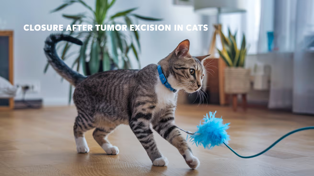
Closure Protocol for Tumor Excision in Cats
Learn the detailed closure protocol for tumor excision in cats, including surgical steps, suture choices, and post-op care for optimal healing.
Feline tumor excision creates the same fundamental closure challenge as in dogs the defect left by the tumor must be closed without compromising the margins that determined the cure rate. But the closure is performed on thinner, less elastic feline skin with fewer reconstruction options than in large-breed dogs.
Understanding how closure decisions are made helps owners interpret what the vet describes after surgery and what to expect during recovery.
Quick answer: Feline tumor excision closure follows the same layered sequence as dogs (deep margin if fascia was included, subcutaneous, skin) but with feline-specific modifications: 4-0 sutures throughout, intradermal Monocryl preferred for skin (feline skin tolerates external sutures poorly), and skin flaps used when primary tension-free closure is not achievable. Injection-site sarcomas in cats require particularly wide margins (often involving en bloc muscle removal) and frequently require reconstructive closure.
Key takeaways
- Feline skin is thinner and less elastic than canine skin, limiting primary closure options after wide-margin excision.
- 4-0 sutures throughout (vs. 3-0 in most medium dogs) feline tissue requires finer suture material.
- Intradermal Monocryl 4-0 is the preferred feline skin closure after tumor excision.
- Injection-site sarcomas (FISS) require the widest margins in feline oncological surgery and most commonly require reconstructive closure.
- Drain placement is particularly important in cats after wide excision, as dead space management is harder with thin subcutaneous tissue.
- Histopathology result timing (typically 5 to 10 business days) means re-excision discussion happens while the primary wound is still healing.
Feline tumor types and their closure implications
Injection-site sarcomas deserve specific mention. WSAVA/VIN (Soft Tissue Sarcoma 2016): "The aim of curative-intent surgery is to widely excise the primary tumor (3 cm wide and 3 cm or a fascial plane deep) and achieve negative histopathological margins."
For FISS, achieving these margins at the interscapular region frequently involves removing the trapezius muscle, spinous processes, or portions of scapula leaving a deep muscular defect that requires multi-layer closure and often a flap for the skin component.
Deep layer closure: when fascia or muscle is included
When the deep margin of excision includes the fascia below the tumor (standard for any tumor with invasion concern), the fascial defect is closed before subcutaneous work.
Pattern: simple interrupted or continuous absorbableMaterial: PDS 2-0 to 3-0
For deep muscular defects after FISS excision: the deep layers are closed in sequence (deep muscle to superficial muscle), using PDS 0 to 2-0. If the defect is too large for primary muscle closure, a mesh or mobilized flap may be used.
Subcutaneous closure and dead space
Feline subcutaneous tissue is thin and provides limited substance for suture purchase compared to dogs. Dead space management is therefore particularly challenging after wide-margin excision.
Options:
- Simple continuous absorbable (Monocryl or Vicryl 3-0 to 4-0) for the subcutaneous layer
- Walking sutures to anchor dermis to fascia when a large dead space pocket exists
- Drain placement (Penrose or closed suction) when suturing alone cannot eliminate the cavity
VCAhospitals (Penrose drain discharge instructions): "A Penrose drain is a latex tube placed into a wound with one or two ends exiting the skin, allowing fluids to drain. In most cases, the drain will exit from a new incision site, not the primary wound site."
For how dead space management relates to the broader closure strategy, see dead space management after cat tumor excision.
Skin closure
Intradermal Monocryl 4-0: the preferred method
Feline skin tolerates external sutures more poorly than canine skin. Suture-track irritation, self-trauma, and suture-mark scarring are more pronounced and more rapidly established in cats.
For routine feline tumor excision closure where tension is manageable, intradermal Monocryl 4-0 is preferred:
- No external material for the cat to lick or chew
- No removal visit required
- Finer, less visible healed scar
When primary closure is under tension
When the wound edges cannot be approximated without tension, the options match those in dogs:
Undermining: releasing skin from underlying subcutaneous tissue and fascia to mobilize it toward the defect. In cats, undermining must be carefully limited thin feline skin can be devascularized more easily than dog skin if undermining is too extensive.
Walking sutures: dermis-to-fascia advancement. Useful for moderate-sized trunk defects. Standard material: 3-0 PDS or Biosyn.
Tension-relieving sutures: horizontal mattress or vertical mattress, used with 3-0 nylon or Prolene.
Skin flap: when primary closure is not achievable. ACVS: "Closure of the defect from the excised mast cell with a small skin flap from adjacent skin near the base of the ear." Skin flaps are more commonly needed in cats than equivalent-weight dogs because feline skin has less laxity.
High-tension locations: face, ears, distal limbs
These locations have minimal adjacent skin available for advancement and carry the highest tension risk. Closure at these sites frequently requires staged reconstruction or accepting second intention healing over small residual defects.
Cosmetic closure after feline tumor excision
For tumor excision in cosmetically sensitive locations (face, visible lateral body), intradermal closure and fine suture sizes minimize visible scarring. For the cosmetic closure approach in cats, see cosmetic closure for feline tumor sites.
Histopathology and re-excision
The excised tissue is sent for histopathology, with results typically returned within 5 to 10 business days. Three possible results:
- Complete margins: no tumor cells at the inked edges. Local recurrence risk is low. No further surgery needed unless the tumor biology warrants adjuvant therapy.
- Close margins: tumor cells within 1 to 2 mm of the edge but not at the ink. Recurrence risk is elevated. Discussion of re-excision or radiation.
- Incomplete margins: tumor cells at the inked edge. Re-excision is recommended when possible.
The re-excision decision is made while the primary wound is in the active healing phase (typically at 7 to 14 days post-surgery). The entire scar track is included in the re-excision specimen meaning closure of the second wound is more complex than the first.
For how tumor excision closure compares in dogs, see tumor excision closure comparison in dogs.
Post-operative monitoring in cats
Cats hide signs of pain and discomfort more effectively than dogs. Behavioral changes (reduced appetite, hiding, abnormal posture) are often the first indicators of complications rather than obvious wound signs.
Specific monitoring for cats after tumor excision:
- Check the wound twice daily for redness, discharge, swelling, or separation
- Cats often groom the wound site even with an E-collar if the collar fits poorly verify fit at each check
- If a drain is present, monitor drainage output and color daily
- Watch for systemic signs (reduced appetite, lethargy, fever) that may precede local wound signs
For the closure checklist applicable to feline tumor excision, see closure checklist for feline tumor excision.
Frequently asked questions
What makes injection-site sarcomas so different to close?
FISS requires the widest margins in feline oncological surgery typically 3 to 5 cm laterally and removing one to two fascial planes deep, sometimes including muscle and portions of bone. The resulting defect is far larger relative to the cat's body than equivalent surgery in a dog. Closure almost always requires reconstruction, and staged surgery (debulking followed by definitive reconstruction) may be necessary.
My cat had a mass removed and the vet said they got "clean margins." Does that mean the cancer is gone?
Clean histopathological margins mean no tumor cells were identified at the edges of the tissue submitted for analysis. For many feline tumors, clean margins significantly reduce local recurrence risk. However, "clean margins" does not address the risk of metastatic spread, which depends on tumor type, grade, and whether staging (chest radiographs, lymph node assessment) was performed. Discuss the complete picture with your vet.
How long will my cat's E-collar need to stay on after tumor surgery?
Minimum until suture removal (10 to 14 days for external sutures). With intradermal closure, the E-collar remains recommended for 10 to 14 days post-surgery not for suture removal purposes, but because the incision site can still be disrupted by licking before it has adequate surface healing. Your vet will advise the specific duration based on wound appearance at the recheck.
Feline tumor excision closure is constrained by the cat's limited skin laxity, the finer tissue that tolerates suture placement, and the requirement not to compromise the margins that determine whether the tumor is fully removed. Meeting all three constraints simultaneously adequate margins, tension-free closure, appropriate suture technique is the challenge that distinguishes feline tumor surgery from routine wound closure.
Resources
- ACVS. Mast Cell Tumors. acvs.org
- VIN (WSAVA 2016). Soft Tissue Sarcoma in Dogs and Cats. vin.com
- VCA Animal Hospitals. Penrose Drain Discharge Instructions for Dogs. vcahospitals.com
- Today's Veterinary Practice. Fundamentals of Surgical Oncology in Small Animals. todaysveterinarypractice.com

Layered Closure Technique in Small Animal Surgery
Learn about the layered closure technique in small animal surgery, its benefits, steps, and tips for optimal healing in pets.
Closing a surgical wound is not a single action. Every incision goes through multiple tissue layers, and each layer needs its own closure before the next one can be addressed.
The layered closure technique is the standard approach in small animal surgery because it mirrors the anatomy of the wound. Each layer is repaired separately, from deep to superficial, restoring both structural integrity and the biological conditions needed for healing.
Quick answer: The layered closure technique closes a surgical wound in sequence from the deepest layer outward, typically: muscle and fascia, subcutaneous tissue, and skin. Each layer uses its own suture material and pattern matched to the tissue's mechanical needs and healing timeline. This method distributes wound tension across all layers, eliminates dead space at each level, and produces stronger, faster-healing wounds than single-layer closure.
Key takeaways
- Each tissue layer is closed separately, from muscle and fascia through to skin.
- Absorbable sutures are used for all internal layers they dissolve as the tissue heals.
- Dead space is eliminated at each layer, not only at the skin level.
- Tension is distributed across all layers, preventing the skin closure from bearing the full load.
- Used in spays, tumor removals, wound repairs, and most soft tissue surgeries in dogs and cats.
- Owner activity restriction is what allows the layered closure to actually adhere and heal.
Why layered closure is the standard
A single-layer closure places all wound tension on one suture line and leaves dead space in every layer below the skin. The result is a wound that has to fight the combined effects of tension, fluid accumulation, and tissue separation all at once.
Layered closure distributes those forces:
- Tension is shared across multiple suture lines
- Dead space is eliminated at each layer rather than accumulating beneath a single closure
- Tissue planes are restored to anatomical contact at every depth
The technique mimics how the body is built. Each layer had its own structure before the incision was made, and it needs its own closure to restore that structure.
The layered sequence: deep to superficial
Layer 1: Muscle and fascia
The deepest functional layer and the one that bears the most structural load after abdominal surgery. The linea alba or muscle fascia must be incorporated in suture bites the muscle belly itself does not hold sutures reliably.
Material: PDS (polydioxanone) or Biosyn, absorbable monofilament, size 0 to 3-0 depending on patient sizePattern: Simple continuous (standard) or interrupted (contaminated or poor-quality tissue)
For muscle layer closure technique in full detail, see muscle layer closure within layered technique.
Layer 2: Fascia (where distinct from muscle)
In some procedures, a separate fascial layer (such as the external rectus sheath) is closed after the muscle layer and before the subcutaneous fat.
Material: PDS, absorbable monofilament, same size range as muscle layerPattern: Simple continuous
For fascial layer closure details, see fascial layer closure within layered technique.
Layer 3: Subcutaneous tissue
The fat and connective tissue layer just beneath the skin. Closing this layer eliminates the dead space where seromas form and reduces tension on the skin edges above.
Material: Monocryl (poliglecaprone 25) or Vicryl (polyglactin 910), absorbable, size 2-0 to 4-0Pattern: Simple continuous
Cats: A 1987 JAVMA study found seroma formation in 9 of 12 cats when subcutaneous tissue was not sutured.
For dog-specific subcutaneous technique, see subcutaneous layer closure in dogs. For cats, see subcutaneous layer closure in cats.
Layer 4: Skin
The final layer. Multiple options are appropriate depending on wound type and patient factors.
For skin layer options and when each is used, see skin layer as the final closure layer.
Suture material by layer: quick reference
Sizes shift down in cats and small dogs, and up in large breed dogs.
Benefits over single-layer closure
The layered approach is not more complex for the sake of complexity. Each step solves a problem that the previous layer alone cannot.
For the principles that guide the decision to use layered closure, see principles that guide layered closure.
Dead space management within layered closure
Dead space forms at every layer where tissue was dissected. Layered closure addresses it at each level rather than leaving it to accumulate beneath the skin.
At the subcutaneous layer specifically, fluid accumulation is prevented by bringing the fat tissue back into contact using continuous absorbable sutures. When extensive dead space remains despite suturing (after large tumor removal or mastectomy), walking sutures or drains may supplement layered closure.
For dead space management strategies when layered closure alone is not enough, see dead space management during layered closure.
When layered closure is used
Layered closure is the standard for nearly all soft tissue surgery in small animals:
- Spay surgery (ovariohysterectomy): linea alba, subcutaneous, skin
- Neuter surgery: subcutaneous and skin at minimum for prescrotal approach
- Tumor excision: adds skin mobilization and walking sutures as needed
- Wound repair: applied after debridement of traumatic wounds
- Laparotomy: linea alba, subcutaneous, skin; same principle regardless of what was done inside
What owners should understand about layered closure
You will see only the skin closure the last 5 to 10 minutes of a procedure that may have taken one to two hours. The layers underneath are doing the structural work that allows the skin to heal correctly.
Your role:
- Activity restriction: the layers only adhere if the dog is rested. Motion disrupts the tissue planes trying to stick together
- E-collar compliance: licking the skin layer does not directly affect the deep layers, but it disrupts the epidermis and introduces bacteria
- No bathing: moisture weakens the skin suture line and creates infection risk before the dermal seal is complete
- Attend rechecks: your vet confirms deep healing at the scheduled follow-up, not just surface healing
Frequently asked questions
How do I know if the deep layers failed even though the skin looks healed?
Signs of deep layer failure include a soft bulge near the incision (seroma or hernia), discharge tracking from a point below the skin, return of lameness after orthopedic surgery, or abdominal discomfort. The skin can appear healed while a deeper problem develops. Report any new lumps or behavioral changes to your vet even after the incision looks fine.
Do cats and dogs get the same layered closure technique?
The sequence is the same, but the materials and sizes differ. Cats have thinner tissue and heal faster, so finer sutures are used at every layer. The subcutaneous closure is particularly important in cats because of their prominent fat layer and high seroma risk when it is omitted.
Why does my dog still need to rest when the skin sutures have been removed?
Skin suture removal at 10 to 14 days reflects external wound healing. The muscle and fascial layers take 4 to 6 weeks to approach functional strength. Full tissue remodeling continues for months. Resuming full activity at suture removal risks the deep layers before they are ready.
Layered closure works because it respects the anatomy of the wound. Each layer that was opened is closed back to its original position. When every layer is repaired with the right material and pattern, the wound heals as the body intended from the inside out, without the complications that arise when shortcuts are taken.
Resources
- Veterian Key. Surgery of the Abdominal Cavity. veteriankey.com
- PubMed. Tissue reaction to suture material in the feline linea alba. JAVMA, 1987. pubmed.ncbi.nlm.nih.gov
- WCVM University of Saskatchewan. Lab 6 Part 4: Incision Closure. wcvm.usask.ca
- Veterinary Surgery Online. Wound Closure: Continued. vetsurgeryonline.com
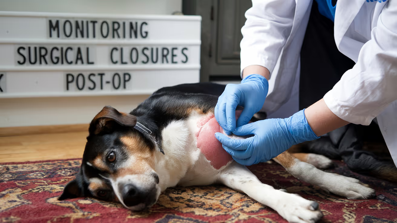
Post-Operative Monitoring of Surgical Closures
Learn essential steps for post-operative monitoring of surgical closures to ensure healing and prevent complications in pets.
What happens at home in the two weeks after surgery determines as much about healing outcomes as what happened in the operating room. The wound that closes perfectly can still fail through licking, jumping, or unrecognized infection.
Owners who know what to check, how often, and what each finding means are far more likely to catch complications early when they are still manageable.
Quick answer: Check the surgical wound twice daily for the first 10 to 14 days. Normal findings: mild swelling and redness for 2 to 3 days, a thin dry crust at the wound margins, gradual improvement day by day. Abnormal findings requiring same-day contact: yellow or green discharge, foul odor, worsening redness beyond the wound margin, increasing swelling after day 3 to 4, wound opening (dehiscence), or any tissue protruding from the wound. Licking is the most preventable cause of post-operative closure failure.
Key takeaways
- Twice-daily wound checks for the full 10 to 14 days are the monitoring standard.
- Normal post-operative swelling and redness peaks at days 2 to 3, then progressively decreases.
- Seroma (soft fluctuant swelling) differs from infection and is usually harmless.
- Dehiscence (wound opening) is a same-day emergency cover with a clean cloth and call immediately.
- Licking is the most common owner-controllable cause of suture failure E-collar compliance is not optional.
- Pale gums, open wound with tissue protruding, or collapse require immediate emergency care.
What to check at each monitoring session
Twice-daily monitoring takes less than two minutes. The same checks, performed consistently, provide the trend data needed to distinguish normal healing from early complication.
Five things to assess at each check:
- Wound edges: closed and apposed at all points? No visible gaps between suture sites?
- Swelling: decreasing compared to the last check? Or increasing?
- Color: pink and normalizing? Or reddening, darkening, or developing pale areas?
- Discharge: none, or only a small amount of dried serous crust at the wound margins?
- Odor: none, or any smell from the wound area?
Metropolitan Veterinary Associates: "The incision should be monitored for redness, swelling, oozing, heat or pain to the touch. Any of these signs may indicate an incisional infection."
Normal findings by timeline
Days 1 to 2
- Mild swelling around the incision: normal the body's inflammatory response is at its peak
- Mild redness at the wound margins: normal
- Small amount of dried bloody crust at suture sites: normal
- Pet may be quiet, less active than usual: normal effect of anesthesia and post-operative pain management
MedVet: "It is normal to see mild lethargy, reduced appetite, or slight discomfort during the first 24 to 72 hours."
Days 3 to 5
- Swelling should begin to decrease from its peak
- Redness should be stabilizing or reducing not spreading
- A small firm ridge along the wound line is normal: this is early collagen deposition
- Pet appetite should be returning toward normal
Days 5 to 10
- Progressive improvement day by day
- The wound surface should look drier, flatter, and less reactive
- Sutures remain intact and wound edges remain fully apposed
Days 10 to 14
- Wound should be fully closed, dry, and healed at the surface
- Suture removal recheck visit (for external sutures)
- Vet assesses wound before removing sutures removal may be deferred if healing is incomplete
For how suture removal timing is assessed at the recheck in dogs, see suture removal timing at the day 10-14 recheck. For cats, see suture removal timing at the feline recheck.
Distinguishing seroma from infection
Both seroma and infection produce swelling near the surgical site. They require different responses.
Liberty Animal Hospital: "Seroma: fluid accumulation at the incision site due to tissue irritation this is normal. Continue with cold compress only until the swelling has gone down. The body will absorb it over time."
If you cannot confidently distinguish a seroma from an early infection, contact your vet. Assessment may require palpation or aspiration to determine fluid character.
For how seromas are prevented at the closure stage, see seroma prevention during closure.
Dehiscence: when the wound opens
Wound dehiscence is the partial or complete opening of a sutured wound. It is a same-day emergency not a wait-and-see situation.
PetPlace: "When sutures break down, the underlying tissues have the potential to protrude through the incision and be exposed to the exterior. This can lead to serious infections, which may be fatal."
What to do if the wound opens:
- Apply a clean towel or cloth gently over the wound
- Do not attempt to replace protruding tissue
- Do not apply ointments, disinfectants, or saline without vet guidance
- Transport to the vet or emergency clinic immediately
Common causes of dehiscence:
- Licking or chewing the sutures
- Jumping, running, or abrupt activity
- Sutures removed too early
- Infection undermining the closure from within
- Underlying tension that was not adequately managed at surgery
For how common closure errors produce dehiscence, see closure errors that lead to dehiscence.
Infection: what to look for
Early surgical site infection typically presents between days 3 and 7. The signs progress from mild to severe as the infection establishes.
Early signs (days 3 to 5):
- Redness extending beyond the immediate wound margin
- Increased warmth at the wound site
- Mild increase in discharge (serous becoming slightly cloudy)
Established infection (days 5 to 10):
- Purulent discharge (yellow, green, or brown)
- Foul odor
- Wound swelling increasing rather than decreasing
- Pet showing systemic signs: lethargy, reduced appetite, fever
Contact your vet the same day if you notice any of the early signs. Do not wait for the established infection stage early treatment is significantly simpler than treating an established surgical site infection.
Activity restriction: enforcing it at home
Metropolitan Veterinary: "Dogs and cats should be kept from jumping up/down on/from high surfaces, running up steps or any other activity that puts tension on the incision. Excess tension can lead to dehiscence."
Practical activity restriction strategies:
- Dogs: leash-only outdoor activity for 10 to 14 days; confine to one room or use a pen when unsupervised
- Cats: confine to a room without high furniture; prevent stair access
- Both: separate from other pets who may play with or groom the wound
The challenge is that pets often feel better before the wound has adequate tensile strength. A dog that seems completely normal at day 5 does not have a day-5 wound the tissue is still in the active repair phase and cannot tolerate the same forces the dog is willing to exert.
E-collar compliance: non-negotiable
Licking introduces oral bacteria directly to the wound surface, mechanically disrupts suture lines, and can remove external sutures within minutes of unsupervised access.
MedVet: "To prevent licking, which can delay healing, cause infection, or lead to the incision opening, use an Elizabethan collar (cone), a cervical collar, or cover the incision with a T-shirt or bandage."
The E-collar must be worn:
- At all times, including during sleep
- When the owner is in the room but not actively watching the pet
- When the pet is in its crate or confined space
Alternatives to the standard E-collar if compliance is difficult: inflatable donut collar (cats often tolerate better), surgical recovery suit, or soft fabric cone.
Emergency signs: call immediately
Some findings do not wait for a scheduled call to the vet. Seek care immediately for:
- Pale or white gums: indicates blood loss or cardiovascular compromise
- Blue-tinged gums or rapid breathing: respiratory distress
- Wound fully open with tissue protruding: cover and transport immediately
- Collapse or inability to stand
- Bright red active bleeding that does not stop within 5 minutes of gentle pressure
MedVet: "Pale gums, which can indicate potential blood loss or poor circulation seek veterinary care immediately, regardless of the time of day."
For the full closure checklist that precedes this monitoring period, see closure checklist prior to discharge. For how drain monitoring integrates with wound monitoring, see monitoring wounds with drains in place.
Frequently asked questions
My dog's wound looks fine but she keeps trying to lick it. Should I be worried?
The licking attempt is the warning sign the wound does not need to look damaged yet for the E-collar to be essential. Licking can remove sutures or disrupt healing within minutes. The fact that she is attempting to lick means the E-collar must be worn consistently, not just when she actively succeeds. Wounds that look fine often look that way because the E-collar has been working.
There is a small bump near the wound that appeared on day 4. How do I know if it is a seroma or infection?
Gently palpate (press softly) the bump. A seroma feels soft and fluid-filled, like a water balloon under the skin. An early infection usually feels firmer, is warmer to the touch, and the pet shows some discomfort when you touch it. If the bump is soft and the pet is otherwise well and eating, a seroma is more likely. Either way, contact your vet at the next business opportunity and immediately if the bump is warm, the pet seems unwell, or discharge is present.
My cat won't eat after surgery. Is that a complication?
Reduced appetite for 24 to 72 hours is common and expected. Cats that refuse to eat for more than 72 hours after surgery require prompt veterinary assessment. Metropolitan Veterinary: "Cats, in particular, cannot tolerate anorexia for long periods. They are predisposed to developing severe liver disease (hepatic lipidosis/fatty liver) within days of complete anorexia."
Post-operative monitoring is owner-controlled quality control for the surgeon's work. The wound that closes well can still fail but usually only if something external disrupts it. Every day of consistent monitoring, E-collar compliance, and activity restriction protects the closure that was placed and gives it the environment it needs to heal.
Resources
- Metropolitan Veterinary Associates. Abdominal Surgery: Post-Operative and Incisional Care. metro-vet.com
- MedVet. Caring for Your Cat or Dog After Surgery. medvet.com
- PetPlace. Postoperative Complications in Dogs. petplace.com
- Liberty Animal Hospital. Post-Surgery Guidelines. libertyanimalhospital.com

Interrupted vs Continuous Suturing in Cats
Learn the differences between interrupted and continuous suturing in cats, including benefits, risks, and best practices for healing wounds.
Cats are not small dogs. This matters in surgery including in how their wounds are closed. Feline skin is thinner, more mobile, and more prone to self-inflicted wound disruption than canine skin.
These properties influence which suture patterns work best and which materials are best tolerated.
This guide explains the main suture patterns used in feline surgery, what research shows about outcomes, and what owners should expect from each type of closure.
Quick answer: Feline surgery most commonly uses buried continuous intradermal (BCID) closure: sutures placed beneath the skin with no external stitches. BCID has significantly lower dehiscence rates than simple interrupted sutures in cats. No suture removal is needed with absorbable intradermal sutures.
Key takeaways
- BCID closure is preferred in most feline surgeries: significantly lower dehiscence rate than simple interrupted external sutures
- ARSHI Veterinary Letters (2024): BCID had significantly fewer complications including dehiscence than simple interrupted in feline OVH
- Intradermal closure eliminates suture removal stress: absorbable sutures dissolve over 60 to 90 days
- Cats are prone to suture self-trauma: external sutures are accessible to grooming; intradermal sutures are not exposed
- Suture size matters in cats: smaller gauges (4-0 or 5-0) minimize tissue drag and inflammatory reaction in feline skin
- Monofilament sutures are preferred over braided for feline skin to reduce bacterial adherence and tissue drag
Why feline suturing differs from canine
Cats have several anatomical and behavioral characteristics that influence closure technique selection:
Thin, mobile skin: feline dermis is thinner than canine dermis. External sutures can cut through the skin edge more easily, particularly if tension is placed across the closure.
Self-grooming behavior: cats groom surgical incisions relentlessly. An E-collar reduces but does not eliminate this behavior. External sutures that protrude through the skin are at higher risk of being chewed or pulled out. Intradermal sutures that sit below the surface are inherently less accessible.
Smaller incisions: many feline procedures (neuters, spays, lumpectomies) involve relatively short incisions where intradermal placement is technically feasible for most surgeons.
Higher inflammatory response to some materials: cats can show more pronounced tissue reaction to certain suture materials than dogs, particularly braided multifilament absorbable sutures.
Suture patterns used in cats
Buried continuous intradermal (BCID)
The standard approach for feline elective surgery skin closure.
NCBi (PMC study in cats): BCID eliminates suture removal, decreases scar formation, reduces tissue inflammation and SSI risk by avoiding percutaneous suture tracts, and reduces self-induced trauma.
ARSHI Veterinary Letters (2024): "This study evaluated two suture techniques, BCID and simple interrupted (SI), in 42 female cats using nonabsorbable nylon sutures.
The results indicated a significantly higher incidence of complications, notably dehiscence, in the SI group. Consequently, BCID is recommended because of its lower complication rate and reduced risk of infection from self-trauma."
How it works: absorbable monofilament suture (poliglecaprone 25 or polydioxanone) is placed within the dermal layer in a continuous running pattern. Knots are buried within the tissue. No suture material protrudes through the skin surface. The incision appears as a clean line with no external stitches visible.
Suture material: NCBi (PMC study): "Monofilament or multifilament suture materials have been used intradermally for closure of skin incisions in dogs, cats, and humans, but there are no studies comparing absorbable and nonabsorbable monofilament suture material for a continuous intradermal closure in small animals."
The comparison of absorbable vs. nonabsorbable for intradermal closure in cats showed both are usable, but absorbable materials have the advantage of not requiring removal.
Simple interrupted
Each suture is a separate unit placed through full skin thickness and tied externally. Standard for skin closure in many species.
SustainableVet: "Simple interrupted sutures: Provide strong closure and allow precise wound edge alignment, commonly used in feline neuters."
NCBi (PMC study on intradermal closure): "Common interrupted external suture patterns in the skin include the simple interrupted and cruciate patterns.
These patterns allow more precise approximation of the skin edges along with more security than continuous buried suture patterns."
Disadvantages in cats: ARSHI (2024) found significantly higher dehiscence rates in cats compared to BCID. External sutures are accessible to grooming. They require a suture removal appointment at 10 to 14 days stressful for cats and owners.
When appropriate: wounds that cannot be closed intradermally (highly contaminated wounds, wounds under significant tension, locations where intradermal technique is not technically feasible), or where the surgeon prefers external closure for monitoring.
Cruciate sutures
A variation of interrupted sutures where each suture crosses in an X pattern. Distributes tension over a wider area; reduces skin edge inversion. Used in higher-tension skin closures in cats.
Ford interlocking (locking continuous)
A continuous pattern where each bite is locked. Used primarily for body wall and fascia closure rather than skin. Provides the speed advantage of a continuous pattern with improved security.
Suture material selection in cats
Gauge
SustainableVet: "Suture size selection: Smaller sizes (4-0 or 5-0) are preferred for delicate feline skin to minimize trauma and promote fine healing."
Smaller gauge sutures cause less tissue drag on passage through feline dermis and produce smaller suture tracts if external sutures are used.
Monofilament vs. braided
SustainableVet: "Monofilament sutures: These have smooth surfaces that reduce tissue drag and lower infection risk compared to braided sutures."
Braided multifilament sutures have a higher surface area for bacterial colonization and more tissue drag. In thin feline skin, monofilament materials are strongly preferred for both skin and subcutaneous layers.
Absorbable materials for internal layers
SustainableVet: "Absorbable sutures: Materials like polydioxanone (PDS) or poliglecaprone (Monocryl) dissolve over time, ideal for internal tissue layers to avoid suture removal stress."
Typical feline surgery layered closure:
- Body wall/linea alba (if abdominal): absorbable, monofilament, continuous
- Subcutaneous layer: absorbable, monofilament, continuous (closes dead space)
- Skin: intradermal, absorbable monofilament 4-0 or 5-0 (no external sutures)
What owners should know
No external sutures visible: with intradermal closure, the incision should appear as a smooth line. This is correct and not a sign that closure was incomplete.
E-collar is still required: even without external sutures, cats can traumatize a healing wound through licking. The E-collar protects the intradermal closure from grooming during the initial healing phase.
No suture removal appointment: absorbable intradermal sutures dissolve without requiring a procedure. If nonabsorbable intradermal sutures were used, your vet will advise on removal.
What to monitor: a small amount of bruising or mild swelling in the first 2 to 3 days is normal. Increasing redness after day 3, any discharge, or opening of the incision line requires veterinary assessment.
For suturing in dogs for comparison, see interrupted vs. continuous suturing in dogs. For tissue adhesives as an alternative, see tissue adhesives in dog and cat surgery.
Frequently asked questions
Why does my cat have no visible stitches after surgery?
Intradermal closure places sutures beneath the skin surface. The incision is closed; the sutures are simply not visible from outside. This is intentional and provides better outcomes in cats than external sutures.
Does my cat still need an E-collar if there are no external stitches?
Yes. Cats can traumatize a healing wound through licking even without external sutures to grab. The E-collar is required throughout the initial healing phase regardless of closure technique.
When will the stitches dissolve?
Absorbable intradermal sutures typically dissolve over 60 to 90 days depending on the material. The dissolution is internal you will not see suture material emerge or fall out.
My cat's incision opened slightly at one end. What should I do?
Contact the vet same day. Minor dehiscence at a closure end is the most common feline complication. Small gaps may close conservatively; larger openings may need reclosure.
Can my cat's incision be closed with tissue glue instead of sutures?
Tissue glue suits very short, low-tension wounds in cats but not longer incisions like spays. Ask your vet whether glue or sutures are more appropriate for the specific wound.
Can staples be used to close cat skin instead of sutures?
Staples are occasionally used in cats but require removal at 10 to 14 days and cause more tissue reaction than fine monofilament sutures. For most elective feline surgery, intradermal sutures are preferred.
Resources
- ARSHI Veterinary Letters. Comparison of Buried Continuous Intradermal and Simple Interrupted Suture Patterns for Skin Closure in Feline Ovariohysterectomy. journal.ipb.ac.id
- NCBi PMC. Comparison of Absorbable and Nonabsorbable Sutures for Intradermal Skin Closure in Cats. ncbi.nlm.nih.gov
- ScienceDirect. Comparison of Continuous Intradermal with Simple Interrupted Suture Pattern in Dogs. sciencedirect.com
- Academia. Comparison of Absorbable and Nonabsorbable Sutures for Intradermal Skin Closure in Cats. academia.edu
Suture Size Selection in Small Animal Surgery
Learn how to choose the right suture size for small animal surgery to ensure optimal healing and minimize complications.
Every surgical suture comes in multiple sizes, and size matters as much as material. A suture that is too large for the tissue causes unnecessary inflammation and delays healing. One that is too small fails to hold under the mechanical forces the wound faces.
Selecting the right size requires understanding how sutures are sized, what each tissue needs, and how patient weight influences the decision.
Quick answer: Suture sizes follow the USP (United States Pharmacopeia) system, where more zeroes mean smaller diameter so 4-0 is smaller than 2-0, and 2-0 is smaller than 0. The guiding principle is to use the smallest size that provides adequate tensile strength for the tissue. For small animals: linea alba in medium dogs takes 0 to 2-0; subcutaneous layers take 2-0 to 3-0; skin takes 3-0 to 4-0. Cats and small dogs use sizes one unit finer throughout.
Key takeaways
- The USP size system runs from 11-0 (smallest) to 7 (largest) more zeroes means smaller diameter.
- Use the smallest size that provides adequate tensile strength larger sizes cause more inflammation.
- Linea alba in medium dogs: 0 to 2-0 PDS or Biosyn.
- Subcutaneous tissue in dogs: 2-0 to 3-0 Monocryl or Vicryl.
- Skin in dogs and cats: 3-0 to 4-0 for external sutures; 4-0 for intradermal.
- Cats and small dogs use sizes one unit finer than dogs of average size throughout.
Understanding the USP suture sizing system
The United States Pharmacopeia (USP) system is the standard for suture sizing in veterinary and human surgery.
How it works:
- Sutures are assigned a numerical designation based on diameter
- Size 1 is larger than size 0
- Below size 0, additional zeroes are added: 0, 2-0, 3-0, 4-0, 5-0, etc.
- Each additional zero indicates a smaller diameter
- 11-0 is the smallest (microsurgery); 7 is the largest (orthopedic/heavy tissue)
Veterinary Surgery Online explains: "The more zeros, the smaller the material, so 6-0 is actually size 000000, and is pronounced 'six ought' or 'six zero'."
AAHA (Oh, Sew Easy, 2022): "Optimal suture size is determined as the smallest size necessary to achieve a tension-free wound closure."
The core principle: smallest effective size
Veterinary Practice News (Dr. Kendra Freeman, DACVS): "The general principle is to use the smallest suture strong enough for the tissue. This allows for less suture material to be present, which may potentially contribute to inflammation and tissue reaction."
What happens when suture is too large:
- More foreign material in the tissue
- Greater inflammatory response
- Higher infection risk at the suture site
- Possible tissue strangulation if pulled too tight
What happens when suture is too small:
- Suture cuts through the tissue under mechanical load
- Wound dehiscence
- Need for re-closure
Published research (Frontiers in Veterinary Science, 2023): A study comparing USP 2-0, 3-0, and 4-0 PGA sutures in canine abdominal incisions found 4-0 suture had lower inflammatory response markers while maintaining adequate mechanical strength. The authors concluded "USP 4-0 PGA suture has more advantages to suturing canine abdominal surgical incisions."
Size guide by tissue type and patient
Linea alba / fascial closure
Veterinary Practice News: "2-0 or 0 suture is appropriate for the linea alba in medium- to large-size animals."
For why the fascial layer specifically requires larger suture than adjacent muscle, see tissue type as a guide for suture size.
Subcutaneous tissue
Gastrointestinal and urinary tract
Veterinary Practice News: "3-0 or 4-0 suture is generally appropriate for the gastrointestinal tract and urinary bladder."
These organs require fine suture because oversized suture causes excessive lumenal reaction and stenosis risk. Taper-point needles are paired with these sizes to minimize tissue injury.
Skin (external interrupted)
Skin (intradermal)
4-0 is the standard for intradermal (subcuticular) closure across all patient sizes. The dermis is a uniform layer that does not vary as much with patient size as deeper structural layers.
Needle size relationship to suture size
Suture size and needle size are linked the needle is sized to match the suture. Common veterinary needle types:
For how needle selection alongside suture size affects closure quality, see needle size alongside suture size.
Common sizing errors
Too large for the tissue:Most common in the skin layer, where surgeons sometimes default to a larger size "for security." The result is more visible suture marks, more inflammation, and longer healing time.
Too small for the load:More common in the linea alba, where a 3-0 suture is used in a large breed dog that needs 0 or 2-0. Tension exceeds the suture's strength before healing occurs.
Mismatched sizing across layers:Each layer needs its own appropriate size. Using the same suture size throughout all layers (a practice shortcut) results in either oversized subcutaneous sutures or undersized fascial sutures.
For how suture size selection connects to material selection across procedures, see size alongside material selection in dogs. For cats, see size alongside material selection in cats.
Frequently asked questions
My vet used 3-0 suture for my large dog's spay. Is that too small?
It depends on the layer. 3-0 for the subcutaneous and skin layers of a medium-large dog is appropriate. 3-0 for the linea alba in a dog over 25 kg would likely be undersized 0 or 2-0 is standard for that layer. If you are concerned, ask your vet specifically which sizes were used at each layer.
Do smaller sutures dissolve faster?
No. Absorption timeline is determined by the suture material, not by its size. A 3-0 Monocryl and a 2-0 Monocryl absorb over the same timeline approximately 90 to 119 days. The difference is tensile strength, not absorption rate.
Can suture sizes vary between different materials of the same USP designation?
Yes, slightly. Veterinary Surgery Online notes: "It is important to note that the USP dimensions for catgut are different to those of other sutures. For example, 2-0 Catgut is larger than 2-0 PDS." When comparing materials, the USP designation is a starting reference, not an exact interchangeable standard across all material types.
Suture size selection follows one rule applied at every layer: the smallest size that adequately holds the tissue under its expected mechanical load. Getting it right means less inflammation, less reaction, and faster healing. Getting it wrong in either direction creates predictable complications that show up in the first two weeks of recovery.
Resources
- Veterinary Practice News. The Must-Read Guide to Selecting Sutures (Dr. Kendra Freeman, DACVS). veterinarypracticenews.com
- Veterinary Surgery Online. Suture Size. vetsurgeryonline.com
- AAHA. Oh, Sew Easy: A Guide to Sutures. aaha.org
- Frontiers in Veterinary Science (2023). Biomechanical and tissue reaction: the effects of varying suture size on canine abdominal wall stitching. frontiersin.org

Cruciate Sutures in Dog and Cat Skin Closure
Learn how cruciate sutures help close dog and cat skin wounds effectively with step-by-step guidance and care tips.
The cruciate suture pattern is simply a simple interrupted suture that crosses back over itself in an X before the knot is tied. That additional crossing pass is what makes it behave differently under tension.
It is one of the more commonly used skin closure patterns in small animal surgery and one of the least explained to owners.
Quick answer: A cruciate suture is an X-shaped interrupted suture pattern where the needle makes two crossing passes across the wound before tying. Published research (JAVMA, 2016, 60 skin specimens from 30 Beagles) found cruciate sutures placed significantly faster than simple interrupted sutures while achieving equivalent tensile strength at failure. Cruciate patterns are preferred when tissues are fragile, sutures are pulling out, or faster closure time is a priority. Both cruciate and simple interrupted patterns significantly outperform intradermal sutures in tensile strength.
Key takeaways
- Cruciate sutures form an X-shape, passing the needle twice across the wound before tying.
- Placement is significantly faster than simple interrupted sutures, per published JAVMA research.
- Tensile strength is equivalent to simple interrupted sutures at failure.
- Preferred over simple interrupted when tissue is fragile and sutures are pulling out with each bite.
- Both patterns outperform intradermal sutures in tensile strength under tension loading.
- Used at 4 to 6 mm spacing, same as simple interrupted, placed 4 to 6 mm from the wound edge.
What a cruciate suture is
A cruciate suture is an interrupted suture pattern. Like simple interrupted, it is placed individually and tied independently failure of one suture does not open the entire wound.
The technique difference from simple interrupted:
- The needle enters the skin 4 to 6 mm from the wound edge on side A
- Crosses the wound and exits 4 to 6 mm from the edge on side B
- Instead of tying here, the needle re-enters side B, crossing back over the wound
- Exits on side A again, offset from the first entry
- The two ends are tied producing an X-shaped crossing over the wound surface
The crossing configuration is what distributes tension differently from simple interrupted. Each arm of the X engages the tissue on a slightly different vector.
Published evidence: JAVMA 2016 study
The most directly relevant published data comes from a JAVMA 2016 study by Kieves et al. (Iowa State University), comparing four closure patterns on 60 skin specimens from 30 Beagles:
Patterns compared: simple interrupted, cruciate, intradermal, subdermal
Key findings:
Placement time: "Mean suture placement time for the cruciate pattern was significantly less than that for other patterns." Cruciate was faster than all three other patterns.
Tensile strength: "Mean tensile strength at skin-edge separation and suture-line failure for the simple interrupted and cruciate patterns were significantly higher than those for the intradermal and subdermal patterns."
Simple interrupted vs. cruciate: no significant difference in tensile strength at failure. The two patterns are mechanically equivalent.
Mode of failure: simple interrupted failed by suture breakage; cruciate, intradermal, and subdermal failed by tissue failure (the suture held; the tissue gave out).
Wound apposition: no significant difference among all four patterns.
Clinical implication: the cruciate pattern closes wounds faster than simple interrupted while providing equivalent mechanical strength. In fragile tissue where the suture-to-tissue interface is the weak point, cruciate distributes that interface differently which is why Veterinary Surgery Online specifically recommends it when tissue is fragile: "If tissues are fragile and suture pulls out easily, use cruciate or horizontal mattress pattern instead of simple interrupted for less stress on each bite."
When cruciate sutures are preferred
Fragile or friable tissue: when the tissue is thin, inflamed, or poor quality, simple interrupted sutures cut through at the entry point. The cruciate distributes the bite force differently, reducing focal stress.
Speed priority: in high-volume settings or time-sensitive cases (very young patients, high anesthetic risk), the faster placement time of cruciate sutures has a practical advantage.
Moderate tension with no mattress pattern: for wounds with moderate tension that do not require the full tension-relieving capacity of a horizontal or vertical mattress, the cruciate provides additional holding strength over simple interrupted without the complexity of a mattress pattern.
Where tissue is moving: joints, limb skin, and areas subject to post-operative movement. The X configuration provides slightly more surface contact with the tissue.
For how cruciate sutures compare to appositional patterns in high-tension contexts, see appositional vs everting closure patterns.
When simple interrupted is preferred over cruciate
Irregular or curved wounds: simple interrupted sutures give more individual control over tension at each point. In irregular wounds, cruciate sutures can be harder to place accurately.
Maximum monitoring need: since each cruciate suture involves more tissue and two crossing passes, failure of one is more visually apparent and potentially more complex than a single interrupted suture. In very high-infection-risk wounds, simple interrupted allows each stitch site to be individually assessed.
When exact edge apposition is critical: the offset crossing passes of a cruciate suture make precise edge alignment slightly more demanding than simple interrupted.
For how cruciate sutures fit within the full skin closure method comparison, see cruciate and other skin closure methods compared.
Comparing key skin closure patterns
Data from JAVMA 2016 (Kieves et al.) and Veterinary Surgery Online.
For the full tension-relieving pattern comparison including how cruciate compares to mattress sutures, see tension-relieving patterns alongside cruciate sutures.
Suture material for cruciate patterns
The same materials used for simple interrupted skin closure are used for cruciate:
- Nylon (Ethilon): most common external skin suture; low tissue reaction; requires removal at 10 to 14 days
- Prolene (polypropylene): similar to nylon; even lower tissue reaction; good for high-movement areas
- Monocryl 4-0: if absorbable cruciate closure is desired to avoid a removal visit
Size: 3-0 for medium dogs; 2-0 for large dogs; 4-0 for cats and small dogs. The same sizing guidance as for simple interrupted applies.
Removal
Cruciate sutures are removed the same way as simple interrupted sutures: scissors cut under one arm of the X, and the suture is pulled through in one motion. The crossing pass does not complicate removal. Timing: 10 to 14 days, per standard skin closure guidance.
For suture removal timing guidance in dogs, see suture removal timing for cruciate sutures in dogs. For cats, see suture removal timing for cruciate sutures in cats.
Frequently asked questions
My dog has X-shaped stitches. Is that a cruciate pattern?
Yes. The visible X crossing the wound surface is the distinguishing feature of a cruciate suture pattern. Each X is tied individually.
Are cruciate sutures stronger than regular stitches?
Published JAVMA data shows no significant difference in tensile strength between cruciate and simple interrupted at failure. Cruciate sutures place faster and distribute their bite force differently which matters in fragile tissue but they are not inherently stronger than simple interrupted.
Can cruciate sutures be used anywhere on the body?
Yes, in most locations. They are particularly useful on the trunk, dorsum, and limb skin in dogs. In cats, they are used in similar situations, though fine-gauge sutures (4-0) are important given feline skin fragility. Intradermal closure is often preferred in cats for cosmetic procedures, but cruciate is appropriate where external sutures are indicated.
The cruciate suture earns its place in small animal surgery primarily through speed placing faster than every other pattern in the JAVMA study while achieving identical tensile strength to simple interrupted. When tissue is fragile enough that simple interrupted sutures are pulling out with each bite, the different bite geometry of cruciate sutures addresses that clinical problem directly.
Resources
- JAVMA 2016 (Kieves et al.). Comparison of Tensile Strength Among Simple Interrupted, Cruciate, Intradermal, and Subdermal Suture Patterns for Incision Closure in Ex Vivo Canine Skin Specimens. pubmed.ncbi.nlm.nih.gov
- Veterinary Surgery Online. Wound Closure Continued. vetsurgeryonline.com
- University of Minnesota Large Animal Surgery. Suturing Skin. open.lib.umn.edu
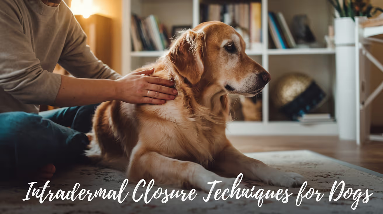
Intradermal Closure in Dogs: Techniques & Benefits
Learn about intradermal closure in dogs, its benefits, techniques, and aftercare for optimal healing and cosmetic results.
Intradermal closure in dogs is a surgical technique used to close skin wounds with minimal scarring and faster healing. This method places sutures within the skin layers, avoiding external stitches that can irritate your dog or require removal. Understanding intradermal closure helps you make informed decisions about your pet's surgical care.
This article explains what intradermal closure is, how it works, its advantages, and how to care for your dog after surgery. You will learn about the materials used, common techniques, and potential complications to watch for during recovery.
What is intradermal closure in dogs?
Intradermal closure is a suturing technique where stitches are placed inside the skin layers, specifically within the dermis. This method hides the sutures beneath the skin surface, reducing irritation and improving cosmetic outcomes. It is commonly used in veterinary surgery for skin incisions and lacerations.
The goal is to bring the skin edges together securely while minimizing external suture exposure. This technique helps wounds heal with less inflammation and reduces the risk of infection or self-trauma by the dog.
- Hidden sutures: Sutures are placed beneath the skin surface, preventing your dog from licking or scratching them, which promotes safer healing.
- Cosmetic benefit: Intradermal closure results in less visible scarring compared to traditional external stitches, improving your dog's appearance after surgery.
- Reduced suture removal: Because sutures are buried, they often do not require removal, reducing stress for both you and your dog.
- Secure wound closure: This technique provides strong wound edge apposition, which helps prevent wound opening and infection.
Intradermal closure is a preferred method in many veterinary surgeries due to these benefits. It requires skill and appropriate suture materials to ensure success.
How is intradermal closure performed in dogs?
The procedure involves placing a continuous or interrupted suture pattern within the dermis layer of the skin. The surgeon carefully aligns the wound edges and uses absorbable sutures to close the incision beneath the surface.
Proper technique is essential to avoid tension on the skin and to ensure the wound edges heal evenly. The choice of suture material and needle type also affects the outcome.
- Suture placement: Sutures are inserted horizontally or vertically within the dermis to bring skin edges together without external exposure.
- Absorbable sutures: Materials like poliglecaprone or polydioxanone are used, which dissolve over time, eliminating the need for removal.
- Needle type: A reverse cutting needle is often preferred to minimize tissue trauma during suture placement.
- Continuous pattern: A running intradermal suture provides even tension distribution and faster closure compared to interrupted stitches.
After suturing, the skin surface appears smooth with no visible stitches. This technique requires careful handling to avoid puckering or uneven closure.
What are the benefits of intradermal closure for dogs?
Intradermal closure offers several advantages over traditional external suturing methods. These benefits improve healing, reduce complications, and enhance cosmetic results for your dog.
Understanding these benefits can help you discuss surgical options with your veterinarian confidently.
- Less irritation: Buried sutures prevent your dog from chewing or scratching the wound, reducing inflammation and discomfort.
- Improved healing: The technique promotes better skin edge alignment, which supports faster and stronger wound healing.
- Lower infection risk: With no external suture exposure, there is less chance for bacteria to enter the wound site.
- Better appearance: The cosmetic outcome is superior, with minimal scarring and no visible stitches after healing.
These benefits make intradermal closure a preferred choice for elective surgeries and wounds in dogs, especially in visible areas like the face or limbs.
When is intradermal closure recommended for dogs?
Veterinarians choose intradermal closure based on the wound type, location, and patient factors. It is most suitable for clean, straight incisions where cosmetic outcome is important.
Not all wounds are ideal for this technique, so your vet will assess the situation before deciding.
- Clean surgical incisions: Intradermal closure is ideal for planned surgeries with minimal contamination risk.
- Superficial wounds: Wounds involving only the skin layers without deep tissue damage are good candidates.
- Areas needing cosmetic care: Locations like the face, neck, or limbs benefit from less visible scarring.
- Stable patients: Dogs that can tolerate anesthesia and have no skin infection are suitable for this closure method.
Your veterinarian will evaluate your dog's specific case to determine if intradermal closure is the best option.
What materials are used for intradermal closure in dogs?
Choosing the right suture material is crucial for successful intradermal closure. Absorbable sutures are preferred to avoid the need for removal and reduce irritation.
The needle type also affects how easily the sutures pass through the skin and how much tissue damage occurs.
- Poliglecaprone sutures: These absorbable sutures dissolve within 90-120 days and cause minimal tissue reaction.
- Polydioxanone sutures: Longer-lasting absorbable sutures that maintain strength for several weeks, ideal for slow-healing wounds.
- Reverse cutting needles: Designed to cut through tough skin with less trauma, improving suture placement accuracy.
- Monofilament sutures: Smooth, single-strand sutures reduce bacterial colonization and tissue drag during closure.
Your veterinarian selects materials based on wound characteristics and healing needs to optimize outcomes.
How should you care for your dog after intradermal closure?
Proper aftercare is essential to ensure your dog's wound heals well after intradermal closure. You must prevent infection, avoid trauma, and monitor for complications.
Following your vet's instructions carefully will help your dog recover quickly and comfortably.
- Limit activity: Restrict your dog's movement to prevent stress on the wound and avoid suture disruption.
- Prevent licking: Use an Elizabethan collar or other devices to stop your dog from licking or chewing the wound area.
- Keep wound clean: Follow your vet’s advice on cleaning the site gently without soaking or harsh chemicals.
- Watch for signs: Monitor for redness, swelling, discharge, or pain, and contact your vet if these occur.
Most wounds heal within 10-14 days with proper care. Absorbable sutures dissolve on their own, so no removal is needed unless complications arise.
What complications can occur with intradermal closure in dogs?
While intradermal closure is generally safe, some complications can happen. Knowing what to expect helps you act quickly if problems arise.
Early detection and veterinary care can prevent minor issues from becoming serious.
- Wound dehiscence: The wound may reopen if sutures fail or if your dog stresses the area excessively.
- Infection risk: Though lower than external sutures, infections can still occur and require prompt treatment.
- Allergic reaction: Some dogs may react to suture materials, causing redness or swelling.
- Excessive scarring: Improper technique or healing can lead to thick or raised scars, affecting appearance and function.
If you notice any unusual changes in your dog's wound, contact your veterinarian immediately for assessment and care.
Conclusion
Intradermal closure in dogs is a valuable surgical technique that offers strong wound closure with minimal scarring and less irritation. It uses absorbable sutures placed beneath the skin surface, improving healing and cosmetic results.
Understanding how this method works, when it is recommended, and how to care for your dog afterward helps ensure the best recovery. Always follow your veterinarian’s advice and watch for any signs of complications to keep your dog safe and comfortable.
FAQs
Is intradermal closure painful for dogs?
The procedure itself is painless due to anesthesia during surgery. Postoperative discomfort is usually mild and manageable with pain medication prescribed by your vet.
How long do intradermal sutures last in dogs?
Absorbable intradermal sutures typically dissolve within 10 to 14 days, depending on the suture material and your dog’s healing rate.
Can intradermal closure be used on infected wounds?
Intradermal closure is generally not recommended for infected or heavily contaminated wounds, as buried sutures may trap bacteria and worsen infection.
Do dogs need a cone after intradermal closure?
Yes, an Elizabethan collar is usually recommended to prevent licking or chewing the wound, which can disrupt sutures and delay healing.
When should I contact my vet after intradermal closure?
Contact your vet if you notice redness, swelling, discharge, foul odor, or if your dog shows signs of pain or the wound opens unexpectedly.

Choosing Closure Technique Based on Tissue Type
Learn how to choose the best closure technique based on different tissue types for optimal healing and minimal complications.
No single closure technique works for every tissue. Skin needs edge apposition with minimal tension. Fascia needs mechanical strength. Hollow organs need watertight inverting patterns that prevent luminal contamination. Subcutaneous tissue needs dead space elimination.
Each tissue layer requires a different approach and matching the technique to the tissue is one of the most fundamental principles in wound closure.
Quick answer: Closure technique is matched to tissue type based on three factors: mechanical load (how much tension the tissue bears), healing timeline (how long the tissue needs support), and contamination risk (whether luminal contents or bacteria can compromise the closure). Fascia uses continuous or interrupted patterns with large-gauge long-retaining material. Hollow organs use inverting patterns (Cushing, Lembert, Connell) to prevent luminal contact. Subcutaneous tissue uses continuous or interrupted absorbable patterns to eliminate dead space. Skin uses interrupted, continuous, intradermal, or staple methods based on tension, cosmetics, and patient factors.
Key takeaways
- Fascia (linea alba) requires continuous or interrupted patterns with PDS or Biosyn the most mechanically critical closure layer.
- Hollow organs use inverting patterns (Cushing, Lembert) to prevent suture ends from contacting luminal contents.
- Muscle belly is generally not sutured fascia closure provides the structural support.
- Subcutaneous tissue uses simple continuous patterns to eliminate dead space and support skin.
- Skin closure technique is chosen based on wound tension, cosmetic needs, and removal feasibility.
- Needle type matches tissue: cutting needles for skin, taper-point for viscera and muscle.
Layer 1: Fascia and linea alba
Why this layer is the most mechanically critical
The linea alba is the aponeurotic junction where the abdominal wall muscles insert. It is the primary load-bearing structure in abdominal wall closure not the muscle itself.
If the fascial closure fails, the muscle bellies on either side retract and the entire abdominal wall opens. This creates incisional hernia at minimum, and evisceration at worst.
Technique
Pattern: simple continuous or simple interrupted.
Continuous is faster and equally strong when the suture material is intact. Interrupted is safer if suture integrity is uncertain failure of one suture does not open the entire closure.
Bite depth and spacing: bites must engage the full fascial thickness. Clinician's Brief (spay closure guide): "Take bites 5 to 10 mm from the incision edge and 5 to 10 mm apart to ensure adequate tissue purchase and tension distribution."
Material: PDS (polydioxanone) or Biosyn (glycomer 631) both monofilament absorbable materials that retain strength for 4 to 6 weeks while the linea alba heals. Size 0 to 2-0 depending on patient size.
For the full fascial closure technique, see fascial layer closure technique.
Layer 2: Muscle belly
In most small animal surgeries, the muscle belly itself is not sutured separately. The fascia that sheathes the muscle provides the structural closure muscle fibers do not hold suture well under load and can tear through.
Exception: deep epaxial muscle closure in dorsal spinal approaches, where muscle belly approximation is performed to reduce dead space. In these cases, a simple continuous pattern with absorbable suture is used, with tension kept minimal.
For how muscle layer closure relates to the layered sequence, see muscle layer as part of layered closure.
Layer 3: Hollow organs (stomach, intestine, bladder, uterus)
Why inverting patterns are used
Hollow organ closures face a unique problem: the luminal surface is in contact with bacteria (intestine), acidic contents (stomach), or urine (bladder). If the suture line protrudes into the lumen, these contents contaminate the closure from the inside. Inverting patterns bury the suture line beneath the seromuscular layer.
Common inverting patterns:
- Cushing: a continuous pattern that takes seromuscular bites parallel to the wound; the bites invert the tissue as the suture is tightened
- Lembert: an interrupted inverting pattern; similar seromuscular bites, tied individually
- Connell: a full-thickness inverting continuous pattern used when complete wall penetration is needed (e.g., some uterine closures)
Material: 3-0 to 4-0 monofilament absorbable on a taper-point needle. Vicryl (braided) should be avoided in the urinary bladder it can lose tensile strength prematurely in an alkaline urine environment.
Two-layer vs. one-layer closure: most enterotomy and gastrotomy closures use two layers a full-thickness first layer followed by an inverting seromuscular second layer for maximum leakage protection.
Layer 4: Subcutaneous tissue
Purpose of this layer
Subcutaneous closure eliminates dead space the gaps between tissue planes that fill with serum and create the ideal environment for seroma formation and bacterial growth.
Pattern: simple continuous absorbable. Bites are placed in the subcutaneous fat perpendicular to the wound, with each loop drawing the fat planes together.
Material: Monocryl (poliglecaprone 25) 2-0 to 3-0, or Vicryl of equivalent size. Monocryl is preferred in contaminated or high-infection-risk cases because its smooth monofilament surface resists bacterial adhesion.
For the full subcutaneous closure technique in dogs, see subcutaneous closure technique in dogs. For cats, see subcutaneous closure technique in cats.
Layer 5: Skin
Skin closure technique is the most variable of all layers influenced by wound tension, cosmetic requirements, patient size, and whether a removal visit is feasible.
For the complete skin closure method comparison, see skin closure methods compared by indication.
Needle selection by tissue type
The needle type must match the tissue. The wrong needle creates unnecessary trauma.
For full needle selection guidance, see needle selection alongside tissue type.
Frequently asked questions
Why did the vet use a different suture for each layer of my dog's surgery?
Because each layer has different mechanical needs and healing timelines. The linea alba needs long-lasting structural support. The subcutaneous layer needs dead space elimination and moderate short-term support. The skin needs surface apposition with or without cosmetic consideration. Using the same material and technique everywhere would mean either overengineering some layers or underprotecting others.
What happens if the wrong pattern is used for a hollow organ?
Using an appositional (non-inverting) pattern on a hollow organ for example, simple interrupted on an enterotomy leaves the full-thickness suture line exposed to the intestinal contents on the luminal side. This increases leakage risk and bacterial contamination of the suture material itself. Inverting patterns are specifically designed to prevent this by burying the closure.
Does tissue type change for cats versus dogs?
The tissue types are the same. The differences are in size cats use finer sutures (typically one size smaller than equivalent-weight dogs), and their thin skin tolerates less tension. Intradermal closure is particularly common in cats because their skin does not tolerate the suture-mark scarring that larger dogs accept.
Closure technique selection is not a preference it is a requirement that each tissue layer imposes on the surgeon. Fascia demands structural strength. Viscera demands contamination prevention. Subcutaneous tissue demands dead space closure. Skin demands surface apposition under the appropriate tension. Meeting each demand with the appropriate technique is what separates reliable healing from unpredictable complications.
Resources
- Clinician's Brief. How to Fine-Tune Suture Choices for Today's Veterinarian. cliniciansbrief.com
- Veterian Key. Selection of Suture Materials, Suture Patterns, and Drains for Wound Closure. veteriankey.com
- Veterinary Practice News. The Must-Read Guide to Selecting Sutures (Dr. Kendra Freeman, DACVS). veterinarypracticenews.com
- AAHA. Oh, Sew Easy: A Guide to Sutures. aaha.org
Interrupted vs Continuous Suturing in Dogs
Learn the differences between interrupted and continuous suturing in dogs, including techniques, benefits, and when to use each method.
When your vet describes "how the incision was closed," they are referring to the suture pattern the specific technique used to bring wound edges together and hold them while tissue heals. Different patterns have different mechanical properties, different risks, and different appropriate indications.
This guide explains the main suture patterns used in dogs, when each is appropriate, how suture material is chosen, and what owners should know about the closure they see (or don't see) on their dog.
Quick answer: Interrupted sutures are individual stitches; if one fails, others hold. Continuous sutures run in a loop; faster but one break risks the whole line. Intradermal sutures sit beneath the skin with no external stitches required.
Key takeaways
- Simple interrupted sutures are the most common skin closure pattern; each stitch is independent
- Continuous (running) sutures close faster and distribute tension more evenly but one break risks the entire line
- Intradermal sutures are placed beneath the skin surface, providing cosmetic closure with no external sutures requiring removal
- Suture material matters: monofilament sutures carry less infection risk than braided multifilament; absorbable sutures dissolve over time; non-absorbable require removal
- Mattress sutures are used for high-tension closures where interrupted sutures would cut through tissue
- The closure pattern does not change owner responsibilities: E-collar, activity restriction, and monitoring apply regardless
Why suture pattern selection matters
SustainableVet (wound closure principles): "The role of sutures in primary wound closure is to approximate tissue edges in order to achieve a functional and cosmetically acceptable scar."
The right pattern for a given location depends on:
- Tension at the wound edges: high-tension closures need patterns that distribute force across a wider area
- Tissue type: skin, subcutaneous tissue, fascia, and internal organs each have different mechanical properties
- Location on the body: areas with movement (over joints, near the perineum) have different requirements than stable trunk locations
- Cosmetic priority: visible areas may favor intradermal patterns; perineal or axillary locations prioritize security over cosmesis
- Time constraints: running patterns are faster; interrupted patterns require more time
Interrupted suture patterns
Simple interrupted
The most commonly used technique for skin closure. Each suture is a separate unit placed, tied, and cut before the next suture is placed.
SustainableVet (interrupted vs continuous article): "Simple interrupted sutures: provide strong closure and allow precise wound edge alignment."
ScienceDirect (comparative study): "The most commonly used technique for skin closure is the simple interrupted suture pattern (SI)."
Advantages:
- If one suture fails, adjacent sutures hold the wound closed
- Individual tension adjustment at each stitch
- Easy to remove individual sutures if an infection develops at one point
- Precise edge-to-edge alignment
Disadvantages:
- More time-consuming than continuous patterns
- More suture knots, each of which is a potential tissue reaction site
Horizontal mattress sutures
The suture passes through tissue on one side, crosses horizontally, passes through the other side, returns, and is tied creating a rectangular pattern that bridges the wound.
SustainableVet: "Cruciate sutures: crossed pattern sutures that distribute tension evenly and reduce skin edge inversion, improving healing."
When used: high-tension closures where standard interrupted sutures would cause tissue necrosis from excessive focal pressure; also used when wound edges tend to invert (fold inward).
Disadvantages: can compromise blood supply to the wound edge if tied too tightly; should not be the primary pattern for routine low-tension skin closure.
Vertical mattress sutures
Similar to horizontal mattress but the loop goes through tissue at two depths one deep bite and one superficial bite on each side. Particularly good at everting (turning outward) wound edges, which promotes primary healing.
Continuous suture patterns
Simple continuous (running)
A single suture is placed from one end of the wound to the other in a running pattern, tied at each end.
SustainableVet: "Continuous sutures: faster to place and distribute tension evenly but risk wound opening if one suture breaks."
SustainableVet (wound closure principles): "Continuous sutures role: Running stitches provide quick closure and distribute tension evenly, reducing tissue trauma."
Advantages:
- Significantly faster than placing individual interrupted sutures
- More even tension distribution along the wound length
- Fewer knots overall
Disadvantages: A failure at any point in the line (suture breakage, knot failure) can lead to the entire closure unzipping. For this reason, simple continuous patterns are less common for primary skin closure where security is paramount.
Ford interlocking (locking continuous)
A variation of the continuous pattern where each bite is locked before proceeding. This prevents the entire line from running if one segment fails each segment is anchored.
When used: closure of fascia and body wall where a running pattern's speed advantage is important but security is still needed.
Intradermal (subcuticular) suture
The most cosmetically appealing closure technique. The suture is placed within the dermis (the layer just beneath the outer epidermis), running horizontally from one end of the wound to the other without penetrating the skin surface.
SustainableVet (intradermal closure article): "Hidden sutures: sutures are placed beneath the skin surface, preventing your dog from licking or scratching them, which promotes safer healing. Cosmetic benefit: intradermal closure results in less visible scarring compared to traditional external stitches. Reduced suture removal: because sutures are buried, they often do not require removal."
ScienceDirect (comparative study): "The continuous intradermal suture pattern (ID) has been lately popularized as a superior method of cosmetic skin closure. It is generally assumed that the intradermal suture pattern has superior cosmetic results, mainly because the epidermis is not penetrated and therefore inflammation is minimal, and a fine approximation of wound edges can be achieved, resulting in minimal scarring."
Suture material used: absorbable monofilament (poliglecaprone 25 or polidioxanone) in a fine gauge (3/0 or 4/0). ScienceDirect: "Intradermal pattern with 4/0 poliglecaprone 25 was superior in terms of cosmetic, clinical, and histologic appearance compared to simple interrupted pattern."
Disadvantages: requires more surgical skill to execute correctly; not appropriate for contaminated wounds or locations where wound tension is high.
Suture material: what it means for healing
Absorbable vs. non-absorbable
Absorbable sutures dissolve over time through hydrolysis (synthetic) or enzymatic digestion (natural materials). They are used for internal layers (subcutaneous tissue, fascia, body wall, internal organs) and for intradermal skin closure. No removal required.
SustainableVet (subcutaneous closure article): "Absorbable sutures like polyglycolic acid are preferred for internal layers to avoid the need for suture removal and reduce irritation risk."
Non-absorbable sutures remain in place indefinitely unless removed. Used for skin closure when suture removal at 10 to 14 days is planned. Examples: nylon (monofilament), polypropylene, polyester.
Monofilament vs. multifilament
Monofilament sutures are single-strand; they move through tissue smoothly and have minimal surface area for bacteria to adhere to.
SustainableVet (wound closure principles): "Monofilament sutures reduce infection risk due to less bacterial trapping compared to braided multifilament sutures."
Multifilament (braided) sutures are stronger and easier to handle but have a greater surface area where bacteria can colonize and form biofilm. For this reason, braided sutures are generally avoided for skin closure of contaminated or infection-prone wounds.
Tissue layers and closure sequence
Most surgical wounds are closed in layers, not just at the skin surface. A typical soft tissue closure:
- Deep layer (fascia or body wall): absorbable suture in a continuous or simple interrupted pattern
- Subcutaneous layer: absorbable suture, often continuous; this eliminates dead space where fluid collects and bacteria proliferate
- Skin: interrupted or intradermal with absorbable (for intradermal) or non-absorbable (for interrupted with planned removal)
SustainableVet (subcutaneous closure): "The subcutaneous tissue is closed first to reduce dead space, followed by skin closure to protect the wound from contamination."
Closing dead space is critical. Dead space is the empty volume left when tissue layers are not approximated it fills with serum, creating a perfect environment for bacterial growth and seroma formation.
What this means for owners
The suture pattern does not change the owner's responsibilities:
- E-collar compliance is required regardless of whether external stitches are visible
- Activity restriction applies regardless of pattern
- Wound monitoring (twice daily, with photographs) is required regardless of pattern
If your dog has an intradermal closure, there are no external sutures to count or check but you still look at the incision line itself for signs of infection or dehiscence.
For the wound care protocol, see wound care after surgery. For signs of complications including wound breakdown, see signs of complications after soft tissue surgery. For the SSI prevention context, see how to prevent surgical site infections in dogs.
Frequently asked questions
My dog has no visible stitches. Is the wound still closed?
Yes. Intradermal or subcuticular closure places sutures beneath the skin surface. The wound is closed; no external sutures are visible. The E-collar is still required, and the wound still needs daily monitoring.
Do intradermal sutures need to be removed?
Not typically. Absorbable intradermal sutures dissolve over 60 to 90 days without requiring a suture removal appointment. If non-absorbable material was used intradermally, your vet will advise on removal.
My dog's sutures look red around the knots. Is that infection?
Mild redness around suture entry points in the first 3 to 5 days is a normal tissue reaction. Yellow or green discharge, redness spreading beyond the knot area, or warmth after day 5 are signs of infection.
What happens if one suture falls out?
For simple interrupted patterns, losing one suture does not typically compromise the closure if wound edges remain apposed. Contact the vet to assess whether replacement is needed. For continuous patterns, any suture failure warrants same-day veterinary assessment.
Why are some incisions closed with staples instead of sutures?
Staples provide fast, secure closure used in orthopedic cases where speed and high tension matter. SustainableVet: "Staples: quick to apply but may cause more skin irritation and require removal after healing."
My dog had internal sutures placed. Can I feel them under the skin?
Absorbable internal sutures are sometimes palpable as small firm knots under the skin, particularly in lean dogs. This is normal and not a sign of complication; they dissolve over 60 to 90 days.
Resources
- SustainableVet. Interrupted vs. Continuous Suturing in Dogs. sustainablevet.org
- SustainableVet. Intradermal Closure in Dogs: Techniques and Benefits. sustainablevet.org
- ScienceDirect. Comparison of Continuous Intradermal with Simple Interrupted Suture Pattern in Dogs. sciencedirect.com
- SustainableVet. Subcutaneous Closure Techniques in Dogs. sustainablevet.org
- SustainableVet. Principles of Wound Closure in Veterinary Surgery. sustainablevet.org
Closure Protocol for Spay Surgery in Dogs
Learn the detailed closure protocol for spay surgery in dogs, including step-by-step suturing techniques and post-op care tips.
The spay incision is one of the most commonly performed surgical wounds in veterinary practice. The closure is routine, but "routine" does not mean it can be done carelessly inadequate linea alba closure is the leading cause of post-spay incisional hernia, and insufficient skin closure is the most common source of early post-operative complications owners observe at home.
Understanding the three-layer protocol helps you know what your dog received and what to expect during recovery.
Quick answer: Canine spay closure proceeds in three layers: (1) linea alba PDS or polyglyconate 0 to 2-0, simple continuous, bites 5 to 10 mm from the incision edge and 5 to 10 mm apart; (2) subcutaneous tissue Monocryl or Vicryl 2-0 to 3-0, simple continuous; (3) skin interrupted nylon 3-0 to 4-0 or intradermal Monocryl 4-0. Secure knots are critical for continuous patterns: minimum 4 throws at the start of the continuous, additional throws at the end knot.
Key takeaways
- Linea alba is the structural layer inadequate closure here causes hernia, not just a surface wound problem.
- Bite dimensions matter: 5 to 10 mm from the incision edge; 5 to 10 mm between bites.
- Knot security is essential in continuous patterns published evidence links terminal knot failure to body wall dehiscence.
- Subcutaneous closure eliminates dead space and reduces skin tension for better wound healing.
- Intradermal skin closure is increasingly standard for routine spays, with no removal visit needed.
- Suture size varies with patient size: small dogs use 2-0 to 3-0; large dogs use 0 to 2-0.
Why the spay closure protocol matters
An ovariohysterectomy creates a ventral midline laparotomy. The linea alba is incised, the abdominal cavity is entered, the ovaries and uterus are removed, and the abdomen is closed in layers.
The closure is not just sealing the skin it is restoring the mechanical integrity of the abdominal wall. A dog that hernias through a spay incision has not had a skin problem; she has had a linea alba failure.
Veterinary Evidence (systematic review, linea alba closure): knot security is significantly affected by suture type, number of throws per knot, and surgeon experience and these factors should all be considered when performing surgery.
Layer 1: Linea alba closure
What the linea alba is
The linea alba is the midline aponeurosis a band of fibrous tissue created where the left and right abdominal wall muscles join. It is the primary mechanical structure of the ventral abdominal wall. The spay incision passes through this tissue to access the abdomen.
Technique
Pattern: simple continuous (preferred for speed and even tension distribution) or simple interrupted (preferred when tissue quality is questionable or infection is a concern).
WVS Academy (canine OVH surgical procedure guide): "Suture bites should be placed in the fascia and muscle 5 to 10 mm from the incision, and 5 to 10 mm apart. Place your first knot in the intact muscle directly adjacent to the incision. This ensures that the knot does not sit within the linea alba creating a gap. Tie a surgeon's knot and then 4 to 5 single-throw knots."
WCVM (University of Saskatchewan, Lab 6): "Take adequate bite (5 to 10 mm) of external rectus fascia or linea alba on either side of abdominal incision. Avoid large amounts of muscle as adds minimal strength and decreases apposition. Avoid fat as prevents healing."
Key principle: bites must be perpendicular to the wound not oblique. Oblique bites create longer bites with a wider gap between the incorporated tissue and the wound edge, reducing the mechanical strength of the closure.
Material
Preferred: PDS (polydioxanone) or polyglyconate (Maxon)
- Both are monofilament absorbable materials that retain strength for 4 to 6 weeks while the linea alba heals
- Monofilament surface minimizes bacterial adhesion compared to braided alternatives
- Size: 0 to 2-0 in most dogs; 2-0 to 3-0 in dogs under 10 kg
Alternative: Vicryl (polyglactin 910), size 0 to 2-0 acceptable for clean spay in a healthy dog; braided structure means slightly higher bacterial adhesion risk but clinically acceptable in routine clean surgery.
Knot security reminder
For continuous patterns, the end knot must be tied with additional throws beyond the start knot. WCVM: "Place appropriate number of throws for material and pattern (e.g., one extra throw at beginning and 2 to 3 throws extra at end of a simple continuous pattern) to ensure knot security."
For how the linea alba closure relates to the layered closure principle, see linea alba closure in the layered closure context.
Layer 2: Subcutaneous closure
Subcutaneous closure serves two functions: eliminating dead space beneath the skin, and reducing the tension on the skin closure.
Pattern: simple continuous absorbable
Material:
- Monocryl (poliglecaprone 25) 2-0 to 3-0: preferred for low tissue reaction
- Vicryl (polyglactin 910) 2-0 to 3-0: acceptable alternative
Technique: bites engage the subcutaneous fat perpendicular to the wound. Each loop draws the fat layers together, eliminating the space below the skin where serum would otherwise accumulate.
For how subcutaneous closure prevents seroma formation, see subcutaneous closure and seroma prevention.
Layer 3: Skin closure
Option 1: Interrupted external sutures (nylon or Prolene 3-0 to 4-0)
Best when:
- Post-operative wound monitoring is a priority
- The dog cannot reliably wear an E-collar (external sutures allow the vet to assess the wound directly at the removal visit)
- Any wound tension exists that exceeds what intradermal can hold
Spacing: 4 to 6 mm between sutures, placed 4 to 5 mm from the wound edge.
Removal: day 10 to 14, at the post-operative recheck.
Option 2: Intradermal (subcuticular) Monocryl 4-0
Increasingly the standard for routine canine spay closure in many practices.
WCVM: "For the intradermal, an absorbable suture material is indicated. A monofilament is typically preferred because of decreased tissue drag. The skin heals rapidly so a rapidly absorbable suture, like poliglecaprone 25, can be used."
Advantages:
- No external material for the dog to lick or chew
- No removal visit required
- Fine, less visible scar
- Knots are buried WCVM notes that "poorly buried knots are frequently associated with excess licking, irritation and increased infection rates"
For the full intradermal technique in the context of canine spay closure, see intradermal closure for spay incisions. For the cat spay closure comparison, see cat spay closure compared to dog spay.
Size-adjusted material guide
Post-operative care: what owners do
Activity restriction: leash walks only for 10 to 14 days. No running, jumping, or rough play. Metropolitan Veterinary Associates: "Dogs should be kept from jumping up/down on/from high surfaces, running up steps or any other activity that puts tension on the incision. Excess tension can lead to dehiscence."
E-collar compliance: non-negotiable, regardless of whether external or intradermal sutures were placed. A dog can disrupt an intradermal closure through licking before the dermal healing is complete.
Wound monitoring: twice daily. Normal: mild swelling and redness for 2 to 3 days, then progressive improvement. Abnormal: increasing redness, swelling after day 3, discharge, wound opening, or odor.
For the full post-operative monitoring protocol applicable to spay recovery, see monitoring the spay closure at home. For suture removal timing, see suture removal at the post-spay recheck.
Frequently asked questions
My dog has no visible stitches after her spay. Does that mean no stitches were used?
No it means intradermal closure was used. The suture runs inside the dermis, is completely buried, and dissolves on its own. There is nothing to remove and no external material to lick. A thin incision line is all that is visible.
The vet said my dog's spay used a "continuous pattern." Is that different from separate stitches?
Yes. Continuous (running) suture means a single thread runs the length of the closure in a sequence of loops. It is faster to place and distributes tension evenly along the entire closure. Separate (interrupted) sutures are placed and tied individually. Both are standard the choice reflects surgeon preference and patient factors.
How do I know if the linea alba closure is holding?
You cannot assess the linea alba directly it is the internal fascial layer below the skin. The external sign that the linea alba has failed is a soft, doughy bulge near the incision that appears weeks to months after surgery this is an incisional hernia. More immediate signs of early closure failure: the incision opening (dehiscence), abdominal contents visible, or your dog showing signs of significant pain or abdominal discomfort. Any of these require immediate veterinary assessment.
The spay closure protocol is a specific instance of the general layered closure principle applied to a clean ventral midline laparotomy. Linea alba for structure, subcutaneous for dead space elimination and skin tension reduction, skin for external protection. Getting each layer right material, bite dimensions, pattern, and knot security is what makes a routine spay a reliably routine recovery.
Resources
- WVS Academy. Canine OVH: Surgical Procedure. wvs.academy
- WCVM University of Saskatchewan. Lab 6 Part 4: Incision Closure. wcvm.usask.ca
- Veterinary Evidence (2017). Choice of Suture Pattern for Linea Alba Closure. veterinaryevidence.org
- Metropolitan Veterinary Associates. Abdominal Surgery: Post-Operative and Incisional Care. metro-vet.com
Post-Operative Monitoring of Surgical Closures
Learn essential steps for post-operative monitoring of surgical closures to ensure healing and prevent complications in pets.
What happens at home in the two weeks after surgery determines as much about healing outcomes as what happened in the operating room. The wound that closes perfectly can still fail through licking, jumping, or unrecognized infection.
Owners who know what to check, how often, and what each finding means are far more likely to catch complications early when they are still manageable.
Quick answer: Check the surgical wound twice daily for the first 10 to 14 days. Normal findings: mild swelling and redness for 2 to 3 days, a thin dry crust at the wound margins, gradual improvement day by day. Abnormal findings requiring same-day contact: yellow or green discharge, foul odor, worsening redness beyond the wound margin, increasing swelling after day 3 to 4, wound opening (dehiscence), or any tissue protruding from the wound. Licking is the most preventable cause of post-operative closure failure.
Key takeaways
- Twice-daily wound checks for the full 10 to 14 days are the monitoring standard.
- Normal post-operative swelling and redness peaks at days 2 to 3, then progressively decreases.
- Seroma (soft fluctuant swelling) differs from infection and is usually harmless.
- Dehiscence (wound opening) is a same-day emergency cover with a clean cloth and call immediately.
- Licking is the most common owner-controllable cause of suture failure E-collar compliance is not optional.
- Pale gums, open wound with tissue protruding, or collapse require immediate emergency care.
What to check at each monitoring session
Twice-daily monitoring takes less than two minutes. The same checks, performed consistently, provide the trend data needed to distinguish normal healing from early complication.
Five things to assess at each check:
- Wound edges: closed and apposed at all points? No visible gaps between suture sites?
- Swelling: decreasing compared to the last check? Or increasing?
- Color: pink and normalizing? Or reddening, darkening, or developing pale areas?
- Discharge: none, or only a small amount of dried serous crust at the wound margins?
- Odor: none, or any smell from the wound area?
Metropolitan Veterinary Associates: "The incision should be monitored for redness, swelling, oozing, heat or pain to the touch. Any of these signs may indicate an incisional infection."
Normal findings by timeline
Days 1 to 2
- Mild swelling around the incision: normal the body's inflammatory response is at its peak
- Mild redness at the wound margins: normal
- Small amount of dried bloody crust at suture sites: normal
- Pet may be quiet, less active than usual: normal effect of anesthesia and post-operative pain management
MedVet: "It is normal to see mild lethargy, reduced appetite, or slight discomfort during the first 24 to 72 hours."
Days 3 to 5
- Swelling should begin to decrease from its peak
- Redness should be stabilizing or reducing not spreading
- A small firm ridge along the wound line is normal: this is early collagen deposition
- Pet appetite should be returning toward normal
Days 5 to 10
- Progressive improvement day by day
- The wound surface should look drier, flatter, and less reactive
- Sutures remain intact and wound edges remain fully apposed
Days 10 to 14
- Wound should be fully closed, dry, and healed at the surface
- Suture removal recheck visit (for external sutures)
- Vet assesses wound before removing sutures removal may be deferred if healing is incomplete
For how suture removal timing is assessed at the recheck in dogs, see suture removal timing at the day 10-14 recheck. For cats, see suture removal timing at the feline recheck.
Distinguishing seroma from infection
Both seroma and infection produce swelling near the surgical site. They require different responses.
Liberty Animal Hospital: "Seroma: fluid accumulation at the incision site due to tissue irritation this is normal. Continue with cold compress only until the swelling has gone down. The body will absorb it over time."
If you cannot confidently distinguish a seroma from an early infection, contact your vet. Assessment may require palpation or aspiration to determine fluid character.
For how seromas are prevented at the closure stage, see seroma prevention during closure.
Dehiscence: when the wound opens
Wound dehiscence is the partial or complete opening of a sutured wound. It is a same-day emergency not a wait-and-see situation.
PetPlace: "When sutures break down, the underlying tissues have the potential to protrude through the incision and be exposed to the exterior. This can lead to serious infections, which may be fatal."
What to do if the wound opens:
- Apply a clean towel or cloth gently over the wound
- Do not attempt to replace protruding tissue
- Do not apply ointments, disinfectants, or saline without vet guidance
- Transport to the vet or emergency clinic immediately
Common causes of dehiscence:
- Licking or chewing the sutures
- Jumping, running, or abrupt activity
- Sutures removed too early
- Infection undermining the closure from within
- Underlying tension that was not adequately managed at surgery
For how common closure errors produce dehiscence, see closure errors that lead to dehiscence.
Infection: what to look for
Early surgical site infection typically presents between days 3 and 7. The signs progress from mild to severe as the infection establishes.
Early signs (days 3 to 5):
- Redness extending beyond the immediate wound margin
- Increased warmth at the wound site
- Mild increase in discharge (serous becoming slightly cloudy)
Established infection (days 5 to 10):
- Purulent discharge (yellow, green, or brown)
- Foul odor
- Wound swelling increasing rather than decreasing
- Pet showing systemic signs: lethargy, reduced appetite, fever
Contact your vet the same day if you notice any of the early signs. Do not wait for the established infection stage early treatment is significantly simpler than treating an established surgical site infection.
Activity restriction: enforcing it at home
Metropolitan Veterinary: "Dogs and cats should be kept from jumping up/down on/from high surfaces, running up steps or any other activity that puts tension on the incision. Excess tension can lead to dehiscence."
Practical activity restriction strategies:
- Dogs: leash-only outdoor activity for 10 to 14 days; confine to one room or use a pen when unsupervised
- Cats: confine to a room without high furniture; prevent stair access
- Both: separate from other pets who may play with or groom the wound
The challenge is that pets often feel better before the wound has adequate tensile strength. A dog that seems completely normal at day 5 does not have a day-5 wound the tissue is still in the active repair phase and cannot tolerate the same forces the dog is willing to exert.
E-collar compliance: non-negotiable
Licking introduces oral bacteria directly to the wound surface, mechanically disrupts suture lines, and can remove external sutures within minutes of unsupervised access.
MedVet: "To prevent licking, which can delay healing, cause infection, or lead to the incision opening, use an Elizabethan collar (cone), a cervical collar, or cover the incision with a T-shirt or bandage."
The E-collar must be worn:
- At all times, including during sleep
- When the owner is in the room but not actively watching the pet
- When the pet is in its crate or confined space
Alternatives to the standard E-collar if compliance is difficult: inflatable donut collar (cats often tolerate better), surgical recovery suit, or soft fabric cone.
Emergency signs: call immediately
Some findings do not wait for a scheduled call to the vet. Seek care immediately for:
- Pale or white gums: indicates blood loss or cardiovascular compromise
- Blue-tinged gums or rapid breathing: respiratory distress
- Wound fully open with tissue protruding: cover and transport immediately
- Collapse or inability to stand
- Bright red active bleeding that does not stop within 5 minutes of gentle pressure
MedVet: "Pale gums, which can indicate potential blood loss or poor circulation seek veterinary care immediately, regardless of the time of day."
For the full closure checklist that precedes this monitoring period, see closure checklist prior to discharge. For how drain monitoring integrates with wound monitoring, see monitoring wounds with drains in place.
Frequently asked questions
My dog's wound looks fine but she keeps trying to lick it. Should I be worried?
The licking attempt is the warning sign the wound does not need to look damaged yet for the E-collar to be essential. Licking can remove sutures or disrupt healing within minutes. The fact that she is attempting to lick means the E-collar must be worn consistently, not just when she actively succeeds. Wounds that look fine often look that way because the E-collar has been working.
There is a small bump near the wound that appeared on day 4. How do I know if it is a seroma or infection?
Gently palpate (press softly) the bump. A seroma feels soft and fluid-filled, like a water balloon under the skin. An early infection usually feels firmer, is warmer to the touch, and the pet shows some discomfort when you touch it. If the bump is soft and the pet is otherwise well and eating, a seroma is more likely. Either way, contact your vet at the next business opportunity and immediately if the bump is warm, the pet seems unwell, or discharge is present.
My cat won't eat after surgery. Is that a complication?
Reduced appetite for 24 to 72 hours is common and expected. Cats that refuse to eat for more than 72 hours after surgery require prompt veterinary assessment. Metropolitan Veterinary: "Cats, in particular, cannot tolerate anorexia for long periods. They are predisposed to developing severe liver disease (hepatic lipidosis/fatty liver) within days of complete anorexia."
Post-operative monitoring is owner-controlled quality control for the surgeon's work. The wound that closes well can still fail but usually only if something external disrupts it. Every day of consistent monitoring, E-collar compliance, and activity restriction protects the closure that was placed and gives it the environment it needs to heal.
Resources
- Metropolitan Veterinary Associates. Abdominal Surgery: Post-Operative and Incisional Care. metro-vet.com
- MedVet. Caring for Your Cat or Dog After Surgery. medvet.com
- PetPlace. Postoperative Complications in Dogs. petplace.com
- Liberty Animal Hospital. Post-Surgery Guidelines. libertyanimalhospital.com

