

Preventing Post-Operative Infections in Dogs
Learn effective ways to prevent post-operative infections in dogs with expert tips on care, hygiene, and monitoring after surgery.

This article is for informational purposes only and is not a substitute for professional veterinary advice. Every case is unique, so always consult your veterinarian for guidance specific to your pet.
This content is intended for veterinary professionals for educational purposes. It does not replace clinical judgment or tailored advice. Always rely on your training, expertise, and the specific context of your patients.
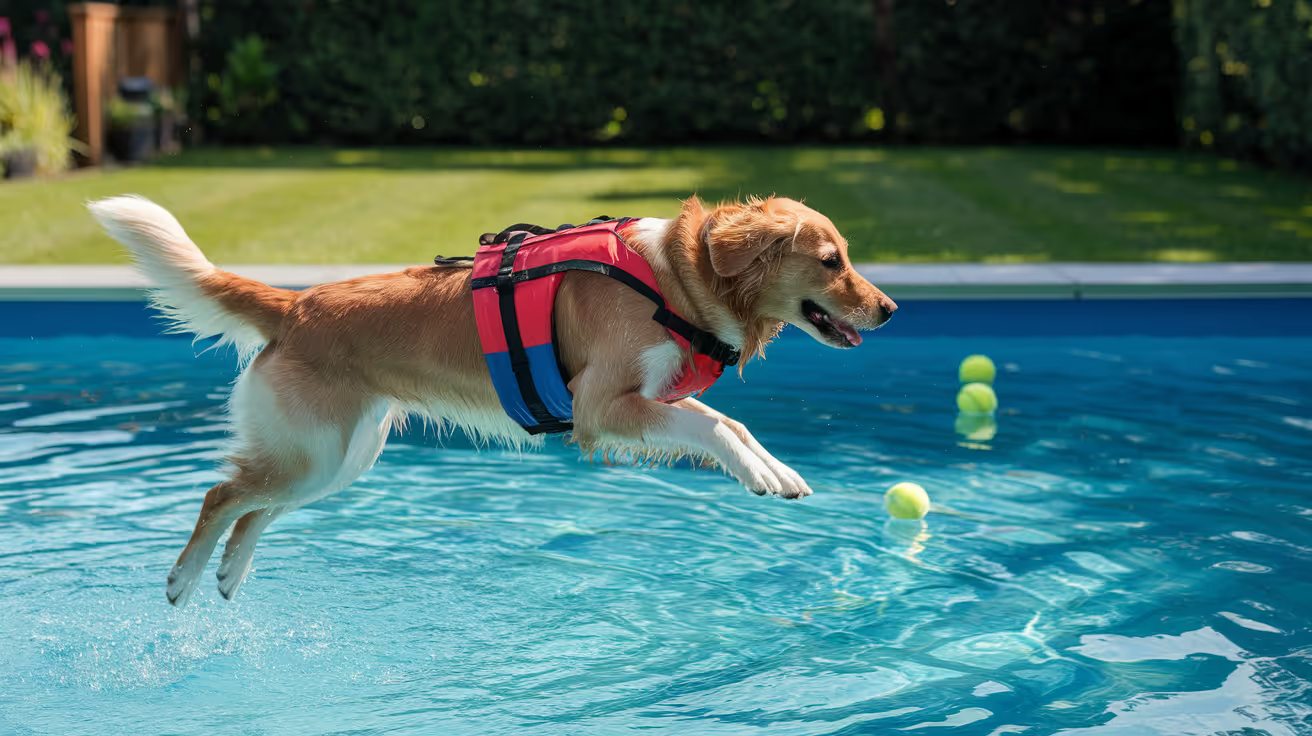
Most post-operative infections in dogs don't happen in the operating room. They happen at home, in the days after surgery, when the wound is healing and the owner is managing recovery.
That's where the risk is, and where you have the most control.
Quick answer: Preventing post-operative infections in dogs requires four owner-controlled actions: keeping the E-collar on at all times to prevent licking, checking the incision daily for early warning signs, restricting activity to protect the healing wound, and completing all prescribed medications on schedule. Missing any of these significantly raises infection risk.
Key takeaways
- Licking is the most preventable cause of post-operative infection in dogs at home.
- E-collar compliance is mandatory: keep it on until the vet confirms the incision is healed.
- Activity restriction protects the wound from mechanical disruption that opens pathways for bacteria.
- Daily incision checks catch early infection before it becomes a deep, harder-to-treat problem.
- Stopping antibiotics early is one of the most common owner errors that causes recurrence.
- The 10-14 day recheck is not optional: it confirms healing and removes sutures aseptically.
The biggest mistake dog owners make after surgery
It's not neglect. It's letting the dog lick.
A dog's mouth contains Staphylococcus pseudintermedius and dozens of other bacteria. When a dog licks a surgical incision, it delivers those bacteria directly to the wound, bypassing every sterile precaution the surgeon took.
The E-collar isn't a punishment. It's the most important infection-prevention tool your dog will use at home.
PetsCare confirms: "Keep your pet's surgical wound clean and dry, restrict activity, prevent licking or scratching, and monitor for signs of infection."
E-collar rules: what owners get wrong
Most owners understand the E-collar concept. What they get wrong is compliance.
When the E-collar must be on:
- At all times when unsupervised
- All night while the dog sleeps
- During leash walks
- Any time you leave the room
When it can come off:
- Eating and drinking : under direct supervision only
- Bathing the unaffected part of the dog : under direct supervision only
Common owner error: Removing the E-collar "just for a few minutes" while the dog looks comfortable. It takes seconds for a dog to lick a wound. One breach can introduce enough bacteria to establish an infection.
Daily wound monitoring: what normal looks like vs. what doesn't
Check the incision at the same time each day in good lighting.
Normal healing progression
Signs that require a same-day vet call
- Spreading redness beyond the incision margin
- Yellow, green, or opaque discharge from the wound
- Foul odor at the wound site
- Increasing swelling after day 3 (should be decreasing, not increasing)
- Sutures separating or the wound reopening
- Dog obsessively pawing at the site despite the E-collar
Activity restriction: why it matters for infection prevention
Activity restriction is not only about protecting the surgical repair. It's about preventing infection.
Running and jumping create mechanical stress on healing tissue. This can:
- Open gaps between wound layers, creating dead space where bacteria multiply
- Disrupt early scar tissue formation, exposing deeper tissue
- Loosen sutures and pull wound edges apart
Standard activity restriction after surgery:
- Weeks 1 to 2: Leash walks for bathroom trips only. No stairs, no jumping, no running.
- Weeks 2 to 4: Short controlled leash walks only. No off-leash activity.
- Weeks 4 to 8: Gradual return to normal activity as cleared by your vet.
For orthopedic procedures, restrictions typically extend to 8 to 12 weeks. Follow your specific surgeon's protocol, not a general timeline.
Medication compliance: the full course matters
Stopping antibiotics when your dog "looks better" is one of the most common owner errors in post-operative care.
Why finishing the course matters:
- Bacteria are not eliminated uniformly; the last few days of treatment kill the most resistant survivors
- Stopping early allows surviving bacteria to multiply and potentially develop resistance
- A second round of antibiotics after early cessation often requires a stronger drug
Rule: Never stop antibiotics before completing the prescribed course, even if the wound looks completely healed.
Pain medications: give these on schedule too. Adequate pain control reduces stress, reduces cortisol (which impairs healing), and keeps the dog calm enough to rest.
Wound cleaning at home: what vets actually recommend
Unless your vet has given specific instructions to clean the wound, don't clean it.
This surprises many owners, but routine wound cleaning can:
- Disrupt the early healing process
- Remove protective fibrin that seals wound edges
- Introduce bacteria from the cleaning materials
What to do instead:
- Keep the wound dry
- Do not apply hydrogen peroxide, rubbing alcohol, or over-the-counter antiseptics
- Do not apply any ointment, cream, or spray unless specifically prescribed
If your vet instructs wound cleaning, use only the solution they specify (typically dilute chlorhexidine) applied with a clean gauze pad; never use cotton balls, which shed fibers.
Recovery environment setup
A clean recovery space reduces environmental bacterial load.
Home setup checklist:
- [ ] Wash all bedding in hot water before your dog comes home
- [ ] Set up a confined recovery area (exercise pen or crate) away from other pets
- [ ] Remove anything the dog can jump on or off
- [ ] Place non-slip mats on hard floors to prevent slipping
- [ ] Keep the recovery area dry: damp environments encourage bacterial growth
- [ ] Change bedding every 2 to 3 days during recovery
For wound-level infection prevention strategies, see wound-level infection prevention. For a complete SSI prevention protocol guide, see specific SSI prevention guide. For what recovery looks like in cats for comparison, see post-operative infection prevention in cats for comparison.
Following up: the rechecks you can't skip
Post-operative rechecks are designed around the infection timeline, not just wound healing.
Missing a recheck delays detection of early infection before it becomes established. Early-stage infections caught at a 10-day recheck are managed far more simply than infections diagnosed at 3 to 4 weeks.
For the full infection timeline and when infections typically appear, see when post-operative infections most commonly occur. For how to distinguish healing from early infection, see how to tell if prevention has worked.
When to go to the emergency vet, not wait
Go immediately, do not wait for a callback, if:
- The wound is bleeding and doesn't stop within 5 minutes
- The wound has opened significantly with visible tissue or fat underneath
- Your dog collapses or can't stand
- Rapidly spreading redness or warmth covers a large skin area
- Your dog has a fever and severe lethargy together
Frequently asked questions
My dog is acting completely normal but the wound looks slightly red. Should I worry?
Mild redness at the wound margin is normal in the first 2 to 3 days. Monitor it. If the redness is spreading outward from the wound, appearing after day 4, or accompanied by any discharge or odor, call your vet that day. Don't wait to see if it gets better.
My dog keeps pawing at the incision through the E-collar. What should I do?
First, confirm the E-collar is correctly sized: it should extend past the dog's nose by several inches. If it's the right size and the dog can still reach the wound, the collar may need to be supplemented with a recovery body suit or a different style of collar. Contact your vet for guidance.
Do I need to clean the stitches?
In most cases, no. Keep them dry. Your vet will give specific instructions if the wound requires cleaning. The most important step is monitoring, not touching.
Post-operative infection prevention is not complicated. It requires discipline. The E-collar stays on. The wound gets checked daily. The medications get finished. The follow-up appointments get kept. These four things, done consistently, prevent the majority of infections that develop after dog surgery.
Resources
- Pooler Veterinary Hospital. Pet Wound Care at Home: Post-Surgery Healing Tips. poolervet.com
- Kainer Vet. Effective Post-Surgical Care for Dogs to Ensure Full Recovery. kainervet.com
- PetsCare. How to Care for Your Pet's Surgical Wound. petscare.com

Get a Free Poster
Enhance your workspace with a high-quality radiographs reference poster, designed for veterinary professionals. This free physical poster will be shipped directly to you—just fill out the form to request your copy.










Taking Great TPLO Radiographs
Click Below to Watch Live Video Demos


We'll send you a Free Wall Poster with all the steps
Step #1
Getting Ready
Ensuring a clean surgical field starts with proper skin preparation. This video demonstrates the best practices for:
- Shaving the patient – Achieving a close, even shave while minimizing skin irritation
- The Dirty Scrub – The initial skin prep step to remove surface debris and reduce bacterial load before the sterile scrub.
Following these techniques helps reduce infection risk and improve surgical outcomes. Watch the video to see how it’s done effectively!


Step #2
Reduce Your Risks
Many surgeons are shocked to find out that their patients are not protected from biofilms and resistant bacteria when they use saline and post-op antibiotics.
That’s Where Simini Comes In.
Why leave these risks and unmanaged? Just apply Simini Protect Lavage for one minute. Biofilms and resistant bacteria can be removed, and you can reduce two significant sources of infection.
Step #3
Take the Course
Preventing surgical infections is critical for patient safety and successful outcomes. This course covers:
- Aseptic techniques – Best practices to maintain a sterile field.
- Skin prep & draping – Proper methods to minimize contamination.
- Antibiotic stewardship – When and how to use perioperative antibiotics effectively.
Stay up to date with the latest evidence-based protocols. Click the link to start learning and earn CE credits!

Things to know
Surgical Site Infection Prevention in Dogs
Learn effective surgical site infection prevention in dogs with expert tips on hygiene, antibiotics, and wound care for safer surgeries.
Surgical site infections are among the most studied complications in veterinary surgery, and the evidence base for preventing them has matured significantly in the last decade.
SSIs are not inevitable. They are the outcome of cumulative failures in preparation, sterile technique, wound management, and post-operative care. Addressing each phase systematically produces the consistently low infection rates that characterize high-quality surgical centers.
Quick answer: Surgical site infection prevention in dogs requires coordinated measures across three phases. Pre-operatively: patient screening and proper skin preparation. Intraoperatively: antibiotic prophylaxis timed 30 to 60 minutes before incision, sterile technique with iodine-impregnated drapes, orthopedic gloves, and intraoperative lavage. Post-operatively: E-collar use, incision monitoring, activity restriction, and follow-up attendance. Extended surgery time is the strongest identified patient-level SSI risk factor in clean surgical procedures.
Key takeaways
- Extended surgery time is the strongest SSI risk factor in clean procedures per published veterinary evidence.
- MRSP carrier status significantly raises SSI risk: one study found a 9x higher odds ratio.
- Perioperative antibiotic prophylaxis without postoperative extension is sufficient for clean procedures.
- Iodophore-impregnated adhesive drapes and orthopedic gloves reduce contamination risk intraoperatively.
- The E-collar at home is the most important owner-controlled variable in post-operative infection prevention.
- SSI rates in canine clean surgery range from 0.8% to 21.3% depending on procedure, center, and protocol quality.
Why SSI prevention matters: the clinical and financial stakes
Surgical site infections in dogs cause measurable harm beyond the immediate complication. A study referenced by Clinician's Brief reported the economic impact of SSIs after TPLO surgery in dogs as ranging from $110.21 to $3,817.12 USD per case, depending on the intervention required. Beyond cost, SSIs extend recovery, increase antibiotic exposure, and in cases involving orthopedic implants, may require hardware removal.
SSI rates in canine surgery vary widely: Clinician's Brief reports published rates ranging from 0.8% to 21.3% of surgical cases depending on procedure type, setting, and protocol quality. This range demonstrates that SSI is not a fixed background risk: it is heavily influenced by practice decisions at every phase of surgical care.
Pre-operative SSI prevention
Patient screening and risk factor management
Not all surgical patients carry equal SSI risk. Identifying and modifying controllable risk factors before surgery reduces baseline risk:
- Active skin infections (pyoderma): must be fully resolved before elective surgery. Skin bacteria can contaminate the surgical field directly.
- MRSP carrier status: a Finnish veterinary teaching hospital study (PMC7495856) identified MRSP carriage as a significant SSI risk factor with an odds ratio of 9.0. Dogs known or suspected to carry MRSP warrant specific pre-operative screening.
- Obesity: impairs blood flow to wound edges and slows healing.
- Metabolic conditions: diabetes, Cushing's disease, and hypothyroidism all compromise immune function and wound repair.
- Higher body temperature: the same Finnish study found higher pre-operative body temperature was associated with increased SSI risk (mean difference of +0.4°C compared to dogs without SSI).
Skin preparation
The skin harbors the microorganisms most likely to contaminate a wound during surgery. Reducing skin flora at the operative site is a fundamental pre-operative measure:
- Hair is clipped, not shaved (shaving creates micro-abrasions that increase bacterial colonization)
- Chlorhexidine gluconate-based prep solution applied in a scrub-then-paint sequence
- The prepared area extends well beyond the planned incision margins
- A minimum skin preparation contact time is observed before draping
The Veterinary Nurse confirms: "Skin preparation and aseptic techniques aim to reduce or eliminate the growth of resident and transient flora at the wound site."
Intraoperative SSI prevention
Antibiotic prophylaxis: timing and duration
The evidence consistently supports perioperative antibiotic prophylaxis without postoperative extension for clean orthopedic and neurosurgical procedures in dogs.
The Finnish study (PMC7495856) found: "Antimicrobial prophylaxis without postoperative antimicrobials is sufficient to maintain the overall rate of SSI at a level similar to published data in canine clean orthopedic and neurosurgeries." Of 406 dogs reviewed, 92.9% received antimicrobial prophylaxis and only 1.1% received postoperative antimicrobials: the SSI rate was comparable to international benchmarks.
The correct protocol: intravenous cefazolin given 30 to 60 minutes before skin incision, repeated every 90 to 120 minutes intraoperatively if surgery exceeds the dosing interval.
For how antibiotic prophylaxis works within the broader SSI prevention framework, see prophylactic antibiotics in SSI prevention.
Sterile field management
Beyond antibiotics, maintaining a sterile intraoperative environment requires:
- Iodophore-impregnated adhesive drapes applied to the skin at the operative field to prevent migration of skin flora into the wound
- Orthopedic surgical gloves for implant procedures to reduce perforation risk
- Double-gloving with glove changes after draping and at the 60-minute mark for extended procedures
- Rigorous instrument handling by all operating room personnel
- Limiting operating room traffic during the procedure
Managing surgery time
Extended surgery time is the only variable identified as a significant SSI risk factor in a large-scale European study of 1,550 dogs undergoing clean procedures (ScienceDirect). Every additional minute of open wound time increases cumulative bacterial exposure. Efficient surgical technique is an infection control measure, not just a quality-of-care issue.
Intraoperative wound lavage
Lavage with sterile isotonic saline before wound closure removes blood, bone debris, and bacteria that accumulate during surgery. This is particularly important in orthopedic procedures where implant surfaces provide an adhesion substrate for bacteria.
For how biofilm forms on implants and why lavage matters, see biofilm as a driver of surgical site infections.
Wound closure technique
Anatomical layer closure without dead space prevents the fluid accumulation that bacteria exploit post-operatively. Antimicrobial suture materials (triclosan-coated) inhibit bacterial colonization of the suture itself. The published TPLO protocol change study (PMID 29878479) demonstrated that replacing staples with antimicrobial intradermal sutures was one component of a bundle that reduced implant-associated infection from 8.5% to 1.3%.
Post-operative SSI prevention
At the hospital before discharge
- Antimicrobial ointment (mupirocin) applied to the wound before bandaging
- Soft-padded bandage protecting the wound during in-hospital recovery
- E-collar placed before the dog recovers from anesthesia
- Single-use gloves for all staff handling the post-operative wound
Owner responsibilities at home
The dog's owner becomes the primary infection control agent once the dog leaves the hospital. The key measures:
E-collar compliance: the most important single owner-controlled factor. Licking delivers oral bacteria directly to the incision and can bypass every intraoperative precaution. The E-collar must remain on whenever the dog is unsupervised.
Incision monitoring: daily visual inspection to identify redness, swelling, discharge, or odor early enough to intervene before deep infection establishes.
Activity restriction: running and jumping create mechanical stress on healing tissue and can open wound layers before they've fully healed, creating dead space and infection opportunity.
Follow-up attendance: post-operative rechecks are not optional. The 10 to 14 day recheck confirms wound healing and removes sutures. The 6 to 8 week recheck (for orthopedic procedures) confirms bone healing and implant stability.
For the practical SSI prevention guide owners use at home, see practical SSI prevention guide. For comparison with SSI prevention in cats, see SSI prevention in cats for comparison. For when infections appear despite prevention, see when surgical site infections typically emerge.
For how SSI prevention in dogs specifically applies to orthopedic cases, see reducing SSI risk in orthopedic cases.
The team culture dimension
The Veterinary Nurse notes: "It is increasingly recognised that team culture can significantly affect the perioperative management of the patient and hence, the SSI risk. A proactive team culture must be cultivated and maintained."
SSI prevention isn't solely a technical matter. Every member of the surgical team must understand their role in maintaining sterile conditions. Compliance with hand hygiene, barrier precautions, and protocol steps must be consistent across all personnel and all cases, not just the highest-profile ones.
Frequently asked questions
What is the most common source of surgical site infections in dogs?
The most common source is translocation of the patient's own skin flora into the wound. The Veterinary Nurse confirms: "Translocation of endogenous microbial flora is the most common route of surgical site infection." This is why skin preparation and sterile draping matter more than environmental contamination, which is a secondary concern.
My dog had SSI after a clean surgery. What likely went wrong?
The most common contributing factors in published veterinary SSI cases are: extended surgery time, inadequate skin preparation, break in sterile technique (often glove perforation or field contamination), failure of E-collar compliance at home leading to licking, and MRSP carriage that wasn't identified pre-operatively. A culture and sensitivity test of the infection identifies the pathogen and guides appropriate treatment.
Does my dog need antibiotics to go home after surgery to prevent SSI?
For clean orthopedic procedures, the published evidence does not support routine post-operative antibiotic courses. Perioperative dosing is sufficient when technique is correct. However, individual circumstances may warrant a post-operative course: contaminated or dirty wounds, immunocompromised patients, or cases where intraoperative contamination occurred. Ask your surgeon for the specific rationale if a post-operative antibiotic course is prescribed.
SSI prevention is a system, not a single action. The surgeon controls most variables intraoperatively, but the perioperative window spans days before and weeks after. When every phase is managed well, consistently low SSI rates are achievable across a broad range of surgical procedures in dogs.
Resources
- Gronkjaer et al. Risk factors for SSI associated with clean surgical procedures in dogs. Vet J, 2021. sciencedirect.com
- Heikkinen et al. Antimicrobial prophylaxis is sufficient in clean orthopaedic and neurosurgeries in dogs. BMC Vet Res, 2020. ncbi.nlm.nih.gov
- Clinician's Brief. Bacterial Biofilms. cliniciansbrief.com
- The Veterinary Nurse. Surgical site infections: preparation, technique and perioperative prevention. theveterinarynurse.com
Asepsis During Abscess Drainage
Learn how to maintain asepsis during abscess drainage to prevent infection and ensure safe healing for your pet.
Abscess drainage is classified as a dirty-infected wound procedure. It involves the deliberate opening of a pre-existing infection, which creates contamination of the drainage site, instruments, gloves, and surrounding tissues at the moment of incision.
The asepsis goal during abscess drainage is not sterility, which is unachievable in this context. It is contamination control: preventing further spread of the infection to adjacent tissues, other patients, and clinical staff.
What this covers: The asepsis principles and practical protocol for abscess drainage in dogs and cats, including patient preparation, incision site selection, lavage, contamination control, wound management post-drainage, and the specific asepsis requirements for different abscess types.Wound class: Dirty-infected (Class IV). Pre-existing infection is present. SSI rates for dirty-infected procedures range from 18 to 27%+ in the absence of appropriate management.Key principle: Because the procedure opens an infected cavity, the emphasis shifts from preventing contamination of the wound (as in clean surgery) to controlling the spread of contamination from the wound to the patient, the staff, and the clinical environment.
Key takeaways
- Abscess drainage is Class IV (dirty-infected); the wound already contains infection.
- The asepsis goal is contamination control, not wound sterility.
- Wide clip margins prevent hair contamination of the drainage site and surrounding area.
- Lavage after drainage is the most important asepsis step for contamination control.
- Instruments must be treated as contaminated after the abscess opens; do not return to the sterile field.
- Staff PPE (gown, gloves, eye protection) is essential; abscess contents aerosolize during incision.
- Environmental disinfection of the procedure area post-drainage must be thorough.
Pre-procedure preparation
Patient assessment and sedation
Abscess drainage requires adequate analgesia and, in many cases, sedation or general anesthesia. Pain and patient movement during incision compromise contamination control.
Assess for:
- Extent and depth of the abscess (palpation, imaging where indicated)
- Signs of systemic involvement (fever, lethargy, regional lymphadenopathy)
- Identifying the most dependent site for drainage
Bite wound abscesses in cats: Cat bites produce small, deep puncture wounds that frequently develop into deep abscesses. Careful palpation and imaging can reveal the full extent before incision.
Perianal abscesses: Perianal fistulae and anal sac abscesses require specific preparation to minimize fecal contamination during drainage.
Clipping
Clip a generous area around the abscess, extending well beyond the anticipated incision site. University of Minnesota Clinical Skills guidance notes: "Be sure to extend clip below the planned draining stab wound to prevent hair from contaminating the drainage hole, and to keep the area easier to clean of drainage material."
The clip area also needs to extend below the drainage site in the animal's normal standing position, because gravity-assisted drainage will track downward and hair in that path will become contaminated and trap purulent material against the skin.
Skin antisepsis
Apply standard antiseptic scrub to the clipped area around the abscess, using centrifugal technique from the intended drainage site outward. Chlorhexidine-alcohol combination is appropriate.
Important: The abscess wall itself cannot be made sterile by skin antisepsis. The purpose of antisepsis here is to reduce surface contamination adjacent to the drainage site, limiting additional organisms from the skin surface entering the wound.
Staff preparation and PPE for abscess drainage
Abscess drainage requires more protective PPE than clean surgical procedures because:
- The procedure opens a pre-existing infected, often under-pressure cavity
- Purulent material may express at force when the abscess is incised, creating aerosol
- Staff hands, face, and clothing may be exposed to infectious material
Required PPE:
- Gloves: Required throughout; change to a fresh pair after the abscess opens if further sterile field work is needed
- Gown or apron: Protects clothing from purulent material splatter
- Eye protection/face shield: Essential; abscess contents frequently aerosolize at incision
- Mask: Reduces inhalation exposure to aerosolized infectious material
Drainage technique and asepsis
Incision site selection
Select the most dependent site in the abscess when the animal is in normal standing position. This allows passive gravity drainage after the incision is made and minimizes abscess pocket residue.
For bite wound abscesses in cats: locate opposing tooth marks (entry and exit wounds). The abscess often tracks between these points.
Incision
Make a stab incision of sufficient size to allow drainage of thick pus. Too small an incision risks the pocket resealing before the contents fully drain.
After the abscess opens:
- Any instrument that entered the abscess is now contaminated
- Gloves contacting the abscess contents are contaminated
- Do not return contaminated instruments to any sterile field
Lavage after drainage
Lavage is the most important asepsis step after the abscess opens.
Purpose: Mechanically remove purulent material, bacteria, and debris from the abscess pocket and surrounding tissue.
Irrigation solution: Sterile saline. Volume: sufficient to produce clear runoff from the cavity. For large abscesses, 100 to 500+ mL may be required.
Dilute antiseptic lavage: Dilute chlorhexidine (0.05%) or dilute PVI (0.1 to 1%) may be used for the initial lavage of an established abscess. Both reduce bacterial load beyond what saline alone achieves. Do not use concentrated antiseptic solutions within the abscess cavity as they damage granulation tissue.
University of Minnesota guidance: "Flush the abscessed area to remove pus and any gross contamination. Flush solution should be tissue-friendly, aid in removal of bacteria, and ideally isotonic to preserve normal function of cells to promote healing."
Culture
Where clinically appropriate, submit purulent material for aerobic and anaerobic culture and sensitivity testing before lavage. This provides the most reliable identification of causative organisms and guides antimicrobial selection.
In cats with bite wound abscesses, culture is often not performed for straightforward cases where Pasteurella and anaerobes are the expected organisms. For recurrent abscesses, non-responsive cases, or immunosuppressed patients, culture is indicated.
For asepsis principles during wound management, including how abscess drainage fits within the broader wound management asepsis framework and the clean technique standards for ongoing wound care after drainage, that guide covers wound management asepsis in detail.
The intraoperative technique framework for abscess drainage, including sterile field management before the abscess opens and the instrument handling and field abandonment protocol once purulent material is encountered, follows the aseptic technique framework adapted to the dirty-infected context. For core aseptic technique applied during drainage, including the sterile field principles and instrument handling standards that apply before and after the abscess opens, that guide covers the technique framework.
Post-drainage wound management
Open or closed management
Open drainage: Leave the incision open to allow continued drainage. This is appropriate when:
- The abscess is extensive and a single drainage is unlikely to resolve it
- Daily lavage through the open stoma is planned
- Risk of premature closure causing re-accumulation is high
Closed drainage: Primary closure with a drain. Used when:
- The abscess is fully drained and lavaged
- The tissue bed is viable
- Suction or passive drainage can be maintained through a Penrose or closed-suction drain
Closed without drainage: Rarely appropriate for established abscesses. Reserved for small, clean-appearing cavities where the surgeon is confident complete drainage was achieved.
Ongoing wound care
For open-managed abscesses, continued clean technique applies at each dressing change:
- Hand hygiene before any wound contact
- Sterile primary dressing or sterile lavage
- Clean outer bandaging layers
Antimicrobial therapy
Abscess drainage (Class IV wound) warrants antimicrobial therapy, not prophylaxis. Selection should be guided by culture results where available.
For cat bite abscesses without culture: Pasteurella multocida and anaerobic organisms are the primary targets. Amoxicillin-clavulanate provides appropriate coverage for most straightforward cat bite abscesses.
For non-responsive or recurrent cases: culture-guided selection is essential.
Environmental decontamination after abscess drainage
The procedure area requires thorough disinfection after abscess drainage because the environment may be contaminated with purulent material, including organisms that could persist on surfaces and transmit to subsequent patients.
Required steps after abscess drainage:
- Table surface: full disinfection with intermediate-level agent; observe contact time
- Any equipment contacted during the procedure: disinfect per material manufacturer guidelines
- Floor below the drainage site: mop with appropriate disinfectant
- Staff gown and gloves: dispose; do not reuse
- Hand hygiene for all staff involved: thorough wash with soap and water
If significant splatter occurred, extend decontamination to walls, adjacent equipment, and any other surfaces with visible contamination.
For errors to avoid during abscess drainage, including the specific aseptic error categories that are most consequential during dirty-infected wound procedures, that guide covers error prevention.
Specific abscess types: additional considerations
Cat bite abscesses
The most common abscess type in small animal veterinary practice. Cats introduced to multi-cat households or free-roaming outdoors are at highest risk.
Asepsis considerations:
- Explore carefully: bite wound tracts can be deep and tortuous
- Multiple drainage points may be needed if the tract extends far from the visible wound
- Elizabethan collar post-drainage is essential in cats; they will groom the drainage site aggressively
Anal sac abscesses
Close proximity to the anus creates ongoing contamination risk from fecal flora.
Asepsis considerations:
- Clip widely; include adequate area caudal to the abscess for drainage clearance
- Position patient to maximize access while minimizing fecal contamination spread
- Copious lavage essential
- Open management typically preferred to allow continued drainage with daily wound flushing
Perianal fistulae (anal furunculosis)
More complex than simple anal sac abscess; involves extensive sinus tracts. Often managed with a combination of immunosuppressive therapy and surgical debridement rather than simple drainage alone. Aseptic technique for any surgical intervention follows the dirty-infected protocol.
For skin antisepsis before drainage procedures, including the skin prep technique and agent selection applicable when antisepsis of the peri-abscess skin is performed before drainage, that guide covers the antisepsis component.
Frequently asked questions
Do abscesses in cats require general anesthesia for drainage?
In most cases, yes. General anesthesia or deep sedation is required for safe, thorough abscess drainage in cats. Inadequate analgesia results in patient movement that compromises contamination control and prevents adequate exploration and lavage. Topical analgesia alone is insufficient for established abscesses.
Should abscess drainage be performed in the OR or a procedure room?
A clean procedure room with appropriate disinfection before and after is sufficient for most abscess drainage procedures. The OR is not required unless the abscess is extensive and requires general anesthesia with the full surgical infrastructure. After the procedure, the area must receive the same enhanced disinfection protocol regardless of which room was used.
Can antibiotics alone resolve an abscess without drainage?
Rarely. The physical presence of purulent material in an abscess cavity provides a protected environment for bacteria that antibiotics cannot adequately penetrate. Drainage removes the bacterial reservoir. Antibiotics address residual infection after drainage. The combination is more effective than either alone.
Abscess drainage is the procedure where the contamination is already there, and the asepsis goal is to stop it spreading further. Generous clip margins, copious lavage after drainage, full PPE for all personnel involved, and rigorous environmental decontamination after the procedure are the components that distinguish controlled abscess management from a procedure that contaminates the patient, the staff, and the clinical environment.
Resources
The following sources were used as reference and background for this article:
- University of Minnesota Clinical Skills Compendium. Abscess Management in Cat/Dog. open.lib.umn.edu
- Merck Veterinary Manual. Management of Specific Wounds in Small Animals. merckvetmanual.com
- Merck Veterinary Manual. Initial Wound Management in Small Animals. merckvetmanual.com
Common Breaks in Surgical Asepsis in Veterinary Clinics
Explore common breaks in surgical asepsis in veterinary clinics and learn how to prevent infections during pet surgeries.
A break in surgical asepsis is a specific event: a moment when sterility in the operative field is compromised. It differs from an aseptic error in precision. An error is a departure from correct technique. A break is the consequence of an error that actually introduces contamination, or creates conditions where contamination is likely.
Understanding the distinction matters because the appropriate response differs. Some errors can be corrected before they produce a break. A break requires immediate remediation regardless of cause.
What this covers: The categories, mechanisms, and consequences of breaks in surgical asepsis in small animal veterinary practice, and the evidence-based framework for detection and response.Key principle: Breaks should be named immediately when observed. The OR culture that normalizes silence in response to a breach is the OR culture with the highest SSI rates.Evidence: Published data confirm that SSI risk increases 3.5-fold when there are lapses in adherence to aseptic principles, including non-obvious events such as OR movement and visitor traffic. Recurring minor breaks are more strongly associated with SSI than single large contamination events.Response framework: Identify, name, assess, remediate, document.
Key takeaways
- Breaks most commonly occur during gowning and gloving: This is the highest-risk phase for human-origin contamination in the operative setting.
- Many breaks go undetected without a designated field monitor: Self-monitoring in the OR is unreliable. External observation changes the rate of detection.
- The response to a break is as important as the break itself: Continuing without correction turns a manageable contamination event into a probable SSI.
- Breaks accumulate: The SSI literature consistently describes a pattern of multiple minor breaks, not single dramatic events, as the primary pathway to infection.
- Instrument breaks are the highest-consequence category: Direct introduction of bacteria into deep tissue bypasses host defense layers entirely.
- OR traffic is an underappreciated source of breaks: Each door opening and person movement increases airborne contamination counts measurably.
Break category 1: Gowning and gloving breaks
Mechanism
During donning of the sterile gown and gloves, the exterior sterile surfaces of both items are in close proximity to non-sterile surfaces including the scrub attire, skin, and surrounding environment.
How breaks occur
- Sterile towel contacts a non-sterile surface during hand drying, then contaminates the gown or gloves
- The gown exterior contacts the scrub attire or table edge during unfolding
- Ungloved skin touches the exterior glove surface during open gloving
- Hands are pushed through gown cuffs before gloving, eliminating closed gloving option
- Personnel touch their face, mask, or hair after gowning and before the procedure
Published incidence
AJVR (2025) observed 96 veterinary surgical procedures and found:
- Contact of sterile towel with non-sterile surfaces: 17.7% of cases
- Contact of gown with non-sterile surfaces: 17.7% of cases
- Touching sterile objects with bare hands: 12.5% of cases
- Overall protocol breach rate during scrubbing, gowning, and gloving: 46.3%
Response protocol
For the errors in aseptic technique that most commonly produce gowning and gloving breaks, including the full error taxonomy and incidence data across all error categories, that guide provides the upstream error context.
Break category 2: Sterile field contamination during setup
Mechanism
The instrument table and sterile field are established before the patient arrives in the OR. Breaks during setup contaminate instruments or surfaces before surgery begins.
How breaks occur
- Non-sterile outer packaging contacts the sterile field during instrument opening
- Circulating nurse touches the sterile field surface when setting items down
- Instruments fall during setup and are returned to the sterile field
- OR staff reach across the sterile field to retrieve items on the far side
- Sterile packs opened without confirming chemical indicator change
Prevention emphasis
Setup breaks are particularly consequential because contaminated instruments will contact the wound for the entire duration of the procedure. Unlike intraoperative breaks, where replacement is possible, setup contamination may go undetected.
The most effective prevention is a sterile field setup protocol where the circulating nurse opens all items toward the scrub technician rather than dropping items from above, and where chemical indicator status is verbalized before any pack is opened.
Break category 3: Intraoperative technique breaks
Mechanism
During the active procedure, the sterile field is maintained by the behavior of all team members. Breaks occur when this behavior departs from correct technique.
How breaks occur
Gowned personnel:
- Turning back to the sterile field (back of gown is non-sterile)
- Allowing hands to drop below waist level
- Leaning across the sterile field rather than stepping around it
- Glove perforation undetected and not replaced
- Touching drape edge with gloved instrument before wound contact
Non-gowned personnel:
- Reaching across the sterile field to hand items to the surgeon
- Standing too close to the sterile field, allowing clothing contact
- Circulating nurse touching the sterile surface while adding items
Equipment:
- Non-sterile cables or tubing contacting the sterile field
- Suction tubing not handled with sterile technique
- Overhead light handle touched by non-sterile hands
For the correct technique breaks deviate from, including the sterile field rules, personnel zones, and instrument handling standards that define correct intraoperative behavior, that guide provides the technical baseline.
Break category 4: OR environment breaks
Mechanism
OR environmental conditions can create contamination events independent of personnel technique, particularly through airborne routes.
How breaks occur
- OR door opened repeatedly during active surgery
- Excessive personnel in OR generating air currents and skin shedding
- HEPA filtration not functioning or not present
- Positive pressure ventilation failure allowing corridor air ingress
- Between-case cleaning not performed, leaving surface contamination from previous procedure
- Wet surfaces aerosolizing bacteria with foot traffic
The significance of door openings
Research in human surgical settings has quantified the contamination effect of OR door openings. Each opening introduces a corridor air pulse and disrupts positive-pressure differential. In procedures requiring strict contamination control, particularly orthopedic and implant surgeries, door traffic is actively counted and minimized as a matter of protocol.
Pre-procedure supply confirmation is the most effective single intervention: confirming all supplies are in the OR before the first incision eliminates the most common cause of door openings during surgery.
For OR standards that prevent breaks, including the physical infrastructure and access control standards that reduce environmental breaks, that guide covers the facility requirements in detail.
Break category 5: Patient preparation breaks
Mechanism
Preparation errors that contaminate the surgical site before it is draped create a break in the planned sterile environment before surgery even begins.
How breaks occur
- Antiseptic scrub performed in the wrong direction (inward rather than outward from incision center)
- Patient transported to OR on a contaminated surface after prep
- Patient positioned in the OR without confirming the prep site is protected
- Draping performed before antiseptic is fully dry
- Drape displaced during patient movement and repositioned rather than replaced
The repositioned drape error
Repositioning a displaced drape is one of the most common and underappreciated breaks in veterinary surgical practice. The underside of any drape that has moved across non-sterile tissue or equipment is contaminated. Placing it back does not restore sterility; it transfers contamination to the surgical field margin.
Correct response: place a new sterile drape over or instead of the displaced one.
Building a culture of break detection and correction
The literature is consistent on this point: technical knowledge of what constitutes a break is not the limiting factor in surgical asepsis performance. The limiting factor is whether breaks are named when they occur.
Structural elements of a correction culture
Designated field monitor:
One named team member, typically the scrub technician or an assigned observer, has explicit responsibility to watch the sterile field and name breaks as they occur. This role does not require seniority. It requires clarity of assignment.
Non-punitive naming:
The correction of a break must be separable from blame. A gloving mistake named and corrected immediately is far less consequential than one left uncorrected because the surgeon is too senior to correct. Training programs should explicitly address this dynamic.
Immediate response expectation:
Every named break generates an immediate response. The response options are limited: replace the contaminated item, reassess the sterile field, or halt and re-establish if the extent of contamination is unclear.
Documentation:
All breaks and responses should be logged in the surgical record. This serves quality improvement, SSI investigation, and medicolegal purposes.
For training that reduces asepsis breaks, including the elements of effective asepsis training programs that address team culture, break naming, and competency assessment, that guide covers the staff development component.
Effective training programs in this area must address the cultural barrier to naming breaks as explicitly as they address technical correct technique. Documented competency assessments with observed performance, rather than written tests alone, are the most reliable method of confirming real-world aseptic behavior.
For auditing to detect recurring breaks, including the audit methods that distinguish individual break events from systematic patterns requiring protocol revision, that guide provides the compliance monitoring framework.
Break response reference
Frequently asked questions
What is the difference between a break and an error in surgical asepsis?
An error is a departure from correct technique. A break is a specific contamination event that results from an error. Not all errors result in breaks. All breaks result from errors. The distinction matters for response: some errors can be corrected before they produce a break. Once a break has occurred, remediation of the contamination is required.
Is it possible to continue surgery after a significant break?
Yes, in most cases, after appropriate remediation. The surgeon must assess whether the contamination affected items or areas that can be replaced, covered, or cleaned. If the extent of contamination is unclear, halting to re-establish the sterile field is the conservative and appropriate choice.
How should the team respond when a senior surgeon makes a break?
With the same immediacy as when anyone else does. Seniority does not reduce the contamination consequence of a break. Clinics that build an explicit expectation of named, non-judgmental break correction regardless of personnel rank consistently maintain better aseptic outcomes than those with hierarchical correction norms.
Breaks in surgical asepsis are a normal feature of surgical practice. They are not evidence of failure; they are evidence that humans are performing complex tasks in a demanding environment. What determines outcomes is not whether breaks occur but whether they are seen, named, and corrected before they translate into patient harm.
Resources
The following sources were used as reference and background for this article:
- AVMA Journals. Aseptic protocol breaches are common among veterinary students. AJVR, 2025. avmajournals.avma.org
- PubMed. Aseptic protocol breaches during SGG in veterinary students. pubmed.ncbi.nlm.nih.gov
- Veterian Key. Principles of Surgical Asepsis. veteriankey.com
- WSAVA 2014. Maintaining a Sterile Operating Environment. vin.com

Veterinary Surgical Hand Scrub Protocol Guide
Learn the step-by-step veterinary surgical hand scrub protocol to ensure sterile surgery and prevent infections in pets.
The surgical hand scrub is performed before every sterile procedure. It removes transient bacteria from the skin and reduces resident flora to a level that the sterile glove can contain.
Skipping it, rushing it, or performing it incorrectly undermines the aseptic barrier regardless of how well everything else is done.
Quick answer: The standard surgical hand scrub uses antiseptic soap applied from fingertips to elbows for a minimum of 5 minutes. Hands stay above elbows throughout. Alcohol-based handrubs (ABHR) are a validated alternative with equivalent or longer-lasting effect.
Key takeaways
- Initial scrubs require a minimum of 5 minutes to maximize skin-to-antiseptic contact time; subsequent same-day scrubs may be shorter
- Hands must be held above elbow level at all times during scrubbing and rinsing to prevent recontamination from forearms
- Fingernails must be short and polish-free: the subungual space harbors the highest bacterial load
- Any contact with a non-sterile surface during scrubbing requires a complete restart
- Alcohol-based handrub (ABHR) is a validated alternative to traditional scrubbing, with equivalent or longer-lasting antimicrobial effect
- Closed gloving is the standard technique for sterile procedures; open gloving is reserved for non-sterile or field situations
Before you scrub: preparation
VetTechPrep: "All jewelry should be removed, and nails should be short."
Veterinary Surgery Online: "Fingernails should be trimmed short to prevent glove damage.
Dirt and bacteria lodged at the base of longer nails are harder to reach during scrubbing.
Nails should also be free of polish (even if clear) as they too can harbor bacteria."
Remove all rings, bracelets, and watches before entering the surgical prep area. These items harbor bacteria and cannot be decontaminated by scrubbing.
Agent selection
Two antiseptic agents are standard:
Chlorhexidine gluconate (4% or 2%):
- Broad-spectrum: effective against gram-positive and gram-negative bacteria, yeasts, and some viruses
- Residual activity: binds to skin proteins, continues working after rinsing
- Less inactivated by organic material than povidone-iodine
Povidone-iodine (7.5–10%):
- Broad-spectrum including bacteria, fungi, viruses, and spores
- Minimal residual activity; effect dissipates quickly after rinsing
- Can be inactivated by blood and organic debris
VetTechPrep: "The two most used scrubs are povidone-iodine (Betadine) and chlorhexidine-gluconate (Nolvasan)."
Veterinary Surgery Online: "Alcohol-based rubs have been shown to have the same immediate effect as chlorhexidine scrub but to last longer (Verwilghen 2011)."
They are associated with similar SSI rates as traditional scrub techniques.
Step-by-step traditional scrub protocol
Step 1: Pre-scrub washWet hands and forearms. Apply antiseptic soap. Wash thoroughly for 1 minute to remove gross contamination soil, organic material, surface bacteria. Rinse.
Step 2: Nail cleaningUse a nail pick under running water to clean under each fingernail. This is the highest-density bacterial area.
Step 3: Systematic scrub with brushApply soap to the brush. Work systematically, spending approximately equal time on each surface.
Veterinary Surgery Online: the sequence is:
- All four surfaces of each finger on one hand
- Back of the hand
- Palm of the hand
- Sides of the hand
- Wrist
- Forearm (in sections working toward elbow)
- Repeat on the other hand
VetTechPrep: "A soap-soaked sterile brush/sponge is used to start a systematic scrub technique.
All four sides of each finger are scrubbed, and special attention to the fingernails should be taken as the area under the fingernails harbors the most bacteria."
Step 4: TimingVetTechPrep: "Initial scrubs for the day should last at least 5 minutes, to maximize skin-to-soap contact time."
Subsequent scrubs the same day (between cases) can be shorter typically 2 to 3 minutes provided the hands remained gloved throughout the previous case without contamination.
Step 5: Rinse hands-firstVeterinary Surgery Online: "Rinse hands and arms by passing them through the water in one direction only, from fingertips to elbow, while maintaining eye contact to avoid touching the sink.
Do not move the arm back and forth through the water."
Veterinary Surgery Online: "Remember to keep your hands up to prevent water from running from the elbow area (which is not as clean) towards your hands."
Step 6: Do not shake handsVeterinary Surgery Online: "Do not shake your hands to remove excess water.
Let the water drip off your arms before returning to the surgery suite to prevent water accumulating on the floor or contaminating your gown pack."
Critical rule throughout: VetTechPrep: "After scrubbing has commenced, the hands and arms can no longer contact non-sterile objects. If this happens, the scrub is completely started over."
Alcohol-based handrub (ABHR): the validated alternative
Veterinary Surgery Online: "Hand rubs are a newer alternative to hand scrubbing.
This technique uses an alcohol-based antiseptic solution that is rubbed onto dry, clean skin and is not scrubbed, rinsed, or dried with a towel."
Veterinary Surgery Online: "These solutions are proven to provide rapid and wide-spectrum antimicrobial activity, to decrease skin irritation, to improve compliance, and to reduce the risk of hand contamination by rinsing water."
ABHR procedure:
- Wash hands with soap and water first (to remove gross contamination and any residual organic material)
- Dry hands completely ABHR is applied to dry skin
- Apply the recommended volume per manufacturer instructions
- Rub in sequence: hands, wrists, forearms to elbows
- Continue rubbing until completely dry do not wipe off
ABHR cannot be used if hands are visibly soiled. The traditional scrub is required in that case.
Gowning after the scrub
After rinsing, the scrubbed team member enters the surgical suite with hands held above elbow level.
The gown pack is opened by the circulating nurse (non-sterile) or from a sterile surface.
The scrubbed person takes the gown by the inner surface only, allows it to unfold, and slides arms into the sleeves without the hands advancing past the cuff.
University of Calgary: "Gowning and gloving is part of aseptic technique, to help decrease the spread of pathogens.
Learning how to properly gown and glove is important in all forms of medicine to decrease the rate of infection after surgeries."
Only the front of the gown from chest to table level and the sleeve cuffs are considered sterile. The back is not. Surgeons never reach behind themselves once gowned.
Closed gloving technique
Closed gloving is performed before advancing the hands through the gown cuffs.
Veterinary Clinical Skills Compendium: "Closed gloving = after 'scrubbing' (sudsing) and drying hands, your hands are within the cuffs of a sterile gown, or within a closed system, you are closed gloving.
This technique looks like you are using 'Puppet hands' to put on gloves over gown."
Why closed gloving:
- The outer surface of the glove is handled entirely through the sterile gown sleeve
- This prevents skin contact with the outside of the glove
- If the glove is perforated later, the scrubbed skin beneath (not bare skin) is what the glove contacts
Open gloving (hands advance through cuffs first) is used when a sterile gown is not available field procedures, some farm situations. It carries a higher contamination risk than closed technique.
For aseptic technique in the broader surgical context, see aseptic technique in dog and cat surgery. For the sterile field this contributes to, see maintaining a sterile field in veterinary surgery.
For SSI prevention overall, see how to prevent surgical site infections in dogs.
Frequently asked questions
How long should a surgical hand scrub take?
VetTechPrep: "Initial scrubs should last at least 5 minutes." Between-case scrubs can be 2 to 3 minutes when hands stayed gloved. ABHR protocols vary by product; follow manufacturer timing instructions.
Can I wear nail polish when performing surgical scrubs?
No. Veterinary Surgery Online: "Nails should be free of polish (even if clear) as they too can harbor bacteria." Clear polish is not an exception.
Polish must be removed before any sterile procedure.
What happens if I accidentally touch the sink during scrubbing?
The scrub must restart entirely. VetTechPrep: "After scrubbing has commenced, the hands and arms can no longer contact non-sterile objects. If this happens, the scrub is completely started over."
There is no partial restart.
Is alcohol-based handrub actually as effective as a traditional scrub?
Yes, when applied correctly to clean hands. Veterinary Surgery Online: ABHR provides "the same immediate effect as chlorhexidine scrub but lasts longer."
It is associated with similar SSI rates and is endorsed by major surgical guidelines.
What is the difference between open and closed gloving?
Closed gloving handles the outer glove only through the sterile gown sleeve. Open gloving advances hands through the cuffs first. Closed is standard for sterile procedures; open is used in field settings.
Can a vet tech or nurse perform the surgical scrub, or only the surgeon?
Any team member who will enter the sterile field performs the surgical scrub: surgeons, surgical assistants, and scrubbed veterinary technicians. The same protocol applies regardless of role.
Resources
- Veterinary Surgery Online. Hand Preparation/Scrubbing. vetsurgeryonline.com
- VetTechPrep. Surgery Prep: Scrubbing, Gloving, Gowning. vettechprep.com
- University of Calgary UCVM. Gowning, Gloving and Scrubbing. vet.ucalgary.ca
- Veterinary Clinical Skills Compendium (UMN). Scrub, Gown, Closed Glove Technique. open.lib.umn.edu
- Clinician's Brief. Preoperative Surgical Site Preparation in Veterinary Medicine. cliniciansbrief.com

Dental Surgical Asepsis in Cats
Learn essential steps and tips for maintaining dental surgical asepsis in cats to ensure safe and effective oral surgery outcomes.
Dental surgical asepsis in cats is crucial to prevent infections during and after oral surgeries. Cats often require dental procedures for issues like tooth extractions, gingivitis, or oral tumors. Maintaining a sterile environment helps protect your cat’s health and promotes faster healing.
This article explains what dental surgical asepsis means for cats, why it matters, and how veterinary teams achieve it. You will learn the key steps to keep the surgical area clean and safe, what instruments and techniques are used, and how you can support your cat’s recovery at home.
What is dental surgical asepsis in cats?
Dental surgical asepsis refers to the methods used to keep the surgical site free from harmful bacteria and contaminants during dental procedures on cats. It involves sterilizing instruments, preparing the cat’s mouth, and maintaining a clean environment throughout surgery.
Proper asepsis reduces the risk of post-surgical infections, which can cause pain, delayed healing, or more serious complications. It is a standard part of veterinary dental care to ensure the best outcomes for feline patients.
- Definition clarity: Dental surgical asepsis means preventing bacteria and germs from entering the surgical site during cat dental procedures to avoid infections.
- Importance explained: Keeping the surgical area sterile helps reduce pain and speeds up healing after dental surgery in cats.
- Scope of asepsis: It includes sterilizing tools, cleaning the cat’s mouth, and controlling the environment where surgery happens.
- Common procedures: Tooth extractions, gum surgery, and oral tumor removals all require strict aseptic techniques in cats.
Understanding the basics of dental surgical asepsis helps pet owners appreciate the care involved in feline dental surgeries and the importance of following veterinary advice.
Why is dental surgical asepsis critical for cats?
Cats have sensitive oral tissues that can easily become infected if bacteria enter during surgery. Dental surgical asepsis protects against these infections, which can cause serious health issues beyond the mouth.
Infections can lead to pain, swelling, and systemic illness in cats. Maintaining asepsis also helps reduce the need for additional treatments and improves surgical success rates.
- Infection prevention: Asepsis stops harmful bacteria from causing infections in the cat’s mouth after surgery, preventing complications.
- Pain reduction: Avoiding infections reduces post-operative pain and discomfort for your cat, improving recovery quality.
- Faster healing: A sterile surgical field promotes quicker tissue repair and less inflammation in feline dental surgeries.
- Overall health protection: Preventing oral infections helps avoid spread to other organs, safeguarding your cat’s general health.
Dental surgical asepsis is a vital part of veterinary care that directly impacts your cat’s wellbeing and recovery after oral procedures.
How do veterinarians prepare cats for dental surgical asepsis?
Preparing a cat for dental surgery involves several steps to ensure the mouth and surrounding area are clean and ready. This preparation minimizes bacteria and contaminants before the procedure begins.
Veterinarians carefully examine the cat, clean the oral cavity, and use antiseptic rinses. They also ensure the cat is properly anesthetized to prevent movement and contamination during surgery.
- Pre-surgical exam: Vets check the cat’s overall health and oral condition to plan safe and effective dental surgery.
- Oral cleaning: Removing plaque and debris from the cat’s teeth reduces bacterial load before surgery starts.
- Antiseptic rinses: Applying chlorhexidine or similar solutions in the mouth helps kill bacteria and disinfect the surgical site.
- Anesthesia use: Proper sedation keeps the cat still, preventing contamination and allowing precise surgical work.
These preparation steps are essential to create a safe environment for dental surgery and protect your cat from infection risks.
What sterilization methods are used for dental instruments in cats?
Dental instruments must be sterile to prevent introducing bacteria into the cat’s mouth during surgery. Veterinary clinics use strict sterilization protocols to clean and disinfect tools.
Common methods include autoclaving, chemical sterilants, and ultrasonic cleaning. Each step ensures instruments are free of microbes before use.
- Autoclaving process: Using high-pressure steam sterilizes dental tools effectively by killing all bacteria, viruses, and spores.
- Chemical sterilants: Soaking instruments in approved disinfectants removes microbes when heat sterilization isn’t suitable.
- Ultrasonic cleaning: Vibrations remove debris and biofilm from instruments before sterilization, enhancing cleanliness.
- Packaging and storage: Sterilized tools are kept in sealed packaging to maintain sterility until the dental procedure.
Proper instrument sterilization is a cornerstone of dental surgical asepsis, ensuring no harmful germs enter the cat’s mouth during surgery.
How is the surgical environment controlled during feline dental surgery?
The surgical environment must remain clean and controlled to maintain asepsis throughout the dental procedure. This includes the surgical room, equipment, and personnel.
Veterinary teams follow strict hygiene protocols, wear sterile gloves and gowns, and use sterile drapes to isolate the surgical site. Air quality and surface cleanliness are also managed carefully.
- Clean surgical room: The operating area is disinfected before and after each procedure to reduce environmental bacteria.
- Sterile attire: Veterinarians and assistants wear gloves, masks, and gowns to prevent contamination of the surgical site.
- Surgical draping: Sterile drapes cover the cat’s body except the mouth, isolating the area and reducing infection risk.
- Air control: Some clinics use filtered air systems to minimize airborne microbes during dental surgery.
Maintaining a controlled environment helps keep the cat safe and supports the success of dental surgical asepsis protocols.
What post-operative care supports dental surgical asepsis in cats?
After dental surgery, proper care helps prevent infections and promotes healing. Owners play a key role in maintaining asepsis at home by following veterinary instructions carefully.
This includes monitoring the surgical site, managing pain, and preventing your cat from disturbing the area. Good oral hygiene and follow-up visits are also important.
- Wound monitoring: Check the cat’s mouth daily for redness, swelling, or discharge that may indicate infection.
- Pain management: Administer prescribed pain medications to keep your cat comfortable and reduce stress on healing tissues.
- Preventing trauma: Use an Elizabethan collar if needed to stop your cat from licking or scratching the surgical site.
- Follow-up visits: Return to the vet for rechecks to ensure the surgical site is healing properly and no infection is present.
Careful post-operative management supports the aseptic environment established during surgery and helps your cat recover fully and comfortably.
Conclusion
Dental surgical asepsis in cats is essential for preventing infections and ensuring successful oral surgeries. It involves careful preparation, sterilization, and environmental control by veterinary teams.
As a cat owner, understanding these steps helps you appreciate the care involved and follow post-operative instructions to support your cat’s healing. Maintaining asepsis protects your cat’s health and comfort during dental treatment.
FAQs
How long does dental surgical asepsis take in cats?
Preparation and sterilization steps usually take 30 to 60 minutes before surgery. The actual dental procedure time depends on the complexity but asepsis is maintained throughout.
Can dental surgical asepsis prevent all infections in cats?
While asepsis greatly reduces infection risk, some infections can still occur due to individual factors. Prompt veterinary care is important if signs of infection appear.
Is anesthesia safe for cats during dental surgery?
Yes, anesthesia is generally safe when administered by trained veterinarians who monitor your cat closely during the procedure.
How can I help maintain asepsis after my cat’s dental surgery?
Follow all veterinary instructions, keep the surgical site clean, prevent your cat from licking wounds, and attend follow-up appointments.
Are there risks if dental surgical asepsis is not followed?
Yes, poor asepsis can lead to infections, delayed healing, pain, and more serious health complications requiring additional treatment.

Auditing Asepsis Compliance in Veterinary Clinics
Learn how to audit asepsis compliance in veterinary clinics to ensure infection control and patient safety effectively.
Asepsis training establishes the standard. Audit determines whether the standard is being met.
Without audit, a clinic cannot distinguish between a team that maintains correct aseptic technique and one that has normalized gradual deviations from it. Both teams may describe their practices as compliant. Only observation-based audit can determine which is actually true.
What this covers: The design and implementation of asepsis compliance auditing in small animal veterinary practice, including observation-based audits, SSI surveillance, autoclave validation, environmental monitoring, and audit cycle management.Evidence base: AJVR 2026 SSI consensus data confirms that implementation of surveillance programs produces a "surveillance effect": the act of monitoring itself increases compliance. Simply participating in an audit program reduces SSI rates independent of any other intervention.Key finding: Higher care-bundle compliance yields fewer SSI cases. A quality improvement study reported a moderate negative correlation (r = −0.31) between care-bundle compliance and SSI incidence, with SSI rates dropping from 0.3% to 0.1% following structured audit implementation.Audit goal: Identify gaps between stated protocol and actual practice, distinguish individual errors from systemic patterns, and drive targeted improvement.
Key takeaways
- Audit detects what self-reporting misses: Personnel consistently overreport their own compliance. Direct observation is the only reliable method for identifying intraoperative technique gaps.
- The surveillance effect is real and significant: Documented in both veterinary and human surgical literature, the mere presence of an active audit program increases compliance independent of the specific findings.
- SSI rate alone is an inadequate compliance metric: SSI is a lagging indicator influenced by many variables. Process compliance metrics are more actionable because they are directly modifiable.
- Audits should distinguish individual from systemic errors: A single practitioner making a specific error is a training issue. Multiple practitioners making the same error repeatedly is a protocol or system design issue.
- Audit findings must be fedback to the team: An audit that produces a report that no one acts on does not improve compliance. Feedback, action planning, and re-audit complete the cycle.
- Autoclave monitoring is a distinct audit domain: Biological indicator validation is the most critical sterilization audit and must be performed regularly, not just when a problem is suspected.
Audit domain 1: Intraoperative technique observation
What it measures
Direct observation of surgical personnel during active procedures, assessing adherence to correct aseptic technique across scrubbing, gowning, gloving, sterile field maintenance, and OR behavior.
Why self-reporting is unreliable
Personnel who are asked whether they followed correct technique will almost universally report yes. AJVR 2025 found that 46.3% of observed procedures involved at least one aseptic protocol breach, yet the breach rate identified by self-report would be substantially lower.
The gap between self-report and observation is not primarily a matter of dishonesty. Most people genuinely do not notice their own minor technique violations. External observation is irreplaceable.
Observation method
Structured observation audit:
- Assign a trained auditor (a senior clinician or designated compliance officer) to observe a sample of procedures
- Use a standardized observation tool with defined scoring criteria for each technique element
- Record all observed deviations without interrupting the procedure (except in cases of significant patient risk)
- Debrief the team after the case, not during, to avoid disrupting active surgery
- Document all findings with date, case type, personnel observed, and specific deviations
Sampling strategy:
- Minimum: observe 10% of surgical procedures per month, distributed across procedure types and personnel
- Higher risk: observe 100% of procedures after an SSI event, a new team member starts, or a significant breach is reported
- Longitudinal: track the same personnel across multiple observations to assess improvement after training
For quality control measures evaluated in audits, including how auditing fits within the broader quality control framework for surgical asepsis, that guide covers the quality assurance architecture.
Audit domain 2: SSI surveillance
What it measures
Post-operative infection rates by procedure type, personnel, patient risk category, and time period.
Why SSI surveillance matters
SSI is the outcome that asepsis is designed to prevent. Tracking SSI rates provides the ultimate performance indicator and can identify temporal clusters that signal a systemic problem.
Key published benchmarks:
- Veterinary SSI rates: 1.5% to 18% depending on procedure type and setting (Veterinary Nurse, 2023)
- Clean wound class expected SSI rate: 1 to 5%
- Clean-contaminated: 5 to 10%
- Contaminated: 10 to 17%
- Dirty-infected: 27%+
A clinic whose clean-wound SSI rate consistently exceeds 5% has a systemic problem that audit should help identify.
SSI definition consistency
The AJVR (2026) published a consensus on SSI definitions for veterinary medicine, noting that implementation of uniform definitions may initially appear to increase SSI rates as surveillance improves. This is the surveillance effect in action: more rigorous definition and counting does not mean more infections, but it does mean more accurate detection.
Clinics should adopt a standardized SSI definition and apply it consistently across all cases to enable meaningful trend analysis.
Surveillance infrastructure
- Post-operative follow-up protocol: All surgical cases should have a defined follow-up contact at 10 to 14 days post-procedure to assess wound status
- SSI recording system: A standardized case record capturing wound class, personnel involved, procedure duration, prophylaxis use, and outcome
- Periodic analysis: Monthly or quarterly review of SSI rate by procedure type and by surgeon
For auditing OR standards compliance, including how SSI surveillance connects to OR environment standards assessment and the physical infrastructure audits that complement technique observation, that guide provides the OR-specific audit components.
Audit domain 3: Autoclave and sterilization validation
What it measures
Whether sterilization equipment is reliably producing sterile loads, and whether loads are being used within validated shelf life.
The three levels of sterilization monitoring
Chemical indicators (Class 1 to 6):
Chemical indicators on the outside and inside of instrument packs change color or appearance when exposed to the sterilization conditions. They confirm exposure to the sterilizing agent but do not confirm sterility.
- External indicators confirm the pack has been through a sterilization cycle
- Internal indicators confirm the agent penetrated the interior of the pack
- Class 5 and 6 integrating indicators provide the closest chemical approximation to a sterility confirmation
Biological indicators (spore tests):
Biological indicators contain Geobacillus stearothermophilus spores that are killed only if sterilization conditions were sufficient to achieve sterility. A killed spore test confirms functional sterility of the autoclave cycle.
Biological indicators are the only method that directly confirms sterilization efficacy.
Recommended frequency: at minimum weekly in active veterinary surgical practices; after any autoclave service or malfunction; after any pack with a failed chemical indicator is identified.
Mechanical monitoring:
Temperature, pressure, and time records from each autoclave cycle should be logged and reviewed. Many modern autoclaves print cycle records automatically. These should be filed and reviewed periodically.
Sterilization audit checklist
- [ ] Biological indicator run within the past week; result documented
- [ ] Chemical indicator status checked on all packs before use
- [ ] Pack integrity (no tears, no moisture) checked before use
- [ ] Expiry dates on all packs confirmed
- [ ] Autoclave service record current
- [ ] Instrument reprocessing log maintained
Audit domain 4: Patient preparation compliance
What it measures
Adherence to clipping, antiseptic agent selection, scrub technique, and transport protocols.
Common preparation compliance gaps
Observation method
Preparation compliance is best audited during the prep phase, not intraoperatively. A designated observer documents whether each preparation step was performed correctly before the patient is transported to the OR.
Audit domain 5: Environmental compliance
What it measures
Between-case disinfection, OR access control, airflow system function, and surface cleanliness.
Environmental audit items
- [ ] Between-case floor mop and surface wipe documented
- [ ] OR door access log (if maintained) reviewed for traffic during procedures
- [ ] HEPA filtration system maintenance current
- [ ] Positive pressure differential confirmed functional
- [ ] Environmental surface swabs taken periodically (quarterly recommended for active ORs)
- [ ] Humidity and temperature log reviewed
For breaks detected through auditing, including the specific break categories that observation-based auditing is most effective at detecting and the response protocols for each, that guide provides the break-level detail that audit findings map to.
Audit cycle management
The audit cycle
An effective asepsis audit program operates as a continuous cycle, not a one-time event:
- Baseline audit: Establish current compliance rates across all audit domains
- Gap identification: Identify specific areas where practice deviates from protocol
- Root cause analysis: Determine whether gaps are individual (training issue) or systemic (protocol or equipment issue)
- Targeted intervention: Training for individual gaps; protocol revision for systemic ones
- Re-audit: Assess whether the intervention produced improvement
- Monitoring: Continue sampling to confirm improvement is sustained
Audit frequency recommendations
Feedback to the team
Audit findings that are not shared with the team do not produce change. Feedback should be:
- Timely: Shared within one to two weeks of the audit observation, while the case is recent
- Specific: Referenced to the specific deviation observed, not generalized
- Non-punitive: Framed as performance data, not disciplinary input
- Actionable: Accompanied by a clear corrective step
For errors identified in audits, including the most common error categories and the evidence for why training and audit together outperform training alone, that guide provides the error-level reference for interpreting audit findings.
The relationship between audit findings and training response should be direct and documented. When an audit identifies a recurring error in a specific category, the training calendar should reflect a targeted refresher for that category within 30 days. This closes the quality loop rather than leaving findings as reports without action.
For training that audits assess for compliance, including how the training program and audit program should be designed as complementary elements of a single quality system rather than independent activities, that guide covers the training-audit integration.
Frequently asked questions
How is an audit different from routine supervision?
Routine supervision is ongoing and informal. An audit is structured, documented, and comparative. Audits use standardized tools, record findings systematically, and compare results to defined standards and to previous audit cycles. Supervision catches problems in the moment; auditing identifies patterns over time.
Who should conduct asepsis audits in a veterinary clinic?
The clinical director, a senior surgeon, or a designated compliance officer. The auditor should be familiar with correct aseptic technique and must be credible to the team being audited. External auditors (visiting specialists or consultants) provide valuable perspective for annual comprehensive reviews.
Should audit findings be used in performance reviews?
With caution. If audit findings are linked to disciplinary consequences, personnel may under-disclose breaches or become resistant to the audit process. The most effective audit programs frame findings as quality improvement data rather than individual performance metrics. Pattern-level findings (repeated errors by the same person despite training) may eventually warrant a performance conversation, but this should not be the primary audit framing.
What should happen when an autoclave biological indicator fails?
Remove all packs sterilized since the last successful biological indicator result from use. Do not use any instruments from those packs. Have the autoclave serviced and repaired. Run a new biological indicator before returning the autoclave to service. Review all cases performed using instruments from potentially non-sterile packs for SSI monitoring. Document the incident and corrective actions taken.
For the checklist used to standardize audits, including the structured verification tool that serves as the reference standard against which audit observations are compared, that guide provides the operational baseline for compliance measurement.
Audit is the discipline that closes the gap between what a clinic believes its aseptic standards are and what they actually are. Without it, compliance is an assumption. With it, compliance becomes a measured, improvable performance metric. The clinics with the best surgical infection rates are invariably those that measure their performance systematically and act on what they find.
Resources
The following sources were used as reference and background for this article:
- AVMA Journals. Surgical site infection definitions consensus in veterinary medicine. AJVR, 2026. avmajournals.avma.org
- NIH/PMC. The effectiveness of aseptic non-touch technique audit cycle implementation on reducing SSI. ncbi.nlm.nih.gov
- NIH/PMC. Quality improvement approach for SSI prevention. ncbi.nlm.nih.gov
- Veterinary Practice. Infection control in the surgical environment. veterinary-practice.com
- The Veterinary Nurse. Surgical site infections: preparation, technique and perioperative prevention. theveterinarynurse.com

Surface Disinfection Protocols in Veterinary Hospitals
Learn effective surface disinfection protocols in veterinary hospitals to prevent infections and ensure pet safety.
Contaminated surfaces in veterinary hospitals can harbor dangerous bacteria for hours, sometimes days. Without consistent disinfection protocols, pathogens like MRSP, Pseudomonas, and E. coli move from surface to patient to surgical wound.
Understanding how veterinary hospitals control environmental contamination helps you evaluate the quality of care your dog receives, and what you can replicate at home after surgery.
Quick answer: Veterinary hospitals use tiered disinfection schedules based on area risk level. Operating rooms are cleaned before and after every surgery. High-touch areas are disinfected multiple times daily. Isolation rooms are cleaned after every patient. The right disinfectant, contact time, and sequence (clean first, then disinfect) determine whether protocols actually work.
Key takeaways
- Operating rooms must be cleaned before and after every surgical procedure to maintain a sterile environment.
- High-touch surfaces like exam tables, door handles, and keyboards need multiple daily disinfections.
- Cleaning before disinfecting is non-negotiable: organic matter blocks disinfectant activity.
- Different disinfectants work against different pathogens: matching the agent to the threat matters.
- 50% or more of hospital surfaces may go untouched during routine cleaning, per published data.
- Home disinfection after MRSP discharge mirrors hospital protocols and reduces reinfection risk.
Why surface disinfection matters in veterinary hospitals
Surgical site infections don't only originate from the surgical field. Environmental contamination in exam rooms, recovery areas, and operating suites contributes to hospital-acquired infections (HAIs).
A Portuguese veterinary teaching hospital study (PMC8240409) found that cages and high-touch human contact surfaces were the most contaminated areas in isolation rooms. Enterococcus spp. were the most frequently isolated pathogens (11.3%), followed by E. coli and Pseudomonas aeruginosa.
One P. aeruginosa isolate in that study was resistant to imipenem, a last-resort antibiotic. Environmental surfaces aren't passive. They are active reservoirs.
Critical finding: Published research on hospital surface disinfection found that 50% or more of surfaces may go uncleaned during standard terminal room disinfection protocols, and 5-30% remain contaminated even after adequate protocols are applied.
The two-step rule: clean first, then disinfect
This is the most commonly skipped step in surface disinfection, and the most consequential.
Disinfectants cannot penetrate organic material (blood, tissue, feces, mucus). Applying disinfectant to a visibly soiled surface does not disinfect it.
The correct sequence:
- Remove visible soiling with a detergent cleaner and disposable cloth
- Rinse the surface if residue remains
- Apply disinfectant and allow full contact time per the label
- Do not wipe off early: contact time is when killing occurs
Skipping step 1 wastes the disinfectant. It's cleaning theater, not actual disinfection.
Common veterinary disinfectants and what they kill
Not all disinfectants work against all pathogens. The AAHA 2018 Infection Control Guidelines provide specific guidance on matching disinfectant class to pathogen type.
Practical rule: Always read the label. Contact time, dilution ratio, and surface compatibility vary significantly between products and formulations.
Disinfection frequency by hospital zone
The AAHA 2018 Infection Control Guidelines establish zone-based cleaning schedules for veterinary practices.
Operating rooms
- Clean and disinfect before and after every surgical procedure
- Between procedures: remove visible debris, re-apply disinfectant, allow full contact time
- Floors mopped after each case
- Air filtration systems (HEPA) and positive pressure ventilation maintained continuously
Isolation rooms
- Full terminal clean after every single patient
- All bedding removed and replaced
- All surfaces (including walls at patient contact height) cleaned then disinfected
- Staff change PPE before and after entering
Exam rooms and high-touch surfaces
High-touch surfaces to disinfect multiple times daily:
- Exam tables (between every patient)
- Door handles and push plates
- Computer keyboards and touchscreens
- Light switches
- Countertops and sink areas
General patient areas
- Daily full disinfection of floors, surfaces, and patient contact points
- Kennels and recovery cages: between every patient occupancy
Operating room-specific protocols
The OR environment requires the most rigorous disinfection because it's where sterile fields are maintained.
Key OR disinfection standards:
- HEPA air filtration removes airborne dust and microbes that would settle on sterile surfaces
- Positive pressure ventilation prevents outside air from entering during surgery
- Restricted access: only essential personnel may enter during and between procedures
- No mops on OR floors during surgery (cross-contamination risk from the mop head itself)
A UV-C disinfection study (PMC6801766) found that pulsed xenon UV-C after standard manual cleaning reduced positive surface samples from 63% to just 18%, demonstrating that UV-C is a powerful adjunct, not a replacement, for manual protocols.
For how the sterile field inside the OR connects to SSI prevention, see environmental infection control.
MRSP-specific disinfection considerations
MRSP (Methicillin-Resistant Staphylococcus pseudintermedius) requires particular attention because it can persist on dry surfaces longer than many other veterinary pathogens.
What works against MRSP on surfaces:
- Accelerated hydrogen peroxide products
- Sodium hypochlorite (1:10 dilution for high-risk areas)
- Potassium peroxymonosulfate (Trifectant/Virkon-S)
What may not be sufficient alone:
- Standard quaternary ammonium compounds (variable efficacy against MRSP biofilm)
The Portuguese veterinary hospital study confirmed that implementing new disinfection protocols reduced bacterial counts by 99.99% in cages and high-contact surfaces, proving that correct protocols work when applied correctly.
For how biofilm on surfaces relates to biofilm in wounds, see biofilm prevention through surface disinfection. For how hospital disinfection connects to SSI prevention in the OR, see SSI prevention in the hospital environment.
What to do at home after your dog is discharged
If your dog was treated for MRSP or another hospital-acquired pathogen, home disinfection matters.
Home disinfection protocol for MRSP cases:
- Wash all bedding in hot water (60°C / 140°F minimum) and dry on high heat
- Disinfect hard surfaces your dog contacts with an accelerated hydrogen peroxide product or dilute bleach solution (1 part bleach to 32 parts water)
- Wash food and water bowls daily with soap and hot water
- Wash hands thoroughly after any contact with wound or discharge
- Limit the dog's contact with other household pets until the infection is cleared
For home hygiene protocols that mirror hospital disinfection, see hygiene protocols that mirror hospital disinfection.
Frequently asked questions
How do I know if a veterinary hospital has good disinfection protocols?
Ask directly. Quality practices will readily describe their cleaning schedules, the disinfectants they use, and how they handle isolation of infectious patients. Accreditation through AAHA also requires adherence to published infection control guidelines.
Can my dog pick up an infection from a vet clinic surface?
Yes, particularly if the dog has open wounds, is immunocompromised, or is a puppy with an immature immune system. This risk is managed through proper surface disinfection and patient segregation. Healthy dogs with intact skin have low risk during routine visits.
Is UV-C disinfection available in veterinary hospitals?
Some specialty and teaching hospitals use UV-C as a supplemental terminal disinfection step. It's not yet standard in general practice but is increasingly adopted where infection control is a priority.
Surface disinfection in veterinary hospitals is a system, not a single product. The correct sequence, the right disinfectant for the right pathogen, the appropriate contact time, and consistent adherence across all zones determine whether environmental contamination is actually controlled.
Resources
- AAHA. 2018 AAHA Infection Control, Prevention, and Biosecurity Guidelines. aaha.org
- Ferreira et al. Controlling bacteriological contamination of environmental surfaces at the biological isolation and containment unit of a veterinary teaching hospital. BMC Vet Res, 2021. ncbi.nlm.nih.gov
- Vianna et al. Evaluation of a UV-C Light-Emitting Device for Disinfection of High Touch Surfaces in Hospital Critical Areas. PMC, 2019. ncbi.nlm.nih.gov
Surgical Site Infection Prevention in Dogs
Learn effective surgical site infection prevention in dogs with expert tips on hygiene, antibiotics, and wound care for safer surgeries.
Surgical site infections are among the most studied complications in veterinary surgery, and the evidence base for preventing them has matured significantly in the last decade.
SSIs are not inevitable. They are the outcome of cumulative failures in preparation, sterile technique, wound management, and post-operative care. Addressing each phase systematically produces the consistently low infection rates that characterize high-quality surgical centers.
Quick answer: Surgical site infection prevention in dogs requires coordinated measures across three phases. Pre-operatively: patient screening and proper skin preparation. Intraoperatively: antibiotic prophylaxis timed 30 to 60 minutes before incision, sterile technique with iodine-impregnated drapes, orthopedic gloves, and intraoperative lavage. Post-operatively: E-collar use, incision monitoring, activity restriction, and follow-up attendance. Extended surgery time is the strongest identified patient-level SSI risk factor in clean surgical procedures.
Key takeaways
- Extended surgery time is the strongest SSI risk factor in clean procedures per published veterinary evidence.
- MRSP carrier status significantly raises SSI risk: one study found a 9x higher odds ratio.
- Perioperative antibiotic prophylaxis without postoperative extension is sufficient for clean procedures.
- Iodophore-impregnated adhesive drapes and orthopedic gloves reduce contamination risk intraoperatively.
- The E-collar at home is the most important owner-controlled variable in post-operative infection prevention.
- SSI rates in canine clean surgery range from 0.8% to 21.3% depending on procedure, center, and protocol quality.
Why SSI prevention matters: the clinical and financial stakes
Surgical site infections in dogs cause measurable harm beyond the immediate complication. A study referenced by Clinician's Brief reported the economic impact of SSIs after TPLO surgery in dogs as ranging from $110.21 to $3,817.12 USD per case, depending on the intervention required. Beyond cost, SSIs extend recovery, increase antibiotic exposure, and in cases involving orthopedic implants, may require hardware removal.
SSI rates in canine surgery vary widely: Clinician's Brief reports published rates ranging from 0.8% to 21.3% of surgical cases depending on procedure type, setting, and protocol quality. This range demonstrates that SSI is not a fixed background risk: it is heavily influenced by practice decisions at every phase of surgical care.
Pre-operative SSI prevention
Patient screening and risk factor management
Not all surgical patients carry equal SSI risk. Identifying and modifying controllable risk factors before surgery reduces baseline risk:
- Active skin infections (pyoderma): must be fully resolved before elective surgery. Skin bacteria can contaminate the surgical field directly.
- MRSP carrier status: a Finnish veterinary teaching hospital study (PMC7495856) identified MRSP carriage as a significant SSI risk factor with an odds ratio of 9.0. Dogs known or suspected to carry MRSP warrant specific pre-operative screening.
- Obesity: impairs blood flow to wound edges and slows healing.
- Metabolic conditions: diabetes, Cushing's disease, and hypothyroidism all compromise immune function and wound repair.
- Higher body temperature: the same Finnish study found higher pre-operative body temperature was associated with increased SSI risk (mean difference of +0.4°C compared to dogs without SSI).
Skin preparation
The skin harbors the microorganisms most likely to contaminate a wound during surgery. Reducing skin flora at the operative site is a fundamental pre-operative measure:
- Hair is clipped, not shaved (shaving creates micro-abrasions that increase bacterial colonization)
- Chlorhexidine gluconate-based prep solution applied in a scrub-then-paint sequence
- The prepared area extends well beyond the planned incision margins
- A minimum skin preparation contact time is observed before draping
The Veterinary Nurse confirms: "Skin preparation and aseptic techniques aim to reduce or eliminate the growth of resident and transient flora at the wound site."
Intraoperative SSI prevention
Antibiotic prophylaxis: timing and duration
The evidence consistently supports perioperative antibiotic prophylaxis without postoperative extension for clean orthopedic and neurosurgical procedures in dogs.
The Finnish study (PMC7495856) found: "Antimicrobial prophylaxis without postoperative antimicrobials is sufficient to maintain the overall rate of SSI at a level similar to published data in canine clean orthopedic and neurosurgeries." Of 406 dogs reviewed, 92.9% received antimicrobial prophylaxis and only 1.1% received postoperative antimicrobials: the SSI rate was comparable to international benchmarks.
The correct protocol: intravenous cefazolin given 30 to 60 minutes before skin incision, repeated every 90 to 120 minutes intraoperatively if surgery exceeds the dosing interval.
For how antibiotic prophylaxis works within the broader SSI prevention framework, see prophylactic antibiotics in SSI prevention.
Sterile field management
Beyond antibiotics, maintaining a sterile intraoperative environment requires:
- Iodophore-impregnated adhesive drapes applied to the skin at the operative field to prevent migration of skin flora into the wound
- Orthopedic surgical gloves for implant procedures to reduce perforation risk
- Double-gloving with glove changes after draping and at the 60-minute mark for extended procedures
- Rigorous instrument handling by all operating room personnel
- Limiting operating room traffic during the procedure
Managing surgery time
Extended surgery time is the only variable identified as a significant SSI risk factor in a large-scale European study of 1,550 dogs undergoing clean procedures (ScienceDirect). Every additional minute of open wound time increases cumulative bacterial exposure. Efficient surgical technique is an infection control measure, not just a quality-of-care issue.
Intraoperative wound lavage
Lavage with sterile isotonic saline before wound closure removes blood, bone debris, and bacteria that accumulate during surgery. This is particularly important in orthopedic procedures where implant surfaces provide an adhesion substrate for bacteria.
For how biofilm forms on implants and why lavage matters, see biofilm as a driver of surgical site infections.
Wound closure technique
Anatomical layer closure without dead space prevents the fluid accumulation that bacteria exploit post-operatively. Antimicrobial suture materials (triclosan-coated) inhibit bacterial colonization of the suture itself. The published TPLO protocol change study (PMID 29878479) demonstrated that replacing staples with antimicrobial intradermal sutures was one component of a bundle that reduced implant-associated infection from 8.5% to 1.3%.
Post-operative SSI prevention
At the hospital before discharge
- Antimicrobial ointment (mupirocin) applied to the wound before bandaging
- Soft-padded bandage protecting the wound during in-hospital recovery
- E-collar placed before the dog recovers from anesthesia
- Single-use gloves for all staff handling the post-operative wound
Owner responsibilities at home
The dog's owner becomes the primary infection control agent once the dog leaves the hospital. The key measures:
E-collar compliance: the most important single owner-controlled factor. Licking delivers oral bacteria directly to the incision and can bypass every intraoperative precaution. The E-collar must remain on whenever the dog is unsupervised.
Incision monitoring: daily visual inspection to identify redness, swelling, discharge, or odor early enough to intervene before deep infection establishes.
Activity restriction: running and jumping create mechanical stress on healing tissue and can open wound layers before they've fully healed, creating dead space and infection opportunity.
Follow-up attendance: post-operative rechecks are not optional. The 10 to 14 day recheck confirms wound healing and removes sutures. The 6 to 8 week recheck (for orthopedic procedures) confirms bone healing and implant stability.
For the practical SSI prevention guide owners use at home, see practical SSI prevention guide. For comparison with SSI prevention in cats, see SSI prevention in cats for comparison. For when infections appear despite prevention, see when surgical site infections typically emerge.
For how SSI prevention in dogs specifically applies to orthopedic cases, see reducing SSI risk in orthopedic cases.
The team culture dimension
The Veterinary Nurse notes: "It is increasingly recognised that team culture can significantly affect the perioperative management of the patient and hence, the SSI risk. A proactive team culture must be cultivated and maintained."
SSI prevention isn't solely a technical matter. Every member of the surgical team must understand their role in maintaining sterile conditions. Compliance with hand hygiene, barrier precautions, and protocol steps must be consistent across all personnel and all cases, not just the highest-profile ones.
Frequently asked questions
What is the most common source of surgical site infections in dogs?
The most common source is translocation of the patient's own skin flora into the wound. The Veterinary Nurse confirms: "Translocation of endogenous microbial flora is the most common route of surgical site infection." This is why skin preparation and sterile draping matter more than environmental contamination, which is a secondary concern.
My dog had SSI after a clean surgery. What likely went wrong?
The most common contributing factors in published veterinary SSI cases are: extended surgery time, inadequate skin preparation, break in sterile technique (often glove perforation or field contamination), failure of E-collar compliance at home leading to licking, and MRSP carriage that wasn't identified pre-operatively. A culture and sensitivity test of the infection identifies the pathogen and guides appropriate treatment.
Does my dog need antibiotics to go home after surgery to prevent SSI?
For clean orthopedic procedures, the published evidence does not support routine post-operative antibiotic courses. Perioperative dosing is sufficient when technique is correct. However, individual circumstances may warrant a post-operative course: contaminated or dirty wounds, immunocompromised patients, or cases where intraoperative contamination occurred. Ask your surgeon for the specific rationale if a post-operative antibiotic course is prescribed.
SSI prevention is a system, not a single action. The surgeon controls most variables intraoperatively, but the perioperative window spans days before and weeks after. When every phase is managed well, consistently low SSI rates are achievable across a broad range of surgical procedures in dogs.
Resources
- Gronkjaer et al. Risk factors for SSI associated with clean surgical procedures in dogs. Vet J, 2021. sciencedirect.com
- Heikkinen et al. Antimicrobial prophylaxis is sufficient in clean orthopaedic and neurosurgeries in dogs. BMC Vet Res, 2020. ncbi.nlm.nih.gov
- Clinician's Brief. Bacterial Biofilms. cliniciansbrief.com
- The Veterinary Nurse. Surgical site infections: preparation, technique and perioperative prevention. theveterinarynurse.com

Quality Control Measures for Surgical Asepsis
Explore essential quality control measures for surgical asepsis to ensure safe, infection-free surgeries and protect patient health.
Quality control in surgical asepsis is the structured system that confirms aseptic standards are being met, not just described.
Training tells the team what to do. Checklists document that it was done. Auditing confirms it was done correctly. SSI surveillance tells the clinic whether the cumulative effect is working. Each component is necessary; none is sufficient alone.
What this covers: The quality control framework for surgical asepsis in small animal veterinary practice, covering the specific QC measures, how they interact, and how quality data should feed back into protocol improvement.Core principle: Quality control in asepsis is a system, not a checklist. The measures described here form an integrated cycle: establish standards, monitor compliance, identify gaps, intervene, and re-assess.Evidence base: AJVR 2026 SSI definitions consensus confirmed the "surveillance effect": the act of participating in an SSI surveillance program increases compliance independently of specific findings. AJVR 2025 breach data confirmed that training alone does not maintain asepsis performance without external monitoring.
Key takeaways
- QC is a cycle, not a checklist: Standards, monitoring, gap identification, intervention, and re-assessment form a continuous loop.
- Auditing is the most sensitive QC tool for technique compliance.
- Biological indicator testing is the most critical QC measure for sterilization.
- SSI surveillance quantifies the patient outcome of all asepsis QC measures combined.
- The surveillance effect is real: monitoring improves compliance independently of findings.
- QC data must feed back into training and protocol revision to close the quality loop.
The quality control framework
Surgical asepsis quality control operates across four interconnected domains:
- Standards: Defined protocols for every asepsis component (patient prep, instrument sterilization, team technique, OR environment)
- Monitoring: Ongoing measurement of compliance against those standards
- Analysis: Identifying gaps between standard and practice, distinguishing individual from systemic failures
- Improvement: Training, protocol revision, and environmental changes that address identified gaps
The framework only works when all four domains are active. Standards without monitoring is assumption. Monitoring without analysis is data collection. Analysis without improvement is documentation of ongoing problems.
QC measure 1: Surgical safety checklists
Checklists convert protocol standards into verified, documented action. They are the most accessible and consistently evidence-supported QC tool for improving surgical safety outcomes.
A Veterinary Practice (2022) publication on infection control in the surgical environment notes that surgical checklists should be used to identify patient risks before surgery and improve team communication, while providing a standardized approach to theatre management.
What checklists quality-control:
- Instrument pack indicator inspection before opening
- Patient prep completion and technique
- Surgical hand antisepsis completion
- Pre-incision time-out including antimicrobial prophylaxis timing
- Instrument count before closure
- Between-case disinfection documentation
For checklists as quality control tools, including the phase-by-phase surgical asepsis checklist and how to implement it as a formal QC document, that guide provides the operational reference.
QC measure 2: Intraoperative auditing
Checklists verify that steps were completed. Auditing verifies that steps were completed correctly.
This distinction is significant. A team can complete a checklist item ("surgical hand antisepsis performed") while the technique used was incorrect. Auditing by a trained observer identifies technique compliance that self-reporting cannot.
Published evidence:
AJVR 2025 found that 46.3% of observed veterinary surgical procedures involved at least one aseptic protocol breach during scrubbing, gowning, and gloving alone. These breaches were largely undetected by the personnel themselves. External observation identified them.
Audit structure:
- Trained observer present during a sample of procedures (minimum 10% of cases per month)
- Standardized observation tool covering hand antisepsis technique, gloving, sterile field maintenance, OR behavior
- Non-punitive debrief after case completion (not during surgery)
- Audit findings documented with date, case type, and specific deviations noted
- Results reviewed monthly to identify patterns
For auditing as a quality control method, including the full audit framework with sampling strategy, observation tools, and feedback protocols, that guide covers intraoperative auditing in comprehensive detail.
QC measure 3: Autoclave validation
Sterilization quality control has its own specific monitoring hierarchy. Instrument sterility is the prerequisite for everything else in surgical asepsis; if sterilization fails, no amount of correct technique downstream compensates.
The three-level autoclave QC system:
- Mechanical monitoring (every cycle): Cycle records confirm temperature, pressure, and time parameters were met
- Chemical indicators (every pack): External and internal indicators confirm the pack was processed and the sterilizing agent penetrated
- Biological indicators (weekly minimum): Spore tests confirm the cycle killed G. stearothermophilus: the only direct confirmation of functional sterility
Class 5 chemical integrating indicators are valuable but cannot substitute for biological indicators. Dispomed (2026): "Class 5 indicators can demonstrate that certain cycle parameters were reached, but they cannot confirm that all microorganisms were killed."
Documentation: Every biological indicator result, positive or negative, must be logged with the date, cycle number, and technician initials. Failed results trigger immediate autoclave removal from service.
For autoclave validation as quality control, including the validation protocol, monitoring schedules, and failed indicator response procedure, that guide covers sterilization QC in full.
QC measure 4: SSI surveillance
SSI surveillance is the outcome measure that validates whether all other QC measures are working. It is also the most powerful single driver of compliance improvement through the surveillance effect.
The surveillance effect:
AJVR 2026 notes that in human healthcare, clear temporal associations between implementation of SSI surveillance programs and decreases in SSI rates have been demonstrated. "Simply the act of participating" in surveillance increases compliance with SSI prevention practices. This surveillance effect operates independently of any specific findings.
SSI surveillance components:
- Standardized SSI definition applied consistently (AVMA Journal 2026 consensus definitions for veterinary use)
- Post-operative follow-up protocol (14-day wound check as minimum)
- SSI data recorded by procedure type, wound class, personnel, and outcome
- Monthly or quarterly SSI rate review
- Trend analysis to identify clusters or increases warranting investigation
Using SSI data:
SSI rate by procedure type provides the clearest signal. A clean-wound SSI rate consistently above 5% in a veterinary practice indicates a systemic asepsis problem requiring investigation across all QC domains.
For errors that quality control aims to prevent, including the most common aseptic error categories with published incidence data and what QC measures are most effective at detecting each, that guide covers the error taxonomy that QC is designed to address.
QC measure 5: Environmental monitoring
OR environmental quality control confirms that the physical environment supports aseptic technique rather than undermining it.
Environmental QC components:
- Surface swabs: Taken from high-contact OR surfaces after between-case disinfection; quarterly baseline with increased frequency after any SSI cluster
- Air sampling: Settle plates or volumetric sampling during occupied OR conditions; provides baseline contamination data
- Positive pressure verification: Smoke pencil or manometer confirmation that the OR maintains positive pressure relative to adjacent spaces
- HVAC maintenance records: Filter replacement, service history, and any pressure differential anomalies documented
Environmental monitoring data should be reviewed alongside SSI rates. An SSI cluster coinciding with a documented HVAC failure or positive pressure loss provides a clear causal relationship requiring investigation.
Connecting QC measures to the improvement cycle
Quality control data has no value unless it drives improvement. The improvement connection requires:
For checklist findings: Any consistently missed checklist item should trigger a review of whether the step is practically achievable within the surgical workflow or requires scheduling/resource changes.
For audit findings: Individual technique deviations → targeted training and observed re-assessment. Systemic pattern across multiple personnel → protocol review or environmental change (e.g., ABHR placement, gloving technique resource).
For autoclave failures: Immediate investigation and repair. Post-repair validation before return to service. Review of all cases in the quarantine window for SSI monitoring.
For SSI rate increases: Structured case review. Cross-reference with audit findings, environmental monitoring, and any protocol changes that preceded the increase. Root cause analysis before intervention.
Documentation of improvement actions: Every identified gap should have a corresponding documented action, responsible person, and follow-up date. Without documentation, quality improvement is anecdotal.
Frequently asked questions
How often should the full QC program be formally reviewed?
Annually at minimum, or whenever a significant SSI event occurs, a new surgical procedure type is introduced, major staff changes occur, or any audit or surveillance data shows a consistent negative trend. An annual comprehensive review covers all four QC domains and assesses whether improvement actions from the prior year produced the expected results.
Should QC findings be shared with all surgical staff?
Yes. Aggregate findings (not individual-level unless performance management is involved) should be shared with the team regularly. Transparency about QC findings builds the culture of quality and accountability that is prerequisite for the surveillance effect to work. Staff who understand QC outcomes and their own contribution to them are more likely to maintain compliance.
Is QC documentation a regulatory requirement?
In many jurisdictions and institutional settings, yes. Autoclave validation records, biological indicator logs, and SSI surveillance data are specifically required by IACUC protocols, veterinary regulatory bodies, and accreditation standards in various countries. Even where not formally required, documentation demonstrates due diligence and supports medicolegal defense if an SSI event results in a complaint or claim.
Quality control in surgical asepsis does not guarantee zero SSI. It guarantees that the practice knows what its aseptic standards are, is actively measuring whether they are being met, and is consistently acting on the gaps it finds. That is the difference between a practice that assumes it is doing well and one that knows it is.
Resources
The following sources were used as reference and background for this article:
- AVMA Journals. Surgical site infection definitions consensus in veterinary medicine. AJVR, 2026. avmajournals.avma.org
- AVMA Journals. Aseptic protocol breaches are common among veterinary students. AJVR, 2025. avmajournals.avma.org
- Dispomed. The Gold Standard of Sterilization: Why Biological Indicators Matter in Veterinary Practice. dispomed.com
- Veterinary Practice. Infection control in the surgical environment. veterinary-practice.com
Surgical Site Preparation in Dogs: Complete Guide
Learn essential steps and tips for surgical site preparation in dogs to ensure safe and infection-free surgeries.
Surgical site preparation is the first line of defense against surgical site infection (SSI).
The goal is to reduce the bacterial load on the skin to the lowest achievable level before the incision is made.
Every step in the preparation protocol matters a single shortcut can undermine the entire chain.
Quick answer: Canine surgical site preparation follows a fixed sequence: clip a wide area in the prep room, perform an initial scrub for gross decontamination, then in the OR perform the aseptic scrub center-to-periphery using chlorhexidine or povidone-iodine, then drape. Never clip or scrub in the OR.
Key takeaways
- Clip a wide margin around the incision site: at minimum 5 to 10 cm on each side; more is better
- Clip in the prep room, not the OR: loose hair and dander are OR contaminants
- Initial scrub removes gross contamination (oils, bacteria, debris) before the aseptic scrub begins in the OR
- Aseptic scrub technique: target pattern center to periphery, discard gauze after each pass, never return toward center
- Chlorhexidine and povidone-iodine are both acceptable: do not mix them on the same patient due to potential chemical incompatibility
- Alcohol rinsing between antiseptic applications improves bacterial kill; contact time for each application must be respected
Why site preparation matters
Translocation of endogenous microbial flora is the most common route of surgical site infection. Skin preparation and aseptic techniques aim to reduce or eliminate the growth of resident and transient flora at the wound site, thereby reducing the morbidity and mortality rates from SSIs.
The skin surface harbors resident flora (permanently colonizing microorganisms) and transient flora (recently deposited organisms). Both can contaminate the wound at the moment of incision if not adequately reduced by preparation.
The preparation protocol addresses both.
Step 1: Pre-clipping assessment
Before clipping, assess the skin in the intended surgical field:
- Note any existing wounds, abrasions, or skin conditions
- Identify regional lymph nodes for palpation
- Confirm the surgical site with the team before hair removal
Step 2: Hair clipping
Clipping is performed in the designated preparation area, not in the operating room. Loose hair shed into the OR environment is a contamination source.
Technique:
- Use electric clippers with a surgical blade (size 40 preferred: finer cut, closer to skin)
- Clip the direction of hair growth first, then against it for a closer result
- Clip a wide field minimum 5 to 10 cm beyond the proposed incision on all sides
- Do not clip the skin itself guard against clipper trauma which creates skin breaks and increases SSI risk
Blade size evidence: studies have evaluated different blade sizes for SSI risk. A size 40 blade (finer cut) has been associated with more SSIs in some studies due to skin microtrauma; a size 10 blade (less close) may reduce this risk. Practice varies; the key is avoiding visible skin irritation.
Male dog prepuce management: if the surgical field includes or borders the prepuce, flush it with 0.05% chlorhexidine diacetate solution for 2 minutes and displace it laterally in the field before draping.
Step 3: Initial scrub (gross decontamination)
Wear examination gloves and remove gross debris from the surgical site using gauze sponges and an antiseptic solution such as chlorhexidine gluconate 4% or povidone-iodine. Scrub the site gently until little to no gross debris remains. The use of clean, but not sterile, supplies for initial surgical site cleaning does not affect infection rates when the skin is intact.
The initial scrub is performed in the prep room before the patient enters the OR. It removes surface oils, organic matter, and loose debris.
This is not the aseptic scrub it prepares the skin for it.
Step 4: Patient transfer to OR
The patient is moved to the OR and positioned on the surgical table. The prepared site must not contact non-sterile surfaces during transfer.
Limbs may be loosely wrapped or positioned to avoid contact.
Step 5: Aseptic scrub in the OR
Once the patient is properly positioned, secured to the operating room table, and connected to all anesthetic monitoring equipment, the aseptic scrub can be performed.
The target pattern:
Begin scrubbing at the center of the proposed incision site, working outward in a circular target pattern until the edge of the clipped area is reached. Use a gentle motion to produce a lather. Discard the gauze and repeat the scrub as needed to allow for the appropriate contact time recommended by the scrub solution manufacturer.
Critical rules:
- Always work center to periphery never return toward the incision center with a used gauze
- Discard each gauze after a single outward pass
- The scrubbing team wears sterile gloves
- Contact time must be respected do not wipe off prematurely
Number of scrub cycles: typically three alternating cycles of antiseptic scrub and alcohol rinse (for chlorhexidine-based protocols) or antiseptic scrub and saline rinse (for povidone-iodine). Confirm your specific protocol with your clinic's surgical guidelines.
Antiseptic selection
Chlorhexidine gluconate (CHG)
Chlorhexidine kills a wide range of bacteria and has residual activity, meaning it continues working after application.
Chlorhexidine gluconate is often considered superior to povidone-iodine because of its longer residual action.
CHG is the more commonly recommended agent for veterinary surgical site preparation based on human medicine data showing superiority over PI, particularly with alcoholic formulations.
Povidone-iodine (PI)
Povidone-iodine is effective against bacteria, viruses, and fungi, and is commonly used in veterinary surgery.
Povidone-iodine has an excellent immediate antimicrobial effect. Most samples collected at post-asepsis did not present bacterial growth, both for animals subjected to povidone-iodine (74%) or chlorhexidine (70%) protocols.
PI has excellent immediate kill but minimal residual activity once dry.
Do not mix antiseptics
There is some evidence to suggest it is important not to mix the two on the same patient do not use chlorhexidine gluconate for initial prep and then povidone-iodine for the final prep. Chlorhexidine gluconate is cationic and povidone-iodine is anionic, which together are chemically unsuited. There is concern that when used together, they may provide limited or no skin antisepsis as the iodine inactivates the chlorhexidine.
Choose one antiseptic and use it throughout the entire preparation protocol.
Step 6: Draping
After the aseptic scrub, the sterile team applies four corner drapes secured with towel clamps, followed by a large fenestrated drape over the patient and table.
Draping rules: drapes can only be moved away from the incision site. A drape moved toward the incision contaminates the sterile field.
For the full draping guide, see draping techniques in small animal surgery. For the surgical hand scrub that precedes site preparation, see veterinary surgical hand scrub protocol guide.
For the SSI prevention overview, see how to prevent surgical site infections in dogs.
Frequently asked questions
Can I clip and scrub in the operating room?
No. Clipping should always be performed in a designated prep area separate from the OR. Loose hair contaminated with skin bacteria sheds into the OR environment and settles on sterile surfaces.
Clipping in the OR is a recognized SSI risk factor.
How wide should the clip area be?
At minimum 5 to 10 cm beyond the proposed incision on all sides.
In practice, clip wider than you think you need running out of prepared skin during surgery is worse than clipping a little more than necessary.
Should I use chlorhexidine or povidone-iodine?
Both are acceptable. Chlorhexidine has longer residual activity; povidone-iodine has excellent immediate kill. Do not mix them.
Most current guidance leans toward chlorhexidine (ideally in alcoholic formulation) based on human surgery data showing superior SSI reduction, though veterinary-specific evidence is more limited.
How many scrub cycles are required?
Protocol varies by clinic. A typical approach is three antiseptic scrub cycles with alcohol rinse between each.
The key is achieving the required contact time for your chosen antiseptic and removing each gauze after a single outward pass. Follow the antiseptic manufacturer's contact time recommendation.
Can I scrub toward the incision center if I see a missed area?
Never. Once a gauze has passed outward, it is contaminated with the organisms it picked up from the skin periphery.
A new gauze must be used for any additional scrubbing of the central area. Returning inward with a used gauze violates the core principle of aseptic scrub technique.
Resources
- Clinician's Brief. Preoperative Surgical Site Preparation in Veterinary Medicine. cliniciansbrief.com
- PMC. Comparative Clinical Effectiveness of Preoperative Skin Antiseptic Preparations of CHG and PI for Preventing SSIs in Dogs. pmc.ncbi.nlm.nih.gov
- PMC. Skin Asepsis Protocols as a Preventive Measure of SSI in Dogs: Chlorhexidine-Alcohol versus Povidone-Iodine. pmc.ncbi.nlm.nih.gov
- The Veterinary Nurse. Surgical Site Infections: Preparation, Technique and Perioperative Prevention. theveterinarynurse.com
- VetNurse. Surgical Skin Preparation: Best Practice Protocol. vetnurse.com.au

Asepsis During Soft Tissue Surgery in Cats
Learn essential asepsis techniques during soft tissue surgery in cats to prevent infections and ensure safe recovery.
Feline soft tissue surgery follows the same asepsis principles as canine soft tissue surgery but requires adjustments at several points. Cats differ physiologically, anatomically, and in their response to certain antiseptic agents. These differences are clinically significant.
What this covers: The perioperative asepsis protocol for soft tissue surgery in cats, including feline-specific patient preparation requirements, antiseptic agent constraints, wound class application to common feline procedures, intraoperative technique, and post-operative wound management.Scope: Applies to all cats undergoing soft tissue surgical procedures, from elective ovariohysterectomy to emergency GI surgery, abscess drainage, and urinary tract procedures.Key feline differences from dogs: Cats are more sensitive to chlorhexidine at high concentrations; cats self-groom and may ingest residual antiseptic post-operatively; feline skin is more delicate than canine skin; stress-related immune suppression is a clinically relevant factor in feline SSI risk.
Key takeaways
- Core asepsis principles are identical to canine; the adjustments are agent-specific and anatomical.
- CHG is safe for cats at correct dilutions; concentrated CHG on wounds or cavities is not.
- Cats self-groom; any residual antiseptic on accessible skin creates ingestion risk.
- Feline skin is more delicate; clipping pressure must be reduced to prevent abrasions.
- Stress-related immunosuppression in cats can elevate SSI risk; minimize stress pre-operatively.
- Ovariohysterectomy (OHH) is the highest-volume feline soft tissue procedure; asepsis is standard.
- Post-operative wound protection (E-collar) is essential; cats can access more wound locations than dogs.
Feline-specific patient risk factors
In addition to the standard SSI risk factors (wound class, procedure duration, concurrent disease), cats present specific considerations:
Retroviral infection
FIV (feline immunodeficiency virus) and FeLV (feline leukemia virus) cause varying degrees of immunosuppression. Retroviral-positive cats undergoing surgery have potentially impaired wound immune defense.
Pre-operative retroviral status should be known before elective surgery. For retroviral-positive cats, enhanced post-operative monitoring is appropriate, and any elevation in wound class (unplanned contamination) warrants more aggressive management.
Stress and corticosteroid-driven immunosuppression
Cats under stress (hospitalization, handling, fear) produce elevated endogenous corticosteroids. Chronic corticosteroid elevation impairs neutrophil function and wound immune defense.
Minimizing pre-operative stress through:
- Reduced hospitalization time before elective surgery
- Feline-friendly handling protocols
- Anxiolytic pre-medication where appropriate
contributes to SSI prevention through immune function preservation.
Urethral obstruction patients
Male cats presenting for perineal urethrostomy or cystotomy after urethral obstruction have often had urinary catheters in place and may have concurrent urinary tract infection. These patients should be classified as Class II or III depending on urine culture results, and therapeutic antimicrobials rather than prophylaxis may be indicated.
Pre-operative preparation: feline-specific protocol
Clipping
Feline skin is more delicate than canine skin. Clipper pressure must be reduced to prevent abrasions that could compromise skin barrier function.
For ovariohysterectomy:
- Midline approach: clip from mid-sternum to pubis, with lateral extension to include bilateral flank
- Flank approach: clip the relevant flank from last rib to hindlimb; generous dorsal and ventral margins
For other soft tissue procedures:
- Clip area extends minimum 5 cm beyond anticipated incision in all directions
- Use a fine blade appropriate for feline coat
- Immediate pre-operative clipping; do not clip the night before
Skin antisepsis
Agent selection:
Chlorhexidine gluconate (CHG) is appropriate for feline surgical skin antisepsis at the correct concentrations:
- Surgical scrub: 2% CHG in 70% ethyl or isopropyl alcohol
- Final solution: 2% CHG-alcohol combination applied and allowed to dry fully
CHG safety constraints for cats:
- Do not use near ear canals: Ototoxic
- Do not use concentrated CHG on wound or cavity contact surfaces: Tissue toxic at surgical concentrations
- Wound irrigation if needed: 0.05% CHG maximum (1 mL CHG 5% to 99 mL sterile water)
- Post-operative grooming risk: Residual CHG on accessible skin creates ingestion exposure; E-collar use post-operatively is particularly important
Povidone-iodine is appropriate for ophthalmic surgical preparation and as an alternative for general sites where CHG is contraindicated.
Application technique:
Three-pass minimum centrifugal scrub (incision center outward; never reversing direction). Contact time: minimum 2 minutes for CHG combinations, 5 minutes for PVI. Full evaporation before draping.
For skin antisepsis preparation for cats, including the complete feline skin antisepsis protocol with the evidence from the 2024 circular vs. linear scrub study, that guide covers the cat-specific preparation protocol in full detail.
Wound classification for common feline soft tissue procedures
Intraoperative asepsis: feline considerations
Sterile field maintenance
Standard sterile field principles apply throughout. Feline patients are smaller than most dogs, which creates:
- Smaller operative field requiring precise draping
- Less dead space in most procedures, simplifying closure
- Different scale instruments: fine tissue forceps, smaller needle drivers, and smaller sutures
These differences do not change the asepsis standard. Every principle of sterile field management applies regardless of patient size.
Tissue handling
Atraumatic tissue handling is particularly important in cats. Feline tissue, especially bowel, mesentery, and subcutaneous fat, is more fragile than the equivalent canine tissue. Excessive trauma increases devitalized tissue at the wound, which increases SSI risk.
Fine instruments, adequate lighting, and a planned approach to tissue layers reduce unnecessary trauma and support faster wound healing.
GI tract procedures in cats
Cats present with intestinal foreign bodies, intussusception, and intestinal lymphoma among other conditions requiring GI surgery. The same tract-isolation and glove-change protocols that apply in dogs apply in cats:
- Isolate the intestinal segment with clamps or tapes before opening
- Instrument change and double-glove change after bowel closure
- Copious lavage before abdominal closure
Intestinal anastomosis in cats: The smaller feline intestinal lumen makes anastomosis technically more demanding. Reduced tissue handling requires sharp technique and fine instruments. Any spillage should be addressed with immediate copious lavage.
Pyometra surgery
Pyometra in cats requires surgical intervention in most cases. Uterine integrity determines wound class:
- Intact (closed pyometra, no rupture): Class II; controlled tract entry under antibiotic coverage
- Ruptured: Class III or IV; therapeutic antibiotics; copious lavage; drain placement may be indicated
Intraoperative culture of the uterine exudate guides post-operative antimicrobial selection.
For core aseptic technique, including the intraoperative sterile field maintenance, instrument handling, and personnel behavior standards that apply across all feline surgical procedures, that guide covers the intraoperative technique framework.
Post-operative wound protection in cats
E-collar or recovery suit use is mandatory after all feline surgical procedures where the cat can access the wound. Cats can access wounds that dogs cannot: notably the dorsal thorax, flank, and many proximal limb sites.
Why this matters more in cats than in some dogs:
- Cats are persistent and motivated groomers
- Cat tongues are barbed and more abrasive than dog tongues
- Cats often access wounds that owners believe are inaccessible
- The ingestion risk from residual antiseptic is eliminated by E-collar use
The E-collar should extend a minimum of 2 to 3 cm past the nose tip. Cats are particularly adept at working around shorter cones.
For asepsis during soft tissue surgery in dogs for comparison, including the canine soft tissue asepsis protocol with wound classification table and tract-specific considerations, that guide covers the canine equivalent in detail.
Frequently asked questions
Is the asepsis protocol for feline OHH different from canine spay?
The core protocol is the same: immediate pre-operative clipping, three-pass centrifugal antiseptic scrub, sterile gown and gloves, sterile draping, sterile instruments. The differences are agent-specific (CHG dilution requirements for cats) and anatomical (flank vs. midline approach options in cats). For a healthy young cat undergoing elective OHH, asepsis requirements are identical in principle to a dog.
Can CHG be used for wound irrigation in cats post-operatively?
Only at 0.05% concentration or below. Concentrated CHG is tissue-toxic. The 0.05% dilution (1 mL CHG 5% to 99 mL sterile water or saline) is within the safe range. Higher concentrations should not be applied to open wounds or body cavities in cats.
Should feline soft tissue cases be scheduled before or after canine cases?
Where possible, feline cases benefit from early scheduling in the surgical day to minimize stress from hospital sounds and smells. From an asepsis standpoint, scheduling contaminated cases (any species) last protects the OR environment for subsequent clean cases.
Is urinary catheterization pre- or intra-operatively relevant to SSI risk?
Yes. Pre-operative urethral catheterization carries SSI risk if performed without aseptic technique. Intra-operative catheterization (e.g., for cystotomy) should be performed using sterile catheter technique, and the catheter site should be kept out of the sterile field. Post-operative urinary catheters are a documented SSI risk factor in cats and should be removed as soon as clinically appropriate.
For surgical asepsis standards that provide the complete five-domain perioperative asepsis framework underlying these procedure-specific guidelines, that guide covers the full surgical asepsis standard.
Consistent execution of the feline soft tissue asepsis protocol requires recognizing which error categories are most likely in this specific clinical context, including CHG concentration errors, clipping micro-abrasion from excessive pressure, and insufficient E-collar use post-operatively.
For common errors specific to feline soft tissue procedures, including the error categories most frequently encountered in small animal surgery with particular relevance to feline cases, that guide covers the error taxonomy.
Feline soft tissue surgery demands the same asepsis standard as canine surgery, applied with awareness of the differences in agent sensitivity, tissue fragility, stress-related immune effects, and post-operative grooming behavior. Getting the CHG concentration right, using appropriately fine instruments, minimizing pre-operative stress, and enforcing E-collar use post-operatively are the feline-specific variables on top of the standard asepsis protocol that every cat patient undergoing soft tissue surgery deserves.
Resources
The following sources were used as reference and background for this article:
- NIH/PMC. Incidence of SSI in dogs undergoing soft tissue surgery: risk factors and economic impact. pmc.ncbi.nlm.nih.gov
- NIH/PMC. Effectiveness of two scrub methods with different chlorhexidine combinations for surgical field antisepsis in cats. pmc.ncbi.nlm.nih.gov
- ATDove. Surgical Site Infection. atdove.org
- The Veterinary Nurse. Surgical site infections: preparation, technique and perioperative prevention. theveterinarynurse.com
- ABCD Cats and Vets. Guideline for Disinfectant Choice in Feline Veterinary Hospitals. abcdcatsvets.org
Asepsis During TPLO Surgery
Learn essential asepsis practices during TPLO surgery to ensure infection control and successful recovery for your pet.
TPLO is one of the most common surgical procedures in veterinary small animal practice. It is also one of the procedures with the highest reported SSI rates.
Published SSI rates for TPLO range from 0.8% to 14.3% in controlled cohort studies, with some reports citing complication rates as high as 28% including all post-operative issues. The procedure is classified as a clean surgery, meaning SSI rates should theoretically be at the lower end of the 1 to 5% range. That they frequently exceed this reflects the specific vulnerability this procedure creates.
What this covers: The TPLO-specific asepsis protocol, SSI risk factors unique to this procedure, intraoperative contamination management, the role of MRSP, and the evidence that stricter asepsis protocols reduce TPLO infection rates.Evidence base: 769-TPLO retrospective cohort (PMC10133455); DVM360 TPLO protocol improvement study (Stine et al., 703 dogs); PMC11946642 perioperative vs. extended antimicrobial study; intraoperative bacterial culture study (PMC11069177).Key clinical finding: Stine et al. (Vet Surg, 2018) demonstrated that implementing protocol changes to reduce implant-associated infection in TPLO produced a significantly lower IAI rate in the modified protocol period. The changes were predominantly asepsis-related, not antibiotic-related.
Key takeaways
- TPLO SSI rates (0.8 to 14.3%) exceed expected rates for a clean surgical class.
- MRSP colonization is an independent risk factor for TPLO SSI.
- The oscillating saw generates bone debris that contaminates the surgical field.
- Stricter asepsis protocols directly reduce TPLO implant-associated infection rates.
- Full-limb prep from groin to paw is required for TPLO procedures.
- Double gloving, strict OR traffic, and intraoperative lavage are standard for TPLO.
- Extended post-operative antibiotics do not reduce SSI rates; perioperative only suffices.
Why TPLO has elevated SSI rates
TPLO is a clean surgical procedure in a healthy dog. By wound classification standards, SSI rates should be 1 to 5%. The documented rates of 7 to 14% (and higher in some series) reflect procedure-specific factors:
1. Implant presence
TPLO uses a locking tibial plate and screws. As with all implant procedures, the presence of foreign material dramatically lowers the minimum infective dose required to establish infection and provides a substrate for biofilm formation.
2. Oscillating saw contamination
The circular osteotomy requires an oscillating saw operating at high speed. This generates:
- Aerosolized bone and marrow contents that settle on the sterile field
- Localized thermal damage to bone and soft tissue that reduces local tissue viability
- Blood and tissue debris that accumulates in the wound and surgical field
Saline irrigation during saw use reduces thermal damage and debris generation. Still, the amount of intraoperative contamination in TPLO exceeds most other clean procedures.
3. Prolonged procedure time
TPLO typically takes 90 minutes or more including arthrotomy, meniscal assessment, osteotomy, and plating. Longer procedures accumulate more contamination opportunities and place sustained demands on OR traffic and sterile field management.
4. MRSP colonization rate in stifle patients
Pre-operative MRSP carrier status has been identified as a significant independent risk factor for TPLO SSI in multiple cohort studies. The MRSP carrier rate in dogs undergoing stifle surgery may be higher than in the general canine population due to prior antibiotic exposure and skin disease that often co-exists with cruciate ligament disease.
5. Sparse soft tissue coverage of the proximal tibia
The medial proximal tibia has thin soft tissue coverage. This creates a challenging wound closure with limited tissue depth between the implant and the skin surface, shortening the distance bacteria must traverse to reach the implant.
The TPLO-specific asepsis protocol
Pre-operative preparation
Patient assessment:
Pre-operative MRSP screening is increasingly supported for TPLO candidates, particularly those with:
- Prior history of skin infection, ear infection, or dermatitis
- Prior antimicrobial treatment within the past three to six months
- Previous SSI at any site
- Known prior MRSP colonization
Clipping:
Clip the entire operated limb from the inguinal region to the distal tarsus. The entire limb clip allows full circumferential draping and access for skin prep to all surfaces that will be manipulated during hanging-limb preparation.
Skin antisepsis (hanging-limb method):
DVM360 documents the modified TPLO protocol at a North Carolina referral center that reduced IAI rates as including: "hanging-limb asepsis with alternating chlorhexidine gluconate solution and alcohol, followed by alternating chlorhexidine and sterile saline."
Standard approach:
- Limb suspended in a sterile stockinette or loop
- Three-pass minimum centrifugal scrub sequence: CHG scrub, alcohol, CHG solution
- Full coverage of the entire clipped limb
- Allow full evaporation before draping
Antimicrobial prophylaxis:
Cefazolin 22 mg/kg IV administered 30 to 60 minutes before incision. Repeated every 90 to 120 minutes intraoperatively for procedures exceeding that interval.
Published evidence (PMC11946642) confirms that perioperative antimicrobial administration alone (without post-operative extension) maintains acceptable SSI rates in TPLO. Extended post-operative antibiotic courses do not reduce SSI rates further and contribute to antimicrobial resistance.
Standard prophylaxis does not cover MRSP. Asepsis is the primary MRSP prevention strategy.
Intraoperative technique
Draping:
Circumferential draping of the limb using sterile stockinette and impervious drapes. The entire limb within the sterile field must be draped such that only the surgical site is exposed.
Double gloving:
Mandatory for all TPLO procedures. Bone work, wire handling, and plate manipulation create multiple glove perforation opportunities. The inner glove provides a second barrier that is typically maintained even when the outer glove is perforated.
OR traffic restriction:
TPLO procedures should have the strictest OR traffic protocol of any procedure in the practice. Pre-operative supply confirmation eliminates the most common reason for door openings during the case. No non-essential personnel should enter during the procedure.
Saw irrigation:
During the circular osteotomy, continuous or pulsed irrigation with sterile saline:
- Reduces thermal damage to the osteotomy site
- Reduces aerosolized bone debris contamination of the sterile field
- Removes blood and tissue debris from the cut surface
Implant handling:
- Plate and screws remain in sterile packaging until immediately before use
- Transferred to the sterile field using sterile technique
- Never contact non-sterile surfaces at any point
- If any implant component is contaminated, it is replaced with a new sterile component
Intraoperative lavage before closure:
Copious saline lavage (minimum 500 mL) after plate application and before closure removes:
- Bone debris from the osteotomy
- Blood clots from the operative field
- Free-floating bacteria accumulated during surgery
For TPLO specifically, where MRSP and biofilm-forming organisms are the primary SSI concern, antiseptic lavage before closure has been used to address residual contamination that saline alone does not eliminate. Non-antibiotic antiseptic lavage options targeting resistant organisms and biofilm have been used in this context.
For implant-specific asepsis during TPLO, including the full rationale for enhanced asepsis in implant procedures and the specific implant handling standards, that guide covers the implant asepsis context.
The intraoperative technique standards that govern the sterile field, double gloving execution, and instrument handling during TPLO are covered in the broader aseptic technique framework. For core aseptic technique applied in TPLO, including the sterile field rules, instrument passing protocol, and technique violation response framework that apply during all surgical procedures, that guide covers the intraoperative technique standard.
Evidence that stricter asepsis reduces TPLO SSI
The DVM360 report of the Stine et al. (Vet Surg, 2018) study at a North Carolina referral center compared 703 dogs and 811 TPLO procedures across two protocol periods:
- Standard protocol period (2006 to 2008): baseline IAI rate
- Modified protocol period (2011 to 2014): stricter asepsis protocol implemented
The modified protocol changes included enhanced skin preparation technique, stricter OR traffic management, and protocol standardization. The IAI rate in the modified protocol period was significantly lower than in the standard period.
This is direct evidence that asepsis protocol changes, not antibiotic changes, drive TPLO SSI rate improvement.
For MRSP prevention through TPLO asepsis, including why MRSP is the dominant SSI pathogen in TPLO cases and how asepsis-based prevention addresses this specifically, that guide covers the MRSP dimension.
Post-operative monitoring for TPLO patients
Monitoring schedule:
- 48 to 72 hour wound check
- Suture/staple removal at 14 days
- Radiographic recheck at 6 to 8 weeks for osteotomy healing assessment
- Full weight-bearing assessment at 10 to 12 weeks
What to monitor:
- Incision: redness, swelling, discharge, warmth
- Systemic signs: fever, lethargy, reduced appetite
- Limb use: sudden non-weight-bearing is the most common sign of deep implant infection
SSI window:
TPLO SSI can develop immediately post-operatively (intraoperative contamination) or weeks to months later (delayed biofilm maturation or hematogenous seeding). Any non-weight-bearing episode in the weeks following a resolved TPLO should prompt veterinary evaluation for late SSI.
For broader orthopedic surgery asepsis in dogs, including the full perioperative asepsis protocol for all canine orthopedic procedures and how TPLO-specific requirements fit within the broader orthopedic asepsis standard, that guide covers the full orthopedic context.
Frequently asked questions
Why is TPLO SSI rate higher than expected for a clean procedure?
Several TPLO-specific factors elevate SSI risk beyond the clean wound class baseline: implant presence reducing the minimum infective dose; oscillating saw contamination; prolonged procedure time; MRSP colonization prevalence in stifle patients; and the thin soft tissue coverage over the proximal tibial plate. These factors make TPLO one of the highest-SSI-risk clean procedures in veterinary surgery.
Does MRSP screening change the surgical plan for TPLO?
MRSP-positive dogs may benefit from targeted decolonization protocols before elective TPLO, enhanced intraoperative asepsis, and post-operative monitoring. For urgent or non-elective cases in MRSP-positive dogs, heightened intraoperative asepsis including antiseptic lavage is particularly important. Consult current veterinary dermatology guidelines for decolonization protocols.
Is post-operative antibiotic prophylaxis required after TPLO?
Current evidence does not support extended post-operative antibiotic prophylaxis for TPLO in dogs without specific risk factors. PMC11946642 found no SSI rate difference between perioperative-only and peri-plus-postoperative antimicrobial groups. Perioperative prophylaxis timed correctly, combined with strict asepsis, represents the evidence-based standard.
For surgical asepsis standards that provide the comprehensive perioperative asepsis framework within which TPLO-specific requirements operate, that guide covers the full five-domain surgical asepsis standard.
TPLO SSI rates are higher than they should be for a clean procedure, and published evidence confirms that stricter asepsis protocols reduce them. The intervention that drives improvement is not the antibiotic choice. It is the combination of correct skin preparation, strict OR traffic control, full-limb draping, implant handling discipline, and intraoperative lavage that constitutes enhanced TPLO asepsis.
Resources
The following sources were used as reference and background for this article:
- NIH/PMC. Surgical site infection after 769 Tibial Plateau Leveling Osteotomies. pmc.ncbi.nlm.nih.gov
- DVM360. Improving Infection Rates After TPLO. dvm360.com
- NIH/PMC. Comparison of SSI Rates in TPLO Using Perioperative vs. Extended Antimicrobial Prophylaxis. ncbi.nlm.nih.gov
- NIH/PMC. Clinical relevance of positive intraoperative bacterial culture in TPLO in dogs. ncbi.nlm.nih.gov
- Veterinary Practice News. Old and New Thoughts on Infection Control. veterinarypracticenews.com

