
Protecting
Pets, People & Planet
Join a group of veterinarians leveraging the latest technologies to deliver excellent care to their patients while being a responsible and positive force for their local and global communities.
100% secure. We do not share your information
Recent Articles
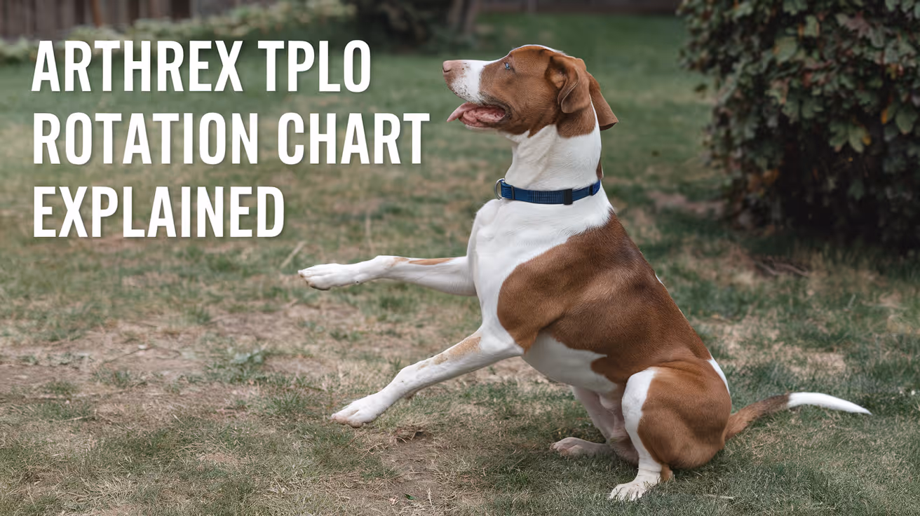
Arthrex TPLO Rotation Chart Explained
Learn how to read and use the Arthrex TPLO rotation chart for precise tibial plateau leveling osteotomy surgery in dogs.
Performing a tibial plateau leveling osteotomy (TPLO) is a common surgical procedure to treat cranial cruciate ligament injuries in dogs. The Arthrex TPLO rotation chart is a vital tool that helps veterinary surgeons determine the exact amount of tibial rotation needed during surgery. Understanding this chart ensures accurate bone alignment and improves surgical outcomes.
This article explains the Arthrex TPLO rotation chart in detail. You will learn how to interpret the chart, why it matters for your pet’s surgery, and how it guides surgeons in achieving the correct tibial plateau angle. This knowledge helps pet owners understand the surgical process better and supports informed discussions with your veterinary surgeon.
What is the Arthrex TPLO rotation chart?
The Arthrex TPLO rotation chart is a reference guide used during TPLO surgery. It shows the degrees of rotation needed to achieve a target tibial plateau angle based on the preoperative measurement. This chart helps surgeons plan and execute the bone cut and rotation precisely.
Using this chart reduces guesswork and improves the accuracy of the surgical correction. It is designed specifically for the Arthrex TPLO surgical system, which includes specialized plates and instruments.
- Rotation guidance: The chart provides exact degrees of tibial rotation required to reach the desired postoperative tibial plateau angle, ensuring surgical precision.
- Preoperative planning: It uses the measured preoperative tibial plateau angle to determine how much rotation is necessary during surgery.
- Standardization tool: The chart standardizes the surgical approach, reducing variability between different surgeons and cases.
- Integration with Arthrex system: It is designed to work with Arthrex-specific implants and instruments for seamless surgical workflow.
Understanding this chart is essential for surgeons performing TPLO with Arthrex equipment and benefits pet owners by improving surgical success rates.
How does the Arthrex TPLO rotation chart improve surgical accuracy?
Accurate rotation of the tibial plateau is critical to restore normal joint mechanics after TPLO surgery. The rotation chart helps surgeons avoid under- or over-rotation, which can lead to poor outcomes or complications.
By providing a clear reference, the chart minimizes errors during the procedure. It also supports consistent results across different patients and surgeons.
- Precise angle correction: The chart ensures the tibial plateau angle is corrected to the target, improving joint stability post-surgery.
- Reduces complications: Proper rotation lowers risks of implant failure, arthritis progression, and abnormal gait after surgery.
- Improves recovery: Accurate alignment supports better healing and faster return to normal activity for dogs.
- Supports training: The chart helps less experienced surgeons perform TPLO with confidence and accuracy.
Using the Arthrex TPLO rotation chart is a key step in achieving the best surgical outcomes for dogs undergoing cruciate ligament repair.
What measurements are needed before using the Arthrex TPLO rotation chart?
Before referencing the rotation chart, the surgeon must measure the tibial plateau angle on preoperative radiographs. This angle indicates how sloped the tibial plateau is, which influences the degree of rotation needed.
Accurate measurement is essential for the chart to provide correct rotation values. The process involves specific radiographic positioning and angle calculation techniques.
- Radiographic positioning: Proper lateral X-rays of the stifle joint are required to visualize the tibial plateau accurately.
- Angle measurement: The tibial plateau angle is measured using anatomical landmarks on the radiograph, typically with digital tools.
- Recording values: The measured angle is noted and used as the input for the rotation chart to find the corresponding rotation degree.
- Repeatability: Consistent measurement technique ensures reliable data for surgical planning and chart use.
Accurate preoperative measurements are the foundation for effective use of the Arthrex TPLO rotation chart during surgery.
How do surgeons use the Arthrex TPLO rotation chart during surgery?
During TPLO surgery, the surgeon references the rotation chart after measuring the tibial plateau angle. The chart indicates how many degrees to rotate the tibial segment after the osteotomy cut.
This rotation changes the slope of the tibial plateau to a safer angle, reducing strain on the cruciate ligament. The chart guides the surgeon to achieve this precisely.
- Osteotomy planning: The surgeon plans the bone cut location and angle based on the chart’s rotation recommendations.
- Rotation execution: After cutting the tibia, the surgeon rotates the bone segment by the degree indicated on the chart.
- Verification: Intraoperative imaging or jigs may be used to confirm the rotation matches the chart’s guidance.
- Plate fixation: The Arthrex TPLO plate is applied to stabilize the rotated tibia, maintaining the corrected angle during healing.
Following the rotation chart during surgery helps ensure the tibial plateau angle is corrected accurately and consistently.
What are the benefits of using the Arthrex TPLO rotation chart for pet owners?
For pet owners, the Arthrex TPLO rotation chart contributes to safer surgeries and better recovery for dogs with cruciate ligament injuries. It supports precise surgical correction, which improves joint function.
Understanding the chart can help owners feel more confident about the procedure and its outcomes.
- Improved surgical outcomes: Accurate rotation reduces complications and improves joint stability after surgery.
- Faster recovery: Correct alignment supports quicker healing and return to normal activity for pets.
- Reduced arthritis risk: Proper tibial plateau angle correction lowers the chance of arthritis development later.
- Enhanced surgeon confidence: The chart helps surgeons perform the procedure with precision, benefiting your pet’s health.
Knowing that the surgeon uses tools like the Arthrex TPLO rotation chart can reassure owners about the quality of care their pet receives.
Are there limitations or challenges with the Arthrex TPLO rotation chart?
While the Arthrex TPLO rotation chart is a valuable tool, it has some limitations. Surgeons must combine chart data with clinical judgment and experience for best results.
Variations in anatomy or measurement errors can affect the chart’s accuracy. Understanding these challenges helps set realistic expectations.
- Measurement variability: Inaccurate preoperative angle measurement can lead to incorrect rotation recommendations.
- Anatomical differences: Individual dog anatomy may require adjustments beyond the chart’s standardized values.
- Technical skill required: Proper use of the chart depends on surgeon experience and surgical technique.
- Not a standalone tool: The chart should be used alongside other surgical planning methods and intraoperative assessments.
Awareness of these limitations ensures the Arthrex TPLO rotation chart is used effectively as part of comprehensive surgical care.
Conclusion
The Arthrex TPLO rotation chart is an essential tool for veterinary surgeons performing tibial plateau leveling osteotomy in dogs. It provides clear guidance on the degree of tibial rotation needed to correct the tibial plateau angle accurately.
By understanding and using this chart, surgeons can improve surgical precision, reduce complications, and support better recovery for pets. Pet owners benefit from knowing their dog’s surgery is planned with advanced, reliable tools like the Arthrex TPLO rotation chart.
What is the Arthrex TPLO rotation chart used for?
The Arthrex TPLO rotation chart is used to determine the exact degrees of tibial rotation needed during TPLO surgery to achieve the desired tibial plateau angle correction.
How do you measure the tibial plateau angle before surgery?
The tibial plateau angle is measured on a properly positioned lateral radiograph of the stifle using anatomical landmarks and digital tools to ensure accuracy.
Can the Arthrex TPLO rotation chart prevent surgical complications?
Yes, by guiding precise tibial rotation, the chart helps reduce risks like implant failure and arthritis progression after TPLO surgery.
Is the Arthrex TPLO rotation chart suitable for all dog breeds?
The chart is designed for general use but surgeons may adjust rotation based on individual anatomical differences in certain breeds.
Do surgeons use imaging during TPLO surgery with the rotation chart?
Yes, intraoperative imaging or jigs are often used to verify that the tibial rotation matches the chart’s recommendations for accuracy.
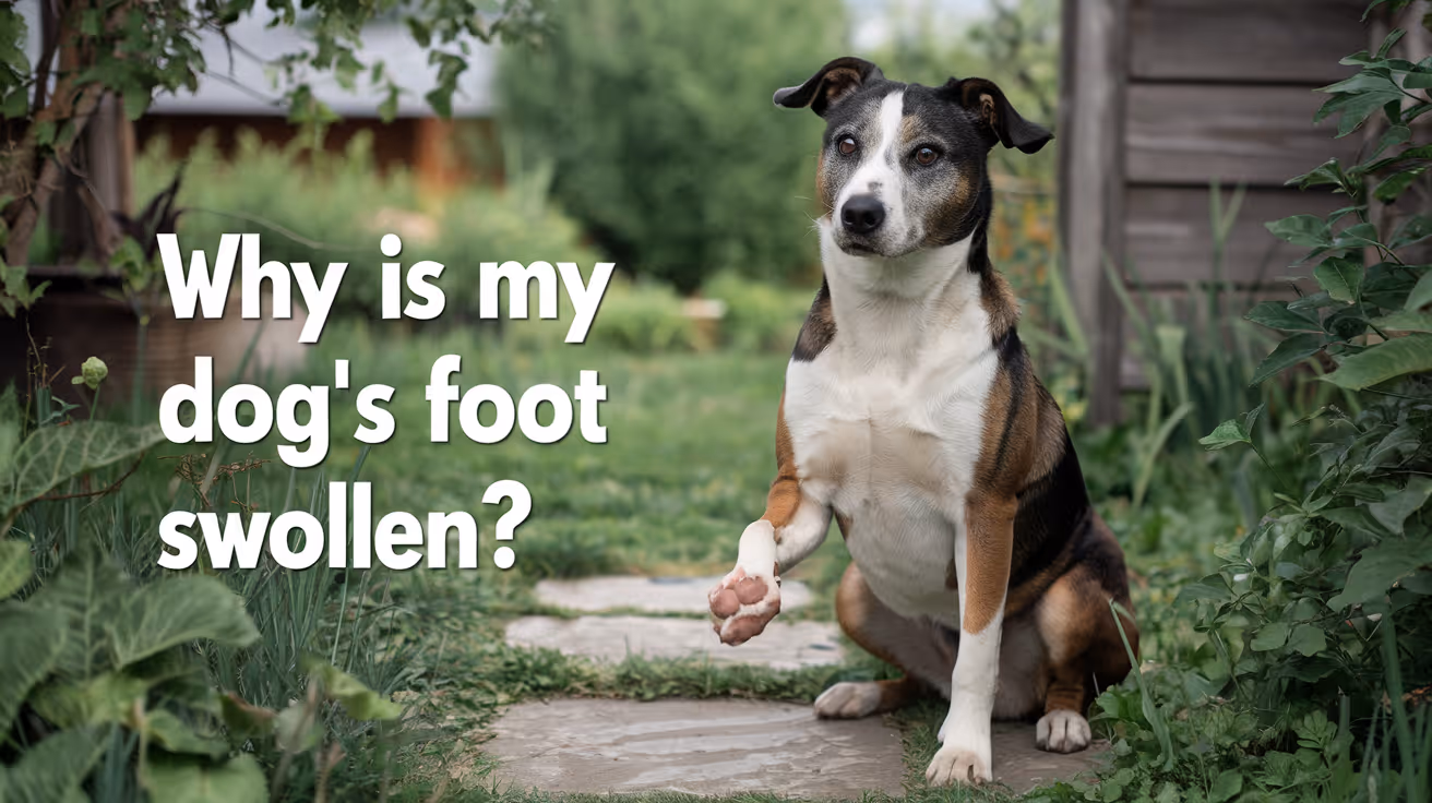
Why Is My Dog's Foot Swollen?
Discover why your dog's foot is swollen, common causes, treatments, and when to see a vet for proper care.
Seeing your dog's foot swollen can be worrying. Swelling in a dog's foot can happen for many reasons, from injuries to infections. Understanding why this happens helps you act quickly and keep your dog comfortable.
This article explains common causes of swollen dog feet, how to spot serious problems, and what treatments work best. You will learn when to treat at home and when to visit a vet for urgent care.
What Causes Swelling in a Dog's Foot?
Swelling in a dog's foot can come from many different problems. It often shows as puffiness, redness, or heat in the paw area. Knowing the cause helps you decide the right care.
Common causes include injuries, infections, allergies, and insect bites. Each cause needs a different approach to treatment.
- Injury or trauma: A cut, sprain, or broken bone can cause swelling due to inflammation and fluid buildup in the foot tissues.
- Infections: Bacterial or fungal infections can cause swelling, redness, and pain, often needing antibiotics or antifungal treatment.
- Allergic reactions: Allergies to plants, chemicals, or insect stings can cause sudden swelling and itching in the foot.
- Foreign objects: Thorns, splinters, or glass stuck in the paw can cause swelling and discomfort until removed.
Identifying the cause early helps prevent complications and speeds healing.
How Can I Tell If My Dog's Foot Swelling Is Serious?
Not all swelling is an emergency, but some signs mean you should see a vet quickly. Serious swelling can affect your dog's ability to walk or cause severe pain.
Look for symptoms like severe limping, open wounds, or signs of infection. These require prompt veterinary care.
- Severe limping or inability to walk: Indicates pain or serious injury needing urgent veterinary evaluation.
- Open wounds or bleeding: Risk of infection and need for cleaning and possibly stitches.
- Fever or lethargy: Signs that infection may have spread and requires medical treatment.
- Rapidly increasing swelling: Could signal an allergic reaction or deep infection needing emergency care.
When in doubt, it is safer to consult your vet to avoid worsening problems.
What Home Treatments Can Help a Swollen Dog Foot?
For mild swelling without serious signs, you can try some home care steps. These help reduce swelling and keep your dog comfortable.
Always watch your dog closely and stop home treatment if symptoms worsen or do not improve.
- Rest and limit activity: Keep your dog from running or jumping to reduce stress on the swollen foot.
- Cold compress application: Apply a cold pack wrapped in cloth for 10-15 minutes to reduce swelling and pain.
- Clean the paw gently: Use warm water to clean dirt or debris, especially if there are small cuts or irritations.
- Prevent licking or chewing: Use an Elizabethan collar if your dog tries to lick or bite the swollen area, which can worsen irritation.
These steps can help minor swelling but do not replace veterinary care for serious cases.
When Should I Take My Dog to the Vet for a Swollen Foot?
Knowing when to seek professional help is important. Some swelling needs medical treatment to avoid complications.
If your dog's swelling is severe, painful, or lasts more than a day or two, a vet visit is necessary. Early treatment can prevent infections or permanent damage.
- Persistent swelling over 48 hours: Indicates that the problem may not resolve without medical intervention.
- Signs of infection: Pus, foul odor, or heat around the swollen area require antibiotics or cleaning by a vet.
- Suspected broken bone or sprain: Needs X-rays and pain management from a veterinary professional.
- Severe allergic reactions: Swelling with difficulty breathing or collapse needs emergency veterinary care immediately.
Your vet can diagnose the cause and recommend the right treatment plan for your dog's recovery.
How Do Vets Diagnose the Cause of a Swollen Dog Foot?
Veterinarians use several methods to find the cause of swelling. A correct diagnosis is key to effective treatment.
They will examine your dog’s foot carefully and may use tests to look deeper into the problem.
- Physical examination: Checking for wounds, foreign objects, and signs of pain or infection in the foot.
- X-rays: Used to detect fractures, bone infections, or foreign bodies inside the paw.
- Skin scrapings or cultures: To identify infections caused by bacteria, fungi, or parasites.
- Blood tests: To check for systemic infection or allergic reactions affecting the swelling.
These tools help your vet create a treatment plan tailored to your dog's needs.
What Treatments Do Vets Use for Swollen Dog Feet?
Treatment depends on the cause of the swelling. Your vet may use medications, procedures, or supportive care to help your dog heal.
Some treatments can be done at home under vet guidance, while others require clinic visits.
- Antibiotics or antifungals: Prescribed to treat infections causing swelling and prevent spread.
- Anti-inflammatory drugs: Help reduce pain and swelling, improving your dog's comfort.
- Wound care and bandaging: Cleaning and protecting open wounds to promote healing and prevent infection.
- Surgery: May be needed to remove foreign objects or repair fractures causing swelling.
Follow your vet’s instructions carefully to ensure the best outcome for your dog.
How Can I Prevent My Dog's Foot from Swelling?
Preventing foot swelling involves protecting your dog from injuries and infections. Regular care and attention can reduce risks.
Simple habits help keep your dog's paws healthy and avoid painful swelling episodes.
- Regular paw inspections: Check your dog's feet daily for cuts, thorns, or swelling to catch problems early.
- Keep nails trimmed: Prevents nails from breaking or causing injury to the foot pads.
- Avoid walking on rough surfaces: Protect paws from sharp objects or hot pavement that can cause injuries.
- Use protective booties: Especially in harsh weather or rough terrain to shield paws from damage.
Good paw care supports your dog’s overall health and comfort.
Conclusion
Swelling in your dog's foot can have many causes, from minor injuries to serious infections. Understanding why your dog's foot is swollen helps you provide the right care quickly.
Always watch for signs of pain, infection, or worsening symptoms. When in doubt, seek veterinary advice to protect your dog's health and comfort. Early treatment can prevent complications and get your dog back on their feet faster.
Why is my dog's foot swollen after walking?
Your dog's foot may swell after walking due to minor injuries, irritation from rough surfaces, or allergic reactions. Rest and paw care usually help reduce swelling quickly.
Can a swollen dog foot heal without a vet?
Mild swelling from minor injuries or irritations can heal at home with rest and care. However, persistent or severe swelling needs veterinary evaluation to avoid complications.
How long does it take for a dog's swollen foot to go down?
Swelling may reduce within a few days with proper care. If swelling lasts more than 48 hours or worsens, consult a vet for treatment.
Is a swollen dog foot painful?
Yes, swelling often causes pain and discomfort. Your dog may limp, lick, or avoid putting weight on the swollen foot.
Can allergies cause a dog's foot to swell?
Yes, allergies to insect bites, plants, or chemicals can cause sudden swelling and itching in a dog's foot, sometimes requiring veterinary treatment.
All Articles

Asepsis for Spay and Neuter Surgery
Learn essential asepsis techniques for spay and neuter surgery to ensure safe, infection-free procedures for your pet.
Spay and neuter surgery is a common procedure in veterinary medicine that requires strict asepsis to prevent infections. Maintaining a sterile environment is crucial to protect your pet from complications during and after surgery. This article explains the key aspects of asepsis for spay and neuter surgery in simple terms.
Understanding asepsis helps you know how veterinarians keep your pet safe. You will learn about the steps taken before, during, and after surgery to maintain cleanliness and reduce infection risks.
What is asepsis in spay and neuter surgery?
Asepsis means keeping the surgical area free from harmful germs. It involves cleaning, sterilizing, and using techniques that stop bacteria from entering the body during surgery. In spay and neuter procedures, asepsis is vital because the surgery opens the abdomen or scrotum, which can easily get infected.
Proper asepsis reduces the chance of wound infections, speeds up healing, and improves overall outcomes for your pet.
- Definition of asepsis: Asepsis is the process of preventing infection by eliminating or controlling pathogens during surgery to protect the patient’s health.
- Importance in surgery: It prevents bacteria from entering the body, reducing risks of complications like abscesses or sepsis after spay or neuter surgery.
- Application in spay/neuter: The surgical site is cleaned, instruments are sterilized, and sterile gloves and gowns are used to maintain a germ-free environment.
- Difference from antisepsis: Asepsis prevents contamination before it happens, while antisepsis involves killing germs on skin or surfaces already exposed.
Understanding asepsis helps you appreciate the care taken to keep your pet safe during surgery. It is the foundation of all surgical procedures, especially spay and neuter.
How do veterinarians prepare the surgical site for asepsis?
Preparing the surgical site is the first step in asepsis. It involves shaving the fur, cleaning the skin, and applying antiseptic solutions to remove bacteria. This reduces the chance of germs entering the body when the skin is cut.
Proper site preparation is essential for a clean surgery and faster healing.
- Fur clipping: Removing hair around the incision site prevents hair from contaminating the wound and allows better antiseptic contact with the skin.
- Skin cleaning: The area is scrubbed with soap and water to remove dirt and oils that can harbor bacteria before antiseptic application.
- Antiseptic application: Solutions like chlorhexidine or povidone-iodine are applied to kill bacteria on the skin surface before surgery.
- Drying and draping: The skin is dried and covered with sterile drapes to create a clean field around the surgical site.
These steps ensure the skin is as clean as possible before the incision, lowering infection risk during spay or neuter surgery.
What sterilization methods are used for surgical instruments?
Sterilizing instruments removes all microorganisms, including bacteria and spores. This is critical because instruments touch internal tissues and must be free of germs to prevent infections.
Veterinary clinics use several sterilization methods to ensure instruments are safe for surgery.
- Autoclaving: Steam under pressure kills all microbes on instruments, making it the most common and effective sterilization method.
- Cold sterilization: Soaking instruments in chemical disinfectants is used for heat-sensitive tools but requires careful timing and rinsing.
- Dry heat sterilization: Instruments are heated in an oven at high temperatures to destroy microorganisms, used less often than autoclaving.
- Packaging: Sterilized instruments are wrapped in sterile packaging to keep them clean until surgery.
Proper sterilization protects your pet by ensuring no germs enter the body during the spay or neuter procedure.
How do surgical staff maintain asepsis during the operation?
During surgery, the veterinary team follows strict rules to keep the environment sterile. This includes wearing sterile gloves, gowns, and masks, and handling instruments carefully.
Maintaining asepsis during the operation prevents contamination and protects your pet’s health.
- Sterile gloves and gowns: Staff wear these to avoid transferring germs from their hands or clothes to the surgical site.
- Hand hygiene: Thorough handwashing and use of antiseptic solutions before gloving reduce bacteria on the skin.
- Instrument handling: Only sterile instruments touch the surgical site, and they are passed carefully to avoid contamination.
- Minimizing traffic: Limiting the number of people and movement in the operating room reduces airborne germs.
These practices help create a safe surgical environment, lowering infection risks during spay and neuter surgeries.
What post-operative care supports asepsis after spay and neuter surgery?
After surgery, proper wound care is essential to keep the site clean and prevent infection. This includes monitoring the incision, preventing licking, and following veterinary instructions.
Good post-operative care helps your pet heal quickly and avoids complications.
- Incision monitoring: Check the surgical site daily for redness, swelling, or discharge that could indicate infection.
- Preventing licking: Use an Elizabethan collar or other devices to stop your pet from licking or biting the wound, which can introduce bacteria.
- Keeping the area dry: Avoid bathing or wetting the incision until fully healed to maintain a clean environment.
- Follow-up visits: Attend veterinary check-ups to ensure proper healing and address any concerns promptly.
Following these steps supports asepsis after surgery and promotes a smooth recovery for your pet.
What are common asepsis challenges in spay and neuter surgeries?
Despite best efforts, some challenges can affect asepsis during spay and neuter procedures. Understanding these helps improve surgical safety.
Veterinary teams work to identify and manage these risks to protect your pet.
- Contamination risk: Accidental contact with non-sterile surfaces or instruments can introduce bacteria during surgery.
- Improper sterilization: Failure to sterilize instruments correctly can lead to infections post-operation.
- Environmental factors: Dust, airflow, and room cleanliness impact the sterile field and must be controlled.
- Patient factors: Pets with skin infections or poor health may have higher infection risks despite aseptic measures.
Awareness and careful management of these challenges help maintain asepsis and ensure successful spay and neuter surgeries.
Conclusion
Asepsis is a vital part of spay and neuter surgery that protects your pet from infections. It involves cleaning, sterilizing, and careful handling before, during, and after surgery to maintain a sterile environment.
By understanding asepsis, you can appreciate the care taken by veterinary teams to keep your pet safe. Following post-operative instructions and monitoring the surgical site help support healing and prevent complications.
What is the difference between asepsis and antisepsis?
Asepsis prevents germs from entering the surgical site by maintaining a sterile environment, while antisepsis kills germs on skin or surfaces already exposed to microbes.
Why is fur clipping important before spay surgery?
Clipping fur removes hair that can carry bacteria and interfere with skin cleaning, reducing infection risk during and after surgery.
How often should the surgical site be checked after neuter surgery?
Check the incision daily for signs of redness, swelling, or discharge to catch infections early and ensure proper healing.
Can pets bathe soon after spay surgery?
Pets should not be bathed or have the incision wet until the wound is fully healed to maintain asepsis and prevent infection.
What should be done if the surgical site looks infected?
If you notice redness, swelling, or discharge, contact your veterinarian immediately for evaluation and treatment to prevent complications.
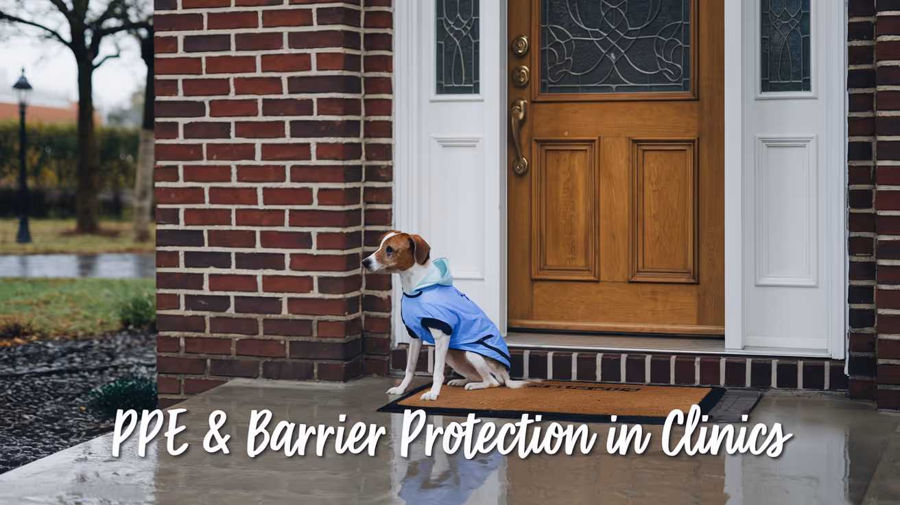
PPE Use and Barrier Protection in Veterinary Clinics
Learn about PPE use and barrier protection in veterinary clinics to keep staff and pets safe from infections and hazards.
Personal protective equipment (PPE) and barrier protection are essential in veterinary clinics to prevent the spread of infections and protect both staff and animals. Proper use of PPE reduces the risk of disease transmission and exposure to harmful substances during veterinary procedures. Understanding the correct PPE and barrier methods helps maintain a safe clinical environment.
This article explains the types of PPE used in veterinary settings, how to apply barrier protection effectively, and best practices for maintaining safety. You will learn how to choose the right equipment, when to use it, and how to dispose of it properly to protect everyone in the clinic.
What is the role of PPE in veterinary clinics?
PPE serves as a physical barrier between veterinary staff and potential hazards such as infectious agents, chemicals, and bodily fluids. It minimizes direct contact and reduces the risk of contamination during animal care and surgical procedures. PPE is a critical component of infection control protocols in veterinary medicine.
Using PPE correctly helps prevent zoonotic diseases, which are infections that can spread from animals to humans. It also protects animals from cross-contamination between patients. Veterinary clinics must implement PPE guidelines to ensure workplace safety and comply with health regulations.
- Infection prevention: PPE blocks pathogens from reaching skin, mucous membranes, and clothing, reducing infection risk for staff and animals.
- Hazard protection: Gloves, masks, and gowns shield workers from chemical exposure and bodily fluids encountered during treatments.
- Cross-contamination control: Barrier use prevents germs from spreading between animals and surfaces in the clinic.
- Regulatory compliance: Proper PPE use meets occupational health standards and legal requirements for veterinary workplaces.
Overall, PPE is vital for maintaining a safe veterinary environment and protecting health.
What types of PPE are commonly used in veterinary settings?
Veterinary clinics use various PPE items depending on the procedure and risk level. Each type offers specific protection tailored to different hazards encountered in animal care. Selecting the right PPE ensures effective barrier protection.
Common PPE includes gloves, masks, gowns, eye protection, and shoe covers. These items help reduce exposure to infectious agents, chemicals, and physical injuries during clinical work.
- Gloves: Disposable gloves protect hands from pathogens, chemicals, and contaminants during exams and surgeries.
- Masks: Surgical or respirator masks prevent inhalation of airborne particles and protect mucous membranes from splashes.
- Gowns: Fluid-resistant gowns shield skin and clothing from blood, saliva, and other fluids.
- Eye protection: Goggles or face shields guard eyes against splashes and debris during procedures.
Using the correct PPE combination based on risk assessment is essential for effective protection in veterinary clinics.
How should PPE be properly worn and removed?
Correct donning and doffing of PPE are crucial to avoid contamination. Improper handling can expose staff to infectious agents or spread pathogens within the clinic. Training and adherence to protocols ensure safety.
Staff must follow step-by-step procedures for putting on and taking off PPE, including hand hygiene before and after use. Removing PPE carefully prevents contact with contaminated surfaces.
- Donning sequence: Put on gown first, then mask, eye protection, and gloves last to cover gown cuffs completely.
- Doffing sequence: Remove gloves first, then eye protection, gown, and mask last to minimize contamination risk.
- Hand hygiene: Wash or sanitize hands before donning and immediately after doffing PPE to reduce pathogen spread.
- Proper disposal: Discard single-use PPE in designated biohazard containers to prevent environmental contamination.
Following these steps protects veterinary staff and maintains a clean clinical environment.
When is barrier protection necessary in veterinary clinics?
Barrier protection is required whenever there is a risk of exposure to infectious materials or hazardous substances. This includes routine exams, surgeries, dental cleanings, and handling of biological samples. Assessing risk helps determine the appropriate level of protection.
Veterinary staff should use barrier methods consistently during high-risk tasks to prevent disease transmission and contamination. This protects both personnel and animal patients.
- Surgical procedures: Full barrier protection with gowns, gloves, masks, and eye protection is essential to maintain sterility.
- Handling infectious cases: Use gloves and gowns to prevent contact with contagious animals or samples.
- Cleaning and disinfection: PPE protects staff from chemical exposure and contaminated surfaces during sanitation.
- Animal restraint: Gloves and protective clothing reduce injury risk and exposure to saliva or blood.
Using barrier protection based on task risk ensures safety and infection control in veterinary clinics.
How can veterinary clinics maintain PPE supply and compliance?
Maintaining adequate PPE stock and ensuring staff compliance are critical challenges in veterinary clinics. Proper management supports continuous protection and reduces infection risks. Clinics must plan and monitor PPE use carefully.
Training, clear policies, and regular audits encourage correct PPE use. Clinics should also establish reliable supply chains to avoid shortages during high demand or emergencies.
- Inventory management: Track PPE stock levels regularly to reorder before supplies run low and avoid interruptions.
- Staff training: Provide ongoing education on PPE importance, correct use, and disposal to improve compliance.
- Policy enforcement: Implement clear PPE protocols and monitor adherence through supervision and audits.
- Supplier relationships: Develop partnerships with trusted vendors to secure timely delivery of quality PPE products.
Effective PPE management promotes a safe workplace and protects veterinary teams and patients.
What are common challenges in PPE use and how to overcome them?
Veterinary clinics face several obstacles in PPE use, including discomfort, communication barriers, and resource limitations. Addressing these challenges improves safety and staff acceptance of protective measures.
Understanding and mitigating difficulties helps clinics maintain consistent PPE use and reduce infection risks. Solutions include ergonomic equipment, training, and workflow adjustments.
- Comfort issues: Select PPE that fits well and allows mobility to reduce fatigue and encourage use during long shifts.
- Communication barriers: Use clear masks or communication aids to facilitate interaction while wearing PPE.
- Resource constraints: Optimize PPE use by prioritizing high-risk tasks and reusing equipment safely when possible.
- Training gaps: Provide regular refresher courses and practical demonstrations to reinforce proper PPE practices.
Overcoming these challenges supports effective infection control and staff well-being in veterinary clinics.
Conclusion
PPE use and barrier protection are fundamental to safety in veterinary clinics. They prevent infections, protect staff and animals, and ensure compliance with health standards. Understanding the types of PPE, correct usage, and when to apply barrier methods is essential for every veterinary professional.
By maintaining proper PPE supplies, training staff, and addressing challenges, clinics can create a safer environment. Consistent use of PPE and barrier protection reduces disease risks and supports high-quality veterinary care.
What PPE should I wear during a routine veterinary exam?
Wear disposable gloves and a mask during routine exams to protect against contact with bodily fluids and respiratory droplets. Use eye protection if splashes are likely.
How do I dispose of used PPE safely in a veterinary clinic?
Dispose of single-use PPE in designated biohazard containers immediately after use. Follow local regulations for hazardous waste to prevent contamination.
Can PPE prevent zoonotic disease transmission in veterinary clinics?
Yes, PPE acts as a barrier to block pathogens from animals to humans, significantly reducing the risk of zoonotic infections in clinical settings.
How often should veterinary staff be trained on PPE use?
Staff should receive PPE training at hiring and refresher sessions at least annually or when protocols change to ensure ongoing compliance and safety.
Is it safe to reuse PPE in veterinary clinics?
Generally, single-use PPE should not be reused. Reuse is only acceptable for specific items after proper cleaning and disinfection, following strict guidelines.

Choosing Closure Technique Based on Tissue Type
Learn how to choose the best closure technique based on different tissue types for optimal healing and minimal complications.
Choosing the right closure technique based on tissue type is critical for successful wound healing. Different tissues require specific methods to ensure strength, reduce infection risk, and promote recovery.
This article explains how to select closure techniques for various tissues, including skin, muscle, fascia, and mucosa. You will learn practical tips to improve surgical outcomes and reduce complications.
What is the importance of selecting closure technique by tissue type?
Each tissue in the body has unique properties such as thickness, vascularity, and healing capacity. Selecting an appropriate closure technique helps maintain tissue integrity and function.
Incorrect closure can lead to wound dehiscence, infection, or poor cosmetic results. Understanding tissue characteristics guides the choice of suture material and method.
- Tissue-specific healing: Different tissues heal at varying rates, so closure methods must match their healing speed to avoid tension or delayed recovery.
- Strength requirements: Some tissues like fascia require stronger closure to withstand mechanical forces, influencing suture type and pattern.
- Infection risk: Certain tissues are more prone to infection, so closure technique must minimize dead space and contamination.
- Cosmetic outcome: Skin closure techniques affect scar appearance, requiring careful selection for visible areas.
Choosing closure technique by tissue type is essential to optimize healing and reduce complications.
How do you choose closure techniques for skin tissue?
Skin is the outermost tissue and requires closure methods that promote rapid healing and minimal scarring. It is exposed to external contaminants and mechanical stress.
Common skin closure techniques include sutures, staples, and adhesive strips. The choice depends on wound size, location, and tension.
- Suture selection: Non-absorbable sutures like nylon are preferred for skin to maintain strength until healing completes.
- Suture pattern: Interrupted sutures allow precise edge alignment and reduce risk of spreading infection.
- Staples use: Staples provide quick closure for scalp or trunk wounds but may cause more scarring.
- Adhesive strips: Useful for small, low-tension wounds to avoid needle trauma and improve cosmetic results.
Proper skin closure reduces infection risk and improves cosmetic outcomes.
What closure techniques suit muscle tissue?
Muscle tissue is highly vascular and contracts during movement. Closure must support healing without restricting mobility or causing ischemia.
Muscle closure often uses absorbable sutures with patterns that distribute tension evenly.
- Absorbable sutures: Polyglycolic acid sutures are commonly used as muscle heals quickly and sutures dissolve safely.
- Interrupted pattern: Allows flexibility and reduces ischemia by avoiding tight continuous sutures.
- Layered closure: Muscle is closed in layers to restore anatomy and reduce dead space.
- Avoid excessive tension: Prevents muscle necrosis and promotes better functional recovery.
Choosing the right muscle closure technique supports healing and preserves function.
How should fascia be closed for optimal healing?
Fascia is a strong connective tissue layer that provides structural support. It requires durable closure to withstand intra-abdominal pressure and movement.
Fascia closure usually involves non-absorbable or slowly absorbable sutures with continuous or interrupted patterns.
- Strong suture material: Nylon or polypropylene sutures provide lasting strength for fascia closure.
- Continuous closure: Distributes tension evenly along the incision, reducing risk of dehiscence.
- Interrupted sutures: Used in contaminated wounds to isolate infection and maintain strength.
- Proper bite size: Large tissue bites prevent suture pull-through and ensure secure closure.
Fascia closure technique is vital to prevent hernias and maintain abdominal wall integrity.
What closure methods are best for mucosal tissue?
Mucosal tissue lines internal cavities and heals rapidly but is delicate and moist. Closure techniques must minimize trauma and promote quick epithelialization.
Absorbable sutures with fine gauge and gentle patterns are preferred for mucosal closure.
- Fine absorbable sutures: Materials like chromic gut dissolve safely without irritation in mucosa.
- Interrupted sutures: Allow precise edge alignment and reduce tension on delicate tissue.
- Minimal handling: Reduces tissue trauma and promotes faster healing.
- Moist environment: Closure must maintain moisture to support epithelial regeneration.
Proper mucosal closure reduces risk of fistulas and promotes functional recovery.
How do suture materials affect closure based on tissue type?
Suture material choice depends on tissue healing time, strength needed, and risk of reaction. Different tissues require specific suture properties.
Matching suture type to tissue optimizes healing and reduces complications like infection or suture failure.
- Absorbable sutures: Ideal for tissues that heal quickly, such as muscle and mucosa, to avoid suture removal.
- Non-absorbable sutures: Used in skin and fascia where long-term strength is necessary.
- Monofilament sutures: Cause less tissue drag and reduce infection risk in delicate tissues.
- Multifilament sutures: Provide better knot security but may harbor bacteria if not handled properly.
Choosing the right suture material enhances closure success for each tissue type.
What are the risks of improper closure technique by tissue type?
Using incorrect closure methods can lead to serious complications such as wound breakdown, infection, or poor function. Each tissue type has specific risks.
Understanding these risks helps prevent postoperative problems and improves patient outcomes.
- Wound dehiscence: Occurs if fascia or muscle closure is weak, risking hernias or muscle damage.
- Infection risk: Poor skin or mucosal closure can allow bacterial entry and delayed healing.
- Scarring and contracture: Improper skin closure may cause unsightly scars or restrict movement.
- Functional impairment: Incorrect muscle or mucosal closure can reduce tissue function and cause pain.
Proper closure technique tailored to tissue type minimizes these risks and supports healing.
Conclusion
Choosing closure technique based on tissue type is essential for optimal healing and minimizing complications. Each tissue has unique needs that guide suture material and method selection.
Understanding these principles helps ensure strong, infection-free wounds with good cosmetic and functional outcomes. Always tailor closure to tissue characteristics for the best surgical results.
FAQs
What suture type is best for skin closure?
Non-absorbable sutures like nylon are best for skin closure as they maintain strength until the skin heals and can be removed easily.
Can absorbable sutures be used for fascia closure?
Absorbable sutures are generally not recommended for fascia closure because fascia requires long-term strength to prevent hernias.
Why is muscle closure done in layers?
Layered muscle closure restores anatomy, reduces dead space, and distributes tension evenly to promote better healing and function.
How does mucosal tissue healing differ from skin?
Mucosal tissue heals faster, is more delicate, and requires absorbable sutures with minimal trauma to support rapid epithelialization.
What happens if closure technique is wrong for a tissue type?
Incorrect closure can cause wound breakdown, infection, poor cosmetic results, and impaired tissue function depending on the tissue involved.

Preventing Dehiscence in Dog Surgical Wounds
Learn how to prevent dehiscence in dog surgical wounds with expert tips on care, suturing, and monitoring to ensure safe healing.
Dehiscence in dog surgical wounds is a serious complication where the wound reopens after surgery. This problem can lead to infection, delayed healing, and additional surgeries. Understanding how to prevent dehiscence is critical for every dog owner and veterinary caregiver.
This article explains the causes of wound dehiscence in dogs and provides clear, practical steps to avoid it. You will learn about proper wound care, surgical techniques, and signs to watch for to keep your dog safe and healthy after surgery.
What is dehiscence in dog surgical wounds?
Dehiscence means the surgical wound edges separate before the tissue has fully healed. This can expose underlying tissues and increase the risk of infection. It usually happens within the first two weeks after surgery when the wound is still fragile.
Understanding dehiscence helps you recognize why prevention is important. It also guides how to care for your dog’s wound properly to avoid complications.
- Definition of dehiscence: It is the reopening or splitting of a surgical wound before complete healing, which can cause serious health risks for your dog.
- Timing of occurrence: Dehiscence most often happens within 7 to 14 days post-surgery when the wound is weakest and healing is incomplete.
- Consequences of dehiscence: It increases the chance of infection, pain, and may require additional surgery to repair the wound.
- Common affected areas: Abdominal and limb wounds are more prone due to movement and tension on the skin during healing.
Knowing what dehiscence is helps you take early action if you notice signs of wound opening in your dog.
What causes surgical wound dehiscence in dogs?
Several factors can cause a surgical wound to reopen in dogs. These include mechanical stress, infection, poor surgical technique, and the dog’s health status. Identifying these causes helps prevent dehiscence effectively.
By controlling these factors, you can reduce the risk of wound complications and support smooth healing.
- Excessive movement: Dogs that are too active or lick their wounds can cause tension that pulls the wound edges apart.
- Infection at the site: Bacterial contamination delays healing and weakens the wound, increasing the chance of reopening.
- Poor suturing technique: Incorrect suture placement or tension can cause the wound to fail under stress.
- Underlying health issues: Conditions like diabetes or poor nutrition impair wound healing and increase dehiscence risk.
Understanding these causes allows you to work closely with your veterinarian to minimize risks during recovery.
How should dog surgical wounds be cared for to prevent dehiscence?
Proper wound care is essential to prevent dehiscence. This includes keeping the wound clean, preventing your dog from disturbing it, and following veterinary instructions carefully.
Good wound care supports tissue repair and reduces complications that lead to wound reopening.
- Keep wound clean: Clean the area gently with prescribed solutions to avoid bacteria buildup and infection.
- Prevent licking or chewing: Use an Elizabethan collar or protective bandages to stop your dog from disturbing the wound.
- Follow medication schedule: Administer antibiotics and pain relief exactly as prescribed to support healing and comfort.
- Limit activity: Restrict your dog’s movement to reduce tension on the wound and allow tissues to heal properly.
Consistent wound care helps maintain a safe environment for healing and reduces the chance of dehiscence.
What surgical techniques reduce the risk of wound dehiscence in dogs?
Veterinarians use specific surgical methods to minimize the chance of wound reopening. These include proper suture selection, tension management, and layered closure techniques.
Understanding these techniques helps you appreciate the care your dog receives during surgery and recovery.
- Appropriate suture material: Using absorbable sutures that match tissue type reduces irritation and supports gradual healing.
- Layered closure: Closing wounds in layers strengthens the repair and distributes tension evenly across tissues.
- Minimal tissue trauma: Gentle handling of tissues during surgery prevents damage that can weaken wound edges.
- Proper suture tension: Sutures should be tight enough to hold edges but not so tight that they cut through tissue.
These surgical practices are vital to create a strong wound closure that resists reopening during healing.
When should you contact a veterinarian about possible wound dehiscence?
Early detection of wound problems can prevent serious complications. You should watch for signs of dehiscence and contact your vet promptly if you suspect an issue.
Knowing when to seek help ensures timely treatment and better outcomes for your dog.
- Visible wound opening: Any gap or separation in the wound edges should be reported immediately to your veterinarian.
- Increased redness or swelling: These signs may indicate infection or inflammation around the wound.
- Discharge or foul odor: Pus or bad smells suggest bacterial infection requiring veterinary care.
- Excessive pain or licking: If your dog shows discomfort or obsessively licks the wound, it may signal a problem.
Prompt veterinary evaluation can prevent worsening and guide appropriate treatment for wound healing.
What role does nutrition play in preventing wound dehiscence in dogs?
Good nutrition supports tissue repair and immune function, which are essential for wound healing. Feeding your dog a balanced diet helps prevent complications like dehiscence.
Understanding nutritional needs during recovery helps you provide the best care for your dog’s healing process.
- Protein intake: Adequate protein supports collagen formation, which strengthens wound tissue during healing.
- Vitamins and minerals: Nutrients like vitamin C, zinc, and vitamin A promote immune function and tissue repair.
- Hydration: Proper water intake maintains skin elasticity and supports cellular functions needed for healing.
- Caloric balance: Sufficient calories provide energy for the body to repair tissues without causing obesity-related stress.
Providing balanced nutrition tailored to your dog’s needs enhances recovery and lowers the risk of wound complications.
How can you manage your dog’s activity to prevent surgical wound dehiscence?
Controlling your dog’s movement after surgery is crucial to avoid stress on the wound. Excessive activity can pull sutures apart and cause dehiscence.
Implementing activity restrictions helps protect the surgical site and supports proper healing.
- Use confinement areas: Restrict your dog to a crate or small room to limit running, jumping, and rough play during recovery.
- Short leash walks: Only allow brief, controlled walks to prevent excessive strain on the wound.
- Monitor interactions: Keep your dog away from other pets that might cause injury or encourage activity.
- Provide mental stimulation: Use toys and gentle training to keep your dog calm and engaged without physical exertion.
Managing activity carefully reduces mechanical stress on the wound and helps prevent reopening during the critical healing phase.
Conclusion
Preventing dehiscence in dog surgical wounds requires careful attention to wound care, surgical technique, nutrition, and activity control. Understanding the causes and signs of dehiscence helps you act quickly to protect your dog’s health.
By following veterinary advice and monitoring your dog closely, you can support safe healing and reduce the risk of wound complications. Proper prevention ensures your dog recovers comfortably and returns to normal activities sooner.
What are the first signs of wound dehiscence in dogs?
Early signs include redness, swelling, discharge, or visible separation of the wound edges. Prompt veterinary evaluation is essential to prevent worsening.
Can wound dehiscence heal without surgery in dogs?
Minor dehiscence may heal with wound care and antibiotics, but larger openings often require surgical repair to close the wound properly.
How long does it take for a dog’s surgical wound to heal?
Most surgical wounds heal within 10 to 14 days, but full strength may take several weeks depending on the wound size and location.
Is it safe to bathe a dog with a surgical wound?
Bathing is usually not recommended until the wound is fully healed to avoid infection and wound opening. Consult your vet for specific instructions.
What suture materials are best to prevent dehiscence in dogs?
Absorbable sutures like polydioxanone or poliglecaprone are preferred as they reduce irritation and support gradual tissue healing without removal.
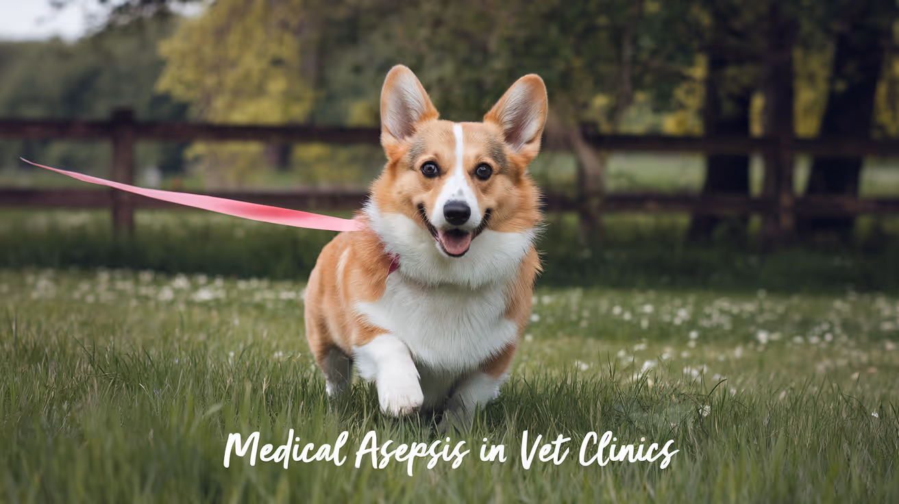
Medical Asepsis in Veterinary Clinics Explained
Learn about medical asepsis in veterinary clinics, its importance, techniques, and best practices to keep pets safe from infections.
Medical asepsis in veterinary clinics is essential to prevent the spread of infections among animals and staff. It involves practices that reduce the number of microorganisms and stop contamination during medical procedures. Understanding medical asepsis helps protect your pet’s health during visits to the vet.
This article explains what medical asepsis means in veterinary settings, why it matters, and how clinics maintain a clean and safe environment. You will learn about common aseptic techniques, equipment sterilization, and how veterinary staff apply these methods daily.
What is medical asepsis in veterinary clinics?
Medical asepsis refers to procedures that reduce or eliminate harmful microorganisms to prevent infection. In veterinary clinics, it focuses on keeping animals and surfaces clean to avoid disease transmission. This differs from surgical asepsis, which aims for complete sterility during operations.
Veterinary staff use medical asepsis to control germs in exam rooms, treatment areas, and during routine care. It helps protect animals with weakened immune systems and prevents outbreaks of contagious diseases.
- Definition clarity: Medical asepsis means reducing germs to safe levels, not complete sterilization, which is critical for everyday veterinary care.
- Scope of use: It applies to cleaning, disinfecting, and handling animals to stop infection spread during exams and treatments.
- Difference from surgical asepsis: Medical asepsis controls microbes, while surgical asepsis requires a sterile field for invasive procedures.
- Importance for animal health: Proper asepsis lowers infection risks, promoting faster recovery and safer clinic visits.
Understanding medical asepsis helps pet owners appreciate the care taken to keep animals safe during veterinary visits.
Why is medical asepsis important in veterinary clinics?
Medical asepsis is crucial to prevent infections that can harm animals and staff. Veterinary clinics treat many animals daily, some carrying contagious diseases. Without proper aseptic techniques, infections can spread quickly.
Infections can delay healing, cause serious illness, or even be fatal. Medical asepsis protects animals undergoing treatment and helps maintain a healthy clinic environment.
- Infection prevention: It reduces the chance of animals catching or spreading infections during visits or procedures.
- Protects vulnerable patients: Sick or young animals have weaker immune systems and need extra protection from germs.
- Staff safety: Proper asepsis lowers the risk of zoonotic diseases spreading to veterinary workers.
- Clinic reputation: Maintaining cleanliness builds trust with pet owners and ensures compliance with health standards.
Medical asepsis is a key part of quality veterinary care that benefits animals, staff, and pet owners alike.
What are common medical asepsis techniques used in veterinary clinics?
Veterinary clinics use several techniques to maintain medical asepsis. These include hand hygiene, cleaning and disinfecting surfaces, using personal protective equipment, and proper waste disposal. Each step helps reduce germs and contamination.
Staff follow strict protocols to ensure these techniques are effective and consistent. Training and monitoring help maintain high aseptic standards.
- Hand hygiene: Frequent hand washing or sanitizing removes germs before and after animal contact, preventing cross-contamination.
- Surface disinfection: Cleaning exam tables, equipment, and floors with approved disinfectants kills microbes and keeps areas safe.
- Personal protective equipment: Gloves, gowns, and masks protect staff and animals from exposure to infectious agents.
- Waste management: Proper disposal of needles, bandages, and biological waste prevents environmental contamination and disease spread.
These techniques work together to create a safer clinic environment and protect animal health.
How do veterinary clinics sterilize equipment for medical asepsis?
Sterilization is a process that destroys all microorganisms on instruments. Veterinary clinics use sterilization to ensure tools are safe for use, especially during invasive procedures. This is a step beyond medical asepsis but supports overall infection control.
Common sterilization methods include autoclaving, chemical sterilants, and dry heat. Clinics select methods based on the equipment type and usage.
- Autoclaving: Uses high-pressure steam to kill all microbes, commonly used for surgical instruments and reusable tools.
- Chemical sterilants: Liquid or gas chemicals disinfect instruments that cannot withstand heat, ensuring safe use.
- Dry heat sterilization: Applies hot air for extended periods, suitable for metal tools sensitive to moisture.
- Packaging and storage: Sterilized instruments are kept in sterile packaging to maintain cleanliness until use.
Proper sterilization prevents infections and supports medical asepsis by ensuring equipment is free of harmful germs.
What role does hand hygiene play in medical asepsis?
Hand hygiene is the single most important practice to prevent infection spread in veterinary clinics. Hands can carry germs from one animal or surface to another, so cleaning them regularly is vital.
Veterinary staff wash hands with soap and water or use alcohol-based sanitizers before and after contact with animals, equipment, or contaminated surfaces.
- Hand washing technique: Proper scrubbing for at least 20 seconds removes dirt and microbes effectively.
- Use of hand sanitizers: Alcohol-based sanitizers quickly kill germs when soap and water are unavailable.
- Glove use: Gloves protect hands but do not replace hand hygiene; hands must be cleaned before and after glove use.
- Preventing cross-contamination: Clean hands reduce the risk of transferring pathogens between animals and surfaces.
Consistent hand hygiene is a simple yet powerful tool to maintain medical asepsis and protect animal health.
How can pet owners support medical asepsis during veterinary visits?
Pet owners play a role in supporting medical asepsis by following clinic guidelines and preparing their pets properly. Cooperation helps reduce infection risks and ensures a smooth visit.
Owners should communicate openly about their pet’s health and follow instructions for appointments and treatments.
- Pre-visit preparation: Bathing pets and cleaning their paws before visits lowers external germs brought into the clinic.
- Following clinic rules: Wearing masks, using hand sanitizer, and maintaining distance help protect everyone in the clinic.
- Informing staff: Reporting any signs of illness or exposure to contagious diseases helps staff take extra precautions.
- Post-visit care: Following discharge instructions and keeping wounds clean supports recovery and prevents infections.
By working with veterinary teams, pet owners help maintain a safe environment and support medical asepsis efforts.
What are common challenges in maintaining medical asepsis in veterinary clinics?
Maintaining medical asepsis in busy veterinary clinics can be challenging due to high patient volume, diverse species, and varying health conditions. Staff must stay vigilant to prevent lapses that could lead to infections.
Resource limitations and human error also affect aseptic practices. Continuous training and monitoring help overcome these challenges.
- High patient turnover: Many animals in a short time increase contamination risk and require rapid cleaning protocols.
- Species differences: Different animals carry different germs, requiring tailored aseptic measures for each case.
- Staff compliance: Ensuring all team members consistently follow aseptic procedures demands ongoing education and supervision.
- Equipment availability: Limited access to sterilized tools or protective gear can compromise asepsis if not managed properly.
Addressing these challenges is vital to uphold medical asepsis and protect animal and staff health in veterinary clinics.
Conclusion
Medical asepsis in veterinary clinics is a fundamental practice to prevent infections and protect animals during medical care. It involves cleaning, disinfecting, hand hygiene, and sterilization techniques that reduce harmful microorganisms.
Understanding and supporting medical asepsis helps pet owners ensure their animals receive safe, high-quality care. Veterinary teams work hard to maintain these standards despite challenges, making clinics safer for all pets and people.
What is the difference between medical asepsis and surgical asepsis?
Medical asepsis reduces germs to safe levels during routine care, while surgical asepsis aims for complete sterility during invasive procedures to prevent all microbial contamination.
How often should veterinary staff perform hand hygiene?
Staff should clean their hands before and after every animal contact, after removing gloves, and after touching contaminated surfaces to prevent infection spread.
Can pet owners bring their own disinfectants to the clinic?
Pet owners should not bring disinfectants as clinics use specific approved products to ensure safety and effectiveness in controlling infections.
Are disposable gloves necessary for all veterinary procedures?
Gloves are required for procedures involving contact with bodily fluids or broken skin but are not always needed for simple exams if hand hygiene is maintained.
What should I do if my pet shows signs of infection after a clinic visit?
Contact your veterinarian promptly to report symptoms like redness, swelling, or discharge, so they can assess and provide appropriate treatment quickly.

Suture Removal Timing in Cats
Learn when and how to safely remove sutures in cats to ensure proper healing and avoid complications.
Suture removal timing in cats is a crucial part of post-surgical care. Knowing when to remove sutures helps prevent infections and supports proper wound healing. Many cat owners worry about the right time to remove stitches and how to do it safely.
This article explains the ideal timing for suture removal in cats, what signs to watch for, and how to care for your cat’s wound during healing. You will learn practical tips to keep your cat comfortable and healthy after surgery.
What is the ideal time to remove sutures in cats?
The timing for suture removal in cats depends on the type of surgery and the location of the wound. Generally, sutures are removed between 7 and 14 days after surgery. This period allows the skin to heal enough to stay closed without stitches.
Waiting too long or removing sutures too early can cause problems. Early removal may lead to wound reopening, while late removal can cause irritation or infection.
- Standard removal window: Most sutures are removed 10 to 14 days after surgery to ensure proper skin healing and strength.
- Location matters: Sutures on areas with more movement, like joints, may need longer healing times before removal.
- Type of suture: Absorbable sutures dissolve on their own and do not require removal, unlike non-absorbable sutures.
- Veterinary advice: Always follow your veterinarian’s specific instructions on timing based on your cat’s surgery and health.
Proper timing helps prevent wound complications and supports your cat’s recovery.
How can you tell if sutures are ready to be removed?
Before removing sutures, you need to check if the wound has healed well. Signs of healing include closed edges, no redness, and no discharge. Your cat should not show pain or swelling around the stitches.
If the wound looks inflamed or your cat is licking or biting the area, it may not be ready for suture removal. In such cases, consult your veterinarian.
- Wound closure: The edges of the wound should be sealed and not open or gaping before removing sutures.
- No redness or swelling: Healthy skin around sutures should look normal without signs of infection or irritation.
- Absence of discharge: There should be no pus, blood, or fluid coming from the wound site.
- Cat’s comfort: Your cat should not show signs of pain or excessive licking near the sutures.
Careful observation helps ensure safe and timely suture removal.
What are the risks of removing sutures too early or too late?
Removing sutures too early can cause the wound to reopen, leading to infection or delayed healing. Removing sutures too late can cause skin irritation, scarring, or suture-related infections.
Both situations can increase discomfort for your cat and may require additional veterinary care.
- Early removal risks: Premature suture removal may cause wound dehiscence, where the skin separates and delays healing.
- Late removal risks: Leaving sutures too long can cause tissue irritation and increase the chance of infection around the stitches.
- Increased scarring: Improper timing can lead to more noticeable scars or skin thickening at the wound site.
- Additional treatment: Complications from poor timing may require antibiotics or further surgery to fix the wound.
Following the correct timing reduces risks and promotes smooth recovery.
How should you care for your cat’s sutures before removal?
Proper wound care before suture removal is essential to prevent infection and support healing. You should keep the area clean, prevent your cat from licking, and monitor for any changes.
Using an Elizabethan collar or bandage can help protect the sutures. Always follow your veterinarian’s instructions on cleaning and care.
- Keep area clean: Gently clean the wound with vet-approved solutions to avoid infection and promote healing.
- Prevent licking: Use an Elizabethan collar to stop your cat from biting or licking the sutures, which can cause damage.
- Monitor daily: Check the wound every day for redness, swelling, or discharge that may indicate problems.
- Follow vet advice: Use any prescribed ointments or medications exactly as directed to support healing.
Good care helps your cat heal faster and reduces the chance of complications.
Can you remove cat sutures at home safely?
Removing sutures at home is generally not recommended. It requires proper tools and knowledge to avoid harming your cat or causing infection. Your veterinarian should perform suture removal to ensure safety.
If you must remove sutures at home due to emergency, use sterile scissors and follow strict hygiene. However, always consult your vet first.
- Professional removal preferred: Veterinarians have the training and tools to remove sutures safely and check wound healing.
- Risk of injury: Improper removal can cause pain, bleeding, or wound reopening in your cat.
- Infection risk: Non-sterile tools or poor technique can introduce bacteria and cause infection.
- Emergency only: Home removal should only be done if vet care is unavailable and with extreme caution.
Always prioritize veterinary care for suture removal to protect your cat’s health.
What signs indicate you should contact your veterinarian about sutures?
Some signs mean your cat’s sutures need veterinary attention. If you notice swelling, redness, discharge, or your cat is in pain, contact your vet immediately. Early treatment prevents serious complications.
Also, if sutures are loose, missing, or the wound reopens, seek veterinary help right away.
- Redness and swelling: Persistent or worsening inflammation around sutures may indicate infection requiring vet care.
- Discharge or pus: Any fluid leaking from the wound suggests infection and needs prompt veterinary attention.
- Excessive pain: If your cat shows signs of pain or discomfort near the sutures, consult your vet immediately.
- Wound reopening: If the wound edges separate or sutures come loose, professional care is necessary to prevent complications.
Timely veterinary intervention ensures your cat heals safely and comfortably.
Conclusion
Suture removal timing in cats is vital for proper wound healing and avoiding complications. Most sutures are removed between 10 and 14 days after surgery, but this depends on the wound and your veterinarian’s advice.
Careful monitoring of the wound and following veterinary instructions will help your cat recover well. Never rush suture removal or try it at home without guidance. If you notice any signs of infection or problems, contact your veterinarian promptly for the best care.
FAQs
When should I remove my cat’s sutures after surgery?
Most cat sutures are removed 10 to 14 days after surgery, depending on the wound location and healing progress. Always follow your veterinarian’s instructions.
Can I remove my cat’s sutures at home?
It is not recommended to remove cat sutures at home due to risks of injury and infection. Have a veterinarian perform the removal safely.
What signs show my cat’s sutures need veterinary attention?
Signs like redness, swelling, discharge, pain, or wound reopening require immediate veterinary care to prevent complications.
How can I care for my cat’s sutures before removal?
Keep the wound clean, prevent licking with an Elizabethan collar, monitor daily, and follow your vet’s care instructions carefully.
What happens if sutures are removed too early or too late?
Removing sutures too early can cause wound reopening, while late removal can cause irritation or infection. Both increase healing problems and discomfort.

Draping Techniques in Small Animal Surgery
Explore essential draping techniques in small animal surgery to ensure sterile fields and reduce infection risks during procedures.
In small animal surgery, maintaining a sterile environment is critical to prevent infections and ensure successful outcomes. Draping techniques play a vital role in creating a clean surgical field by isolating the operative site from surrounding contamination. Understanding proper draping methods helps veterinary professionals protect both patients and staff during surgery.
This article covers the key draping techniques used in small animal surgery. You will learn why draping is important, the types of drapes available, how to prepare the surgical site, and best practices for applying drapes effectively. This knowledge helps improve surgical safety and patient recovery.
What is the purpose of draping in small animal surgery?
Draping creates a sterile barrier that protects the surgical site from bacteria and contaminants. It isolates the area where the incision will be made and prevents contact with non-sterile surfaces. This reduces the risk of postoperative infections and complications.
Proper draping also helps organize the surgical field, giving the surgeon clear access and visibility. It supports infection control protocols and maintains aseptic technique throughout the procedure.
- Sterile barrier creation: Draping forms a physical barrier that blocks bacteria and debris from reaching the surgical site, minimizing infection risk during surgery.
- Field isolation: It isolates the incision area from surrounding skin and fur, which may harbor microorganisms harmful to the patient.
- Improved visibility: Drapes help define the surgical field clearly, allowing the surgeon to focus on the operative site without distractions.
- Supports aseptic technique: Draping reinforces sterile practices by maintaining separation between sterile and non-sterile zones in the operating room.
Overall, draping is essential for patient safety and surgical success in veterinary medicine.
What types of drapes are used in small animal surgery?
Several types of drapes are available for small animal surgery, each designed for specific purposes. Choosing the right drape depends on the procedure, patient size, and surgeon preference. Common drapes include disposable, reusable, fenestrated, and non-fenestrated options.
Understanding drape materials and designs helps ensure proper coverage and sterility during surgery.
- Disposable drapes: Made from synthetic materials, these drapes are single-use and reduce cross-contamination risks by being discarded after surgery.
- Reusable drapes: Typically made from woven fabrics, these drapes can be sterilized and reused multiple times, offering cost savings but requiring careful handling.
- Fenestrated drapes: These drapes have a pre-cut opening to expose the surgical site while covering surrounding areas, allowing precise access and protection.
- Non-fenestrated drapes: Solid drapes without openings, used to cover large areas or as additional layers to maintain sterility around the surgical field.
Selecting appropriate drapes improves surgical efficiency and infection control.
How do you prepare the surgical site before draping?
Preparing the surgical site is a critical step before applying drapes. It involves cleaning and disinfecting the area to remove dirt, hair, and microbes. Proper preparation reduces the bacterial load and enhances the effectiveness of the draping barrier.
Following a systematic approach ensures the site is ready for a sterile procedure.
- Clipping hair: Remove hair around the incision site using clippers to reduce contamination and improve drape adhesion.
- Skin cleaning: Use antiseptic solutions like chlorhexidine or povidone-iodine to scrub the skin thoroughly, lowering microbial presence.
- Rinsing and drying: Rinse the antiseptic off with sterile saline and dry the area with sterile gauze to prevent irritation and ensure drape adherence.
- Marking incision site: Optionally, mark the planned incision location with sterile ink to guide precise draping and surgery.
Thorough preparation supports a sterile environment and reduces infection risks.
What are the best practices for applying drapes in small animal surgery?
Applying drapes correctly is essential to maintain sterility and protect the surgical site. The process requires attention to detail and adherence to aseptic technique. Proper draping minimizes contamination and provides a stable field for surgery.
Following best practices helps avoid common mistakes and ensures patient safety.
- Use sterile gloves: Always wear sterile gloves when handling drapes to prevent transferring bacteria to the surgical field.
- Apply drapes from sterile packs: Open drapes carefully and place them without touching non-sterile surfaces to maintain their sterility.
- Cover surrounding areas: Extend drapes beyond the incision site to protect adjacent skin and surfaces from contamination.
- Secure drapes properly: Use towel clamps or adhesive strips to keep drapes in place and prevent shifting during surgery.
Consistent technique and careful handling are key to effective draping.
How do draping techniques differ for various small animal surgeries?
Draping approaches vary depending on the type and location of the surgery. Different procedures require specific draping methods to optimize access and maintain sterility. Understanding these variations helps tailor draping to each case.
Adjusting draping techniques ensures the surgical field is appropriate for the procedure and patient anatomy.
- Orthopedic surgeries: Often require fenestrated drapes that expose limbs while covering the rest of the body to allow precise access and minimize contamination.
- Abdominal surgeries: Use large non-fenestrated drapes to cover the entire abdomen and surrounding areas, creating a broad sterile field.
- Thoracic surgeries: Require careful draping to isolate the chest area, often using multiple drapes to protect vital structures and maintain sterility.
- Dental procedures: Smaller drapes or towels may be used to cover the head and neck, focusing on the oral cavity while protecting other regions.
Customizing draping techniques improves surgical outcomes and safety.
What are common mistakes to avoid during draping in small animal surgery?
Errors during draping can compromise sterility and increase infection risks. Recognizing and avoiding common mistakes helps maintain a safe surgical environment. Awareness of pitfalls supports better surgical practice.
Preventing these errors protects patients and enhances procedural success.
- Touching non-sterile surfaces: Contact with unsterile areas can contaminate drapes, so avoid touching anything outside the sterile field.
- Inadequate coverage: Failing to cover enough surrounding area leaves skin exposed, increasing contamination risk during surgery.
- Improper drape placement: Misaligned drapes can expose the incision site or shift during surgery, compromising sterility.
- Using damp drapes: Wet drapes can allow bacteria to pass through, so ensure drapes are dry before application.
Careful technique and vigilance prevent draping errors and maintain asepsis.
Conclusion
Draping techniques in small animal surgery are fundamental for creating a sterile surgical field and preventing infections. Proper draping protects the patient and supports a smooth surgical process. By understanding the purpose, types, preparation, and best practices of draping, veterinary teams can improve surgical safety and outcomes.
Avoiding common mistakes and tailoring draping to specific procedures further enhances infection control. Mastering these techniques is essential for any veterinary professional involved in small animal surgery.
What materials are best for reusable surgical drapes?
Reusable drapes are usually made from woven cotton or polyester blends that withstand sterilization. These materials are durable, breathable, and maintain barrier properties after multiple uses.
How long should the surgical site be scrubbed before draping?
The surgical site should be scrubbed with antiseptic for at least 5 minutes to effectively reduce microbial load before rinsing and drying.
Can drapes be repositioned once placed on the patient?
Drapes should not be repositioned after placement to avoid contamination. If adjustment is necessary, use sterile technique or replace the drape.
Are adhesive drapes recommended for small animal surgery?
Adhesive drapes can provide secure coverage and reduce drape movement, but they must be used carefully to avoid skin irritation or damage.
How do you handle drapes if they become contaminated during surgery?
If drapes become contaminated, they must be removed and replaced immediately to maintain a sterile field and prevent infection.

Closure Protocol for Orthopedic Incisions in Dogs
Learn the best closure protocol for orthopedic incisions in dogs to ensure optimal healing and reduce complications.
Orthopedic surgeries in dogs often require precise closure protocols to promote healing and prevent complications. Proper closure of orthopedic incisions is critical for restoring mobility and ensuring the success of surgical interventions. Understanding the best practices for incision closure helps pet owners and veterinarians work together to support recovery.
This article explains the closure protocol for orthopedic incisions in dogs, covering the materials, techniques, and care necessary for optimal healing. You will learn how to manage the wound, what to expect during recovery, and how to avoid common problems.
What is the recommended suture material for orthopedic incisions in dogs?
Choosing the right suture material is essential for strong wound closure and minimizing tissue reaction. Orthopedic incisions require sutures that provide adequate strength and support during healing.
Different suture types have unique properties that influence healing outcomes. Selecting the appropriate suture depends on the location and tension of the incision.
- Absorbable sutures: These sutures dissolve over time, reducing the need for removal and are preferred for deep tissue layers to support healing internally.
- Non-absorbable sutures: Used mainly for skin closure, these sutures provide prolonged support but require removal after healing to prevent irritation.
- Monofilament sutures: These sutures cause less tissue trauma and reduce infection risk due to their smooth surface and minimal bacterial adherence.
- Size selection: Using an appropriate suture size, such as 2-0 or 3-0, balances strength and minimizes tissue damage for orthopedic incisions.
Proper suture selection helps maintain wound integrity and reduces complications like dehiscence or infection, which are critical in orthopedic surgery recovery.
How should the layers of an orthopedic incision be closed in dogs?
Orthopedic incisions involve multiple tissue layers that require careful closure to restore function and prevent complications. Each layer has specific closure needs to ensure strength and minimize dead space.
Layered closure supports healing by aligning tissues properly and reducing tension on the skin.
- Deep fascia closure: This layer provides the main strength and should be closed with strong absorbable sutures to support the underlying structures.
- Subcutaneous tissue: Closing this layer reduces dead space and helps prevent fluid accumulation, using absorbable sutures for gradual support.
- Skin closure: Skin can be closed with non-absorbable sutures or staples to provide a secure barrier against infection.
- Muscle layer: If involved, muscle should be closed carefully with absorbable sutures to restore function and reduce bleeding.
Following a layered closure protocol helps maintain stability and promotes faster, complication-free healing of orthopedic incisions.
What are the best techniques for skin closure in orthopedic incisions?
Skin closure techniques affect wound healing, cosmetic outcome, and infection risk. Choosing the right method depends on the incision size, location, and expected tension.
Proper technique ensures the skin edges align well and heal without excessive scarring or complications.
- Simple interrupted sutures: This technique offers precise edge alignment and allows removal of individual sutures if needed, ideal for most orthopedic skin closures.
- Continuous sutures: Faster to place and distribute tension evenly but may risk wound opening if one part fails.
- Staples: Provide quick closure and are useful for long incisions but may cause more skin irritation than sutures.
- Subcuticular sutures: Placed under the skin surface, these provide a cosmetic closure with less scarring and no external sutures visible.
Choosing the appropriate skin closure technique helps reduce infection risk and improves healing quality in orthopedic surgeries.
How should postoperative care support orthopedic incision healing in dogs?
Postoperative care is vital to protect the incision site and promote healing after orthopedic surgery. Proper management reduces complications like infection, swelling, and wound opening.
Owners and veterinarians must collaborate to ensure the incision remains clean and protected during recovery.
- Prevent licking and chewing: Use an Elizabethan collar or protective garments to stop the dog from disturbing the incision and causing damage or infection.
- Keep the incision clean: Avoid bathing or wetting the area until the veterinarian approves to prevent bacterial contamination.
- Monitor for signs of infection: Watch for redness, swelling, discharge, or odor and report concerns to the veterinarian promptly.
- Limit activity: Restrict the dog's movement to prevent stress on the incision and allow proper tissue healing.
Following these care steps helps ensure the incision heals without complications and supports a smooth recovery.
When should sutures or staples be removed after orthopedic surgery in dogs?
Timing suture or staple removal is important to avoid wound reopening or delayed healing. The removal time depends on the incision location and the dog's healing rate.
Veterinarians assess the wound before removal to confirm adequate healing.
- Typical removal period: Sutures or staples are usually removed 10 to 14 days after surgery when the skin has healed sufficiently.
- Location factors: Areas with more tension or movement may require longer support before removal.
- Signs of readiness: The wound should be closed, dry, and free from swelling or discharge before removal.
- Delayed removal risks: Leaving sutures too long can cause irritation or infection, while early removal risks wound dehiscence.
Following the veterinarian's guidance on removal timing ensures the incision remains secure and heals properly.
What complications can arise from improper closure of orthopedic incisions in dogs?
Improper closure of orthopedic incisions can lead to serious complications that affect healing and the dog's overall recovery. Recognizing these risks helps prevent long-term problems.
Understanding potential complications guides better surgical and postoperative care decisions.
- Wound dehiscence: This occurs when the incision reopens due to poor closure technique or excessive tension, delaying healing and increasing infection risk.
- Infection: Improper closure can allow bacteria to enter, causing redness, swelling, discharge, and systemic illness requiring antibiotics.
- Seroma or hematoma formation: Dead space from inadequate closure can lead to fluid or blood accumulation, causing swelling and discomfort.
- Delayed healing and scarring: Poor technique may result in prolonged recovery, excessive scar tissue, or reduced mobility in the affected limb.
Proper closure protocols and attentive postoperative care minimize these complications and support successful orthopedic surgery outcomes.
Conclusion
Following a proper closure protocol for orthopedic incisions in dogs is essential to ensure strong healing and reduce complications. Using the right suture materials, layered closure techniques, and appropriate skin closure methods supports the surgical repair.
Postoperative care and timely suture removal further promote recovery. Understanding and applying these principles helps veterinarians and pet owners achieve the best outcomes for dogs undergoing orthopedic surgery.
FAQs
What suture type is best for closing deep tissues in orthopedic surgery?
Absorbable sutures are best for deep tissues as they provide strong support and dissolve over time, eliminating the need for removal and reducing tissue irritation.
Can staples be used for skin closure in dogs after orthopedic surgery?
Yes, staples can be used for quick and secure skin closure but may cause more irritation than sutures and require removal once healing is adequate.
How can I prevent my dog from licking the incision site?
Using an Elizabethan collar or protective clothing prevents licking and chewing, which helps avoid infection and wound reopening during healing.
When should I contact the vet about incision problems?
Contact your vet if you notice redness, swelling, discharge, foul odor, or if the incision opens, as these signs may indicate infection or complications.
Is it normal for the incision to be slightly swollen after surgery?
Mild swelling is normal initially but should decrease over time. Persistent or worsening swelling needs veterinary evaluation to rule out infection or fluid buildup.
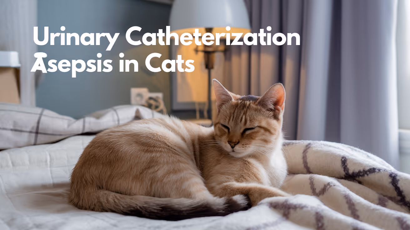
Asepsis for Urinary Catheterization in Cats
Learn essential asepsis techniques for urinary catheterization in cats to prevent infections and ensure safe catheter use.
Urinary catheterization in cats is a common veterinary procedure used to relieve urinary obstruction or collect sterile urine samples. However, improper aseptic technique during catheterization can lead to serious infections and complications. Understanding and applying correct asepsis methods is crucial to protect your cat's health during this delicate process.
This article explains the importance of asepsis for urinary catheterization in cats. You will learn step-by-step how to maintain a sterile environment, prepare your cat and equipment, and reduce infection risks effectively.
What is asepsis in urinary catheterization for cats?
Asepsis means preventing contamination by harmful bacteria or pathogens during medical procedures. For urinary catheterization in cats, asepsis focuses on keeping the urinary tract and catheter sterile to avoid urinary tract infections (UTIs).
Maintaining asepsis involves careful preparation, sterile equipment, and proper technique. This reduces the chance of introducing bacteria into the bladder, which can cause painful infections or worsen your cat's condition.
- Definition of asepsis: Asepsis is the practice of preventing infection by eliminating microbes during catheter insertion and handling.
- Importance in cats: Cats have sensitive urinary tracts that can easily become infected if aseptic techniques are not followed.
- Goal of asepsis: The main goal is to keep the catheter and urinary tract free from bacteria to prevent UTIs.
- Common risks: Without asepsis, bacteria from skin or environment can enter the bladder causing inflammation and infection.
Understanding asepsis helps you appreciate why strict hygiene and sterile tools are essential when catheterizing cats.
How do you prepare for aseptic urinary catheterization in cats?
Proper preparation is the first step to ensure asepsis during catheterization. This includes preparing the environment, your hands, the cat, and all equipment.
Preparation reduces contamination risks and makes the procedure smoother and safer for your cat.
- Clean environment: Perform catheterization in a clean, quiet area to minimize airborne contaminants and stress for the cat.
- Hand hygiene: Wash hands thoroughly with soap and water before and after the procedure to remove bacteria.
- Cat preparation: Clip fur around the urethral opening and clean the area with antiseptic solution to reduce skin bacteria.
- Sterile equipment: Use a sterile urinary catheter, gloves, lubricant, and antiseptic wipes to maintain sterility.
Taking time to prepare properly helps prevent infections and improves catheterization success.
What equipment is needed for aseptic urinary catheterization in cats?
Using the right equipment is essential to maintain asepsis. All items should be sterile or properly disinfected before use.
Having all equipment ready before starting reduces procedure time and contamination risks.
- Sterile urinary catheter: Choose the correct size and type (usually 3.5 to 5 French) designed for cats.
- Sterile gloves: Wear gloves to avoid transferring bacteria from your hands to the catheter or cat.
- Antiseptic solution: Use chlorhexidine or povidone-iodine to clean the catheterization site.
- Sterile lubricant: Apply to the catheter tip to ease insertion and reduce tissue trauma.
Proper equipment choice and sterility are key to successful and safe catheterization.
How should you perform aseptic technique during catheter insertion in cats?
Following a strict aseptic technique during catheter insertion prevents contamination and injury. The procedure must be gentle and precise.
Each step should minimize contact with non-sterile surfaces and avoid introducing bacteria into the urinary tract.
- Wear sterile gloves: Always put on gloves after hand washing to keep hands clean during insertion.
- Clean catheterization site: Thoroughly disinfect the area around the urethral opening before inserting the catheter.
- Use sterile lubricant: Lubricate the catheter tip to reduce friction and discomfort during insertion.
- Gentle insertion: Slowly and carefully insert the catheter into the urethra without force to avoid trauma.
Adhering to these steps reduces infection risk and improves catheter placement success.
What are the common aseptic challenges during urinary catheterization in cats?
Despite best efforts, some challenges can compromise asepsis during catheterization. Recognizing these helps you prevent or address them promptly.
Being aware of these issues allows you to maintain strict aseptic control throughout the procedure.
- Cat movement: Cats may struggle or move suddenly, increasing contamination risk or injury during catheterization.
- Equipment contamination: Touching non-sterile surfaces can contaminate gloves or catheter, leading to infection.
- Improper site cleaning: Inadequate antiseptic use can leave bacteria on the skin near the urethra.
- Repeated catheterization: Multiple attempts increase trauma and infection risk, requiring careful technique and patience.
Managing these challenges with calm handling and strict hygiene improves outcomes.
How do you care for a urinary catheter aseptically after placement in cats?
Post-catheterization care is vital to maintain asepsis and prevent infections while the catheter remains in place.
Proper monitoring and cleaning routines help keep the urinary tract sterile and reduce complications.
- Secure catheter: Fix the catheter gently to prevent movement that can introduce bacteria or cause injury.
- Regular cleaning: Clean the catheter insertion site daily with antiseptic to remove debris and bacteria.
- Monitor for infection: Watch for signs like redness, swelling, or discharge around the catheter site.
- Maintain closed system: Keep the urine collection system closed and below bladder level to prevent backflow and contamination.
Good catheter care supports healing and reduces the chance of urinary tract infections.
What are the signs of infection after urinary catheterization in cats?
Early detection of infection after catheterization helps you seek veterinary care promptly to avoid serious complications.
Knowing the signs allows you to monitor your cat closely and act quickly if problems arise.
- Redness and swelling: Inflammation around the catheter site indicates possible infection or irritation.
- Discharge or odor: Pus or foul-smelling fluid from the catheter area suggests bacterial infection.
- Changes in urination: Straining, frequent attempts, or blood in urine may signal urinary tract infection.
- Lethargy or fever: General signs of illness can accompany infection and require immediate veterinary attention.
If you notice any of these signs, contact your veterinarian for evaluation and treatment.
Conclusion
Asepsis for urinary catheterization in cats is essential to prevent urinary tract infections and ensure safe catheter use. Proper preparation, sterile equipment, and careful technique protect your cat’s health during this sensitive procedure.
By understanding and applying these aseptic principles, you can help your cat recover comfortably and avoid complications. Always consult your veterinarian for guidance and support during catheterization and care.
FAQs
How often should the catheter site be cleaned in cats?
The catheter site should be cleaned at least once daily with an antiseptic solution to reduce bacterial buildup and prevent infection.
Can I perform urinary catheterization at home for my cat?
Urinary catheterization should only be done by a trained veterinarian or under their direct guidance to avoid injury and infection risks.
What size catheter is best for cats?
Typically, a 3.5 to 5 French sterile urinary catheter is used for cats, but your veterinarian will select the appropriate size based on your cat’s size and condition.
How long can a urinary catheter stay in a cat?
Catheters can remain in place for 24 to 72 hours, but duration depends on the cat’s condition and veterinary instructions to minimize infection risk.
What should I do if my cat shows signs of infection after catheterization?
If signs of infection appear, contact your veterinarian immediately for assessment and treatment to prevent serious complications.

Closing High-Tension Surgical Wounds in Dogs
Learn effective methods for closing high-tension surgical wounds in dogs to promote healing and reduce complications.
High-tension surgical wounds in dogs present a significant challenge for veterinarians and pet owners. These wounds occur when the skin and underlying tissues are stretched tightly during surgery or injury repair, making closure difficult and increasing the risk of complications such as wound dehiscence or delayed healing.
Proper closure techniques are essential to ensure the wound heals well and the dog recovers quickly. This article explains the best practices for closing high-tension surgical wounds in dogs, including suture choices, techniques, and postoperative care.
What are high-tension surgical wounds in dogs?
High-tension wounds happen when the skin edges are under excessive stress or pull during closure. This tension can cause the sutures to cut through the tissue or the wound to reopen after surgery.
Understanding the nature of these wounds helps in selecting the right closure method to minimize complications.
- Definition of high-tension wounds: Wounds where skin edges are stretched tightly, increasing the risk of suture failure and delayed healing in dogs.
- Common causes: Large skin defects, surgical removal of tumors, or injuries in areas with limited skin mobility like joints or limbs.
- Risks involved: Increased chance of wound dehiscence, infection, and prolonged healing time if not managed properly.
- Impact on healing: Excessive tension reduces blood flow to wound edges, slowing tissue repair and increasing scar formation.
Recognizing these factors is crucial for planning an effective closure strategy that supports healing.
What suture materials are best for high-tension wounds in dogs?
Choosing the right suture material is key to supporting the wound under tension. Sutures must be strong, cause minimal tissue reaction, and maintain their strength during healing.
Different suture types offer various benefits depending on the wound location and tension level.
- Monofilament sutures: These cause less tissue drag and reduce infection risk, making them ideal for high-tension closures in dogs.
- Non-absorbable sutures: Provide long-term strength, useful in areas where prolonged support is needed to prevent wound reopening.
- Absorbable sutures: Suitable for internal layers where long-term strength is less critical, helping reduce the need for suture removal.
- Suture size selection: Larger suture sizes (e.g., 2-0 or 3-0) offer greater tensile strength necessary for high-tension wounds.
Consulting with your veterinarian about the best suture choice ensures optimal wound support and healing.
Which surgical techniques help close high-tension wounds effectively?
Several surgical methods can reduce tension on the wound edges and improve closure success. These techniques aim to redistribute tension and protect the skin during healing.
Using the right technique depends on the wound size, location, and available skin.
- Undermining skin edges: Loosening the skin around the wound allows easier approximation without excessive tension on sutures.
- Use of tension-relieving sutures: Techniques like mattress or pulley sutures help distribute tension evenly across the wound.
- Skin flaps and grafts: Transferring nearby skin or tissue can cover large defects and reduce tension on the primary closure.
- Layered closure: Closing deep tissue layers separately supports the skin and reduces surface tension on the wound edges.
Applying these techniques improves wound stability and promotes faster, complication-free healing.
How to manage postoperative care for high-tension wounds in dogs?
Proper postoperative care is vital to protect the wound and support healing after closure. This includes monitoring for signs of complications and preventing stress on the wound.
Owners play a key role in ensuring the dog follows recovery guidelines.
- Restricting activity: Limiting movement prevents excessive strain on the wound, reducing the risk of reopening.
- Using protective devices: Elizabethan collars or bandages protect the wound from licking, biting, or contamination.
- Regular wound inspection: Checking for redness, swelling, or discharge helps detect infections early.
- Follow-up veterinary visits: Scheduled appointments allow professionals to assess healing and remove sutures if needed.
Adhering to these care steps ensures the wound heals properly and complications are minimized.
What complications can arise from improper closure of high-tension wounds?
Failing to close high-tension wounds correctly can lead to several problems that delay healing and affect the dog's health.
Understanding these risks highlights the importance of proper surgical technique and care.
- Wound dehiscence: The wound edges separate due to suture failure or excessive tension, requiring additional treatment.
- Infection risk: Open wounds are more prone to bacterial contamination, causing inflammation and delayed healing.
- Excessive scarring: Poor closure can lead to thick, fibrous scar tissue that limits skin flexibility and appearance.
- Delayed healing: Complications extend recovery time and may require further surgical intervention.
Prompt veterinary attention and proper closure techniques reduce these risks significantly.
How can veterinarians prepare for closing high-tension wounds in dogs?
Preparation before surgery is essential to handle high-tension wounds successfully. This includes planning the closure method and having necessary materials ready.
Veterinarians use assessment and planning to improve surgical outcomes.
- Preoperative wound assessment: Evaluating wound size, location, and skin mobility guides closure strategy selection.
- Choosing appropriate suture materials: Selecting strong, suitable sutures ensures the wound withstands tension during healing.
- Planning tension-relief techniques: Deciding on undermining, flaps, or special sutures before surgery improves efficiency and results.
- Preparing postoperative care instructions: Educating owners on wound care and activity restrictions supports recovery.
Thorough preparation helps veterinarians close high-tension wounds effectively and reduces complications.
Conclusion
Closing high-tension surgical wounds in dogs requires careful technique, appropriate suture selection, and diligent postoperative care. These wounds pose challenges due to the stress on skin edges, but with the right approach, healing can proceed smoothly.
Understanding wound tension, using tension-relieving methods, and following veterinary guidance will help your dog recover safely and comfortably from surgery or injury.
What are the signs of wound infection in dogs?
Signs include redness, swelling, heat, discharge, and pain around the wound. If you notice these, contact your veterinarian promptly to prevent worsening infection.
How long does it take for high-tension wounds to heal in dogs?
Healing time varies but generally takes 10 to 14 days for skin closure, with deeper tissues requiring longer. Proper care can speed recovery and reduce complications.
Can I bathe my dog with a high-tension surgical wound?
Bathing is usually not recommended until the wound fully heals to avoid infection and suture damage. Always follow your veterinarian’s advice on wound care.
When should sutures be removed after closing a high-tension wound?
Sutures are typically removed 10 to 14 days post-surgery, depending on healing progress and wound location. Your veterinarian will advise the best timing.
Are there alternatives to sutures for closing high-tension wounds?
Yes, options like staples, skin adhesives, or tension-relieving devices may be used depending on the wound. Your veterinarian will choose the best method for your dog’s needs.

Operating Room Asepsis Standards in Veterinary Hospitals
Learn about operating room asepsis standards in veterinary hospitals to ensure safe surgeries and prevent infections in pets.
Operating room asepsis standards in veterinary hospitals are critical for preventing infections during surgical procedures. Maintaining a sterile environment protects your pet from harmful bacteria and ensures successful recovery. Understanding these standards helps pet owners appreciate the care taken during surgery.
This article explains what operating room asepsis means, why it matters, and how veterinary hospitals implement strict protocols. You will learn about sterilization methods, staff hygiene, and environmental controls that keep surgical areas safe.
What is operating room asepsis in veterinary hospitals?
Operating room asepsis refers to the practices used to keep the surgical environment free from harmful microorganisms. It involves cleaning, sterilizing, and controlling the environment to reduce infection risks during surgery.
In veterinary hospitals, asepsis is vital because animals are prone to infections that can complicate healing. The goal is to create a safe space where surgery can proceed without contamination.
- Definition of asepsis: Asepsis means the absence of bacteria, viruses, and other pathogens in the surgical area to prevent infection.
- Importance in surgery: Maintaining asepsis reduces the chance of post-operative infections that can delay healing or cause serious illness.
- Scope of asepsis: It includes sterilizing instruments, preparing the surgical site, and controlling the operating room environment.
- Veterinary-specific concerns: Animals have different skin flora and behaviors that require tailored aseptic protocols compared to human medicine.
Understanding asepsis helps pet owners trust the surgical process and the efforts taken to protect their pets.
How do veterinary hospitals sterilize surgical instruments?
Sterilizing surgical instruments is a key part of asepsis. Veterinary hospitals use several methods to ensure tools are free from microorganisms before use.
Proper sterilization prevents bacteria from entering the pet’s body during surgery, which can cause severe infections.
- Autoclaving process: Autoclaves use high-pressure steam at 121-134°C to kill all microbes on instruments effectively.
- Cold sterilization: Some delicate instruments are soaked in chemical sterilants for a specified time to avoid heat damage.
- Instrument packaging: Sterilized tools are wrapped in special materials that maintain sterility until use.
- Regular monitoring: Hospitals use biological indicators to confirm sterilization cycles are effective and safe.
These sterilization steps ensure that every instrument used in surgery is safe and clean, protecting your pet from infection.
What hygiene practices do veterinary staff follow in the operating room?
Veterinary staff follow strict hygiene protocols to maintain asepsis during surgery. These practices minimize the risk of contaminating the sterile field.
From handwashing to wearing protective clothing, every step is designed to keep germs away from the surgical site.
- Hand hygiene: Staff wash hands thoroughly with antimicrobial soap before and after surgery to remove pathogens.
- Surgical scrubs and gowns: Wearing sterile scrubs and gowns prevents bacteria from clothing contaminating the area.
- Glove use: Sterile gloves are worn and changed if torn or contaminated to maintain a barrier against microbes.
- Face masks and caps: Masks and caps reduce the spread of respiratory droplets and hair that could carry bacteria.
These hygiene measures are essential to protect both the surgical team and the patient from infections.
How is the operating room environment controlled for asepsis?
Controlling the operating room environment is crucial to prevent airborne contamination. Veterinary hospitals design their surgical suites to support aseptic conditions.
Environmental controls include air filtration, cleaning protocols, and limiting access during surgery.
- Air filtration systems: High-efficiency particulate air (HEPA) filters remove dust and microbes from the air to reduce contamination.
- Positive pressure ventilation: This system pushes clean air out of the operating room, preventing outside air from entering.
- Regular cleaning schedules: Floors, walls, and surfaces are disinfected before and after surgeries to remove pathogens.
- Restricted access: Only essential personnel enter the operating room to minimize the introduction of contaminants.
These controls create a clean and safe environment that supports successful surgical outcomes.
What protocols are used for preparing the surgical site on animals?
Preparing the surgical site on your pet is a critical step to reduce infection risk. Veterinary staff follow detailed protocols to clean and disinfect the area before surgery.
This preparation helps remove dirt, hair, and bacteria from the skin surface.
- Shaving the area: Fur is clipped around the surgical site to expose clean skin and reduce bacteria hiding in hair.
- Skin cleansing: The area is scrubbed with antiseptic solutions like chlorhexidine or iodine to kill microbes.
- Use of sterile drapes: Sterile drapes cover the surrounding area to isolate the surgical site from contamination.
- Minimizing skin trauma: Gentle techniques are used to avoid skin damage that could increase infection risk.
Proper site preparation is essential for preventing infections and promoting healing after surgery.
How do veterinary hospitals monitor and improve asepsis standards?
Veterinary hospitals continuously monitor asepsis standards to ensure patient safety. They use audits, training, and infection tracking to improve practices.
Ongoing evaluation helps identify risks and implement better protocols.
- Regular staff training: Teams receive updates on aseptic techniques and infection control best practices.
- Infection surveillance: Hospitals track surgical site infections to detect trends and address causes promptly.
- Environmental testing: Periodic sampling of air and surfaces checks for microbial contamination levels.
- Quality improvement programs: Data from audits and infection rates guide changes to improve asepsis protocols.
These efforts maintain high standards and protect pets during surgery.
Conclusion
Operating room asepsis standards in veterinary hospitals are essential to protect pets from infections during surgery. Strict sterilization, hygiene, environmental control, and site preparation work together to create a safe surgical environment.
Understanding these standards helps pet owners feel confident in the care their animals receive. Veterinary hospitals continuously monitor and improve asepsis to ensure the best outcomes for every patient.
What is the difference between sterilization and disinfection in veterinary surgery?
Sterilization kills all microorganisms on instruments, while disinfection reduces harmful microbes on surfaces. Sterilization is required for surgical tools, disinfection for operating room surfaces.
How often should surgical instruments be sterilized in veterinary hospitals?
Instruments must be sterilized before every surgery to ensure they are free of pathogens and safe for use on each patient.
Can pets develop infections despite aseptic surgery protocols?
Yes, infections can still occur due to factors like the pet's health or environment, but aseptic protocols greatly reduce this risk.
Why is hand hygiene so important for veterinary surgical staff?
Hand hygiene removes bacteria and viruses that staff may carry, preventing them from contaminating the sterile field during surgery.
Are there special aseptic considerations for emergency surgeries in veterinary hospitals?
Emergency surgeries may have limited preparation time, so staff follow rapid but strict aseptic measures to minimize infection risks as much as possible.

Skin Antisepsis Protocol for Cats
Learn the essential skin antisepsis protocol for cats to ensure safe and effective preparation before surgery or wound care.
Preparing a cat's skin before surgery or wound treatment is crucial to prevent infections. Skin antisepsis protocol for cats involves carefully cleaning and disinfecting the skin to reduce bacteria and contaminants. This process helps protect your cat from complications during and after medical procedures.
This article explains the step-by-step skin antisepsis protocol for cats. You will learn why it matters, what products to use, and how to perform the procedure safely and effectively to keep your cat healthy.
What is the purpose of skin antisepsis in cats?
Skin antisepsis aims to remove dirt, oils, and microbes from the cat's skin surface. This reduces the risk of infection during surgery or wound care. Proper antisepsis helps promote faster healing and prevents complications caused by bacteria entering the body.
Understanding the purpose helps you appreciate why following the protocol carefully is important for your cat's safety.
- Infection prevention: Skin antisepsis lowers the number of bacteria on the skin, reducing the chance of surgical site infections and wound contamination.
- Improved healing: Clean skin supports the body's natural healing process by minimizing infection risks that can delay recovery.
- Reduced complications: Proper antisepsis decreases the likelihood of post-procedure problems such as abscesses or sepsis.
- Enhanced surgical outcomes: Surgeons rely on antisepsis to maintain a sterile field, ensuring safer and more successful operations.
By understanding these benefits, you can see why skin antisepsis is a critical step in veterinary care for cats.
Which antiseptic agents are safe and effective for cats?
Choosing the right antiseptic agent is vital because cats have sensitive skin and can react to harsh chemicals. Veterinarians typically select antiseptics that are both effective against microbes and safe for feline use.
Knowing which products to use helps you avoid skin irritation or toxicity while ensuring proper disinfection.
- Chlorhexidine gluconate: A broad-spectrum antiseptic that is effective and generally safe for cats when diluted properly.
- Povidone-iodine: An iodine-based antiseptic that kills bacteria and fungi but should be used carefully to avoid irritation.
- Alcohol-based solutions: Often avoided alone due to skin dryness and irritation but may be used in combination with other agents.
- Saline solution: Used for initial cleaning to remove debris before applying antiseptics, safe and gentle on cat skin.
Consult your veterinarian to select the best antiseptic agent tailored to your cat’s needs and skin sensitivity.
How do you prepare a cat for skin antisepsis?
Proper preparation is essential to ensure the antisepsis process is effective and safe. This includes handling your cat gently and preparing the area to be cleaned.
Preparation reduces stress for your cat and improves the antiseptic’s ability to work effectively.
- Restrain gently: Use calm, gentle restraint to keep your cat still without causing distress or injury during the procedure.
- Clip hair: Carefully clip hair around the surgical or wound site to expose the skin and allow better antiseptic contact.
- Clean visible dirt: Remove dirt and debris with a damp cloth or saline solution before applying antiseptics.
- Check skin condition: Inspect the skin for cuts, rashes, or infections that might affect antiseptic use or require veterinary attention.
Following these steps prepares your cat’s skin for optimal antiseptic action and reduces procedure complications.
What is the step-by-step skin antisepsis procedure for cats?
The antisepsis procedure involves systematic cleaning and disinfecting to ensure the skin is as free from microbes as possible. Each step must be done carefully and in order.
Following a clear procedure ensures thorough antisepsis and minimizes risks.
- Initial cleaning: Use sterile saline or warm water with a soft cloth to gently clean the area, removing surface dirt and oils.
- Apply antiseptic: Use a sterile gauze or cotton soaked in antiseptic solution to wipe the skin in circular motions from the center outward.
- Repeat application: Perform at least three antiseptic wipes, using fresh gauze each time to maximize microbial reduction.
- Allow drying time: Let the antiseptic dry completely before proceeding with surgery or dressing to ensure effectiveness and avoid dilution.
Adhering to these steps helps maintain a sterile field and protects your cat from infections.
How do you handle complications or adverse reactions during skin antisepsis?
Sometimes cats may react to antiseptic agents or the procedure itself. Recognizing and managing these reactions promptly is important to avoid further harm.
Being prepared for complications ensures quick response and better outcomes for your cat.
- Watch for irritation: Look for redness, swelling, or discomfort during or after antiseptic application, which may indicate sensitivity.
- Stop if allergic reaction: If your cat shows signs of allergy like hives or excessive scratching, stop the procedure and consult a vet immediately.
- Use alternative agents: If irritation occurs, your vet may recommend a different antiseptic that is gentler on your cat’s skin.
- Provide supportive care: Keep the area clean and monitor for infection signs, seeking veterinary help if symptoms worsen.
Prompt recognition and management of complications help keep your cat safe during antisepsis.
How often should skin antisepsis be performed in cats?
The frequency of skin antisepsis depends on the situation, such as surgery preparation or wound care. Overdoing antisepsis can harm the skin, while too little increases infection risk.
Understanding when and how often to perform antisepsis helps maintain skin health and prevent infections.
- Pre-surgical use: Skin antisepsis is done once immediately before surgery to prepare the site.
- Wound care: Antisepsis may be repeated daily or as directed by a veterinarian to keep wounds clean.
- Chronic skin conditions: Frequent antisepsis is usually not recommended to avoid skin damage unless advised by a vet.
- Post-operative care: Follow your vet’s instructions on antiseptic use to prevent infection without overuse.
Always follow veterinary guidance on antisepsis frequency tailored to your cat’s specific health needs.
What safety precautions should you take during skin antisepsis?
Safety is essential for both your cat and yourself during antisepsis. Proper precautions reduce risks of injury, contamination, or adverse effects.
Knowing and applying safety measures ensures a smooth and safe antisepsis process.
- Wear gloves: Use disposable gloves to protect yourself and prevent cross-contamination between animals or surfaces.
- Use sterile materials: Employ sterile gauze, cotton, and antiseptic solutions to maintain cleanliness and effectiveness.
- Avoid ingestion: Prevent your cat from licking the antiseptic area until it is fully dry to avoid toxicity.
- Dispose properly: Safely discard used materials and antiseptic containers to maintain hygiene and safety.
Following these precautions protects both you and your cat during skin antisepsis procedures.
Conclusion
Skin antisepsis protocol for cats is a vital step in preventing infections during surgery or wound care. It involves careful cleaning, selecting safe antiseptics, and following a precise procedure to protect your cat’s health.
By understanding and applying this protocol, you help ensure safer medical treatments and faster healing for your cat. Always consult your veterinarian for personalized advice and guidance on skin antisepsis.
FAQs
Can I use human antiseptic products on my cat?
Some human antiseptics like diluted chlorhexidine may be safe, but many are not suitable for cats. Always consult your vet before using any human product on your cat’s skin.
How long should I wait after antisepsis before surgery?
Allow the antiseptic to dry completely, usually 2 to 3 minutes, to ensure maximum effectiveness and avoid dilution during surgery.
What if my cat has sensitive skin or allergies?
Inform your vet about any skin sensitivities. They can recommend gentler antiseptics or alternative cleaning methods to avoid irritation or allergic reactions.
Is it necessary to shave the fur before antisepsis?
Yes, clipping the fur around the surgical or wound site helps expose the skin for better antiseptic contact and reduces contamination risk.
Can I perform skin antisepsis at home?
Minor wound antisepsis can be done at home with vet guidance. For surgeries or major wounds, professional veterinary antisepsis is recommended to ensure safety and sterility.
Sustainable Vet | Copyright ©2026 | All Rights Reserved

