
Protecting
Pets, People & Planet
Join a group of veterinarians leveraging the latest technologies to deliver excellent care to their patients while being a responsible and positive force for their local and global communities.
100% secure. We do not share your information
Recent Articles
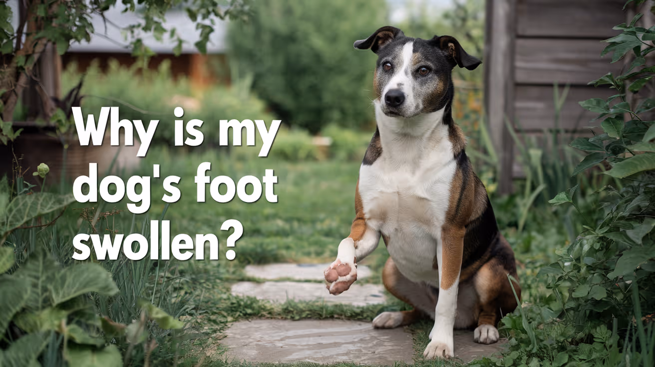
Why Is My Dog's Foot Swollen?
Discover why your dog's foot is swollen, common causes, treatments, and when to see a vet for proper care.
Seeing your dog's foot swollen can be worrying. Swelling in a dog's foot can happen for many reasons, from injuries to infections. Understanding why this happens helps you act quickly and keep your dog comfortable.
This article explains common causes of swollen dog feet, how to spot serious problems, and what treatments work best. You will learn when to treat at home and when to visit a vet for urgent care.
What Causes Swelling in a Dog's Foot?
Swelling in a dog's foot can come from many different problems. It often shows as puffiness, redness, or heat in the paw area. Knowing the cause helps you decide the right care.
Common causes include injuries, infections, allergies, and insect bites. Each cause needs a different approach to treatment.
- Injury or trauma: A cut, sprain, or broken bone can cause swelling due to inflammation and fluid buildup in the foot tissues.
- Infections: Bacterial or fungal infections can cause swelling, redness, and pain, often needing antibiotics or antifungal treatment.
- Allergic reactions: Allergies to plants, chemicals, or insect stings can cause sudden swelling and itching in the foot.
- Foreign objects: Thorns, splinters, or glass stuck in the paw can cause swelling and discomfort until removed.
Identifying the cause early helps prevent complications and speeds healing.
How Can I Tell If My Dog's Foot Swelling Is Serious?
Not all swelling is an emergency, but some signs mean you should see a vet quickly. Serious swelling can affect your dog's ability to walk or cause severe pain.
Look for symptoms like severe limping, open wounds, or signs of infection. These require prompt veterinary care.
- Severe limping or inability to walk: Indicates pain or serious injury needing urgent veterinary evaluation.
- Open wounds or bleeding: Risk of infection and need for cleaning and possibly stitches.
- Fever or lethargy: Signs that infection may have spread and requires medical treatment.
- Rapidly increasing swelling: Could signal an allergic reaction or deep infection needing emergency care.
When in doubt, it is safer to consult your vet to avoid worsening problems.
What Home Treatments Can Help a Swollen Dog Foot?
For mild swelling without serious signs, you can try some home care steps. These help reduce swelling and keep your dog comfortable.
Always watch your dog closely and stop home treatment if symptoms worsen or do not improve.
- Rest and limit activity: Keep your dog from running or jumping to reduce stress on the swollen foot.
- Cold compress application: Apply a cold pack wrapped in cloth for 10-15 minutes to reduce swelling and pain.
- Clean the paw gently: Use warm water to clean dirt or debris, especially if there are small cuts or irritations.
- Prevent licking or chewing: Use an Elizabethan collar if your dog tries to lick or bite the swollen area, which can worsen irritation.
These steps can help minor swelling but do not replace veterinary care for serious cases.
When Should I Take My Dog to the Vet for a Swollen Foot?
Knowing when to seek professional help is important. Some swelling needs medical treatment to avoid complications.
If your dog's swelling is severe, painful, or lasts more than a day or two, a vet visit is necessary. Early treatment can prevent infections or permanent damage.
- Persistent swelling over 48 hours: Indicates that the problem may not resolve without medical intervention.
- Signs of infection: Pus, foul odor, or heat around the swollen area require antibiotics or cleaning by a vet.
- Suspected broken bone or sprain: Needs X-rays and pain management from a veterinary professional.
- Severe allergic reactions: Swelling with difficulty breathing or collapse needs emergency veterinary care immediately.
Your vet can diagnose the cause and recommend the right treatment plan for your dog's recovery.
How Do Vets Diagnose the Cause of a Swollen Dog Foot?
Veterinarians use several methods to find the cause of swelling. A correct diagnosis is key to effective treatment.
They will examine your dog’s foot carefully and may use tests to look deeper into the problem.
- Physical examination: Checking for wounds, foreign objects, and signs of pain or infection in the foot.
- X-rays: Used to detect fractures, bone infections, or foreign bodies inside the paw.
- Skin scrapings or cultures: To identify infections caused by bacteria, fungi, or parasites.
- Blood tests: To check for systemic infection or allergic reactions affecting the swelling.
These tools help your vet create a treatment plan tailored to your dog's needs.
What Treatments Do Vets Use for Swollen Dog Feet?
Treatment depends on the cause of the swelling. Your vet may use medications, procedures, or supportive care to help your dog heal.
Some treatments can be done at home under vet guidance, while others require clinic visits.
- Antibiotics or antifungals: Prescribed to treat infections causing swelling and prevent spread.
- Anti-inflammatory drugs: Help reduce pain and swelling, improving your dog's comfort.
- Wound care and bandaging: Cleaning and protecting open wounds to promote healing and prevent infection.
- Surgery: May be needed to remove foreign objects or repair fractures causing swelling.
Follow your vet’s instructions carefully to ensure the best outcome for your dog.
How Can I Prevent My Dog's Foot from Swelling?
Preventing foot swelling involves protecting your dog from injuries and infections. Regular care and attention can reduce risks.
Simple habits help keep your dog's paws healthy and avoid painful swelling episodes.
- Regular paw inspections: Check your dog's feet daily for cuts, thorns, or swelling to catch problems early.
- Keep nails trimmed: Prevents nails from breaking or causing injury to the foot pads.
- Avoid walking on rough surfaces: Protect paws from sharp objects or hot pavement that can cause injuries.
- Use protective booties: Especially in harsh weather or rough terrain to shield paws from damage.
Good paw care supports your dog’s overall health and comfort.
Conclusion
Swelling in your dog's foot can have many causes, from minor injuries to serious infections. Understanding why your dog's foot is swollen helps you provide the right care quickly.
Always watch for signs of pain, infection, or worsening symptoms. When in doubt, seek veterinary advice to protect your dog's health and comfort. Early treatment can prevent complications and get your dog back on their feet faster.
Why is my dog's foot swollen after walking?
Your dog's foot may swell after walking due to minor injuries, irritation from rough surfaces, or allergic reactions. Rest and paw care usually help reduce swelling quickly.
Can a swollen dog foot heal without a vet?
Mild swelling from minor injuries or irritations can heal at home with rest and care. However, persistent or severe swelling needs veterinary evaluation to avoid complications.
How long does it take for a dog's swollen foot to go down?
Swelling may reduce within a few days with proper care. If swelling lasts more than 48 hours or worsens, consult a vet for treatment.
Is a swollen dog foot painful?
Yes, swelling often causes pain and discomfort. Your dog may limp, lick, or avoid putting weight on the swollen foot.
Can allergies cause a dog's foot to swell?
Yes, allergies to insect bites, plants, or chemicals can cause sudden swelling and itching in a dog's foot, sometimes requiring veterinary treatment.
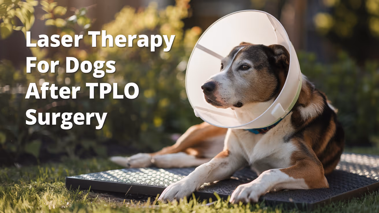
Laser Therapy for Dogs After TPLO Surgery
Learn how laser therapy helps dogs recover faster and with less pain after TPLO surgery for cruciate ligament repair.
TPLO surgery is a common procedure to fix torn cruciate ligaments in dogs. After surgery, many pet owners seek ways to help their dogs heal faster and feel less pain. Laser therapy is one treatment option that has gained popularity for post-TPLO recovery.
This article explains what laser therapy is, how it works for dogs after TPLO surgery, and what benefits you can expect. You will learn important details about safety, treatment schedules, and how to combine laser therapy with other care methods.
What is laser therapy for dogs after TPLO surgery?
Laser therapy uses focused light energy to stimulate healing in tissues. It is non-invasive and painless. After TPLO surgery, laser therapy targets the surgical site and surrounding tissues to reduce inflammation and promote repair.
The light penetrates the skin and affects cells at a deeper level. This encourages faster cell growth and blood flow, which are essential for healing the bone and soft tissues after surgery.
- Non-invasive treatment: Laser therapy does not require needles or surgery, making it a gentle option for post-operative care in dogs recovering from TPLO.
- Cell stimulation: The laser light encourages cells to produce energy and repair themselves faster, which helps the surgical site heal more quickly.
- Inflammation reduction: Laser therapy decreases swelling and redness around the surgery area, which reduces pain and discomfort for your dog.
- Improved blood flow: The treatment increases circulation, delivering oxygen and nutrients needed for tissue repair after TPLO surgery.
Overall, laser therapy supports the natural healing process after TPLO surgery by enhancing cellular function and reducing symptoms that slow recovery.
How does laser therapy help with pain management after TPLO surgery?
Pain control is a major concern after TPLO surgery. Laser therapy can reduce pain by calming nerve endings and lowering inflammation. This helps dogs feel more comfortable during their recovery.
Laser treatment can also reduce the need for high doses of pain medications, which sometimes have side effects. It works alongside medications to provide a balanced approach to pain relief.
- Nerve calming effect: Laser light decreases nerve sensitivity, which lowers the sensation of pain around the surgical site after TPLO surgery.
- Reduced inflammation: By lowering swelling, laser therapy helps relieve pressure on nerves and tissues that cause pain in dogs.
- Medication support: Laser therapy can reduce reliance on pain drugs, minimizing potential side effects from long-term medication use.
- Comfort improvement: Dogs often show better mobility and less limping after laser treatments, indicating effective pain relief.
Using laser therapy as part of a pain management plan can improve your dog's comfort and speed up their return to normal activity after TPLO surgery.
When should laser therapy start after TPLO surgery?
The timing of laser therapy after TPLO surgery depends on your veterinarian’s advice and your dog’s condition. Usually, treatment begins within a few days after surgery once the incision starts healing.
Early laser therapy can prevent excessive swelling and reduce pain. However, the surgical site must be stable enough to avoid irritation. Your vet will decide the best time to start based on healing progress.
- Early initiation: Starting laser therapy 2 to 3 days post-surgery can help control inflammation and pain early in recovery.
- Incision healing check: Therapy begins only after the surgical wound shows no signs of infection or excessive drainage.
- Regular sessions: Laser treatments are often scheduled 2 to 3 times per week for several weeks to maximize healing benefits.
- Veterinary guidance: Always follow your vet’s instructions on timing and frequency to ensure safe and effective laser therapy.
Proper timing ensures laser therapy supports healing without interfering with the surgical site’s recovery after TPLO surgery.
How many laser therapy sessions does a dog need after TPLO surgery?
The number of laser therapy sessions varies depending on your dog’s individual healing and response to treatment. Most dogs benefit from multiple sessions over several weeks.
Typically, veterinarians recommend 6 to 12 sessions spaced out over 2 to 4 weeks. This schedule allows consistent stimulation of healing while monitoring progress.
- Typical session count: Dogs often receive between 6 and 12 laser therapy treatments after TPLO surgery for optimal recovery support.
- Session frequency: Treatments are usually given 2 to 3 times per week to maintain steady healing stimulation.
- Progress evaluation: Your vet will assess healing and adjust the number of sessions based on your dog’s improvement.
- Individual variation: Some dogs may need more or fewer sessions depending on age, health, and surgery complexity.
Following a recommended session plan helps ensure your dog gets the full benefits of laser therapy after TPLO surgery.
Is laser therapy safe for dogs after TPLO surgery?
Laser therapy is generally safe when performed by trained professionals. It is non-invasive and has few side effects. However, proper use and precautions are important to avoid risks.
Dogs with certain conditions or sensitivities may require special care. Your veterinarian will evaluate your dog’s health before starting laser therapy to ensure it is appropriate.
- Non-invasive safety: Laser therapy does not break the skin or cause pain, making it a low-risk treatment option after TPLO surgery.
- Minimal side effects: Most dogs tolerate laser therapy well, with rare cases of mild redness or warmth at the treatment site.
- Professional administration: Treatments should be done by trained veterinary staff to ensure correct dosage and avoid eye exposure to the laser.
- Health screening: Dogs with cancer, photosensitivity, or certain infections may not be suitable candidates for laser therapy.
When used correctly, laser therapy is a safe and effective way to support healing and comfort after TPLO surgery.
Can laser therapy replace other post-TPLO treatments?
Laser therapy is a helpful addition but does not replace other important post-TPLO care. Surgery recovery requires a combination of treatments for best results.
Physical therapy, pain medications, rest, and controlled exercise all play vital roles. Laser therapy complements these by enhancing healing and reducing pain.
- Complementary treatment: Laser therapy works best alongside physical rehabilitation and medication, not as a sole treatment after TPLO surgery.
- Physical therapy importance: Controlled exercises and massage improve joint mobility and muscle strength during recovery.
- Medication role: Pain and anti-inflammatory drugs manage symptoms that laser therapy alone cannot fully address.
- Rest and care: Proper rest and restricted activity prevent complications and support healing after surgery.
Using laser therapy as part of a comprehensive recovery plan helps your dog heal faster and regain function after TPLO surgery.
Conclusion
Laser therapy is a valuable tool to help dogs recover after TPLO surgery. It reduces pain, inflammation, and speeds tissue healing through safe, non-invasive light treatment.
While laser therapy supports recovery, it should be combined with other treatments like physical therapy and medication. Following your veterinarian’s guidance ensures the best outcome for your dog’s post-surgical healing journey.
FAQs
How soon after TPLO surgery can laser therapy begin?
Laser therapy typically starts 2 to 3 days after surgery once the incision begins healing and shows no infection signs.
Is laser therapy painful for dogs?
No, laser therapy is painless and non-invasive. Most dogs tolerate it well and may even find it soothing.
How long does each laser therapy session last?
Sessions usually last 5 to 15 minutes depending on the size of the treatment area and the laser device used.
Can laser therapy reduce the need for pain medications?
Yes, laser therapy can lower inflammation and pain, potentially reducing the amount of pain medication needed.
Are there any risks with laser therapy after TPLO surgery?
Risks are minimal when done properly. Avoid use on dogs with cancer or photosensitivity, and always have a vet supervise treatment.
All Articles
Common Breaks in Surgical Asepsis in Veterinary Clinics
Explore common breaks in surgical asepsis in veterinary clinics and learn how to prevent infections during pet surgeries.
Surgical asepsis is critical in veterinary clinics to prevent infections during pet surgeries. However, breaks in aseptic technique can occur, risking patient safety and recovery. Understanding these common breaks helps improve surgical outcomes and protect animal health.
This article explains the typical ways surgical asepsis can be compromised in veterinary settings. You will learn about causes, prevention strategies, and best practices to maintain sterile environments during surgery.
What are the most frequent breaks in surgical asepsis in veterinary clinics?
Breaks in surgical asepsis happen when sterile technique is not properly followed. These breaches can introduce bacteria into the surgical site, causing infections. Identifying frequent breaks helps clinics focus on key areas for improvement.
- Improper hand hygiene: Failing to thoroughly scrub hands and arms before surgery allows microbes to contaminate sterile fields.
- Incorrect glove use: Touching non-sterile surfaces after donning gloves or using damaged gloves compromises sterility.
- Contaminated instruments: Using instruments that are not properly sterilized can transfer pathogens directly into the surgical site.
- Inadequate surgical site preparation: Poor clipping or skin disinfection leaves bacteria on the patient’s skin before incision.
Recognizing these common breaks is the first step to preventing surgical site infections in veterinary patients.
How does improper hand hygiene affect surgical asepsis?
Hand hygiene is the foundation of aseptic technique. Veterinary staff must remove transient and resident microbes from hands and forearms before surgery. Failure to do so increases infection risk.
- Incomplete scrubbing: Not following recommended scrubbing time or technique leaves microbes on the skin.
- Touching non-sterile objects: Contact with door handles or equipment after scrubbing reintroduces contamination.
- Using damaged gloves: Gloves with tears do not protect against microbial transfer during surgery.
- Skipping hand hygiene: Rushing or skipping handwashing before surgery directly compromises sterility.
Proper hand hygiene protocols and staff training are essential to maintain a sterile surgical environment.
What role do surgical instruments play in maintaining asepsis?
Surgical instruments must be sterile to prevent introducing bacteria into the patient. Breaks in instrument sterility are a common cause of surgical infections.
- Improper sterilization: Using autoclaves incorrectly or skipping sterilization cycles leaves instruments contaminated.
- Storage contamination: Storing instruments in unclean or damp environments allows microbial growth.
- Handling errors: Touching sterile instruments with non-sterile gloves or surfaces breaks asepsis.
- Reusing disposable instruments: Using single-use tools multiple times increases infection risk.
Strict sterilization protocols and careful instrument handling are vital to surgical asepsis.
How can surgical site preparation lead to aseptic breaks?
Preparing the patient’s skin before surgery reduces bacteria at the incision site. Poor preparation can leave microbes that cause infections.
- Inadequate clipping: Leaving hair near the incision traps bacteria and debris.
- Insufficient skin cleaning: Using ineffective antiseptics or skipping cleaning steps allows microbes to persist.
- Recontamination: Touching the prepared site with non-sterile gloves or instruments after cleaning breaks asepsis.
- Failure to isolate the site: Not using sterile drapes exposes the area to environmental contaminants.
Following strict protocols for clipping, cleaning, and draping helps maintain a sterile surgical field.
What environmental factors contribute to breaks in surgical asepsis?
The surgical environment must be controlled to minimize contamination. Environmental lapses can introduce pathogens into sterile fields.
- Improper operating room cleaning: Failing to disinfect surfaces between surgeries allows bacteria to accumulate.
- Poor air quality: Lack of proper ventilation or filtration increases airborne contaminants.
- Traffic flow issues: Excessive personnel movement in and out of the operating room raises contamination risk.
- Inadequate sterilization of surgical linens: Using unsterile drapes or gowns compromises asepsis.
Maintaining a clean, controlled environment is essential for preventing surgical site infections.
How does staff behavior impact surgical asepsis?
Staff actions and awareness directly affect aseptic technique. Training and discipline reduce breaks in sterility during surgery.
- Lack of training: Staff unfamiliar with aseptic protocols are more likely to make errors.
- Improper gowning and gloving: Incorrect donning techniques lead to contamination.
- Distractions during surgery: Interruptions can cause lapses in sterile technique.
- Failure to speak up: Staff not addressing observed breaks allows contamination to continue.
Ongoing education and a culture of safety help maintain high aseptic standards.
What are effective strategies to prevent breaks in surgical asepsis?
Preventing aseptic breaks requires a combination of protocols, training, and monitoring. Veterinary clinics must implement comprehensive measures.
- Standardized protocols: Clear, written aseptic procedures ensure consistency among staff.
- Regular training: Frequent education sessions keep staff updated on best practices and new guidelines.
- Checklists and audits: Using surgical checklists and monitoring compliance reduces errors.
- Proper equipment maintenance: Routine checks and servicing of sterilizers and surgical tools prevent contamination.
By adopting these strategies, veterinary clinics can significantly reduce surgical infections and improve patient outcomes.
Maintaining surgical asepsis in veterinary clinics is vital for safe and successful surgeries. Common breaks such as poor hand hygiene, instrument contamination, and environmental lapses increase infection risks. Understanding these issues helps clinics implement effective prevention measures. With proper training, strict protocols, and vigilant monitoring, veterinary teams can protect their patients and ensure the best surgical care.
By focusing on the causes and prevention of aseptic breaks, you can help your veterinary clinic maintain a sterile environment. This protects pets from infections and supports faster, complication-free recoveries after surgery.
What is surgical asepsis in veterinary clinics?
Surgical asepsis is the practice of keeping the surgical area and instruments free from all microorganisms to prevent infections during veterinary surgeries.
How often should surgical instruments be sterilized?
Instruments must be sterilized before every surgery using validated methods like autoclaving to ensure complete elimination of microbes.
Can gloves be reused in veterinary surgeries?
No, gloves are single-use only. Reusing gloves increases the risk of contamination and surgical site infections.
What is the best way to prepare a surgical site on an animal?
Clip hair carefully, clean the skin with antiseptic solutions, and use sterile drapes to isolate the site before incision.
How can veterinary staff reduce environmental contamination in operating rooms?
By cleaning and disinfecting surfaces regularly, controlling room traffic, and ensuring proper air filtration and ventilation.

Aseptic Technique in Dog and Cat Surgery
Learn the essentials of aseptic technique in dog and cat surgery to prevent infections and ensure safe surgical outcomes.
Surgery in dogs and cats requires strict aseptic technique to prevent infections and promote healing. Aseptic technique means using methods to keep the surgical area free from harmful germs. Without proper aseptic care, pets risk serious complications after surgery.
This article explains what aseptic technique is and why it matters in dog and cat surgery. You will learn the key steps veterinarians take to keep surgeries clean and safe, including preparation, sterilization, and handling of surgical tools and tissues.
What is aseptic technique in dog and cat surgery?
Aseptic technique is a set of procedures used to prevent contamination by microorganisms during surgery. It helps protect pets from infections that can delay healing or cause severe illness. In veterinary surgery, aseptic technique covers everything from cleaning the surgical site to sterilizing instruments.
Understanding aseptic technique helps pet owners appreciate the care taken during their pet’s surgery. It also highlights why following pre- and post-surgery instructions is important for recovery.
- Definition and purpose: Aseptic technique aims to keep the surgical environment free of harmful bacteria and fungi to avoid infections in pets.
- Scope of practice: It includes skin preparation, sterilizing tools, wearing sterile gloves, and maintaining a clean operating area.
- Importance in veterinary care: Proper aseptic technique reduces post-surgical complications and improves healing outcomes in dogs and cats.
- Difference from antiseptic: Aseptic technique prevents contamination, while antiseptic refers to substances that kill or inhibit microbes on tissues or surfaces.
Maintaining asepsis is a continuous process during surgery. Every step matters to keep pets safe and healthy.
How do veterinarians prepare the surgical site on dogs and cats?
Preparing the surgical site is the first critical step in aseptic technique. It involves cleaning and disinfecting the area where the incision will be made. This reduces the number of microbes on the skin and lowers infection risk.
Veterinarians follow strict protocols to ensure the site is ready for surgery. This preparation varies slightly depending on the pet’s size, coat type, and surgery type.
- Clipping hair: Removing hair around the incision site prevents bacteria from hiding in fur and contaminating the wound.
- Skin cleaning: The skin is washed with antiseptic solutions like chlorhexidine or iodine to kill surface microbes effectively.
- Use of sterile drapes: After cleaning, sterile drapes cover the surrounding area to create a barrier against contamination.
- Minimizing contact: Only sterile gloves and instruments touch the prepared site to maintain cleanliness throughout surgery.
Proper site preparation is essential to reduce infection risks and promote faster healing in pets.
What sterilization methods are used for surgical instruments?
Surgical instruments must be sterile before use to prevent introducing bacteria into the pet’s body. Veterinarians use several sterilization methods to ensure instruments are free from all microbes.
Choosing the right sterilization method depends on the instrument type and material. Some methods are better for delicate tools, while others suit heat-resistant instruments.
- Autoclaving: Using high-pressure steam at 121–134°C kills all microorganisms and spores, making it the most common sterilization method.
- Gas sterilization: Ethylene oxide gas sterilizes heat-sensitive instruments without damaging them, but requires long aeration times.
- Chemical sterilants: Solutions like glutaraldehyde disinfect instruments that cannot tolerate heat or gas sterilization.
- Dry heat sterilization: High temperatures without moisture sterilize metal tools but need longer exposure times than autoclaving.
Proper sterilization ensures instruments do not carry infectious agents into the surgical site, protecting the pet’s health.
How do surgeons maintain aseptic technique during surgery?
Maintaining aseptic technique throughout surgery is vital to prevent contamination. Surgeons and surgical staff follow strict protocols to keep the environment sterile from start to finish.
This includes wearing sterile clothing, handling instruments properly, and avoiding unnecessary contact with non-sterile surfaces.
- Sterile gloves and gowns: Surgeons wear sterile gloves and gowns to create a barrier between their skin and the surgical site.
- Controlled movements: Minimizing movement and avoiding touching non-sterile objects reduces contamination risk during surgery.
- Instrument handling: Instruments are passed carefully using sterile techniques to prevent contact with non-sterile surfaces.
- Maintaining sterile field: The surgical area and instruments are kept within a sterile zone, and any breach requires immediate correction.
Strict adherence to these practices helps keep the surgery safe and lowers infection chances.
What role does the surgical environment play in aseptic technique?
The surgical environment significantly affects the success of aseptic technique. A clean, controlled operating room reduces airborne and surface contamination risks.
Veterinary clinics design surgical suites to support aseptic procedures, including air filtration, surface cleaning, and restricted access.
- Clean operating room: Regular cleaning and disinfecting of floors, walls, and surfaces minimize microbial presence in the environment.
- Air filtration systems: HEPA filters reduce airborne particles and microbes, improving air quality during surgery.
- Restricted access: Limiting personnel and movement in the surgical area reduces contamination chances.
- Proper lighting and equipment layout: Good lighting and organized instruments help surgeons work efficiently without breaking sterility.
A well-maintained surgical environment supports all aseptic measures and improves surgical outcomes.
How can pet owners support aseptic technique after surgery?
Pet owners play a key role in maintaining aseptic conditions after surgery. Proper wound care and hygiene at home prevent infections and promote healing.
Following veterinary instructions carefully helps protect the surgical site and avoid complications.
- Keep the incision clean: Avoid dirt, water, or debris on the wound to prevent bacterial growth and infection.
- Prevent licking or chewing: Use an Elizabethan collar or other devices to stop pets from disturbing the surgical site.
- Follow medication schedules: Administer antibiotics and pain medications exactly as prescribed to support healing.
- Limit activity: Restrict running, jumping, or rough play to avoid stress or injury to the surgical area.
By supporting aseptic care at home, pet owners help their dogs and cats recover safely and comfortably.
What are common complications from poor aseptic technique?
Poor aseptic technique can lead to infections and other serious complications after surgery. Recognizing these risks helps emphasize the importance of strict asepsis.
Veterinarians monitor pets closely to detect and treat any issues early for the best outcomes.
- Surgical site infections: Bacterial contamination causes redness, swelling, pain, and discharge at the incision site.
- Delayed healing: Infection or contamination slows tissue repair, prolonging recovery time and discomfort.
- Systemic infections: In severe cases, bacteria can enter the bloodstream causing fever, lethargy, and life-threatening conditions.
- Increased costs and risks: Treating infections requires extra veterinary visits, medications, and sometimes additional surgery.
Maintaining aseptic technique is essential to avoid these complications and ensure a smooth recovery for your pet.
Conclusion
Aseptic technique in dog and cat surgery is critical for preventing infections and ensuring successful healing. It involves careful preparation, sterilization, and maintenance of a sterile environment throughout the procedure.
Understanding these practices helps pet owners appreciate the care taken during surgery and supports proper wound care at home. Following veterinary advice and maintaining aseptic conditions after surgery protects your pet’s health and promotes a quick recovery.
What is the difference between aseptic and antiseptic techniques?
Aseptic technique prevents contamination by keeping the surgical area and instruments sterile, while antiseptic technique uses chemicals to kill microbes on skin or surfaces.
How long does it take to sterilize surgical instruments?
Autoclaving typically takes 15–30 minutes depending on the cycle, while gas sterilization requires several hours including aeration time.
Can I bathe my pet before surgery?
Bathing is usually recommended 24 hours before surgery to reduce skin bacteria, but follow your veterinarian’s specific instructions.
Why do veterinarians clip hair before surgery?
Clipping hair removes fur that can harbor bacteria and interfere with skin cleaning, reducing infection risk at the incision site.
What signs indicate a post-surgical infection in pets?
Look for redness, swelling, discharge, foul odor, pain, or fever. Contact your vet immediately if these signs appear.
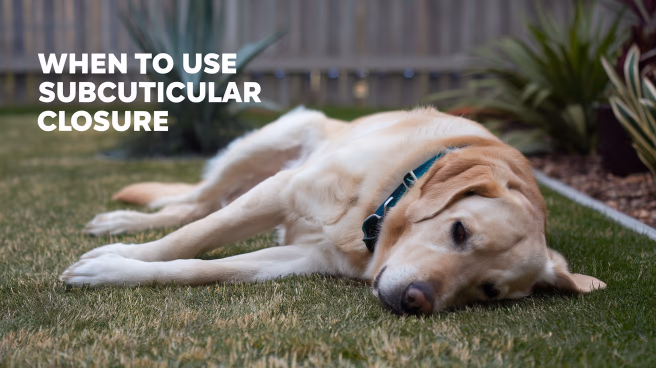
When to Use Subcuticular Closure in Dogs
Learn when to use subcuticular closure in dogs, its benefits, techniques, and care tips for optimal healing after surgery.
Surgical wound closure is a critical step in veterinary surgery for dogs. Choosing the right closure technique affects healing, infection risk, and cosmetic outcomes. One common method is subcuticular closure, which places sutures just under the skin surface to minimize scarring and improve healing.
Subcuticular closure is ideal for many types of surgical wounds in dogs. This article explains when to use subcuticular closure, its advantages, how it compares to other methods, and how to care for your dog’s incision after surgery.
What is subcuticular closure in dogs?
Subcuticular closure is a surgical technique where sutures are placed beneath the skin’s surface, within the dermal layer. This method avoids external stitches, reducing irritation and visible scarring. It is commonly used in veterinary surgery for dogs after procedures such as spays, mass removals, or skin biopsies.
The sutures run horizontally in a continuous pattern under the skin edges, pulling them together evenly. This creates a smooth skin surface and promotes better cosmetic healing compared to traditional interrupted skin sutures.
- Definition and technique: Subcuticular closure involves placing continuous sutures just under the skin surface to align wound edges without external knots or stitches.
- Common uses: It is often used for clean, straight surgical incisions in dogs where cosmetic appearance and healing speed are priorities.
- Suture materials: Absorbable sutures like poliglecaprone or polydioxanone are preferred to avoid the need for suture removal.
- Healing benefits: This method reduces skin tension and irritation, lowering infection risk and improving scar appearance.
Understanding this technique helps pet owners appreciate why their veterinarian may choose subcuticular closure for certain surgeries.
When should veterinarians choose subcuticular closure for dogs?
Veterinarians select subcuticular closure based on wound type, location, and patient factors. It is best suited for clean, surgical wounds with minimal contamination. The skin edges should be healthy and easy to approximate without tension.
Subcuticular closure is ideal for areas where cosmetic results matter, such as the abdomen or limbs. It is less suitable for wounds under high tension or contaminated wounds that require drainage.
- Clean surgical wounds: Best used on fresh, uncontaminated incisions to promote rapid healing and reduce infection risk.
- Low tension areas: Effective when skin edges can be easily brought together without excessive pulling or gaps.
- Cosmetic importance: Preferred for visible areas where minimal scarring is desired, such as the abdomen or flank.
- Patient health status: Dogs with good immune function and no skin disease heal better with this closure method.
Veterinarians assess these factors before deciding if subcuticular closure is the best choice for your dog’s surgery.
What are the benefits of subcuticular closure compared to other methods?
Subcuticular closure offers several advantages over traditional interrupted skin sutures or staples. It provides a smoother skin surface and reduces the risk of suture-related irritation or infection.
This technique also eliminates the need for suture removal when absorbable materials are used, making aftercare easier for pet owners. The cosmetic outcome is generally superior, with less visible scarring.
- Improved cosmetic results: The continuous suture under the skin creates a neat, less noticeable scar compared to external stitches.
- Reduced infection risk: No external suture knots reduce bacterial colonization and irritation at the skin surface.
- Less discomfort: Dogs experience less itching and irritation since there are no external stitches to catch or pull.
- Absorbable sutures: Using absorbable materials avoids the need for suture removal visits, reducing stress for dogs and owners.
These benefits make subcuticular closure a preferred choice for many elective surgeries in dogs.
How is subcuticular closure performed in dogs?
The procedure requires skill and sterile technique. After the surgeon completes the internal layers, the skin edges are aligned carefully. A fine absorbable suture is used to place a continuous stitch just beneath the skin surface.
The needle passes horizontally through the dermis on alternating sides of the wound. The suture is tightened to bring edges together evenly without gaps or tension. The ends are secured with buried knots.
- Preparation: The surgical site is cleaned and draped to maintain sterility before closure begins.
- Suture selection: Absorbable monofilament sutures like poliglecaprone 25 are commonly chosen for strength and minimal tissue reaction.
- Suturing technique: The needle passes horizontally through the dermis in a continuous pattern to approximate skin edges smoothly.
- Final knot placement: Knots are buried under the skin to avoid irritation and maintain a smooth surface.
Proper technique ensures the wound heals well with minimal scarring and complications.
What aftercare is needed for dogs with subcuticular closure?
After surgery, proper care helps prevent infection and supports healing. Dogs with subcuticular closure usually have less irritation but still need monitoring and protection.
Owners should keep the incision clean and dry, prevent licking or chewing, and watch for signs of infection or swelling. Follow your veterinarian’s instructions closely.
- Prevent licking: Use an Elizabethan collar or protective clothing to stop the dog from licking or biting the incision site.
- Keep incision dry: Avoid bathing or wetting the wound until fully healed to reduce infection risk.
- Monitor for complications: Check daily for redness, swelling, discharge, or opening of the wound and report concerns to your vet.
- Follow-up visits: Attend scheduled veterinary checks to ensure proper healing and address any issues early.
Good aftercare improves outcomes and helps your dog recover comfortably from surgery.
Are there any risks or limitations to subcuticular closure in dogs?
While subcuticular closure has many benefits, it is not suitable for all wounds. High-tension wounds or contaminated injuries may require other closure methods to prevent complications.
Improper technique can lead to wound dehiscence or infection. Also, absorbable sutures may sometimes cause mild tissue reaction or delayed absorption.
- Not for high tension wounds: Wounds under excessive tension may separate if closed only with subcuticular sutures.
- Unsuitable for contaminated wounds: Dirty or infected wounds need drainage and different closure to reduce infection risk.
- Technical skill required: Surgeons must be experienced to place sutures correctly and avoid complications.
- Possible suture reaction: Some dogs may develop mild inflammation or granulomas from absorbable suture material.
Discuss your dog’s specific case with your veterinarian to understand if subcuticular closure is the best option.
Conclusion
Subcuticular closure is a valuable technique for closing surgical wounds in dogs. It offers improved cosmetic results, reduced infection risk, and less discomfort compared to traditional skin sutures.
Choosing this method depends on the wound type, location, and patient factors. Proper surgical technique and diligent aftercare are essential for the best healing outcomes. Consult your veterinarian to learn if subcuticular closure is right for your dog’s surgery.
What types of surgeries in dogs commonly use subcuticular closure?
Subcuticular closure is often used in spays, mass removals, skin biopsies, and other clean surgical procedures where cosmetic healing is important.
How long does it take for a subcuticular closure to heal in dogs?
Healing typically takes 10 to 14 days, but full skin strength may take several weeks depending on the dog’s health and wound care.
Can subcuticular sutures be removed in dogs?
Usually no, because absorbable sutures are used that dissolve on their own, eliminating the need for suture removal visits.
Is subcuticular closure painful for dogs?
The technique reduces skin irritation and discomfort compared to external stitches, making it generally less painful during healing.
What signs indicate a problem with a subcuticular closure in dogs?
Watch for redness, swelling, discharge, wound opening, or excessive licking, which may indicate infection or complications needing veterinary attention.
Environmental Asepsis and Airflow in Vet Surgery
Learn how environmental asepsis and airflow control improve safety in veterinary surgery rooms for pets.
Environmental asepsis and airflow control are critical in veterinary surgery to prevent infections and ensure safe procedures. Maintaining a sterile environment reduces the risk of contamination that can harm your pet during surgery.
This article explains how veterinary clinics manage asepsis and airflow. You will learn about the techniques used to keep surgical areas clean and how airflow systems help protect your pet during operations.
What is environmental asepsis in veterinary surgery?
Environmental asepsis means keeping the surgical area free from harmful microorganisms. It involves cleaning, disinfecting, and controlling the environment to reduce infection risks for pets undergoing surgery.
Veterinary staff follow strict protocols to maintain asepsis. These include sterilizing instruments, wearing clean surgical attire, and preparing the surgery room properly.
- Cleaning protocols: Veterinary teams use hospital-grade disinfectants to clean all surfaces before and after surgery to kill bacteria and viruses effectively.
- Sterile instruments: Surgical tools are sterilized using autoclaves or chemical methods to ensure no microbes remain on them.
- Staff hygiene: Surgeons and nurses wear sterile gowns, gloves, and masks to prevent transferring germs to the surgical site.
- Controlled access: Only authorized personnel enter the surgery room to limit contamination from outside sources.
Maintaining environmental asepsis is essential to protect pets from post-surgical infections and promote faster healing.
How does airflow affect infection control in vet surgery?
Airflow in veterinary surgery rooms helps remove airborne contaminants that could infect the surgical site. Proper airflow design reduces the number of bacteria and dust particles in the air.
Ventilation systems create a clean air environment by filtering and directing airflow to minimize contamination risks during surgery.
- Laminar airflow: This system provides a steady, unidirectional flow of filtered air over the surgical area to push contaminants away from the wound.
- HEPA filters: High-efficiency particulate air filters trap microscopic particles, including bacteria and viruses, improving air quality in surgery rooms.
- Positive pressure rooms: These rooms maintain higher air pressure inside than outside, preventing unfiltered air from entering the sterile area.
- Air exchange rates: Frequent air changes per hour dilute airborne contaminants and maintain a clean atmosphere for surgery.
Effective airflow control is vital to reduce airborne infection risks and maintain a safe environment for veterinary surgery.
What are the key design features of a vet surgery room for asepsis?
Veterinary surgery rooms are designed to support asepsis through layout, materials, and equipment choices. These features help maintain cleanliness and control airflow effectively.
Designing the room with infection control in mind reduces contamination risks and improves surgical outcomes for pets.
- Smooth surfaces: Walls, floors, and counters use non-porous materials that are easy to clean and disinfect thoroughly.
- Minimal clutter: Surgery rooms avoid unnecessary equipment or furniture to reduce dust and make cleaning easier.
- Separate zones: Designated areas for clean and dirty instruments prevent cross-contamination during surgery preparation.
- Airflow placement: Air vents and filters are strategically located to create optimal airflow patterns over the surgical field.
These design elements work together to create a sterile and safe environment for veterinary surgeries.
How do veterinary teams maintain asepsis during surgery?
Maintaining asepsis during surgery requires strict protocols and teamwork. Every step from patient preparation to instrument handling is controlled to prevent infection.
Veterinary teams are trained to follow aseptic techniques that protect the surgical site and ensure the best care for pets.
- Patient preparation: The surgical site is shaved and disinfected thoroughly before surgery to reduce skin bacteria.
- Surgical attire: Staff wear sterile gowns, gloves, masks, and caps to prevent shedding microbes into the environment.
- Instrument handling: Sterile instruments are handled only by gloved hands and kept in sterile fields until used.
- Monitoring environment: The team monitors airflow systems and room conditions to ensure asepsis is maintained throughout the procedure.
Following these steps helps minimize infection risks and supports successful surgical outcomes.
What role does air filtration play in veterinary surgery rooms?
Air filtration removes harmful particles from the air, reducing the chance of airborne infections during surgery. It is a key component of environmental control in veterinary clinics.
Proper filtration improves air quality and protects both pets and staff from contaminants.
- HEPA filters: These filters capture 99.97% of particles 0.3 microns or larger, including bacteria and fungal spores.
- Pre-filters: They trap larger dust and debris, extending the life of HEPA filters and maintaining airflow efficiency.
- Regular maintenance: Filters are inspected and replaced on schedule to ensure continuous effective filtration.
- Filter placement: Filters are installed in air handling units and vents to clean air before it reaches the surgery room.
Effective air filtration is essential for maintaining a clean surgical environment and reducing infection risks.
How can pet owners support asepsis and airflow safety before surgery?
Pet owners play an important role in preparing their pets for surgery to support asepsis and airflow safety. Proper preparation helps reduce infection risks and improves recovery.
Following veterinary instructions carefully ensures the surgical environment remains safe and sterile for your pet.
- Pre-surgery fasting: Follow fasting guidelines to reduce anesthesia risks and prevent vomiting during surgery.
- Bathing pets: Give your pet a bath as advised to reduce skin bacteria before surgery.
- Arriving on time: Timely arrival helps staff prepare the surgical area and maintain asepsis protocols without rush.
- Informing health issues: Share any recent illnesses or medications with the vet to adjust surgical plans and infection control.
By cooperating with the veterinary team, pet owners help maintain a safe surgical environment and support their pet’s health.
Conclusion
Environmental asepsis and airflow control are vital to safe veterinary surgery. They reduce infection risks and protect your pet during important procedures.
Understanding how veterinary clinics maintain asepsis and manage airflow can give you confidence in your pet’s surgical care. Following pre-surgery instructions and trusting the veterinary team helps ensure the best outcomes for your pet’s health.
What cleaning methods ensure environmental asepsis in vet surgery?
Veterinary clinics use hospital-grade disinfectants and sterilization techniques like autoclaving to clean surfaces and instruments thoroughly, preventing microbial contamination.
How does laminar airflow benefit veterinary surgeries?
Laminar airflow provides a steady, filtered air stream over the surgical site, pushing contaminants away and reducing airborne infection risks during surgery.
Why are positive pressure rooms important in vet surgery?
Positive pressure rooms keep clean air inside by preventing unfiltered outside air from entering, maintaining a sterile environment for surgery.
What should pet owners do before their pet’s surgery to support asepsis?
Owners should follow fasting and bathing instructions, arrive on time, and inform vets of any health changes to help maintain a safe surgical environment.
How often should air filters be replaced in veterinary surgery rooms?
Air filters should be checked regularly and replaced according to manufacturer guidelines to ensure effective removal of airborne contaminants during surgery.
Managing Dead Space During Surgical Closure
Learn how to manage dead space during surgical closure to prevent complications and promote healing in pets.
Dead space during surgical closure is a common challenge that can lead to complications such as fluid accumulation and infection. Managing this space properly is crucial for successful healing after surgery in pets.
This article explains what dead space is, why it matters, and how veterinary surgeons manage it effectively. You will learn practical techniques and tips to understand this important surgical concept.
What is dead space in surgical closure?
Dead space refers to the empty area left between tissue layers after surgery. This space can fill with blood or fluid, increasing the risk of infection and delayed healing.
Understanding dead space helps you appreciate why surgeons take extra care during closure to minimize it.
- Definition of dead space: The gap between tissue layers after surgery that can trap fluid and cause complications if not managed properly.
- Causes of dead space: Tissue removal, trauma, or swelling during surgery can create spaces that do not naturally close.
- Risks of dead space: Fluid accumulation in dead space can lead to seromas, hematomas, or infections.
- Importance in healing: Minimizing dead space promotes better tissue adhesion and faster recovery.
Proper management of dead space is a key part of surgical technique to ensure the best outcome for your pet.
Why is managing dead space important in veterinary surgery?
Managing dead space prevents complications that can affect your pet’s recovery. It reduces the chance of fluid buildup and infection, which can cause pain and delay healing.
Surgeons focus on dead space to improve surgical success and reduce the need for additional treatments.
- Prevents fluid buildup: Closing dead space stops blood or serum from collecting under the skin, reducing swelling and discomfort.
- Reduces infection risk: Fluid pockets can harbor bacteria, so managing dead space lowers infection chances.
- Enhances tissue healing: Eliminating gaps helps tissues bond firmly, speeding recovery.
- Minimizes complications: Proper closure decreases the risk of wound breakdown or delayed healing.
Effective dead space management is essential for a smooth postoperative course and better pet health.
What techniques are used to manage dead space during closure?
Veterinary surgeons use several techniques to reduce dead space during surgical closure. These methods help tissues stay close and prevent fluid accumulation.
Choosing the right technique depends on the surgery type, location, and tissue involved.
- Layered closure: Closing tissues in layers brings each layer together, reducing gaps and supporting healing.
- Use of sutures: Placing sutures strategically helps approximate tissue edges and eliminate space.
- Placement of drains: Drains remove fluid that might collect, preventing dead space complications.
- Tissue apposition: Careful alignment of tissue edges ensures tight closure and less dead space.
Combining these techniques allows surgeons to manage dead space effectively for each patient.
How do surgical drains help in managing dead space?
Surgical drains are tubes placed to remove fluid from dead space after surgery. They help prevent fluid buildup that can cause swelling and infection.
Drains are especially useful when complete closure of dead space is difficult or when fluid production is expected.
- Drain function: Drains allow continuous removal of blood or serum from the surgical site.
- Types of drains: Passive drains use gravity, while active drains use suction to remove fluid.
- Placement timing: Drains are placed during surgery before final closure to manage fluid effectively.
- Drain care: Proper monitoring and timely removal of drains prevent complications and promote healing.
Drains are an important tool to control dead space and support recovery in many surgeries.
What complications arise if dead space is not managed?
Failure to manage dead space can lead to serious complications that affect your pet’s health and recovery. Recognizing these risks highlights the importance of proper surgical technique.
Complications may require additional treatment, increasing stress and cost for pet owners.
- Seroma formation: Fluid accumulation in dead space causes swelling and discomfort at the surgical site.
- Hematoma development: Blood pooling in dead space can cause painful bruising and delay healing.
- Infection risk: Fluid pockets provide a breeding ground for bacteria, leading to wound infections.
- Wound dehiscence: Excess fluid pressure can cause the wound to open, requiring further surgery.
Proper dead space management minimizes these risks and supports a smooth recovery.
How can pet owners support healing related to dead space after surgery?
Pet owners play a key role in supporting healing after surgery by following care instructions that help manage dead space complications.
Understanding what to watch for and how to care for the surgical site improves outcomes and comfort for your pet.
- Follow veterinary instructions: Adhere to wound care and activity restrictions to prevent stress on the surgical site.
- Monitor for swelling: Watch for lumps or swelling that may indicate fluid buildup in dead space.
- Keep incision clean: Prevent infection by maintaining hygiene around the wound area.
- Report concerns promptly: Contact your vet if you notice redness, discharge, or your pet shows pain or lethargy.
Active involvement in postoperative care helps reduce dead space complications and speeds healing.
What materials and sutures are best for closing dead space?
Choosing the right suture material and technique is important for effectively closing dead space. Different materials offer benefits depending on the tissue and surgery type.
Surgeons select sutures to balance strength, absorption, and tissue reaction to optimize healing.
- Absorbable sutures: These dissolve over time, reducing the need for removal and minimizing tissue irritation.
- Non-absorbable sutures: Used when long-term support is needed, but may require removal later.
- Suture patterns: Interrupted or continuous patterns help distribute tension and close dead space securely.
- Suture size: Smaller sutures minimize tissue trauma while providing adequate strength for closure.
Proper suture choice and technique are key to eliminating dead space and promoting successful healing.
Conclusion
Managing dead space during surgical closure is vital to prevent complications such as fluid buildup, infection, and delayed healing in pets. Understanding dead space helps you appreciate the care surgeons take to ensure a smooth recovery.
By learning about techniques like layered closure, drain use, and suture selection, you can better support your pet’s healing process. Following postoperative care instructions reduces risks and promotes comfort after surgery.
What is dead space in surgical closure?
Dead space is the empty gap between tissue layers after surgery that can fill with fluid and cause complications if not managed properly.
Why is managing dead space important in veterinary surgery?
Managing dead space prevents fluid buildup, reduces infection risk, and helps tissues heal faster, improving surgical outcomes for pets.
How do surgical drains help in managing dead space?
Drains remove fluid from dead space, preventing swelling and infection, and are placed during surgery to aid healing.
What complications arise if dead space is not managed?
Unmanaged dead space can cause seromas, hematomas, infections, and wound opening, leading to pain and delayed recovery.
How can pet owners support healing related to dead space after surgery?
Owners should follow vet instructions, monitor for swelling, keep the incision clean, and report any concerns promptly to support healing.
Autoclave Monitoring and Validation in Vet Practice
Learn how autoclave monitoring and validation ensure safe sterilization in veterinary practices to protect pets and staff.
Autoclave monitoring and validation are critical processes in veterinary practices to ensure instruments and equipment are properly sterilized. Without effective monitoring, there is a risk of infection transmission among pets and veterinary staff. Understanding how to monitor and validate autoclave performance helps maintain a safe clinical environment.
This article explains what autoclave monitoring and validation involve, why they matter, and how veterinary clinics can implement best practices. You will learn about different monitoring methods, validation protocols, and troubleshooting tips to keep your sterilization process reliable and compliant.
What is autoclave monitoring in veterinary practice?
Autoclave monitoring is the routine process of checking sterilization cycles to confirm they meet required parameters. It helps verify that temperature, pressure, and time are sufficient to kill all microorganisms on instruments. Monitoring is essential to prevent the spread of infections during veterinary procedures.
Veterinary clinics use various monitoring tools to track autoclave performance. These tools provide immediate or delayed feedback on sterilization effectiveness.
- Physical monitoring: Involves checking autoclave gauges and printouts for correct temperature, pressure, and cycle duration to ensure proper sterilization conditions.
- Chemical indicators: Use color-changing strips or tapes that react to heat or steam exposure, providing quick visual confirmation that sterilization parameters were met.
- Biological indicators: Contain resistant bacterial spores and are the gold standard for monitoring; they confirm sterilization by showing whether spores survive the cycle.
- Record keeping: Maintaining logs of each sterilization cycle helps track performance trends and identify potential issues early.
Regular autoclave monitoring helps veterinary staff catch problems before contaminated instruments are used, protecting animal patients and clinic workers.
Why is autoclave validation important in a veterinary clinic?
Autoclave validation is a formal process to prove that the sterilizer consistently achieves sterilization standards. Validation goes beyond daily monitoring by confirming the autoclave’s overall reliability and performance under different conditions.
Validation is important because veterinary instruments vary in size, shape, and material, which can affect sterilization effectiveness. Validation ensures that all items receive adequate steam penetration and heat exposure.
- Ensures patient safety: Validation confirms that sterilization kills all pathogens, reducing infection risk during surgeries and treatments.
- Regulatory compliance: Many veterinary boards and health authorities require documented validation to meet safety standards and avoid penalties.
- Identifies equipment issues: Validation tests can reveal mechanical or operational faults that daily monitoring might miss, prompting timely repairs.
- Improves staff confidence: Knowing the autoclave is validated helps veterinary teams trust their sterilization process and focus on patient care.
Validation is a key step to maintaining high-quality sterilization and protecting the health of animals and staff in veterinary settings.
How often should autoclave monitoring and validation be performed?
Frequency of autoclave monitoring and validation depends on the veterinary practice’s workload, autoclave type, and regulatory guidelines. Regular monitoring is essential to catch any sterilization failures promptly.
Validation is typically done less often but must be repeated whenever the autoclave is installed, repaired, or relocated. Following a schedule ensures consistent sterilization quality.
- Daily monitoring: Physical and chemical indicators should be checked with every sterilization cycle to verify correct parameters each time.
- Weekly biological testing: Running biological indicators weekly provides a sensitive check for sterilization effectiveness and spore kill.
- Annual validation: A full validation study should be performed at least once a year or after any major autoclave maintenance or changes.
- After repairs or relocation: Validation must be repeated after any autoclave repair, modification, or move to confirm continued performance.
Adhering to these schedules helps veterinary clinics maintain safe sterilization practices and comply with health regulations.
What are the common methods used for autoclave monitoring?
Veterinary clinics use a combination of physical, chemical, and biological methods to monitor autoclave sterilization. Each method provides different information about the sterilization process.
Using multiple methods together offers the best assurance that sterilization is effective and consistent.
- Physical monitoring: Checking gauges and cycle printouts confirms that temperature, pressure, and time meet sterilization standards during each run.
- Chemical indicators: Color-changing tapes or strips placed inside or outside instrument packs provide quick visual confirmation of steam exposure.
- Biological indicators: Spore test vials or strips are incubated after sterilization to detect any surviving spores, indicating sterilization failure if positive.
- Integrators: These combine chemical and physical parameters in one indicator, providing more reliable confirmation of sterilization conditions.
Combining these methods helps veterinary staff detect problems early and maintain a safe sterilization environment.
How can veterinary staff validate autoclave performance effectively?
Effective autoclave validation involves systematic testing and documentation to prove the sterilizer meets required standards. Veterinary staff should follow established protocols and use appropriate tools.
Validation requires planning, training, and cooperation among clinical and technical teams to ensure accurate results.
- Use biological indicators: Place spore test vials in challenging locations inside the autoclave load to confirm steam penetration and kill of resistant spores.
- Perform multiple test cycles: Run validation tests under different load conditions to ensure consistent sterilization across all instrument types.
- Document results thoroughly: Keep detailed records of validation tests, including dates, operator names, and outcomes for regulatory compliance.
- Train staff regularly: Educate veterinary personnel on validation procedures, indicator use, and interpreting results to maintain quality control.
Following these steps helps veterinary clinics maintain validated autoclave performance and ensure patient safety.
What are common problems in autoclave monitoring and how to troubleshoot?
Autoclave monitoring can reveal issues such as incomplete sterilization, equipment malfunction, or user error. Identifying and fixing these problems promptly is critical in veterinary practice.
Understanding common problems helps veterinary teams respond effectively and maintain sterilization quality.
- Incorrect temperature or pressure: Faulty sensors or controls can cause cycles to run below required parameters, risking sterilization failure.
- Poor steam penetration: Overloading or improper packaging can block steam flow, preventing effective sterilization of instruments.
- Expired or damaged indicators: Using old or compromised chemical or biological indicators can give false results, misleading staff about sterilization quality.
- Inadequate maintenance: Lack of regular cleaning and servicing can cause mechanical failures or contamination inside the autoclave chamber.
Regular maintenance, proper loading techniques, and careful indicator use help prevent these problems and ensure reliable autoclave performance.
Conclusion
Autoclave monitoring and validation are essential to maintain safe and effective sterilization in veterinary practices. They help prevent infections by ensuring instruments are properly sterilized before use. Understanding and implementing proper monitoring methods and validation protocols protect both animal patients and veterinary staff.
By performing daily monitoring, regular biological testing, and annual validation, veterinary clinics can maintain compliance with health standards and provide high-quality care. Proper training, documentation, and troubleshooting further enhance autoclave reliability and clinic safety.
What types of indicators are best for autoclave monitoring in veterinary clinics?
Chemical indicators like color-changing tapes provide quick visual checks, while biological indicators using spore tests offer the most reliable confirmation of sterilization effectiveness.
How often should biological indicator tests be run in a veterinary practice?
Biological indicator tests should be performed weekly to ensure the autoclave consistently kills resistant spores and maintains sterilization quality.
What steps should be taken if an autoclave fails validation?
If validation fails, stop using the autoclave, investigate mechanical or operational issues, repair as needed, and repeat validation before resuming use.
Can improper autoclave loading affect sterilization results?
Yes, overloading or incorrect packaging can block steam penetration, leading to incomplete sterilization and increased infection risk.
Is record keeping important for autoclave monitoring and validation?
Yes, keeping detailed logs of monitoring and validation results helps track performance, identify trends, and comply with regulatory requirements.
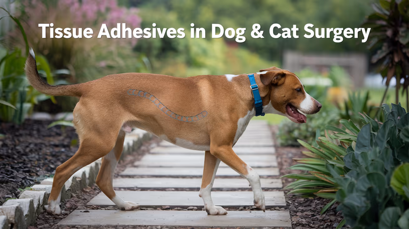
Tissue Adhesives in Dog and Cat Surgery
Explore the use, benefits, and safety of tissue adhesives in dog and cat surgery for better healing and less pain.
Surgery in dogs and cats often requires precise wound closure to promote healing and prevent infection. Tissue adhesives have become a popular alternative to traditional sutures and staples in veterinary surgery. These adhesives help seal wounds quickly and reduce trauma to delicate tissues.
Tissue adhesives are medical glues designed to bond skin and tissues during surgery. They offer a fast, effective, and less painful way to close surgical wounds in dogs and cats. This article explains what tissue adhesives are, how they work, their benefits, risks, and practical use in veterinary surgery.
What are tissue adhesives in dog and cat surgery?
Tissue adhesives are special glues used to close wounds or surgical incisions in animals. They form a strong bond that holds the skin edges together while the tissue heals underneath. These adhesives are made from biocompatible materials safe for pets.
They come in different types, such as cyanoacrylate-based glues, which are the most common in veterinary use. These adhesives polymerize quickly when applied, creating a flexible, waterproof seal over the wound.
- Definition and purpose: Tissue adhesives are medical glues used to close surgical wounds, reducing the need for stitches and speeding up recovery in dogs and cats.
- Common types: Cyanoacrylate adhesives are widely used due to their fast bonding and strong hold suitable for skin closure in pets.
- Biocompatibility: These adhesives are designed to be safe and non-toxic, minimizing tissue irritation or allergic reactions in animals.
- Application forms: Tissue adhesives are available as liquids or gels, allowing precise application on different wound types and sizes.
Understanding what tissue adhesives are helps pet owners appreciate their role in modern veterinary surgery. They offer a less invasive way to close wounds compared to traditional sutures.
How do tissue adhesives work in veterinary surgery?
Tissue adhesives work by bonding the edges of a wound or incision together. When applied, the adhesive quickly polymerizes and forms a strong film that holds the skin in place. This seal protects the wound from dirt and bacteria.
The glue also creates a waterproof barrier, which helps keep the wound clean and reduces the risk of infection. The adhesive naturally sloughs off as the skin heals underneath.
- Polymerization process: The adhesive hardens rapidly upon contact with moisture, creating a strong bond that holds wound edges securely.
- Wound sealing: The glue forms a flexible, waterproof layer that protects the surgical site from external contaminants.
- Healing support: By holding the skin edges together, adhesives promote faster tissue regeneration and reduce scar formation.
- Natural shedding: The adhesive film naturally falls off as the skin heals, eliminating the need for removal in most cases.
This mechanism makes tissue adhesives an efficient and less painful alternative to sutures, especially for superficial skin wounds in dogs and cats.
What are the benefits of using tissue adhesives in dog and cat surgery?
Using tissue adhesives offers several advantages over traditional sutures or staples. These benefits improve the surgical experience for both pets and veterinarians.
Adhesives reduce surgery time, lower infection risk, and cause less discomfort to animals. They also improve cosmetic outcomes and simplify post-operative care.
- Faster wound closure: Applying adhesives takes less time than stitching, reducing anesthesia duration and surgical stress for pets.
- Less pain and trauma: Adhesives avoid needle punctures, minimizing tissue damage and post-surgical discomfort in animals.
- Reduced infection risk: The waterproof seal protects wounds from bacteria, lowering chances of infection after surgery.
- Improved cosmetic results: Adhesives create smooth, neat closures that often heal with less noticeable scarring.
These benefits make tissue adhesives a valuable tool in veterinary surgery, especially for small or superficial wounds where quick healing is desired.
Are tissue adhesives safe for dogs and cats?
Tissue adhesives are generally safe when used correctly by trained veterinary professionals. They are made from materials tested for biocompatibility and minimal toxicity in animals.
However, some risks exist, such as allergic reactions or improper application leading to poor wound healing. It is important to follow veterinary guidance for their use.
- Biocompatibility testing: Adhesives undergo rigorous testing to ensure they do not cause harmful reactions in dog and cat tissues.
- Minimal toxicity: The materials used are designed to break down safely without releasing harmful substances into the body.
- Possible allergic reactions: Rarely, some pets may develop sensitivity to adhesive components, requiring alternative closure methods.
- Proper application needed: Incorrect use can cause wound gaps or delayed healing, emphasizing the need for veterinary expertise.
Overall, tissue adhesives provide a safe option for wound closure when applied by experienced veterinarians following proper protocols.
When should tissue adhesives be used in dog and cat surgery?
Tissue adhesives are best suited for specific types of wounds and surgical procedures. They work well for clean, superficial skin closures but are not ideal for deep or high-tension wounds.
Veterinarians decide on adhesive use based on wound location, size, and the animal’s health status to ensure optimal healing.
- Superficial skin wounds: Adhesives are ideal for closing small, clean incisions or cuts on the skin surface of dogs and cats.
- Low-tension areas: They work best where the skin edges do not experience strong pulling forces during movement.
- Supplement to sutures: Adhesives can be used alongside sutures to seal and protect the wound surface for added security.
- Not for deep wounds: Tissue adhesives are not suitable for closing deep tissue layers or wounds under high mechanical stress.
Choosing the right cases for tissue adhesive use helps ensure successful healing and reduces complications in veterinary surgery.
How should pet owners care for wounds closed with tissue adhesives?
After surgery with tissue adhesives, proper wound care is essential to support healing and prevent infection. Pet owners should follow veterinary instructions closely.
Care involves keeping the wound clean, dry, and protected from licking or scratching by the animal.
- Keep wound dry: Avoid bathing or wetting the adhesive area until it naturally falls off to maintain the waterproof seal.
- Prevent licking: Use an Elizabethan collar or other barriers to stop pets from licking or chewing the wound site.
- Monitor for infection: Watch for redness, swelling, or discharge and contact your vet if signs of infection appear.
- Avoid strenuous activity: Limit your pet’s movement to prevent stress on the wound and allow proper healing.
Following these care steps helps ensure the tissue adhesive remains effective and the wound heals smoothly without complications.
Conclusion
Tissue adhesives have become a valuable option for closing surgical wounds in dogs and cats. They offer a fast, safe, and less painful alternative to traditional sutures for many skin closures.
By understanding how tissue adhesives work, their benefits, and proper care, pet owners can support their animals’ recovery after surgery. Always consult your veterinarian to determine if tissue adhesives are suitable for your pet’s specific surgical needs.
What types of tissue adhesives are used in veterinary surgery?
Cyanoacrylate-based adhesives are the most common in veterinary surgery due to their fast bonding and strong hold on skin wounds in dogs and cats.
Can tissue adhesives replace sutures completely in pet surgery?
Tissue adhesives are suitable for superficial skin closures but cannot replace sutures for deep or high-tension wounds requiring stronger support.
Are tissue adhesives painful for pets during application?
Applying tissue adhesives is generally painless and less traumatic than suturing, reducing discomfort during and after surgery.
How long does it take for tissue adhesives to fall off after surgery?
The adhesive film naturally sloughs off within 5 to 14 days as the skin heals, usually without needing removal.
What should I do if my pet’s wound looks infected after adhesive closure?
If you notice redness, swelling, discharge, or foul odor, contact your veterinarian immediately for evaluation and treatment.

Layered Closure Technique in Small Animal Surgery
Learn about the layered closure technique in small animal surgery, its benefits, steps, and tips for optimal healing in pets.
Layered closure technique in small animal surgery is a vital method used to close surgical wounds in pets like dogs and cats. Proper wound closure helps prevent complications such as infection and dehiscence, ensuring faster healing and better outcomes for your pet. Understanding this technique can help you appreciate the care your veterinarian provides during surgery.
This article explains what the layered closure technique is, why it is important, and how it is performed. You will also learn about the materials used, common challenges, and aftercare tips to support your pet's recovery after surgery.
What is the layered closure technique in small animal surgery?
The layered closure technique involves closing a surgical wound in multiple layers, each repaired separately to restore the skin and underlying tissues. This method mimics the natural anatomy and provides strength and protection to the wound site.
By closing the wound in layers, veterinarians reduce tension on the skin and minimize the risk of wound opening or infection. This approach is standard in many soft tissue surgeries in small animals.
- Definition of technique: It is a method of suturing wounds by closing each tissue layer individually to promote proper healing and strength.
- Purpose of layering: Layering distributes tension evenly across the wound, preventing skin tearing and reducing scar formation.
- Tissues involved: Common layers include the muscle, subcutaneous tissue (fat), and skin, each requiring different suture materials and patterns.
- Common surgeries: This technique is used in spays, tumor removals, and traumatic wound repairs in dogs and cats.
Understanding the layered closure technique helps pet owners know why their veterinarian uses specific sutures and methods during surgery. It also highlights the importance of proper wound care after surgery.
Why is the layered closure technique important in pet surgery?
Using a layered closure technique improves surgical outcomes by providing a strong, stable wound closure that heals well. It reduces the risk of complications such as wound dehiscence, infection, and delayed healing.
Proper closure also helps maintain the pet’s comfort and reduces postoperative pain by minimizing tension and movement at the wound site.
- Prevents wound opening: Layered closure strengthens the wound to avoid separation of edges during healing.
- Reduces infection risk: Closing each layer properly limits dead space where bacteria can grow, lowering infection chances.
- Improves healing speed: Proper tissue alignment supports faster and more organized tissue repair.
- Enhances cosmetic results: Layered closure reduces scarring and improves the appearance of the healed wound.
Overall, this technique is essential for the health and comfort of your pet after surgery, ensuring a smooth recovery process.
What materials and sutures are used in layered closure?
Different suture materials and needle types are chosen for each tissue layer based on strength, absorption rate, and tissue reaction. The goal is to support healing without causing irritation or prolonged foreign body presence.
Veterinarians select sutures carefully to match the tissue type and expected healing time, balancing durability and biocompatibility.
- Absorbable sutures: Used for muscle and subcutaneous layers to avoid the need for removal and reduce irritation.
- Non-absorbable sutures: Sometimes used for skin closure when longer support is needed and removal is planned.
- Suture size choice: Smaller sizes (e.g., 3-0, 4-0) are common for delicate tissues to minimize trauma.
- Needle types: Curved, reverse cutting needles are preferred for skin to reduce tissue damage during suturing.
Using the right materials ensures each layer heals properly and the overall wound remains secure during recovery.
How is the layered closure technique performed step-by-step?
The surgeon closes the wound starting from the deepest tissue layer and moves outward to the skin. Each layer is sutured separately with appropriate technique and tension.
This stepwise approach restores the anatomy and function of the tissues, supporting optimal healing.
- Step 1 - Muscle closure: Sutures are placed in the muscle layer to restore strength and support the overlying tissues.
- Step 2 - Subcutaneous closure: The fat layer is closed to eliminate dead space and reduce tension on the skin.
- Step 3 - Skin closure: The skin edges are aligned and sutured carefully to promote good cosmetic healing.
- Step 4 - Final inspection: The surgeon checks for proper tension, bleeding, and alignment before finishing the procedure.
Following these steps carefully helps prevent complications and ensures the wound heals well.
What are common complications of layered closure and how to avoid them?
While layered closure is effective, complications can occur if the technique or aftercare is inadequate. Recognizing and preventing these issues is important for your pet’s recovery.
Proper surgical technique and good postoperative care reduce risks significantly.
- Wound dehiscence: Occurs if sutures are too tight or tissues are under excessive tension, causing the wound to open.
- Infection risk: Poor aseptic technique or contamination can lead to infection at the surgical site.
- Seroma formation: Fluid accumulation in dead space can delay healing and cause swelling.
- Suture reactions: Some pets may develop inflammation or irritation from suture material.
Following your veterinarian’s instructions and monitoring the wound closely can help avoid these complications.
How should pet owners care for wounds after layered closure surgery?
After surgery, proper wound care at home is crucial to support healing and prevent problems. Owners should follow specific guidelines provided by their veterinarian.
Monitoring the wound and protecting it from trauma or infection helps ensure the best outcome.
- Keep wound clean: Avoid dirt and moisture around the incision to reduce infection risk.
- Prevent licking: Use an Elizabethan collar if needed to stop the pet from licking or chewing the wound.
- Limit activity: Restrict running and jumping to avoid stress on the wound and sutures.
- Watch for signs: Check daily for redness, swelling, discharge, or opening of the wound and report concerns promptly.
Good aftercare supports the layered closure technique’s success and helps your pet heal comfortably and quickly.
What are the benefits of layered closure over single-layer closure?
Layered closure offers several advantages compared to closing the wound in just one layer. These benefits improve healing quality and reduce complications.
Understanding these benefits helps explain why veterinarians prefer this technique for many surgeries.
- Stronger wound support: Multiple layers provide better mechanical strength than a single-layer closure.
- Reduced tension on skin: Layering decreases stress on the skin edges, lowering the risk of tearing or scarring.
- Lower infection risk: Closing dead space in subcutaneous layers reduces fluid accumulation and bacterial growth.
- Improved healing alignment: Each tissue type is repaired in its natural position, promoting organized tissue regeneration.
These benefits make layered closure the preferred technique for most soft tissue surgeries in small animals.
Conclusion
The layered closure technique in small animal surgery is a careful, stepwise method to close wounds in multiple tissue layers. This approach strengthens the wound, reduces complications, and supports faster healing in pets.
Understanding this technique helps pet owners appreciate the surgical care their animals receive. Following proper aftercare instructions ensures the best recovery and comfort for your pet after surgery.
FAQs
How long does it take for a layered closure wound to heal in pets?
Most layered closure wounds heal significantly within 10 to 14 days, but full healing may take several weeks depending on the surgery and pet’s health.
Can I bathe my pet after layered closure surgery?
Bathing is usually not recommended until sutures are removed and the wound is fully healed, typically after 10 to 14 days, to prevent infection.
What signs indicate a problem with my pet’s surgical wound?
Watch for redness, swelling, discharge, foul odor, or wound opening, as these may indicate infection or dehiscence requiring veterinary attention.
Are stitches always removed after layered closure surgery?
Absorbable sutures in deeper layers do not require removal, but skin sutures may need removal 10 to 14 days post-surgery depending on the material used.
Is layered closure technique painful for pets?
The technique itself is not painful due to anesthesia, and proper closure reduces postoperative pain by minimizing wound tension and irritation.
Closure Protocol for Neuter Surgery in Dogs
Learn the detailed closure protocol for neuter surgery in dogs to ensure safe healing and prevent complications.
What is the closure protocol for neuter surgery in dogs?
Closure protocol for neuter surgery in dogs involves the steps taken to properly close the surgical site after removing the testicles. This process is crucial to promote healing and prevent infection or complications.
The protocol includes suturing techniques, materials used, and post-operative care to ensure the incision heals well and the dog recovers quickly.
- Suturing layers: The protocol requires closing the tunica vaginalis, subcutaneous tissue, and skin separately to ensure strength and reduce dead space.
- Suture materials: Absorbable sutures are preferred for internal layers to avoid removal stress, while non-absorbable or staples may be used for skin closure.
- Technique choice: Simple interrupted or continuous sutures are common, chosen based on surgeon preference and tissue type.
- Post-op care: Proper wound monitoring and preventing licking or trauma are essential for successful healing.
Following this closure protocol reduces risks of swelling, infection, and dehiscence, ensuring a smooth recovery for your dog.
Why is proper closure important after neuter surgery in dogs?
Proper closure after neuter surgery is vital to protect the surgical site from infection and to promote quick healing. The scrotal area is prone to contamination, so secure closure helps prevent complications.
Without correct closure, dogs can develop swelling, bleeding, or wound opening, which can lead to pain and require further treatment.
- Infection prevention: Tight closure limits bacterial entry, reducing the chance of post-surgical infections that delay healing.
- Hemostasis support: Closing layers properly helps control bleeding by compressing blood vessels.
- Wound strength: Layered suturing provides mechanical strength to withstand movement and prevent wound opening.
- Comfort and mobility: Proper closure minimizes swelling and pain, allowing dogs to move comfortably soon after surgery.
Thus, following a strict closure protocol is essential for the dog’s health and well-being after neuter surgery.
What suture materials are recommended for neuter surgery closure in dogs?
Choosing the right suture material is key for successful wound closure in neuter surgery. The materials must be strong enough to hold tissues together but also safe and comfortable for the dog.
Absorbable sutures are generally preferred for internal layers, while skin closure may use absorbable or non-absorbable sutures depending on the surgeon’s approach.
- Absorbable sutures: Materials like polyglycolic acid or poliglecaprone dissolve over time, eliminating the need for suture removal.
- Non-absorbable sutures: Nylon or polypropylene may be used for skin closure but require removal after healing.
- Monofilament vs. multifilament: Monofilament sutures reduce infection risk due to less bacterial trapping compared to multifilament.
- Suture size: Sizes 3-0 or 4-0 are commonly used for delicate tissues to balance strength and minimal tissue trauma.
Using appropriate suture materials helps ensure the incision heals well without irritation or complications.
How should the layers be closed during neuter surgery in dogs?
Closing the surgical site in layers is critical to restore normal anatomy and provide wound strength. Each layer has a specific role and requires proper technique.
The main layers to close are the tunica vaginalis, subcutaneous tissue, and skin. Each layer should be sutured separately to avoid dead space and promote healing.
- Tunica vaginalis closure: This layer is closed with absorbable sutures to prevent herniation and fluid accumulation.
- Subcutaneous tissue closure: Closing this layer reduces dead space and supports skin edges, minimizing swelling.
- Skin closure: The skin is closed with interrupted or continuous sutures or staples to protect the wound from external contamination.
- Aseptic technique: Maintaining sterility during closure prevents introducing bacteria into the wound.
Proper layered closure ensures a strong, clean surgical site that heals efficiently without complications.
What post-operative care supports closure healing after neuter surgery in dogs?
Post-operative care is essential to protect the surgical closure and support healing after neuter surgery. Owners must follow veterinary instructions carefully to avoid complications.
This includes monitoring the incision, preventing licking or biting, and managing activity levels to avoid stress on the wound.
- Incision monitoring: Check daily for redness, swelling, discharge, or opening that may indicate infection or dehiscence.
- Elizabethan collar use: Prevents the dog from licking or chewing the incision, which can damage sutures and delay healing.
- Restricted activity: Limit running, jumping, and rough play for at least 10-14 days to avoid wound stress.
- Follow-up visits: Scheduled veterinary checks ensure the wound is healing properly and sutures can be removed if needed.
Good post-op care helps the closure heal securely and reduces the risk of complications after neuter surgery.
What complications can arise from improper closure after neuter surgery in dogs?
Improper closure of the surgical site can lead to several complications that affect the dog’s recovery and health. Recognizing these risks highlights the importance of following the closure protocol.
Complications may require additional treatment and cause discomfort or longer healing times for the dog.
- Wound dehiscence: The surgical site may reopen if sutures fail or closure is weak, exposing tissues to infection.
- Infection: Poor closure can allow bacteria to enter, causing redness, swelling, pain, and possible abscess formation.
- Hematoma or seroma: Fluid or blood may accumulate under the skin if dead space is not properly closed.
- Excessive swelling: Inadequate closure or trauma can cause inflammation that delays healing and causes discomfort.
Prompt veterinary care is necessary if any signs of complications appear to ensure proper treatment and recovery.
Conclusion
Closure protocol for neuter surgery in dogs is a critical step that ensures the surgical site heals safely and effectively. Proper suturing techniques, materials, and layered closure all contribute to a strong, infection-free wound.
Following the closure protocol and providing diligent post-operative care helps prevent complications such as infection, swelling, or wound opening. This leads to a smoother recovery and better outcomes for your dog after neuter surgery.
What type of sutures are best for closing the skin after neuter surgery in dogs?
Non-absorbable sutures like nylon or polypropylene are often used for skin closure to provide strength and are removed after healing, but absorbable sutures can also be used to avoid removal stress.
How long does it take for the incision to heal after neuter surgery in dogs?
The incision usually heals within 10 to 14 days, but full internal healing may take longer; following post-op care instructions is essential for timely recovery.
Can dogs lick their incision after neuter surgery?
Dogs should not lick their incision as it can cause infection or wound opening; using an Elizabethan collar helps prevent this behavior during healing.
When should sutures be removed after neuter surgery in dogs?
Sutures are typically removed 10 to 14 days after surgery, once the incision has healed sufficiently, but this depends on the suture type and veterinary advice.
What signs indicate a problem with the surgical closure after neuter surgery?
Signs include redness, swelling, discharge, bleeding, or wound opening; these require prompt veterinary evaluation to prevent serious complications.
Isolation Protocols for Infectious Dogs
Learn effective isolation protocols for infectious dogs to protect your pets and household from contagious diseases.
When a dog becomes infectious, it poses a significant risk to other pets and humans in the household. Isolation protocols for infectious dogs are essential to prevent the spread of contagious diseases such as parvovirus, kennel cough, or canine influenza. Understanding how to properly isolate your dog helps protect your entire family and other animals from infection.
This article explains the key steps and best practices for isolating an infectious dog. You will learn how to set up a safe isolation area, manage hygiene, and monitor your dog's health during the isolation period. Following these guidelines ensures effective containment and supports your dog's recovery.
What are isolation protocols for infectious dogs?
Isolation protocols are specific measures taken to separate an infectious dog from healthy animals and people. These protocols reduce the risk of disease transmission by controlling contact and contamination. They include physical separation, hygiene practices, and monitoring procedures.
Proper isolation helps break the chain of infection and protects vulnerable pets and family members. It is a critical part of managing contagious diseases in dogs.
- Physical separation: Keeping the infectious dog in a separate room or area away from other pets and people limits direct and indirect contact.
- Dedicated supplies: Using separate food bowls, bedding, and toys prevents cross-contamination between the sick dog and others.
- Hygiene control: Regular hand washing and disinfecting surfaces reduce the spread of infectious agents.
- Health monitoring: Observing the dog's symptoms and progress helps detect worsening conditions or complications early.
Implementing these protocols consistently is vital for effective infection control and the safety of all household members.
How do you set up an isolation area for an infectious dog?
Creating a proper isolation area involves selecting a suitable space and preparing it to minimize infection risks. The area should be easy to clean and away from common living spaces.
Setting up the isolation space correctly ensures the infectious dog is contained safely while receiving care and comfort.
- Choose a separate room: Select a room with a door that can close fully to prevent the dog from roaming freely.
- Use washable surfaces: Floors and walls should be easy to clean and disinfect regularly to remove pathogens.
- Provide essential supplies: Include food, water, bedding, and waste disposal items dedicated solely to the isolated dog.
- Limit access: Restrict entry to the isolation area to only necessary caregivers to reduce contamination risk.
Maintaining a well-prepared isolation area supports both infection control and the dog's wellbeing during recovery.
What hygiene practices are essential during dog isolation?
Hygiene is a cornerstone of isolation protocols. Proper cleaning and disinfection prevent the spread of infectious agents from the dog to people or other animals.
Adhering to strict hygiene routines protects everyone involved and helps contain the infection within the isolation area.
- Hand washing: Wash hands thoroughly with soap and water before and after contact with the dog or its environment.
- Disinfect surfaces: Clean floors, bedding, and feeding areas daily with pet-safe disinfectants to kill germs.
- Use protective gear: Wear disposable gloves and gowns when handling the dog or cleaning its area to minimize contamination.
- Proper waste disposal: Dispose of feces, urine, and contaminated materials promptly in sealed bags to prevent pathogen spread.
Consistent hygiene practices reduce the risk of spreading infection and support a safe environment for recovery.
How long should a dog remain in isolation?
The duration of isolation depends on the specific infectious disease and the dog's recovery progress. Some diseases require longer isolation to ensure the dog is no longer contagious.
Following veterinary guidance on isolation length is crucial to prevent premature reintegration and further spread of infection.
- Disease-specific timing: Different infections like parvovirus or kennel cough have recommended isolation periods based on contagiousness.
- Symptom resolution: Isolation should continue until the dog shows no signs of illness and has recovered fully.
- Veterinary clearance: A veterinarian should confirm when it is safe to end isolation based on clinical evaluation and testing.
- Gradual reintegration: Reintroduce the dog slowly to other pets and people to monitor for any relapse or residual infection.
Adhering to the recommended isolation timeframe ensures the safety of your household and other animals.
What signs indicate an infectious dog needs veterinary care during isolation?
Monitoring your dog's health during isolation is essential to detect worsening conditions or complications. Certain signs require prompt veterinary attention.
Recognizing these signs early helps ensure timely treatment and prevents severe illness or spread of infection.
- Persistent vomiting or diarrhea: Ongoing gastrointestinal symptoms can lead to dehydration and need veterinary intervention.
- High fever: A sustained elevated temperature may indicate worsening infection or secondary complications.
- Difficulty breathing: Respiratory distress requires immediate veterinary evaluation and care.
- Loss of appetite or lethargy: Significant changes in behavior or eating habits can signal serious illness needing medical attention.
Regularly observing your dog and contacting your veterinarian if these signs appear is critical during isolation.
How can you protect other pets and family members during dog isolation?
Protecting other pets and people involves minimizing exposure to the infectious dog and maintaining strict hygiene and safety measures.
Taking these precautions reduces the risk of disease transmission and keeps your household safe.
- Separate living spaces: Keep healthy pets and family members away from the isolation area to avoid contact with infectious agents.
- Personal hygiene: Encourage everyone in the household to wash hands frequently and avoid touching their face after contact with the dog.
- Disinfect shared areas: Clean common spaces regularly, especially if the infectious dog has access or contamination is suspected.
- Limit visitors: Avoid having guests or other animals visit during the isolation period to prevent spreading the infection.
Following these protective measures helps contain the infection and safeguards your entire household.
Conclusion
Isolation protocols for infectious dogs are vital to prevent the spread of contagious diseases within your home. Properly isolating your dog, maintaining hygiene, and monitoring health help protect other pets and family members from infection.
By following these guidelines and working closely with your veterinarian, you can support your dog's recovery and keep your household safe. Effective isolation is a key step in managing infectious diseases in dogs.
What is the best place to isolate an infectious dog?
The best place is a separate room with a door that closes, easy-to-clean surfaces, and limited access to reduce contamination risks.
How often should you clean the isolation area?
Clean and disinfect the isolation area daily, including floors, bedding, and feeding supplies, to minimize pathogen spread.
Can children interact with an infectious dog during isolation?
Children should avoid contact with the infectious dog to prevent disease transmission and should practice good hand hygiene if exposure occurs.
When can an infectious dog be reintroduced to other pets?
Only after full recovery and veterinary clearance, with gradual reintroduction while monitoring for any signs of illness.
What protective equipment should caregivers use during isolation?
Caregivers should wear disposable gloves and gowns when handling the dog or cleaning its area to reduce contamination risks.

Closure Considerations in Obese Dogs
Learn key closure considerations in obese dogs after surgery to ensure safe healing and reduce complications.
Obesity in dogs presents unique challenges during surgical closure. Excess fat and skin tension increase the risk of wound complications, making closure techniques critical for successful healing. Understanding these considerations helps pet owners and veterinarians improve outcomes in obese canine patients.
This article explains closure considerations in obese dogs, covering surgical techniques, wound care, and prevention of complications. You will learn how obesity affects closure and what steps can optimize recovery after surgery.
Why is closure more challenging in obese dogs?
Obese dogs have thicker fat layers and loose skin, which complicate surgical closure. These factors increase tension on sutures and reduce blood supply to the wound edges, raising the chance of wound breakdown.
Additionally, the extra weight on the incision site can cause more stress during healing. Understanding these challenges is key to selecting proper closure methods.
- Increased tissue tension: Excess fat and skin create higher tension on sutures, risking wound dehiscence or reopening during healing.
- Reduced blood flow: Fatty tissue has poorer blood supply, slowing healing and increasing infection risk at the closure site.
- Greater dead space: Thick fat layers create pockets under the skin that can fill with fluid, causing seromas or infections.
- Mechanical stress: Extra body weight puts pressure on the incision, increasing the chance of suture failure or wound opening.
These factors make closure in obese dogs more complex and require careful surgical planning to avoid complications.
What surgical techniques improve closure in obese dogs?
Choosing the right closure technique helps manage the challenges of obesity. Surgeons often use layered closure and tension-relieving methods to support the wound.
Proper suture material and pattern also play a role in maintaining wound integrity under stress.
- Layered closure: Closing the wound in multiple layers distributes tension and reduces stress on the skin sutures.
- Tension-relieving sutures: Techniques like mattress or far-near-near-far sutures help reduce tension on the skin edges.
- Use of absorbable sutures: Absorbable sutures in deeper layers avoid the need for removal and reduce irritation in fatty tissue.
- Subcutaneous drains: Placing drains can prevent fluid accumulation in dead space, lowering seroma risk.
These techniques improve wound strength and reduce complications in obese dogs.
How does wound care differ for obese dogs after closure?
Postoperative wound care is critical in obese dogs to support healing and prevent infection. Extra attention is needed due to the higher risk of complications.
Owners and vets must monitor the incision closely and manage factors that could impair healing.
- Regular inspection: Frequent checks for redness, swelling, or discharge help detect infection early.
- Keeping the site clean: Gentle cleaning prevents bacterial buildup without damaging delicate tissue.
- Restricting activity: Limiting movement reduces tension on the wound and prevents suture failure.
- Use of protective bandages: Bandages shield the incision from dirt and trauma, especially in active dogs.
Good wound care supports recovery and reduces the chance of complications in obese dogs.
What complications are common with closure in obese dogs?
Obese dogs have a higher risk of wound complications after closure. Recognizing these helps in prevention and early treatment.
Complications can delay healing and cause discomfort or further surgery.
- Wound dehiscence: The incision reopens due to tension or suture failure, requiring prompt veterinary care.
- Seroma formation: Fluid collects under the skin in dead space, causing swelling and risk of infection.
- Infection: Poor blood supply and fat tissue increase infection risk, needing antibiotics and wound management.
- Delayed healing: Reduced circulation and tissue stress slow the repair process, extending recovery time.
Awareness and early intervention reduce the impact of these complications.
How can nutrition affect closure outcomes in obese dogs?
Nutrition plays a vital role in wound healing for obese dogs. Proper diet supports tissue repair and immune function.
Managing obesity through diet before and after surgery improves closure success and overall health.
- Balanced protein intake: Protein supports collagen formation and tissue repair essential for wound healing.
- Controlled calorie intake: Avoiding excess calories helps reduce fat and tension on the wound over time.
- Vitamin and mineral support: Nutrients like vitamin C and zinc enhance immune response and healing.
- Weight management: Gradual weight loss before surgery lowers surgical risks and improves closure outcomes.
Good nutrition complements surgical care to optimize healing in obese dogs.
What role does owner compliance play in closure success?
Owner involvement is crucial for managing closure in obese dogs. Following veterinary instructions ensures proper healing and reduces complications.
Owners must understand the special needs of obese dogs after surgery to provide the best care.
- Following activity restrictions: Limiting exercise prevents stress on the incision and supports suture integrity.
- Administering medications: Giving antibiotics or pain relief as prescribed helps prevent infection and discomfort.
- Monitoring wound changes: Reporting any swelling, discharge, or redness to the vet allows early treatment.
- Maintaining hygiene: Keeping the incision clean and dry reduces infection risk and promotes healing.
Active owner participation is key to successful closure and recovery in obese dogs.
What preventive measures reduce closure complications in obese dogs?
Prevention is better than treatment when it comes to closure in obese dogs. Several steps can minimize risks before and after surgery.
These measures improve surgical outcomes and long-term health.
- Preoperative weight loss: Reducing obesity before surgery lowers tension and improves healing capacity.
- Careful surgical planning: Choosing appropriate closure techniques tailored to the dog's body condition reduces complications.
- Postoperative monitoring: Close follow-up detects problems early and allows timely intervention.
- Owner education: Teaching owners about wound care and signs of complications supports better outcomes.
Implementing these steps helps ensure safe closure and recovery in obese dogs.
Conclusion
Closure considerations in obese dogs are essential to prevent wound complications and promote healing. The extra fat and skin tension require special surgical techniques and careful postoperative care.
By understanding these challenges and following veterinary advice on closure methods, wound care, nutrition, and owner compliance, you can help your obese dog recover safely and comfortably after surgery.
FAQs
What suture materials are best for obese dogs?
Absorbable sutures are preferred for deeper layers to avoid irritation, while strong, tension-relieving patterns help maintain skin closure under stress in obese dogs.
How long should activity be restricted after surgery in obese dogs?
Activity should be limited for at least 10 to 14 days post-surgery to reduce tension on the wound and allow proper healing in obese dogs.
Can weight loss before surgery improve closure outcomes?
Yes, gradual weight loss reduces fat tension and improves blood flow, lowering the risk of wound complications and enhancing closure success.
When should I contact the vet about wound problems?
Contact your vet immediately if you notice redness, swelling, discharge, or if the wound opens, as these signs indicate possible infection or dehiscence.
Are drains always necessary in obese dog surgeries?
Drains are often recommended to prevent fluid buildup in thick fat layers, but the decision depends on the surgery type and the surgeon’s assessment.
Asepsis for Cesarean Section in Dogs
Learn essential asepsis techniques for cesarean sections in dogs to ensure safe surgery and reduce infection risks.
A cesarean section in dogs is a common surgical procedure performed to deliver puppies safely when natural birth is risky or impossible. Maintaining asepsis during this surgery is critical to prevent infections that can threaten the lives of both the mother and her puppies. Understanding proper aseptic techniques helps ensure a successful operation and smooth recovery.
This article explains what asepsis means for cesarean sections in dogs, why it is important, and how veterinary teams maintain a sterile environment. You will learn step-by-step methods to prepare the surgical site, instruments, and personnel to minimize infection risks during the procedure.
What is asepsis in cesarean section surgery for dogs?
Asepsis refers to the practice of preventing contamination by harmful microorganisms during surgery. In cesarean sections, it means creating and maintaining a sterile field to protect the mother and puppies from infections. This involves sterilizing instruments, disinfecting the surgical site, and using sterile gloves and gowns.
Proper asepsis reduces the chance of postoperative complications such as wound infections, delayed healing, or systemic infections. It is a vital part of surgical care that every veterinary team must follow strictly.
- Definition of asepsis: Asepsis is the absence of bacteria, viruses, and other pathogens in the surgical environment to prevent infection during and after surgery.
- Importance in cesarean sections: Preventing contamination protects the mother’s uterus and puppies from infections that can cause serious illness or death.
- Difference from antisepsis: Asepsis prevents contamination entirely, while antisepsis reduces microorganisms on living tissues like skin.
- Goal of asepsis: To maintain a sterile field from the start to the end of surgery, minimizing infection risks.
Understanding asepsis helps veterinary teams plan and execute cesarean sections safely. It also reassures pet owners about the care their dogs receive during surgery.
How do you prepare the surgical site for asepsis in a dog cesarean?
Preparing the surgical site is a key step to reduce bacteria on the dog's skin before making the incision. This preparation involves clipping hair, cleaning, and disinfecting the area around the abdomen where the surgery will occur.
Proper site preparation lowers the risk of bacteria entering the uterus during surgery, which could cause infection in the mother or puppies.
- Hair clipping: Remove all hair from the abdomen to expose clean skin and reduce places where bacteria can hide.
- Initial cleaning: Use sterile gauze soaked in warm water to wipe away dirt and debris before disinfecting.
- Disinfectant application: Apply an antiseptic solution such as chlorhexidine or povidone-iodine in a circular motion starting from the incision site outward.
- Repeat scrubbing: Perform at least three rounds of scrubbing with antiseptic to ensure thorough skin disinfection.
After preparing the site, keep the area dry and avoid touching it with non-sterile objects. This helps maintain the sterile field until surgery begins.
What sterilization methods are used for instruments in canine cesarean sections?
Sterilizing surgical instruments is essential to kill all microorganisms before use. Veterinary clinics use several methods to ensure instruments are sterile and safe for surgery.
Choosing the right sterilization technique depends on the instrument type and availability of equipment. Proper sterilization prevents infections caused by contaminated tools.
- Autoclaving: Uses high-pressure steam at 121–134°C to sterilize metal instruments effectively and quickly.
- Cold sterilization: Soaking instruments in chemical disinfectants like glutaraldehyde for long periods when autoclaving is unavailable.
- Dry heat sterilization: Heating instruments in an oven at 160–180°C for 1–2 hours, suitable for heat-resistant tools.
- Packaging instruments: Sterile packs or wraps keep instruments clean until surgery and prevent contamination.
After sterilization, instruments must be handled with sterile gloves and stored properly to maintain their sterility until use during the cesarean section.
How should the surgical team maintain asepsis during the cesarean procedure?
The surgical team plays a vital role in maintaining asepsis throughout the cesarean section. This includes wearing sterile clothing, using proper hand hygiene, and following strict protocols to avoid contamination.
Every team member must understand and follow aseptic techniques to protect the patient and ensure a successful surgery.
- Surgical hand scrub: Team members must scrub hands and forearms with antiseptic soap for at least 5 minutes before gloving.
- Sterile gowns and gloves: Wearing sterile gowns and gloves prevents bacteria from the skin or clothes contaminating the surgical field.
- Minimal movement: Limiting unnecessary movement and talking reduces airborne contamination in the operating room.
- Proper instrument handling: Only sterile instruments should touch the surgical site, and contaminated tools must be replaced immediately.
Maintaining asepsis requires constant attention and teamwork. Any break in sterile technique can increase infection risks, so vigilance is essential.
What postoperative care supports asepsis after a dog’s cesarean section?
After the surgery, proper wound care and monitoring help prevent infections and promote healing. Postoperative asepsis includes keeping the incision clean and preventing the dog from licking or scratching the area.
Good postoperative care reduces complications and supports the mother’s recovery and the puppies’ health.
- Wound inspection: Check the incision daily for redness, swelling, discharge, or signs of infection.
- Cleaning the site: Use sterile saline or prescribed antiseptics to gently clean the wound if needed.
- Preventing licking: Use an Elizabethan collar to stop the dog from licking or biting the incision site, which can introduce bacteria.
- Administering antibiotics: Follow the veterinarian’s instructions on antibiotics to control or prevent infections after surgery.
Following these steps helps ensure the surgical site heals properly and reduces the chance of postoperative infections.
What are common asepsis challenges during cesarean sections in dogs?
Despite best efforts, some challenges can compromise asepsis during cesarean sections. Recognizing and addressing these issues helps improve surgical outcomes.
Veterinary teams must be prepared to manage these challenges to maintain a sterile environment throughout the procedure.
- Emergency surgeries: Urgent cesareans may limit preparation time, increasing infection risks if aseptic steps are rushed.
- Contaminated environment: Operating in non-ideal or poorly cleaned areas can introduce bacteria despite precautions.
- Multiple puppies: Handling many puppies increases the chance of contamination if instruments or gloves are not changed appropriately.
- Patient factors: Dogs with skin infections or poor health may have higher bacterial loads, requiring extra aseptic care.
Understanding these challenges allows veterinary teams to plan better and take extra precautions during cesarean surgeries.
Conclusion
Asepsis for cesarean section in dogs is essential to prevent infections and ensure the safety of both the mother and her puppies. It involves careful preparation of the surgical site, sterilization of instruments, and strict sterile techniques by the surgical team.
By following proper aseptic protocols before, during, and after surgery, veterinary teams can reduce complications and support a smooth recovery. Pet owners should understand the importance of asepsis and trust their veterinary team to provide the best care during this critical procedure.
FAQs
How long should the surgical hand scrub be before a dog cesarean?
The surgical hand scrub should last at least 5 minutes using antiseptic soap to remove bacteria and reduce infection risks during surgery.
Can I bathe my dog before a cesarean to improve asepsis?
Bathing your dog before surgery can reduce skin bacteria but should be done at least 12 hours prior to avoid skin irritation and allow the skin to dry.
What antiseptic solutions are best for preparing the surgical site?
Chlorhexidine and povidone-iodine are commonly used antiseptics that effectively disinfect the skin before surgery.
Is it necessary to change gloves during the cesarean procedure?
Yes, changing gloves between handling puppies and the surgical site helps prevent cross-contamination and maintains asepsis.
How soon after surgery can the dog resume normal activity?
Dogs usually resume normal activity within 10 to 14 days after surgery, once the incision has healed and the veterinarian approves.
Sustainable Vet | Copyright ©2026 | All Rights Reserved

