

TPLO Surgery Cost in Raleigh
Explore TPLO surgery cost in Raleigh, including price ranges and factors like dog size, surgeon skill, and rehab that affect pricing.

This article is for informational purposes only and is not a substitute for professional veterinary advice. Every case is unique, so always consult your veterinarian for guidance specific to your pet.
This content is intended for veterinary professionals for educational purposes. It does not replace clinical judgment or tailored advice. Always rely on your training, expertise, and the specific context of your patients.

TPLO surgery is a common procedure to repair a torn cranial cruciate ligament (CCL) in dogs. This surgery stabilizes the knee joint, helping dogs regain mobility and reduce pain. Understanding the cost of TPLO surgery is important for pet owners planning for their dog’s health needs.
Costs vary widely depending on factors such as the city, surgeon experience, diagnostic tests, dog size, implants used, and post-operative rehabilitation. In Raleigh, we will explore typical price ranges, what the cost usually includes and excludes, key cost drivers, and helpful tips for pet owners.
Typical TPLO Surgery Cost in Raleigh
TPLO surgery costs in Raleigh vary by clinic, surgeon expertise, and the size of your dog. Prices reflect the quality of care and resources available at each facility.
- Low estimate in Raleigh — Some clinics offer TPLO surgery starting around $3,000. These lower prices may reflect less experienced surgeons or fewer included services. While affordable, owners should confirm what is included to avoid unexpected expenses.
- Average cost range in Raleigh — Most pet owners pay between $4,000 and $6,000 for TPLO surgery. This range typically covers surgery by experienced surgeons, standard implants, and basic post-op care, representing the common market rate in Raleigh.
- High-end TPLO specialists in Raleigh — Premium clinics with board-certified surgeons and advanced surgical technology may charge $6,500 or more. These facilities often provide comprehensive care, including detailed diagnostics and specialized rehabilitation.
What the Cost Usually Covers
TPLO surgery packages generally include several key components essential for a successful outcome. Understanding these helps owners know what to expect from the quoted price.
- Surgery itself — The procedure involves cutting and rotating the tibia to stabilize the knee. This complex surgery requires skilled surgeons and specialized equipment, contributing to the overall cost.
- Anaesthesia + monitoring — General anaesthesia is critical for pain-free surgery. Continuous monitoring during the procedure ensures the dog’s safety, adding to the expense.
- Implants/plates — Stainless steel or titanium plates and screws are used to secure the bone. Titanium implants tend to cost more but offer benefits like reduced weight and better biocompatibility.
- Post-op care and follow-up exams — Follow-up visits monitor healing progress and address complications. These visits are usually included in the surgery package to support recovery.
What Might Not Be Included
Some costs are often overlooked by pet owners when budgeting for TPLO surgery. Being aware of these helps avoid surprises.
- Pre-surgical diagnostics — X-rays, blood work, and other tests before surgery may be billed separately. These diagnostics are essential to plan the surgery safely.
- Post-surgical rehab therapy — Physical therapy or hydrotherapy after surgery can improve recovery but often comes at an additional cost. Raleigh clinics may offer packages or refer to specialists.
- Medications beyond standard pain control — Some dogs require extra medications for infection or inflammation, which may not be included in the initial quote.
- Additional cost if both legs need surgery — Bilateral TPLO surgeries double the surgical effort and materials, significantly increasing the total cost.
Key Cost Drivers in Raleigh
Even within Raleigh, TPLO surgery prices vary due to several important factors. Understanding these helps owners make informed decisions.
- Dog size/weight — Larger dogs require bigger implants and longer surgery times, increasing costs. Smaller dogs may have lower fees due to simpler procedures.
- General vet vs board-certified surgeon — Board-certified surgeons have advanced training and may charge more, but often provide higher success rates and fewer complications.
- City living costs + overhead in Raleigh — Clinic rent, staff salaries, and equipment maintenance in Raleigh influence pricing. Higher overhead leads to higher surgery fees.
- Implant brand and surgical technology used — Premium implant brands and modern surgical tools add to the cost but can improve outcomes and reduce recovery time.
- Complication or infection risk — Cases with complications require extra care, medications, or additional surgeries, increasing the total expense.
Tips for Pet Owners in Raleigh
Careful financial planning and research can help pet owners manage TPLO surgery costs effectively in Raleigh.
- Ask for a detailed itemised estimate — Request a full breakdown of costs before surgery. This helps identify what is included and avoid hidden fees.
- Clarify if quote is for one leg or both — Some dogs need surgery on both knees. Confirm whether the price covers one or both legs to budget accurately.
- Explore pet insurance or financing options — Pet insurance may cover part of the surgery cost. Financing plans can spread payments over time, easing financial strain.
- Compare multiple clinics and ask about success rates — Research clinics’ reputations and outcomes. Choosing experienced surgeons can reduce complications and additional costs.
- Ask about rehab or physiotherapy packages — Some clinics offer bundled rehab services at discounted rates. Early rehab improves recovery and long-term mobility.
Conclusion
TPLO surgery is a significant financial decision for dog owners in Raleigh. Costs vary widely depending on clinic, surgeon expertise, and dog-specific factors. Planning ahead and understanding pricing helps prevent unexpected expenses.
Comparing quotes and confirming what is included before choosing a clinic ensures the best care for your dog and peace of mind for you. Thoughtful preparation supports a smooth surgery and recovery journey.
Frequently Asked Questions
How much does TPLO surgery cost in Raleigh?
TPLO surgery in Raleigh typically costs between $3,000 and $6,500. Prices depend on the clinic, surgeon experience, dog size, and included services. Premium clinics with board-certified surgeons may charge more.
Is TPLO worth the cost for dogs with CCL tears?
TPLO surgery is often worth the cost as it stabilizes the knee, reduces pain, and improves mobility. It can prevent arthritis and long-term joint damage, enhancing your dog’s quality of life.
Can pet insurance cover TPLO surgery in Raleigh?
Many pet insurance plans cover TPLO surgery, but coverage varies. It is important to check your policy details and pre-authorization requirements to understand your benefits.
How do I know if a TPLO quote is reasonable?
A reasonable TPLO quote includes surgery, anaesthesia, implants, and follow-up care. Comparing multiple detailed estimates and checking surgeon credentials helps ensure fair pricing.

Get a Free Poster
Enhance your workspace with a high-quality radiographs reference poster, designed for veterinary professionals. This free physical poster will be shipped directly to you—just fill out the form to request your copy.




Taking Great TPLO Radiographs
Click Below to Watch Live Video Demos


We'll send you a Free Wall Poster with all the steps
Step #1
Getting Ready
Ensuring a clean surgical field starts with proper skin preparation. This video demonstrates the best practices for:
- Shaving the patient – Achieving a close, even shave while minimizing skin irritation
- The Dirty Scrub – The initial skin prep step to remove surface debris and reduce bacterial load before the sterile scrub.
Following these techniques helps reduce infection risk and improve surgical outcomes. Watch the video to see how it’s done effectively!


Step #2
Reduce Your Risks
Many surgeons are shocked to find out that their patients are not protected from biofilms and resistant bacteria when they use saline and post-op antibiotics.
That’s Where Simini Comes In.
Why leave these risks and unmanaged? Just apply Simini Protect Lavage for one minute. Biofilms and resistant bacteria can be removed, and you can reduce two significant sources of infection.
Step #3
Take the Course
Preventing surgical infections is critical for patient safety and successful outcomes. This course covers:
- Aseptic techniques – Best practices to maintain a sterile field.
- Skin prep & draping – Proper methods to minimize contamination.
- Antibiotic stewardship – When and how to use perioperative antibiotics effectively.
Stay up to date with the latest evidence-based protocols. Click the link to start learning and earn CE credits!

Things to know
DeAngelis vs TPLO Surgery for Dogs
Compare DeAngelis and TPLO surgery for dogs, exploring benefits, risks, recovery, and costs to help pet owners choose the best treatment.
DeAngelis surgery also called lateral suture, extracapsular repair, or ELSS (extracapsular lateral suture stabilization) is one of the oldest and most widely performed CCL repair techniques.
TPLO is the most commonly recommended procedure by the ACVS for dogs over 60 lbs.
Decades of research strongly favor TPLO for larger, more active dogs while supporting extracapsular repair as a cost-effective option for smaller dogs.
Quick answer: TPLO produces superior outcomes in medium and large breed dogs: better limb loading at 8 weeks, higher 1-year function, and slower arthritis progression. DeAngelis surgery is effective for small dogs under 15 to 20 lbs and costs $800 to $1,500 vs $3,000 to $6,000 for TPLO.
Key takeaways
- TPLO produces superior outcomes at 8 weeks and 1 year in size-matched studies: more symmetric loading, higher vertical force, higher owner satisfaction
- DeAngelis surgery is effective in small dogs under 15 to 20 lbs: success rates above 90% reported; significantly worse in larger dogs
- Extracapsular repair relies on periarticular fibrosis for stability; the suture eventually stretches or fails in larger dogs
- DeAngelis surgery costs significantly less: $800 to $1,500 vs $3,000 to $6,000 for TPLO
- Arthritis progression is faster after extracapsular repair than TPLO in multiple studies; TPLO slows OA more effectively
- 10 to 20% of extracapsular repair dogs will require additional treatment for lameness during their lifetime per Today's Veterinary Practice
How each procedure works
DeAngelis surgery (lateral suture / extracapsular repair)
DeAngelis surgery uses a heavy monofilament nylon suture (or, in the TightRope variation, a fiber-tape through bone tunnels) placed outside the joint to mimic the function of the torn CCL.
The suture runs from the lateral fabella behind the femur around to the tibial crest, restraining the cranial drawer movement that occurs with CCL loss.
The suture is not a permanent replacement.
DVM360 confirms that extracapsular stabilization relies on periarticular fibrosis for long-term stability the suture eventually stretches or fails, and joint stability depends on scar tissue formed around the joint during recovery.
DeAngelis surgery does not require bone cutting, specialized equipment, or the steep learning curve associated with TPLO. It is technically accessible to general practice surgeons with appropriate training.
TPLO (Tibial Plateau Leveling Osteotomy)
TPLO addresses CCL rupture by changing joint geometry rotating the tibial plateau so the stifle is mechanically stable without any functioning CCL.
The stability provided by TPLO is bone-based and does not depend on scar tissue or suture integrity over time.
TPLO requires specialized equipment, a radiolucent table, intraoperative radiographs, and surgeon training in the procedure. It is primarily performed at specialty hospitals and by board-certified orthopedic surgeons.
What the clinical evidence shows
Short-term outcomes
DVM360's evidence review found TPLO dogs bear more weight sooner; extracapsular repair dogs hold the leg up for 1 to 2 weeks.
A force plate gait study (ECR n=23, TPLO n=15) found more symmetric limb loading at 8 weeks in TPLO patients compared to extracapsular repair patients at both walk and trot.
Long-term outcomes
Today's Veterinary Practice's evidence-based review reports:
- A randomized blinded clinical trial of 80 dogs found TPLO resulted in higher peak vertical force and higher owner satisfaction at 1 year post-surgery compared to lateral fabellar suture stabilization
- A more recent study showed TPLO outcomes were always better than lateral suture outcomes at any time point up to 1 year
DVM360 notes one early study found no difference at 2 years, but more recent studies consistently show TPLO outperforming extracapsular repair.
Arthritis progression
Today's Veterinary Practice confirms that extracapsular stabilization may result in increased progression of radiographic osteoarthritis compared to TPLO.
Restoring joint mechanics (TPLO) slows cartilage degradation more effectively than temporary stabilization with subsequent fibrosis (extracapsular repair).
Long-term failure
Today's Veterinary Practice reports that 10 to 20% of dogs with extracapsular stabilization will require treatment or reoperation for lameness during their lifetimes, sometimes years after original surgery.
This rate is higher than the expected rate of reoperation-requiring complications after TPLO.
Head-to-head comparison
Which dogs are best suited to each?
DeAngelis surgery is appropriate for:
- Small dogs under 15 to 20 lbs with CCL rupture
- Dogs with significant health conditions making a longer anesthetic or more invasive procedure higher risk
- Cases where cost is the primary determining factor
- Older, less active dogs with lower functional demands
TPLO is recommended for:
- Dogs over 30 lbs, especially active and large breeds
- Dogs where long-term joint function and minimal OA progression are the goals
- Working dogs, sport dogs, or highly active pets
- Dogs with steep tibial plateau angles that make extracapsular repair less reliable
For the full surgical alternatives overview, see alternatives to TPLO surgery for dogs. For the TPLO pros and cons, see TPLO surgery pros and cons for dogs.
For the lateral suture detailed comparison, see lateral suture vs TPLO: what's the difference?.
Frequently asked questions
Is DeAngelis surgery as good as TPLO?
For small dogs under 15 to 20 lbs, DeAngelis surgery can be as effective as TPLO.
For medium and large breed dogs, the evidence clearly favors TPLO: better short-term function, better 1-year outcomes, slower arthritis progression, and lower long-term reoperation rates.
How much less does DeAngelis surgery cost than TPLO?
Typically $2,000 to $4,500 less per leg. DeAngelis surgery costs approximately $800 to $1,500 at general practices vs $3,000 to $6,000 for TPLO at specialist centres.
For bilateral cases (both knees), the cost difference is doubled.
Does DeAngelis surgery fail more often than TPLO?
In large dogs, yes. The suture stretches and fails in larger, more active patients because the periarticular fibrosis cannot compensate for the higher joint forces.
Today's Veterinary Practice estimates 10 to 20% of extracapsular repair dogs will need further treatment for lameness during their lifetime.
Can my large dog have DeAngelis surgery instead of TPLO?
Technically yes, but the evidence does not support it as the primary recommendation for dogs over 30 lbs.
DVM360 confirms it is possible that larger dogs treated with lateral suture technique may have significantly worse outcomes than those treated with TPLO.
If DeAngelis surgery fails, can TPLO be done afterward?
Yes. TPLO is a standard revision option when extracapsular repair fails or when the dog develops ongoing instability and lameness.
Discuss this with a board-certified orthopedic surgeon if your dog's original repair has not produced adequate results.
Resources
- DVM360. Cranial Cruciate Ligament Repair: One Size Does NOT Fit All. dvm360.com
- Today's Veterinary Practice. Canine Cranial Cruciate Disease: An Evidence-Based Look at Current Treatment Modalities. todaysveterinarypractice.com
- DVM360. Journal Scan: Ruptured CCL Surgery: TPLO vs Extracapsular Repair. dvm360.com
- Walkerville Vet. Cruciate Surgery: TPLO, TTA, MMP, DeAngelis. walkervillevet.com.au
- ACVS. Cranial Cruciate Ligament Disease. acvs.org
What to Expect After TPLO Surgery in Dogs
Learn what to expect after TPLO surgery, including pain, swelling, healing time, and care tips for your dog’s smooth recovery.
TPLO recovery is a structured 8 to 12 week process. The surgery takes 1.5 to 2 hours; the recovery takes months.
Understanding what is normal at each stage allows owners to support recovery confidently, recognize complications early, and avoid mistakes that extend recovery or cause setbacks.
Quick answer: After TPLO surgery, expect 8 to 12 weeks of graduated activity restriction. Weeks 1 to 2: crate rest, short walks, e-collar on. Weeks 3 to 8: progressive walks and range-of-motion exercises. Weeks 8 to 12: confirm bone healing and resume normal activity. Full muscle recovery takes 6 months.
Key takeaways
- The 8 to 12 week bone healing period is non-negotiable: the plate holds the osteotomy until new bone consolidates; premature high-impact activity before radiographic healing can cause plate failure
- Weeks 1 and 2 are highest-risk for infection and wound complications: the incision is healing; activity and wound management must be strictly controlled
- Most dogs begin partial weight bearing within the first few days: early weight bearing maintains muscle mass and supports bone healing per TPLO Info
- The 2-week recheck, 6-week radiograph, and 12-week radiograph are essential milestones: each has a specific purpose; missing them risks missing early complications
- Full muscle recovery takes up to 6 months after bone healing is confirmed; bone union and functional recovery are not the same endpoint
- The e-collar must stay on for 10 to 14 days without exception: licking is the leading owner-controlled cause of TPLO surgical site infection
Immediately after surgery: days 1 to 3
Coming home
Dogs are typically discharged 24 to 48 hours after TPLO surgery, or sometimes the same day at facilities with same-day discharge protocols.
The dog will be sedated, disoriented from anesthesia, and may have bandaging on the surgical leg.
Expect:
- Groggy, disoriented behavior for 12 to 24 hours from anesthesia
- Some whining or restlessness (normal -- from anesthesia, not necessarily pain)
- Refusal to bear weight on the operated leg
- Mild to moderate swelling around the incision, peaking at days 2 to 3
Pain management
Give prescribed medications on the exact schedule provided. Typical medications include NSAIDs (meloxicam, carprofen, or similar) and gabapentin. Give NSAIDs with food to reduce gastric irritation.
TPLO Info confirms: dogs may experience slight discomfort from surgical inflammation in the days following surgery; this should be managed with appropriate pain medications given on schedule.
Confinement
Strict crate rest or confinement in a small, non-slippery room. No stairs, no jumping, no access to furniture. Leash walks outdoors for bathroom purposes only, 3 to 5 minutes, multiple times daily.
Weeks 1 to 2: incision healing phase
This is the highest-risk period for wound complications and infection.
Incision care:
- E-collar must be on at all times except supervised eating and drinking
- Keep the incision dry -- no baths, no wet grass walks
- Inspect the incision daily for spreading redness, cloudy or malodorous discharge, or wound dehiscence
- Normal: a small amount of clear serum discharge in the first 2 to 3 days; mild redness at the incision line fading from day 5 onward
- Abnormal: spreading redness, cloudy discharge, increasing warmth, or wound edges separating -- contact your vet immediately
Activity:
- Leash walks only, 5 minutes maximum, multiple times daily
- No running, jumping, stairs, or playing with other pets
- No unsupervised time without the e-collar
Weight bearing:
- Many dogs begin toe-touching within the first few days
- Some dogs refuse weight bearing for the full two weeks -- this is also normal
- Encouraging gentle, slow walking on a leash helps maintain muscle mass
2-week recheck:SustainableVet.org confirms: the 2-week visit confirms incision healing, removes sutures or staples if healing is complete, and reviews pain management and early mobility status.
Weeks 3 to 6: progressive walking phase
The incision is closed; the focus shifts to controlled progressive activity increase and gentle range of motion.
Activity:
- Increase leash walk duration gradually -- TPLO Info recommends increasing by up to 5 minutes per week from the week 2 baseline
- No off-leash activity, no running, no jumping, no stairs without a ramp
- No playing with other pets
Range-of-motion exercises:
- Passive range-of-motion (PROM) exercises can begin in most dogs at weeks 2 to 4 if the vet approves
- Gentle flexion and extension of the stifle for 5 to 10 repetitions, 2 to 3 times daily
- Supports joint flexibility and prevents stiffness during the bone healing period
Weight bearing:
- By weeks 3 to 4, most dogs are consistently bearing weight on the operated leg
- Occasional toe-touching or three-legged walking during activity is still normal
What to watch for:
- Lameness that worsens after a period of improvement (trajectory reversal)
- New swelling at the plate site, separate from the healed incision
- Any discharge from the healed wound area
Weeks 6 to 8: early consolidation phase
6-week radiograph (critical milestone):SustainableVet.org confirms: the 6-week visit includes follow-up radiographs to evaluate bone alignment and early plate stability; if healing looks good, limited physical therapy or short walks may begin.
If the radiograph shows good progression of bone healing, activity is increased. If healing is delayed, restrictions continue with a re-radiograph at 8 weeks.
Activity (if healing is confirmed):
- Leash walks increasing to 15 to 20 minutes twice daily
- Rehabilitation exercises as directed
- Hydrotherapy (if available) is an excellent low-impact option for muscle building
Weeks 8 to 12: bone consolidation and return to activity
Follow-up radiograph at 8 to 10 weeks:Medcovet confirms: a follow-up radiograph is commonly performed at weeks 8 to 10 to assess bone healing; if healing is good, the vet may approve a gradual increase in activity.
12-week visit (major milestone):SustainableVet.org confirms: the 12-week visit includes final radiographs to confirm bone fusion and plate integrity; the vet may lift most activity restrictions at this stage.
Activity (if healing confirmed at 12 weeks):
- Progressive return to normal activity
- Running, jumping, and play can gradually resume over weeks 12 to 16
- Supervised play and controlled activity continue until full muscle recovery
Animal Outpatient Surgery confirms: full muscle recovery takes up to 6 months to restore full strength and coordination in the operated leg.
Medications and supplements
NSAIDs: given for 2 to 4 weeks post-surgery or as directed. Never exceed the prescribed dose; never use human NSAIDs (ibuprofen, aspirin) -- they are toxic to dogs.
Gabapentin: helps with pain and anxiety in the early recovery period; typically tapered off after 2 to 4 weeks.
Joint supplements: glucosamine and omega-3 fatty acids are commonly recommended for long-term joint health. Starting them at surgery is reasonable; discuss with your vet.
Signs requiring immediate veterinary contact
At any stage of recovery, contact your vet immediately if you see:
- Spreading redness or warmth at the incision or plate site
- Cloudy, yellow, green, or malodorous discharge from the wound
- Wound edges separating
- Severe worsening of lameness
- Fever, lethargy, or loss of appetite beyond the first 2 to 3 days
- Any draining tract opening near the surgical site
For the week-by-week rehabilitation exercises guide, see TPLO recovery exercises for dogs. For the infection prevention guide, see how can TPLO infections be prevented post-operatively?.
For the PROM exercises guide, see PROM exercises for dogs after TPLO surgery. For the earliest infection signs, see earliest signs of TPLO infection.
Frequently asked questions
When will my dog be back to normal after TPLO?
Bone healing is typically confirmed by radiograph at 8 to 12 weeks. Most activity restrictions are lifted at the 12-week mark.
Full muscle recovery and functional normalization takes up to 6 months from surgery.
TPLO has a high success rate (more than 90%) and most dogs return to their pre-injury activity level after full recovery.
My dog is not using the leg at all after surgery. Is that normal?
Some dogs refuse to bear weight for the full first 2 weeks. This is within normal range.
If the dog was bearing some weight and has now stopped completely after day 5, contact your vet to rule out a complication.
PROM exercises and gentle encouraged walking help prompt early weight bearing.
Can my dog go up stairs during recovery?
Not during weeks 1 to 6. Stairs generate impact and torsion on the healing osteotomy. Carry small dogs over stairs or use a ramp.
Larger dogs may need a sling or harness to assist navigating a small number of essential steps.
When can my dog swim or do hydrotherapy?
Hydrotherapy (underwater treadmill or controlled pool swimming) typically begins after incision healing is confirmed at 2 weeks. Check with your vet.
Swimming in open water (lakes, rivers) is not appropriate until the vet explicitly clears the dog, typically after the 12-week radiograph.
When should I be concerned about my dog's pain during recovery?
Increasing pain after initial improvement -- the dog more lame or more distressed today than yesterday -- is the key warning sign. Normal recovery pain decreases progressively.
Worsening pain warrants veterinary assessment to rule out infection, implant complication, or late meniscal injury.
Resources
- TPLO Info. What to Expect After Your Dog's TPLO Surgery. tploinfo.com
- Animal Outpatient Surgery. What to Expect After Your Dog's TPLO Surgery. animaloutpatientsurgery.com
- Medcovet. Essential Guide to Dog Recovery TPLO Surgery. medcovet.com
- VCA Animal Hospitals. Tibial Plateau Leveling Osteotomy (TPLO) in Dogs. vcahospitals.com
Alternatives to TPLO Surgery for Dogs
Explore safe and effective alternatives to TPLO surgery for dogs with knee injuries, including treatments, benefits, and recovery options.
TPLO is the most commonly recommended surgical procedure for CCL rupture in medium and large breed dogs, but it is not the only option and it is not always the right option.
Some dogs are better served by a different surgical technique; others by non-surgical management. Understanding the full menu of alternatives helps owners have more informed conversations with their veterinarians.
Quick answer: Surgical alternatives to TPLO include TTA, extracapsular repair, Tightrope, MMP, and CBLO — most achieving 90 to 93% good function in the right patient. Non-surgical options produce variable outcomes and are not recommended for complete CCL rupture in large dogs.
Key takeaways
- Surgical alternatives achieve comparable outcomes in the right patient: TTA and Tightrope show 90 to 93% good function at one year
- Extracapsular repair is appropriate for dogs under 15 to 35 lbs: above 90% success in small dogs; significantly worse in larger breeds
- Conservative management is not recommended for large dogs with complete CCL rupture: outcomes are worse; appropriate only for comorbidities or small partial tears
- Knee braces do not have strong evidence of restoring joint stability; current studies show no significant benefit per Vetamac
- MMP and CBLO are newer alternatives with growing evidence: MMP uses a titanium wedge; CBLO uses a different osteotomy geometry
- The best alternative to TPLO is determined by dog size, activity level, surgeon training, and individual clinical factors not by owner preference alone
Surgical alternatives
TTA (Tibial Tuberosity Advancement)
TTA advances the tibial tuberosity cranially to change the angle of the patellar tendon relative to the tibial plateau.
This achieves mechanical neutralization of cranial tibial thrust through a different geometric approach than TPLO.
TTA is appropriate for large breed dogs, similar to TPLO. Studies show comparable outcomes at one year.
DVM360 notes that TTA recovery may be faster in the early post-operative period than TPLO, though TPLO tends to show better objective outcomes in size-matched longer-term comparisons.
For the full TTA vs TPLO comparison, see TPLO, CBLO, and TTA: a guide to cruciate surgery options.
Extracapsular repair (DeAngelis/lateral suture/ELSS)
Extracapsular repair uses a heavy suture outside the joint to stabilize the stifle.
It is the oldest and most widely available CCL repair technique and is appropriate for small dogs under 15 to 35 lbs.
Simon Veterinary Surgical confirms extracapsular repair achieves excellent results in appropriately sized patients (typically under 50 lbs), with many dogs living full, active lives.
In larger, more active dogs, the suture eventually stretches or fails, and outcomes are consistently inferior to TPLO.
Today's Veterinary Practice confirms TPLO produces higher peak vertical force and higher owner satisfaction at one year compared to extracapsular repair in size-matched dogs.
For the full comparison, see DeAngelis vs TPLO surgery for dogs.
Tightrope procedure
Tightrope uses a synthetic fiber-tape passed through bone tunnels in the femur and tibia to stabilize the stifle.
It is more anatomically positioned than extracapsular repair and achieves comparable outcomes to TPLO at 6 months in published comparative studies.
Tightrope is appropriate for small to medium dogs and as a less expensive alternative to TPLO. Dr.
Buzby's rehabilitation veterinary guide lists Tightrope among the established surgical options alongside TPLO, TTA, ELSS, and CBLO.
For the full comparison, see Tightrope vs TPLO surgery for dogs.
Modified Maquet Procedure (MMP)
MMP is an evolution of TTA that uses a porous titanium foam wedge instead of a cage to advance the tibial tuberosity.
It is less invasive and faster than TPLO, with comparable 6-month outcomes in the primary randomized study.
MMP is appropriate for medium and large breed dogs in the hands of surgeons trained in the technique.
For the full comparison, see Modified Maquet Procedure vs TPLO surgery.
CBLO (CORA-Based Leveling Osteotomy)
CBLO is a newer osteotomy technique that applies a different geometric principle to achieve tibial plateau leveling.
Rather than a radial osteotomy (as in TPLO), CBLO uses the CORA (center of rotation of angulation) to guide a wedge osteotomy.
Published success rates of 85 to 95% good function have been reported.
CBLO is an emerging alternative with growing evidence. For the full comparison, see CORA-based leveling osteotomy vs TPLO in dogs.
Zlig
Zlig is a synthetic implant placed inside the joint to replace the function of the CCL.
It is a less commonly discussed alternative with limited long-term veterinary evidence compared to the established osteotomy techniques.
For the full comparison, see Zlig vs TPLO: which surgery is better for dogs?.
Non-surgical alternatives
Conservative management
Conservative management refers to non-surgical approaches: strict rest and activity restriction, physical therapy and rehabilitation, weight management, NSAIDs for pain and inflammation control, and joint supplements.
Vetamac confirms: the main non-surgical treatments for CCL rupture include restricting activity, promoting weight loss, and physical therapy.
NSAIDs help manage pain and inflammation, although lameness persists as long as joint instability remains.
Simon Veterinary Surgical's guide notes: non-surgical outcomes are described as achieving "pasture soundness" comfortable for daily activities but the dog may not return to high-impact sports.
Consistency with physical therapy and weight management is the biggest predictor of good conservative outcomes.
When conservative management is appropriate:
- Dogs with serious concurrent medical conditions that make anesthesia high-risk
- Small dogs (under 10 to 15 lbs) with partial CCL tears
- As a temporary bridge to surgery when surgery is delayed
- Dogs whose owners cannot afford surgical options
When conservative management is not appropriate:
- Complete CCL rupture in dogs over 15 to 20 lbs lameness persists, arthritis progresses faster, and meniscal injury risk remains
- Active working dogs or dogs with high functional demands
For the full non-surgical guide, see non-surgical alternatives to TPLO surgery for dogs.
Orthopedic knee braces
Custom orthopedic braces provide external support to the stifle. They are sometimes used in dogs that cannot undergo surgery or during the waiting period before surgery.
Vetamac notes directly: current studies do not show significant benefits from orthopedic braces for CCL rupture.
They may improve comfort for some dogs but do not restore joint stability or prevent ongoing meniscal damage and arthritis progression. They are not a reliable substitute for surgical stabilization.
Regenerative medicine
Platelet-rich plasma (PRP) and stem cell therapy are emerging adjuncts.
Evidence for repairing a ruptured CCL is limited. These approaches may reduce inflammation but cannot restore the structural integrity that surgery provides.
Choosing between alternatives
For the lateral suture detailed comparison, see lateral suture vs TPLO: what's the difference?. For the full surgical options guide, see TPLO, CBLO, and TTA: a guide to cruciate surgery options.
Frequently asked questions
What is the best alternative to TPLO for a large dog?
TTA and MMP produce comparable outcomes to TPLO in large breed dogs and are appropriate alternatives.
Tightrope is less commonly recommended for very large dogs due to concerns about long-term stability at high joint forces.
The best choice depends on the individual dog, the surgeon's training and preference, and the specific clinical findings.
Can small dogs avoid surgery altogether?
Sometimes. Small dogs under 10 to 15 lbs with partial CCL tears sometimes achieve acceptable function with conservative management. However, surgery produces better outcomes even in small dogs.
If conservative management is chosen, strict weight management and physical therapy are essential.
Is a knee brace a good alternative to surgery?
Not as a primary treatment for complete CCL rupture. Braces may provide comfort and are sometimes used as a temporary measure or for dogs that truly cannot undergo surgery.
Vetamac confirms that current evidence does not support braces as an effective alternative to surgical stabilization.
How do I know which surgery is right for my dog?
A board-certified orthopedic surgeon (ACVS diplomate) is the most qualified person to advise on surgical selection for your specific dog.
Factors include body weight, tibial plateau angle, activity level, surgeon experience, and cost. General practitioners often refer to specialists for this decision.
Can a dog have a different surgery if the first one fails?
Yes. TPLO is a common revision option when extracapsular repair fails.
Different surgical approaches can be used as revisions depending on the original procedure, the current clinical state, and the degree of healing that has occurred.
Resources
- Vetamac. TPLO Surgery for Dogs: Success Rates and Alternative Options. vetamac.com
- Dr. Buzby's ToeGrips. Dog Torn ACL Treatment Without Surgery. toegrips.com
- Simon Veterinary Surgical. Alternatives to TPLO Surgery for Dogs. simonvetsurgical.com
- Animal Outpatient Surgery. Alternatives to TPLO Surgery for Dogs. animaloutpatientsurgery.com
- ACVS. Cranial Cruciate Ligament Disease. acvs.org
When to Start Physical Therapy After TPLO Surgery
Learn when to start physical therapy after TPLO surgery, which exercises help recovery, and how to avoid early-stage complications.
Physical therapy after TPLO begins on day one -- not after the bone heals. The question is not whether to start, but which type is appropriate at each stage.
Early rehabilitation is about pain management and encouraging weight bearing. Later rehabilitation is about restoring muscle mass, range of motion, and functional strength.
The timing for each phase is defined by healing biology, not by how the dog appears to feel.
Quick answer: Physical therapy begins on day 1 with leash walks and cold therapy. Passive range-of-motion exercises start at days 3 to 5. Hydrotherapy begins at weeks 2 to 3. Strengthening increases from weeks 4 to 8. Plans complete by weeks 8 to 12; full recovery takes 4 to 6 months after TPLO surgery.
Key takeaways
- Leash walking begins within 24 hours of surgery: TPLO Info confirms short 5-minute walks multiple times daily from day 1; early weight bearing maintains muscle mass and bone healing
- Passive range-of-motion exercises start at days 3 to 5: gentle manual flexion and extension of the stifle maintains joint mobility during restricted activity
- Hydrotherapy can begin around weeks 2 to 3 after incision healing is confirmed; underwater treadmill provides resistance and buoyancy simultaneously
- Professional rehabilitation offers advantages over at-home protocols: certified therapists provide therapeutic laser, TENS, underwater treadmill, and individualized monitoring
- Strengthening exercises begin around weeks 3 to 4 when the dog consistently bears weight; cavaletti poles, weight-shifting, and balance work rebuild lost muscle
- Free running and high-impact activities resume only after radiographic bone healing confirmation at weeks 8 to 12; earlier return risks osteotomy failure
Phase 1: immediate post-surgical period (days 1 to 14)
What begins immediately
Leash walking: begins within 24 hours of surgery. TPLO Info confirms short 5-minute leash walks multiple times daily from day one. These walks are bathroom trips only in the first week -- not exercise walks. The goal is to maintain the neural stimulus for weight bearing and to prevent the complete disuse that leads to rapid muscle atrophy.
Veterinary Partner VIN confirms: Phase 1 begins immediately after surgery; slow, controlled leash walks 3 to 4 times daily for 5 to 10 minutes are the primary activity.
Cold therapy (icing): ice applied to the incision site for 10 to 15 minutes after each walk during the first 3 to 5 days. TPLO Info recommends icing the incision directly for the first 3 to 5 days post-surgery. Cold therapy reduces local inflammation and pain, making the dog more comfortable and more likely to bear weight.
Edmonton Veterinary TPLO protocol confirms: time physical therapy exercises approximately 30 minutes after pain medications are given so the dog is most comfortable during exercise.
Passive range-of-motion exercises (days 3 to 5)
PROM exercises are gentle manual flexion and extension of the stifle joint. SustainableVet.org confirms: gentle passive exercises usually start within 3 to 5 days post-surgery.
How to perform PROM:
- Hold the thigh and lower leg, not the joint itself
- Slowly flex the stifle toward the chest (as far as comfortable without resistance)
- Slowly extend the stifle toward straight
- 5 to 10 repetitions, 2 to 3 times daily
- Never force the range -- stop at the point of resistance or discomfort
PROM maintains joint cartilage health, prevents fibrous tissue formation in the joint, and maintains range of motion during the period when the dog is resting.
Phase 2: early rehabilitation (weeks 2 to 4)
What begins at the 2-week recheck
The 2-week recheck confirms incision healing and reviews early recovery. If healing is proceeding normally, Phase 2 activities begin.
Increased leash walk duration: TPLO Info recommends increasing walk duration by up to 5 minutes per week from the 2-week baseline. Vetplayas confirms: by week 2, controlled leash walks should be extended to promote muscle strength and flexibility.
Hydrotherapy: SustainableVet.org and the Vetplayas recovery guide both confirm hydrotherapy can begin around week 2 to 3, once the incision is healed. Underwater treadmill therapy provides three simultaneous benefits:
- Buoyancy reduces joint loading while still requiring weight bearing
- Water resistance builds muscle without impact
- Warmth promotes circulation and reduces stiffness
Professional hydrotherapy (certified facility with underwater treadmill) is preferable to open-water swimming in the early phase -- controlled depth and therapist supervision improve safety.
Balance training (from week 3 to 4): TPLO Info confirms that once the dog is consistently bearing weight on the operated leg (typically around weeks 3 to 4), weight-shifting exercises can begin. Stand the dog on a non-slip surface and gently push the hindquarters from side to side -- the dog responds by shifting weight to maintain balance, which activates the hip stabilizers.
Phase 3: progressive strengthening (weeks 4 to 8)
Core strengthening and higher-level exercises
Veterinary Partner VIN confirms: Phase 2 (which overlaps with what most owners call weeks 3 to 8) involves core strengthening once sufficient basic strength is achieved.
Activities that begin in weeks 4 to 8 (vet-directed):
- Cavaletti poles: slow stepping over low poles placed on the ground; encourages deliberate limb placement and hip flexion
- Hill walking: gentle inclines on a leash (begin by week 6 in most protocols)
- Sit-to-stand exercises: transitions from sitting to standing strengthen hip extensors
- Balance board or wobble board exercises: builds proprioception and stabilizer strength
WM-Referrals TPLO physiotherapy protocol confirms: start hydrotherapy, cavaletti poles, and balance board work at week 6; progress to 30 to 40 minute walks by this phase.
6-week radiograph checkpoint: activity level in Phase 3 is calibrated against radiographic findings at the 6-week visit. If bone healing is progressing well, activities increase as above. If healing is delayed, Phase 3 activities are deferred.
Phase 4: return to function (weeks 8 to 12 and beyond)
Radiographic confirmation at 8 to 12 weeks
SustainableVet.org confirms: most structured plans complete within 8 to 12 weeks. By 4 to 6 months, most dogs regain full mobility including running and playing.
After the 12-week radiograph confirms bone healing:
- Progressive jogging on leash
- Longer off-leash exploration in controlled environments
- Return to normal activity for most dogs
- Athletes and working dogs continue structured rehab for 4 to 6 months
Professional rehabilitation vs. at-home protocols
When professional rehab is recommended
TPLO Info confirms: ideally every dog would follow up with a rehabilitation therapist so a doctor can develop an individualized protocol. Professional rehabilitation is particularly beneficial when:
- The dog is slow to bear weight
- Significant muscle atrophy is present at the 6-week recheck
- The dog is a working dog, sports dog, or agility competitor
- Progress has stalled at any phase of recovery
What professionals offer
Certified canine rehabilitation therapists (CCRPs) provide:
- Therapeutic laser (reduces inflammation, promotes tissue healing)
- Transcutaneous electrical nerve stimulation (TENS) for pain management
- Controlled underwater treadmill protocols
- Individualized exercise prescription based on daily assessment
- Early detection of complications (inappropriate gait patterns, asymmetric muscle development)
At-home protocols
At-home rehabilitation following the vet's guidelines is appropriate for most dogs.
The core elements -- short leash walks, PROM exercises, cold therapy, gradual activity increase, and balance work -- are all achievable at home with proper instruction.
For the recovery timeline overview, see what to expect after TPLO surgery in dogs. For PROM exercises specifically, see PROM exercises for dogs after TPLO surgery.
For range-of-motion exercises, see TPLO range-of-motion exercises for dogs. For rehab exercise detail, see TPLO rehab exercises for dogs.
Frequently asked questions
Can physical therapy start before the incision heals?
Yes -- the early elements (leash walking, cold therapy, and PROM exercises) begin before the incision is fully healed, typically from day 1 to 5 onward.
Hydrotherapy and balance work require incision healing confirmation, typically at the 2-week recheck.
Is professional physical therapy necessary for TPLO recovery?
Not strictly necessary for all dogs, but beneficial for those recovering slowly, those with significant muscle atrophy, and working or sport dogs. At-home protocols following vet instructions are appropriate for most dogs.
Professional rehab adds precision and advanced modalities.
How often should my dog have hydrotherapy sessions?
Typically 1 to 3 times per week during the active rehabilitation phase (weeks 2 to 8). Frequency depends on the dog's response, transportation constraints, and cost.
Each session's therapeutic benefit persists for several days, so daily hydrotherapy is not required.
What if my dog refuses to use the leg despite physical therapy?
Persistent non-weight-bearing beyond 3 to 4 weeks warrants veterinary investigation. Possible causes include inadequate pain control, implant complication, late meniscal tear, or infection.
Physical therapy cannot proceed normally until the underlying cause of non-weight-bearing is addressed.
Can I use a swimming pool at home for hydrotherapy?
Only with vet guidance and with the dog supervised at all times. Open water swimming is less controlled than an underwater treadmill.
Risks include the dog launching themselves off the pool edge (impact on landing), inability to control depth, and water contamination of the incision if not fully healed.
Resources
- TPLO Info. Rehabilitation After TPLO Surgery. tploinfo.com
- Veterinary Partner VIN. Physical Rehabilitation of Dogs Following TPLO. veterinarypartner.vin.com
- SustainableVet. TPLO Rehab Exercises for Dogs. sustainablevet.org
- WM Referrals. Post-operative Rehabilitation After TPLO. wm-referrals.com
Laser Therapy for Dogs After TPLO Surgery
Learn how laser therapy helps dogs recover faster and with less pain after TPLO surgery for cruciate ligament repair.
Laser therapy also called photobiomodulation (PBMT) or low-level laser therapy (LLLT) is one of the most commonly offered adjunct treatments during TPLO recovery. Many specialist and rehabilitation centres include it routinely.
But the evidence for its benefits is more nuanced than the marketing suggests, and owners deserve an honest picture of what it does and does not reliably achieve.
Quick answer: Laser therapy after TPLO uses specific light wavelengths to reduce inflammation and support tissue healing. Evidence for early pain reduction is moderately supported; evidence for improved radiographic bone healing is weak. It is a safe adjunct but should not replace rehabilitation exercises, pain medication, or activity restriction.
Key takeaways
- Laser therapy reduces postoperative inflammation and may improve gait scores: a TPLO study found better hindlimb function at 8 weeks in treated dogs
- Evidence for improving radiographic bone healing is weak: three controlled studies found no statistically significant difference in healing time
- The 2024 AVMA randomized trial found no significant difference in CRP, weight bearing, pain scores, or SSI rates between PBMT and sham groups
- Sessions typically begin within the first few days of surgery and continue through the rehabilitation phase
- Laser therapy is safe with few contraindications: avoid eyes and active tumour sites; safe over the TPLO incision once closed
- It works best as part of a multimodal plan: exercise therapy and pain medication carry stronger evidence than laser alone
What laser therapy does
Laser therapy for dogs, also known as photobiomodulation, involves using specific wavelengths of light to penetrate tissues and promote cellular regeneration and healing. The laser light stimulates the production of ATP (adenosine triphosphate), enhancing cell repair and growth, reducing inflammation, and increasing blood circulation.
Photobiomodulation therapy has been shown to decrease inflammation, and increase analgesia, vascularization, and tissue healing after musculoskeletal injury or surgery.
The mechanism is photochemical: light energy at specific wavelengths (typically 630 to 980 nm) is absorbed by mitochondria.
This increases ATP production, modulates reactive oxygen species, and influences gene expression related to inflammation and healing.
The effects are local confined to the tissue depth the light reaches.
For post-TPLO use, the targets are: the surgical incision, the osteotomy site in the proximal tibia, and the surrounding periarticular soft tissues.
What the clinical evidence shows
Pain and function
Research following TPLO surgery showed that dogs receiving LLLT had better hindlimb function and gait scores at 8 weeks compared to controls. This is especially valuable in orthopedic recovery, where early weight-bearing can prevent muscle atrophy and joint stiffness.
In a controlled veterinary study, dogs with surgical incisions treated with laser therapy exhibited significantly less inflammatory cell infiltration and tissue necrosis within the first week post-op compared to untreated controls.
The 2024 randomized trial
54 client-owned dogs with CCL rupture undergoing unilateral TPLO surgery were enrolled. The study population was randomly assigned to either a treatment group receiving PBMT (24 dogs) or a control group (30 dogs). PBMT was performed immediately after induction, and at 6 hours, 24 hours, 48 hours, and 8 weeks postoperatively. Evaluation of CRP, pain scores, evidence of SSI, and percentage weight bearing were assessed at all time points.
The trial found the therapy showed promise but no statistically significant difference between groups on any primary outcome measure.
Bone healing
Three studies compared LLLT to a control and concluded that LLLT treatment did not make a significant difference in improving radiographic bone healing. The studies collectively provide weak evidence for this outcome.
This is an important distinction: laser therapy may support soft tissue healing, pain, and early function but it does not appear to accelerate the osteotomy healing visible on radiographs.
When to start and how often
Laser therapy uses focused light energy on the surgical site to support healing.
Most rehabilitation programmes begin laser therapy within the first 1 to 3 days after TPLO surgery, often at the surgical centre before discharge or at the first rehabilitation visit.
Typical post-TPLO laser protocol:
- Frequency: 3 to 5 sessions per week in the first 2 to 3 weeks
- Frequency: 1 to 2 sessions per week from weeks 3 to 8
- Session duration: 5 to 15 minutes depending on the laser system and dosing protocol
- Total sessions: typically 6 to 12 in the first 8-week recovery phase
The protocol varies by laser system, power output, and the individual patient's response.
Realistic expectations
Laser therapy is a useful adjunct in TPLO recovery. It is not a substitute for the treatments with stronger evidence: pain medication, activity restriction, and structured rehabilitation exercises.
Laser therapy could be particularly helpful for dogs with weight-bearing and gait issues while recovering from TPLO surgery after a cruciate injury.
Dogs with significant early swelling, wound sensitivity, or slow initial weight-bearing progress may benefit most. Dogs recovering well with standard multimodal analgesia and rehabilitation may show less measurable difference.
For the bone healing timeline that laser therapy supports during recovery, see TPLO bone healing time in dogs explained.
For the full recovery plan that laser therapy fits into, see 10 essential TPLO recovery tips for pet owners.
For the physical therapy that is the primary evidence-based adjunct, see when to start physical therapy after TPLO surgery.
For swelling management in the recovery period, see how long does swelling last after TPLO surgery.
Frequently asked questions
Is laser therapy safe over the TPLO incision?
Yes, once the incision is closed. Laser therapy is safe over sutured incisions and can be applied at the surgical site from the first post-operative day in most protocols.
Avoid direct application over open wounds or actively infected tissue.
How many laser sessions does a dog need after TPLO?
Typically 6 to 12 sessions across the first 8 weeks.
Start at 3 to 5 sessions per week for the first 2 to 3 weeks, tapering to 1 to 2 per week through the rehabilitation phase.
The exact protocol depends on the laser system and rehabilitation plan.
Does laser therapy replace pain medication after TPLO?
No. Laser therapy is an adjunct to pharmaceutical pain management, not a replacement. Post-TPLO pain management requires NSAIDs, and often gabapentin or other analgesics.
Laser therapy may reduce the pain burden and support earlier mobility but does not provide sufficient analgesia on its own.
Can I do laser therapy at home with a consumer device?
Consumer-grade red light therapy devices exist but operate at lower power densities than veterinary therapeutic lasers. The clinical evidence discussed in this article relates to veterinary-grade PBMT devices.
Home devices may offer some benefit but cannot replicate the dosing of professional equipment. Discuss with your rehabilitation veterinarian before purchasing.
Will laser therapy prevent my dog from needing more medication?
Possibly. If laser therapy reduces post-operative inflammation and pain, some dogs may need lower doses of pain medication or taper off sooner.
This should be guided by your veterinarian based on your dog's individual recovery trajectory.
Resources
- AVMA Journal. Photobiomodulation Therapy in Dogs Undergoing TPLO After Cranial Cruciate Ligament Rupture. avmajournals.avma.org
- Veterinary Evidence. Does LLLT Improve Radiographic Healing for Dogs with CCL Rupture Undergoing TPLO Surgery? veterinaryevidence.org
- AKC. Laser Therapy For Dogs: Uses, Side Effects, and Alternatives. akc.org
- Erchonia. Laser Therapy for Post-Surgical Recovery in Pets. erchonia.com
Arthritis After TPLO Surgery in Dogs
Learn about arthritis after TPLO surgery in dogs, its causes, symptoms, and management to help your pet recover comfortably.
TPLO surgery is not a cure for arthritis it is a mechanical correction that stabilizes the stifle and slows the rate at which arthritis progresses.
Understanding what happens to the joint long-term is one of the most important conversations an owner can have with their veterinarian.
Quick answer: All dogs develop some OA after CCL rupture, and it continues after TPLO. Research shows OA progresses more slowly after TPLO than after TTA or non-surgical management. Long-term management includes weight control, joint supplements, NSAIDs, and controlled exercise.
Key takeaways
- OA develops in all dogs with CCL rupture: TPLO stops instability that accelerates OA but does not eliminate existing arthritis
- TPLO slows OA progression: a PubMed study found OA progressed significantly more after TTA than TPLO (p=0.003)
- Radiographic OA scores increase significantly at 24 months post-TPLO in dogs with complete CCL rupture
- MedVet confirms: all dogs develop arthritis within weeks of ligament tearing, but knee stabilization slows the progression
- Weight management is the most impactful owner intervention: excess weight increases stifle load and accelerates cartilage wear
- Long-term NSAID use requires veterinary guidance: kidney and liver monitoring is required for dogs on chronic NSAID therapy
Why arthritis develops after TPLO
CCL rupture damages the joint before surgery even begins. The instability the tibia sliding forward under the femur with each step causes:
- Cartilage microtrauma from abnormal loading
- Synovial membrane inflammation, releasing enzymes that break down cartilage
- Medial meniscus injury (30 to 40% of CCL dogs have concurrent meniscal damage)
- Subchondral bone remodeling in response to abnormal forces
TPLO corrects the mechanical instability, but the cartilage damage already present does not reverse.
The joint has a permanently altered surface that is more susceptible to progressive OA than an intact, never-injured stifle.
Surgical trauma from the TPLO procedure itself also contributes.
The operation creates local inflammation that can accelerate cartilage breakdown in the first months, even as it creates the long-term stability that slows OA over years.
What research shows about OA progression after TPLO
Rate of progression
A prospective radiographic study (Rayward et al., 40 dogs, JSAP) found a significant increase in mean osteophyte score between the preoperative assessment and 6-month post-surgical examination.
However, in the majority of dogs in this study, there was no further radiographic progression of osteophytosis during the study period.
A larger long-term study (94 TPLO dogs, 133 stifles, followed to 3+ years) found OA progressed more after TTA (p=0.003) and in dogs with bilateral stifle surgery (p=0.022).
Dogs treated with TPLO had less pain and fewer mobility issues than TTA dogs at long-term follow-up, including better scores for stiffness, limping, jumping, climbing, and quality of life.
Functional outcomes vs. radiographic findings
Radiographic OA does not always correlate directly with clinical signs. Many dogs have significant osteophyte formation on radiographs but remain comfortable and functional with appropriate management.
Conversely, some dogs with relatively mild radiographic changes show significant clinical signs.
Early signs of arthritis after TPLO
Signs typically appear gradually rather than acutely. Watch for:
Morning stiffness: reluctance to get up or moving stiffly for the first few minutes after resting, improving with gentle activity.
Reduced activity: decreased willingness to run, jump, climb stairs, or sustain exercise at previous levels.
Lameness after exercise: the dog is comfortable on a short leash walk but noticeably lame after a longer walk or play session.
Behavioral changes: irritability, reluctance to be touched on the operated limb, reduced interaction with family members.
Muscle loss: loss of thigh muscle mass on the operated limb as the dog offloads it.
Joint swelling: persistent low-grade swelling around the stifle due to chronic synovial inflammation.
Long-term management of post-TPLO arthritis
Weight management
The most impactful single intervention. Every pound of excess body weight multiplies the compressive force on the stifle joint with each step.
Dogs at ideal body condition score 4 to 5 out of 9 consistently have better long-term joint outcomes than overweight dogs.
Controlled, low-impact exercise
Regular, low-impact exercise maintains muscle mass, supports joint fluid circulation, and prevents joint stiffness. Leash walks on flat terrain, swimming, and underwater treadmill work are ideal.
High-impact activities ball chasing, jumping, sustained running accelerate cartilage wear.
Joint supplements
Glucosamine and chondroitin sulfate support cartilage health and reduce inflammation. Omega-3 fatty acids (EPA and DHA) have anti-inflammatory properties with evidence for benefit in canine OA.
These are maintenance-level interventions appropriate for all post-TPLO dogs as lifelong support.
NSAIDs
Non-steroidal anti-inflammatory drugs (meloxicam, carprofen, grapiprant) are the most effective pharmaceutical interventions for OA pain.
They are used on a needs basis for dogs with intermittent discomfort, or continuously for dogs with chronic significant OA pain.
Kidney and liver function monitoring is required for dogs on long-term NSAID therapy.
Disease-modifying osteoarthritis agents
Librela (bedinvetmab), an anti-nerve growth factor monoclonal antibody approved for canine OA pain, is a newer option for dogs that do not respond adequately to NSAIDs or cannot tolerate them.
Discuss with your veterinarian if your dog's arthritis is not well-controlled with standard approaches.
Physical rehabilitation
Certified canine rehabilitation practitioners can provide individualized programs combining therapeutic exercises, hydrotherapy, laser therapy, and acupuncture to maintain muscle mass, joint mobility, and overall comfort.
For the full recovery guide that sets the foundation for arthritis prevention, see 10 essential TPLO recovery tips for pet owners. For the long-term outcomes data, see long-term outcomes of TPLO surgery.
For the complications overview including OA, see 15 long-term effects of TPLO surgery on dogs. For the lameness guide covering OA-related limping, see lameness after TPLO surgery in dogs.
Frequently asked questions
Does every dog get arthritis after TPLO?
Yes. All dogs that rupture a CCL develop some degree of OA in that stifle the process begins at the time of ligament injury and is not fully stopped by surgery.
TPLO significantly slows the rate of progression compared to no treatment or less effective surgeries, but it does not prevent OA entirely.
How quickly does arthritis progress after TPLO?
Research shows a significant increase in radiographic OA scores at 6 months post-TPLO, then a more gradual increase over the following 24 to 36 months.
In many dogs, radiographic progression stabilizes after 2 to 3 years. Clinical signs depend on individual pain tolerance and concurrent management.
Can I see signs of arthritis before X-rays show it?
Yes. Dogs show clinical signs (stiffness, reduced activity, gait changes) often before radiographic changes are apparent.
This is why monitoring behavior and function not just waiting for X-ray evidence is important for timely intervention.
Is arthritis the same as TPLO failure?
No. Arthritis progression is expected and does not constitute surgical failure. TPLO failure refers to mechanical problems implant complications, non-union, persistent instability.
OA progression is a separate, expected degenerative process that is managed over the dog's lifetime.
What is the single best thing I can do to slow arthritis after TPLO?
Maintain your dog at an ideal lean body weight.
This single factor has the greatest impact on the rate of cartilage wear, joint inflammation, and long-term comfort of any intervention within owner control.
Resources
- PubMed. Extended Long-Term Radiographic and Functional Comparison of TPLO vs TTA. pubmed.ncbi.nlm.nih.gov
- PubMed. Progression of Osteoarthritis Following TPLO Surgery: A Prospective Radiographic Study of 40 Dogs. pubmed.ncbi.nlm.nih.gov
- MedVet. Dogs and Knee Surgery: Understanding TPLO Surgery. medvet.com
- ACVS. Cranial Cruciate Ligament Disease. acvs.org
- VCA Animal Hospitals. Tibial Plateau Leveling Osteotomy (TPLO) in Dogs. vcahospitals.com
10 Essential TPLO Recovery Tips for Pet Owners
Help your dog heal faster after TPLO surgery with these 10 simple, vet-approved recovery tips every pet owner should know
TPLO recovery takes 8 to 12 weeks of structured care. Most owners know the broad requirements -- rest, medications, vet visits -- but the specific actions that protect recovery are less obvious.
These 10 tips translate the key principles of TPLO recovery into concrete, actionable guidance.
Quick answer: The 10 most essential TPLO recovery tips: keep the e-collar on; give medications on schedule; confine in weeks 1 to 2; inspect the incision daily; apply cold therapy for 3 to 5 days; use a leash indoors; provide mental stimulation; clean bedding weekly; attend check-ups; call the vet early if something looks wrong.
Key takeaways
- E-collar compliance is the highest-priority single action: VetSurgInfo confirms most TPLO incision infections are caused by licking; the e-collar must stay on at all times for 10 to 14 days
- Medication timing matters as much as compliance: giving NSAIDs and gabapentin at the exact scheduled intervals maintains therapeutic blood levels; gaps allow pain to break through
- Strict confinement in weeks 1 to 2 prevents plate failure: jumping, slipping, or running applies forces to the plate that risk displacement before the osteotomy heals
- Daily incision inspection detects infection before it becomes deep: the window between superficial and deep infection is days; daily inspection closes that window
- Cold therapy in the first 72 hours is the most underused home tool: 10 to 15 minutes, 3 to 4 times daily, reduces swelling and pain in the acute phase
- Mental stimulation replaces physical activity: a bored dog becomes restless and moves; food puzzles, lick mats, and scent games provide neural reward without mechanical risk
Tip 1: Keep the e-collar on without exception
The e-collar is not a suggestion.
A single licking episode can inoculate the incision with S. pseudintermedius from the dog's oral cavity, introducing infection that may require antibiotics or, in the worst case, plate removal.
VetSurgInfo confirms: most incision infections are due to licking; the cone is the mainstay for avoiding incision infections.
The best test: place peanut butter on the dog's leg before surgery and check whether they can reach it with the collar on. If they can, the collar is not effective.
The e-collar must stay on during sleep, during unsupervised periods, and any time the dog is not under direct visual supervision. Remove only for supervised eating and drinking, then immediately replace it.
Tip 2: Give all medications on schedule
Pain medications work through consistent blood levels. An NSAID given 2 hours late allows a pain gap that makes the dog uncomfortable.
Discomfort leads to restlessness, inability to settle, and -- critically -- attempts to move around or adjust position that stress the plate.
Medcovet confirms: a strong TPLO surgery recovery plan includes pain management with prescribed pain medication to ensure comfort during the initial recovery phase.
Practical guidance:
- Set phone alarms for each medication
- Give NSAIDs with food to reduce gastric irritation
- Never double dose if a dose is missed -- contact your vet for guidance
- Time gabapentin/trazodone about 30 minutes before anticipated stressful periods (vet visits, times when other family members arrive home)
Tip 3: Confine strictly in weeks 1 to 2
The first 14 days are the highest-risk period for plate displacement.
The osteotomy site has the least mechanical strength at this stage and cannot tolerate the forces generated by running, jumping, or sudden twisting.
SustainableVet.org confirms: dogs typically need 6 to 8 weeks of restricted activity; strict confinement ensures the bone plate stabilizes and heals properly.
Confinement means:
- Crate or exercise pen at all times when not supervised
- Bathroom leash walks of 5 minutes maximum, 3 to 5 times daily
- No stairs, no furniture access, no running
- All other pets separated from the recovering dog
Tip 4: Inspect the incision daily
A 30-second daily inspection at the same time each day catches early infection before it progresses. Normal healing shows consistently fading redness, no discharge, and a healing incision line.
Early infection shows spreading redness, cloudy or malodorous discharge, or increasing warmth.
Medcovet confirms: a strong recovery plan includes wound care and close monitoring of the surgical site.
What to look for:
- Redness: fading daily from day 5 onward is normal; spreading is not
- Discharge: clear serum only in the first 2 to 3 days is normal; anything cloudy or malodorous at any stage is not
- Warmth: mild for the first week; increasing after day 5 is a warning sign
Tip 5: Do cold therapy in the first 3 to 5 days
Cold therapy reduces swelling, pain, and inflammation during the acute post-surgical phase. It is the most effective home intervention for managing early post-TPLO swelling and the least frequently done consistently.
SustainableVet.org confirms: cold therapy, like applying an ice pack wrapped in a towel for 10 to 15 minutes several times a day during the first 72 hours, helps reduce inflammation.
Wrap an ice pack in a thin cloth. Apply to the incision for 10 to 15 minutes, 3 to 4 times daily. Begin day 1 and continue through day 5.
Never place ice directly on skin.
Tip 6: Leash the dog indoors
A house leash held by the owner prevents sudden movements that cause plate failure: slipping, running to the door, or startling at a noise.
Every uncontrolled moment indoors is a risk. The house leash gives the owner instant control.
Tip 7: Provide mental stimulation instead of physical exercise
A dog with no cognitive engagement will self-stimulate physically. Mental stimulation provides the same dopamine reward as physical activity without mechanical impact on the healing plate.
SustainableVet.org confirms: redirect energy through mental stimulation like puzzle feeders or scent games; a mentally stimulated dog is less likely to feel the physical restrictions.
Effective options:
- Frozen Kong: stuffed with wet food or peanut butter (xylitol-free) and frozen
- Lick mat: spread wet food or peanut butter on a textured mat
- Scatter feeding: spread kibble across a towel or crate mat
- Sniff games: hide treats in a muffin tin covered with tennis balls
Tip 8: Keep bedding clean weekly
Dirty bedding accumulates S. pseudintermedius and other bacteria from the dog's skin. The healing incision is in close proximity to bedding throughout recovery.
Contaminated bedding is a low-grade but continuous source of bacteria that can reach the incision.
TPLO Info confirms: dogs do not like dirty bedding; wash crate items at least once a week to keep them clean and free of dog fur.
Wash bedding in hot water with laundry detergent. Air dry or use a hot dryer cycle. Replace immediately with clean bedding.
Tip 9: Attend all scheduled vet appointments
The 2-week, 6-week, and 12-week checkpoints each have a specific clinical purpose. Missing them means missing the opportunity to catch complications before they become serious.
Texas A&M VetMed confirms: complications are easier to manage when caught early; minor issues may be resolved with oral medications and rest, but evaluation and X-rays may still be needed.
What happens at each visit:
- 2-week: incision assessment, suture removal, pain management review
- 6-week: radiograph of osteotomy, activity level adjustment based on healing
- 12-week: final radiograph, clearance for return to full activity
Tip 10: Call the vet early, not late
The most common mistake owners make is waiting to see if something resolves on its own.
A sign that is concerning today and is still concerning tomorrow warrants a call -- not continued observation.
Texas A&M VetMed confirms: if anything seems off during recovery, it is always best to contact the surgical team immediately rather than waiting.
Call same day or immediately if:
- Any cloudy, yellow, green, or malodorous wound discharge
- Redness spreading beyond the incision line
- Fever, lethargy, or loss of appetite
- Lameness worsening after prior improvement
- Wound edges separating
For the full recovery timeline, see what to expect after TPLO surgery in dogs. For confinement setup, see how to confine your dog after TPLO surgery.
For keeping the dog calm, see how to keep a dog calm after TPLO surgery. For the infection prevention guide, see post-operative care mistakes that increase TPLO infection risk.
Frequently asked questions
How strict does the activity restriction need to be?
Very strict in weeks 1 to 2. The dog cannot run, jump, use stairs, or play with other pets. Leash walks of 5 minutes for bathroom purposes only.
This is not an exaggeration -- a single running episode in the first 2 weeks can displace the plate and require revision surgery.
My dog seems completely fine by week 2. Can I relax the restrictions?
No. Clinical improvement (the dog appears comfortable and energetic) does not mean the osteotomy is healed.
The plate holds the rotated tibial plateau in position while new bone grows across the cut -- a process that takes 8 to 12 weeks regardless of how well the dog appears.
Radiographic confirmation of healing is required before any restriction is relaxed.
Which medication side effects should I watch for?
NSAIDs: vomiting, diarrhea, reduced appetite, dark stools (suggests GI bleeding). Gabapentin: sedation (which is expected), unsteadiness (monitor and report if severe). Trazodone: sedation (expected), vomiting in some dogs.
Contact your vet if any medication appears to be causing significant side effects -- alternatives are usually available.
Can I give my dog human supplements like fish oil or turmeric during recovery?
Fish oil (omega-3 fatty acids) is generally considered safe and potentially beneficial for joint health.
However, do not add any supplement without discussing with your vet first -- some supplements interact with NSAIDs or affect bleeding. Turmeric in high doses has similar concerns. When in doubt, ask.
My dog is crying or whining a lot. Should I give extra pain medication?
No -- never give extra medication beyond what is prescribed without vet guidance. Contact your vet and describe the pain signs.
They may adjust the dosing schedule, add a medication, or ask you to bring the dog in.
Whining can also reflect anesthesia effects, anxiety, or opioid dysphoria rather than uncontrolled pain -- your vet can help distinguish these.
Resources
- VetSurgInfo. TPLO Recovery Tips. vetsurginfo.com
- Medcovet. Essential Guide to Dog Recovery TPLO Surgery. medcovet.com
- Texas A&M VetMed. TPLO Recovery: Setting Pets Up for Success. vetmed.tamu.edu
- SustainableVet. 10 Essential TPLO Recovery Tips for Pet Owners. sustainablevet.org
How Long Does TPLO Surgery Take?
Learn how long TPLO surgery takes, what to expect during the procedure, and recovery timelines for your dog's knee surgery.
"Failure rate" is used loosely when owners discuss TPLO, which can cause significant confusion. A complication is not the same as a failure.
Understanding what the published data actually shows and distinguishing minor complications from true surgical failures gives owners a much clearer picture of what to expect.
Quick answer: The overall TPLO complication rate is 10 to 34% across studies; most are minor. Major complications requiring reoperation occur in 2 to 7%. Infection is the most common major complication. Long-term good outcomes are reported in 90 to 93% of dogs.
Key takeaways
- Overall complication rate is 10 to 34% across published studies; most complications are minor and resolve without additional surgery
- Major complication rate requiring reoperation is 2 to 7%: a 1,000-dog study found 14.8% overall with 6.6% major complications
- SSI is the most common major complication: 2.9% to 25.9% across studies; giant breeds over 50 kg are at highest risk
- Small dogs under 15 kg have significantly lower complication rates: 4.44% in one 90-dog study, with no revisions
- Good to excellent long-term outcomes in 90 to 93% of dogs: the complication rate does not equal the failure rate
- Surgeon experience is the most modifiable risk factor: complication rates are consistently lower at high-volume specialty centers with board-certified surgeons
Defining "failure" vs. "complication"
This distinction is critical for interpreting the literature and for setting owner expectations accurately.
Complication: any deviation from the expected post-operative course. This includes minor findings like superficial wound dehiscence, mild swelling, or radiographic evidence of delayed osteotomy healing with no clinical signs. Many publications include these minor events in their complication rates.
Major complication: an event requiring additional surgical intervention or causing extended lameness. Major complications represent the subset of complications that meaningfully affect recovery and outcome.
Failure: the most restrictive definition surgery that did not achieve its goal of stifle stabilization, resulting in persistent instability and requiring revision with a different technique. True failure in this sense is uncommon after TPLO.
What published complication data shows
Overall complication rates
Vetamac confirms: complication rates following TPLO surgery range from 14% to 34%, with less than 10% of cases requiring a second surgery.
PMC (intraoperative bacterial culture study) confirms: the complication rate of TPLO varies between 10% and 34%, with 2 to 4% requiring surgical revision.
Today's Veterinary Practice reports a 14.8% complication rate in 1,000 TPLO patients (6.6% major — requiring repeat surgery or extended lameness), including 2.8% late meniscal injury and 6.6% infections.
Infection rates
Infection is the most common major complication and the one with the greatest variation by dog size and surgical setting.
PMC confirms: SSI rate after TPLO is reported to be 2.9% to 25.9% across studies, which is higher than other clean orthopedic surgeries (2.0 to 6.7%).
Surgical site infection rates of 21.3% to 25.9% have been reported specifically for giant-breed dogs weighing over 50 kg.
The implant removal rate due to infection is 3 to 7.4% per published literature (PMC PRP study; ResearchGate implant removal study).
Complication rates by dog size
Dog size is a consistent predictor of complication risk.
PLOS One retrospective (90 dogs under 15 kg): overall complication rate 4.44%, with no complications requiring surgical revision.
The study confirms: the complication rate in dogs under 15 kg is less than that historically reported in heavier dogs.
PubMed retrospective (small and medium dogs, 2.0 and 2.7 mm plates): overall complication rate 36%.
The most common complications were radiographic evidence of delayed healing (9%), patellar tendon thickening (8%), and tibial tuberosity fracture (6%). Only 7% of complications were major.
PLOS One confirms: the TPLO complication rates range from 10% to 34% and vary in severity from swelling and bruising to fractures and osteomyelitis.
Implant-related complications reportedly occur in under 10% of all TPLO procedures.
Long-term success rates
Complication rates must be interpreted alongside long-term success rates.
The retrospective cohort study comparing TPLO, Tightrope, and TTA found: function greater than 1 year after surgery was 93.1% for TPLO, 92.7% for Tightrope, and 89.2% for TTA.
SustainableVet.org confirms: good to excellent long-term outcomes are achieved in 90.4% of patients up to 6.8 years after TPLO surgery; 80 to 90% of owners are satisfied with their dog's long-term recovery.
A dog can have a complication even a significant one like an infection and still achieve a good long-term outcome after the complication is resolved.
The complication rate and the long-term success rate address different aspects of the outcome.
Factors that affect complication and failure rates
Surgeon experience and volume
This is the most consistently cited modifiable risk factor. High-volume specialty centers and board-certified surgeons consistently report lower complication rates than general practitioners performing occasional TPLO procedures.
Surgical efficiency, technique precision, and post-operative protocol standardization all contribute.
Dog size and body weight
Larger dogs generate higher joint forces, have larger dead space at the surgical site, and place more mechanical demand on the implant during healing.
These factors contribute to the higher complication rates seen in large and giant breeds.
Excessive tibial plateau angle (TPA)
Dogs with TPA above 35 degrees (excessive TPA, or eTPA) have higher complication rates.
Today's Veterinary Practice notes: owner-perceived outcome was superior for eTPA group dogs when TPLO resulted in TPA of 14 degrees or less compared to those with TPA greater than 14 degrees.
Postoperative complications were more common in dogs with eTPA.
Post-operative activity restriction compliance
Dogs that are over-active during recovery have higher rates of implant complications, seroma formation, and wound complications.
Strict adherence to the prescribed restriction protocol is the owner's primary contribution to reducing complication risk.
For the complications overview, see 15 common complications after TPLO surgery in dogs. For the infection signs guide, see TPLO plate infection signs and treatment.
For the long-term outcomes guide, see long-term outcomes of TPLO surgery. For the plate removal guide, see TPLO plate removal recovery guide.
Frequently asked questions
What is the TPLO failure rate?
Depends on how "failure" is defined. If failure means any complication: 10 to 34% across studies. If failure means major complications requiring surgery or causing extended lameness: 2 to 7%.
If failure means persistent instability requiring revision with a different technique: uncommon and not consistently reported across large series.
Is TPLO safe for large breed dogs?
Yes, but with a higher complication risk than small dogs. Vetamac confirms: for large breeds, TPLO is still the gold standard, providing better long-term stability than alternatives.
The higher complication rate in large dogs reflects the greater mechanical demands and surgical challenges, not a fundamental unsafety of the procedure.
What percentage of TPLO dogs need a second surgery?
Across published studies, 2 to 10% require a second surgical intervention. Most second surgeries address infection (requiring plate removal), late meniscal tears (requiring partial meniscectomy), or implant complications.
Does my dog's vet experience affect failure rate?
Yes, substantially. Surgeons performing fewer than a set number of TPLO procedures per year have higher complication rates than high-volume specialists.
This is one reason referral to a board-certified orthopedic surgeon (ACVS diplomate) is recommended for TPLO.
If my dog has a complication, does that mean the TPLO failed?
Not necessarily. Many dogs who develop complications including infections requiring plate removal still achieve good to excellent long-term function after the complication is resolved.
The complication rate and the long-term outcome are different measures.
Resources
- Today's Veterinary Practice. Canine Cranial Cruciate Disease: Evidence-Based Look at Current Treatment Modalities. todaysveterinarypractice.com
- PLOS One. Risk Factors for Short-Term Postoperative Complications in Dogs Weighing Less Than 15 kg. journals.plos.org
- PubMed. Short-Term Complications Associated With TPLO in Dogs Using 2.0 and 2.7 mm Plates. pubmed.ncbi.nlm.nih.gov
- PMC. Clinical Relevance of Positive Intraoperative Bacterial Culture in TPLO in Dogs. ncbi.nlm.nih.gov
- Vetamac. TPLO Surgery for Dogs: Success Rates and Alternative Options. vetamac.com

13 Signs Your Dog May Need TPLO Surgery
Is your dog limping or slowing down? Discover 13 early signs that may indicate your dog needs TPLO surgery for a torn cruciate ligament
CCL (cranial cruciate ligament) rupture is one of the most common orthopedic injuries in dogs.
Canine CCL rupture often develops gradually through progressive degeneration. Early signs are subtle and easy to dismiss as aging or a minor sprain.
Recognizing the signs early improves outcomes because partial tears can progress to complete ruptures if not addressed.
Quick answer: Common signs a dog may need TPLO surgery: persistent rear limb limping, stiffness after rest, toe-touching gait, sitting with one rear leg extended (positive sit test), stifle swelling and warmth, and sudden non-weight bearing. A drawer test and radiographs confirm CCL rupture.
Key takeaways
- Hind limb lameness is the most common presenting sign: any dog with a persistent rear leg limp warrants veterinary evaluation; CCL rupture is the most common orthopedic cause
- The classic sitting posture change is highly specific: a dog now sitting with one rear leg extended to the side rather than tucked under is compensating for stifle pain (positive sit test)
- Intermittent lameness that comes and goes may indicate a partial tear: partial tears progress to complete ruptures in most dogs without treatment
- Stiffness after rest or sleep is a common early sign: the dog seems fine after moving around but is stiff when first rising
- Non-weight bearing or acute severe lameness after activity indicates a complete CCL rupture; the dog holds the leg entirely off the ground
- Both stifles are at risk: 30 to 40% of dogs develop CCL rupture in both knees over time; monitor the opposite side in any dog with known CCL disease
The 13 signs
1. Hind limb limping after exercise
The dog walks normally at rest but limps noticeably after running, playing, or climbing stairs. The limping may resolve after 10 to 30 minutes of rest.
This pattern suggests the joint can handle low-level loading but is symptomatic under increased mechanical stress.
SustainableVet.org confirms: occasional limping may come and go, especially after exercise or long walks; some days may seem better than others, but this inconsistency is a sign of joint problems.
2. Toe-touching gait (barely bearing weight)
The dog walks with most weight on three legs, barely resting the toes of the affected rear limb on the ground.
The toe touches down for stability and ground contact but carries almost no weight.
SustainableVet.org confirms: the dog may lightly place just the toes of the affected leg on the ground without putting full weight on it; this is a common sign of knee pain.
Coldwater Online confirms: sitting with the leg kicked out to the side is a classic signal.
3. Sitting with one rear leg extended to the side (positive sit test)
A dog with stifle pain cannot comfortably flex the knee to a normal sitting position. Instead, the affected leg extends out to the side while the dog sits.
This is reproducible and highly suggestive of stifle pathology.
AESC confirms: dogs with CCL tears often sit with one leg stuck out to the side rather than tucked normally.
4. Stiffness after rest or sleep
The dog gets up from sleeping or lying and is noticeably stiff and lame for the first few minutes. After moving around, the stiffness resolves.
This is the classic pattern of joint inflammation: fluid redistribution during rest causes stiffness that is mobilized by movement.
Coldwater Online confirms: stiffness after rest, especially in the morning or after a nap, is a classic early signal.
5. Reluctance to go up or down stairs
Climbing stairs requires repetitive stifle flexion and load bearing. A dog with a partial or complete CCL tear shows hesitation or refusal to use stairs, often pausing at the base or top.
6. Reluctance to jump into the car or onto furniture
Loading into the car from the ground requires a pushing-off motion from the rear legs with sudden stifle extension. Dogs with CCL pain become reluctant to perform this movement.
7. Muscle atrophy of the rear leg
The quadriceps and hamstring muscles of the affected leg visibly reduce in size compared to the opposite leg. This occurs because the dog guards the painful limb and reduces loading over time.
Noticeable asymmetry in thigh circumference is a sign of chronic or progressive CCL disease.
8. Swelling at the stifle joint
The stifle (knee) joint becomes palpably enlarged.
The medial compartment (inside of the knee) often shows a visible soft tissue swelling called a medial buttress -- fibrous tissue that develops in response to chronic joint instability.
The joint may feel warm compared to the opposite stifle.
MetLife Pet Insurance confirms: the stifle area may appear swollen or feel warm to the touch.
9. Audible popping or clicking from the knee
Meniscal damage accompanies CCL rupture in approximately 40 to 60% of chronic cases.
When the unstable tibia slides across the femoral condyles, it may crush or tear the meniscus, producing an audible clicking or popping sound.
AESC confirms: popping noises as the knee joint moves may be heard and are a sign of possible meniscal involvement.
10. Sudden complete non-weight bearing after activity
The dog was running, playing, or jumping, then suddenly cried out and completely stopped using the rear leg. This acute presentation indicates a sudden complete CCL rupture.
Unlike gradual partial tears, complete acute ruptures are painful and dramatic at onset.
Sandringham Vet confirms: most dogs with a CCL tear will avoid putting weight on the affected leg; difficulty standing up or sitting from a resting position is common after complete rupture.
11. Chronic intermittent lameness with improvement then worsening
The dog limps, seems to improve, then worsens again. This cyclical pattern often reflects a partial tear progressing with activity then settling with rest.
Without treatment, partial tears progress to complete ruptures in most dogs.
12. Both rear legs showing signs simultaneously
Some dogs present with bilateral CCL disease -- both stifles partially or completely ruptured.
These dogs show a characteristic bunny-hopping gait (using both rear legs together rather than alternating) or severe difficulty rising.
Midvalley Animal Clinic notes 30 to 40% of patients will develop CCL rupture in both knees over time.
13. Hindquarters weakness or instability during activity
The dog's rear end seems weak or gives way during turning, running, or on uneven terrain. This reflects dynamic instability of the stifle under load rather than pain at rest.
How the diagnosis is confirmed
Signs alone are not sufficient for definitive diagnosis. Veterinary confirmation requires:
Physical examination tests
Tibial thrust test: the vet applies cranial-directed force to the hock while stabilizing the femur. In a CCL-deficient stifle, the tibia slides forward under this force.
Drawer test: with the dog sedated or relaxed, the vet grasps the femur and tibia and attempts to slide them in opposite directions. Forward movement of the tibia relative to the femur (cranial drawer) confirms CCL rupture.
Chewy confirms: your veterinarian will check for tibial thrust -- abnormal sliding of femur and tibia. A positive drawer test confirms CCL rupture is very likely.
Radiographs
X-rays assess joint fluid accumulation (which causes fat pad displacement on the lateral view -- the "fat pad sign"), early arthritis changes, and tibial plateau angle (TPA) measurement for surgical planning.
Radiographs do not directly image the CCL but confirm the secondary changes of CCL disease.
Arthroscopy or arthrotomy
At the time of surgery, the surgeon directly inspects the CCL and menisci. This provides the definitive diagnosis and allows concurrent meniscal treatment.
For the TPLO surgery overview, see what causes TPLO surgery to be needed in dogs. For alternatives, see alternatives to TPLO surgery for dogs.
For TPLO vs lateral suture, see lateral suture vs TPLO for dogs. For surgery preparation, see preparing for your dog's TPLO surgery.
Frequently asked questions
Can a dog with a CCL tear recover without surgery?
Partial tears in very small dogs (under 10 to 15 kg) sometimes stabilize with strict rest and conservative management.
For most dogs, particularly those over 15 to 20 kg, conservative management does not restore normal joint stability and the lameness persists or worsens.
Complete ruptures in any dog over about 15 kg typically require surgery for full functional recovery.
How quickly does a CCL tear progress if untreated?
A partial tear typically progresses to a complete rupture within weeks to months if activity is not restricted. Arthritis develops progressively from the moment the joint becomes unstable.
Early treatment reduces arthritis progression and improves long-term outcomes.
My dog limped once and then seemed fine. Should I go to the vet?
Yes, if the limp was in a rear leg and involved guarding or reduced weight bearing. A single episode of rear limb lameness lasting more than a few minutes warrants evaluation.
Early CCL tears can produce episodic lameness that resolves with rest -- evaluation while the dog appears normal can still reveal joint instability or effusion on examination.
Does the dog need sedation for the drawer test?
Sedation often improves accuracy. A dog in pain or a large, tense dog may resist examination sufficiently to produce a false-negative drawer test.
Sedation allows full relaxation and more reliable assessment of drawer and tibial thrust.
Which breeds are most at risk for CCL rupture?
Dispomed confirms high-risk breeds include Rottweilers, Newfoundlands, Staffordshire Terriers, Mastiffs, Akitas, Saint Bernards, Labrador Retrievers, and Chesapeake Bay Retrievers. Neutered dogs are at higher risk than intact dogs of the same breed.
Obesity and poor physical condition also increase risk.
Resources
- SustainableVet. 13 Signs Your Dog May Need TPLO Surgery. sustainablevet.org
- AESC Parker. TPLO Surgery in Dogs: Frequently Asked Questions. aescparker.com
- MetLife Pet Insurance. TPLO Surgery for Dogs. metlifepetinsurance.com
- ACVS. Cranial Cruciate Ligament Disease. acvs.org

TPLO Plate Rejection Symptoms in Dogs
Learn to recognize TPLO plate rejection symptoms in dogs, including signs, causes, and treatment options for better recovery.
TPLO plate rejection is one of the less common but more serious complications that can follow tibial plateau leveling osteotomy surgery.
The term "rejection" is sometimes used loosely distinguishing between true immune-mediated rejection, infection, and implant loosening matters because the treatment differs significantly for each.
Quick answer: True TPLO plate rejection is rare — implants are highly biocompatible. It produces persistent swelling, heat, pain over the plate, and sometimes a draining tract. Infection produces identical symptoms. Contact your vet if lameness worsens or discharge appears weeks to months after surgery.
Key takeaways
- True immune-mediated plate rejection is rare: TPLO plates are made from highly biocompatible titanium or stainless steel
- Symptoms overlap significantly with infection: pain, swelling, heat, and discharge occur in both; bacterial culture distinguishes them
- Infection is the most common implant complication: TPLO infection rates range from 0.8 to 14.3%; true rejection is a small subset
- Signs typically appear weeks to months after surgery: worsening lameness after initial improvement is the classic red flag
- A draining tract (fistula) over the plate is a hallmark sign of implant-associated infection or rejection
- True rejection requires plate removal: once the osteotomy heals (at least 12 weeks), the plate is removed; bone heals without it
What is TPLO plate rejection?
The TPLO plate and screws are made from medical-grade titanium or stainless steel.
These materials are designed to be biologically inert the body should recognize them as neutral foreign material and wall them off without reaction.
Laguna Hills Animal Hospital explains that in some cases, the TPLO plate is rejected by the dog's body because it is perceived as a foreign object.
True immune-mediated rejection occurs when the immune system mounts an inflammatory response against the implant material itself. This is distinct from:
- Periprosthetic infection: bacteria colonize the implant surface and cause chronic infection around the plate; this is far more common than true rejection
- Mechanical failure: the plate bends, breaks, or screws loosen due to excessive force; this is a structural problem, not an immune reaction
- Normal post-surgical inflammation: expected in the first 2 to 3 weeks; should be improving, not worsening
In clinical practice, the distinction between true rejection and low-grade infection is often made by bacterial culture of the fluid or tissue at the site.
True aseptic rejection produces inflammation without bacterial growth; periprosthetic infection produces the same symptoms with positive cultures.
Symptoms of TPLO plate rejection
Dog Discoveries' veterinary reference describes plate rejection as: the knee becomes inflamed and sore around the plate, the dog feels constant pain and irritation, and the dog gets worse rather than better.
Signs of trouble are generally seen weeks to months after surgery rather than in the immediate post-operative period.
Key symptoms to monitor:
Worsening or persistent lameness: a dog that was improving and then regresses, or one whose lameness fails to improve beyond the expected baseline, is showing a key red flag. This is distinct from the expected early post-TPLO lameness that should be progressively improving week by week.
Localized swelling over the plate: visible or palpable swelling directly over the plate site (proximal medial tibia) that does not resolve with rest and is not reducing over time.
Warmth and heat at the surgical site: warmth beyond the first 2 to 3 post-operative weeks suggests ongoing inflammation.
Draining tract (fistula): a small hole in the skin near the surgical site that produces persistent clear, bloody, or purulent discharge. This is a hallmark of implant-associated complications either infection or reaction. Vet Playas confirms persistent drainage or discharge, especially with a foul odor, may indicate infection or plate rejection.
Pain on palpation: the dog reacts to direct pressure over the plate site.
Failure to heal: the dog is not progressing as expected at radiographic rechecks; osteotomy bridging is delayed or absent.
How plate rejection is diagnosed
Clinical examination: systematic palpation of the plate site, assessment of lameness grade, and comparison to expected recovery trajectory.
Radiographs: assess osteotomy healing, implant position, screw integrity, and periimplant bone quality. Periprosthetic bone loss suggests infection or chronic reaction.
Culture and sensitivity: fluid or tissue from the surgical site is cultured to determine whether bacteria are present and which antibiotics are effective. Culture-negative inflammation supports true rejection; culture-positive results confirm infection.
Blood work: elevated inflammatory markers (C-reactive protein, white blood cell count) confirm systemic involvement.
Treatment
For infection (most common implant complication)
Culture-directed antibiotic therapy is initiated. If the osteotomy is not yet healed, the plate must remain in place removing the plate before the osteotomy heals causes the bone segments to lose support.
Targeted antibiotics and local wound management may control infection until healing is sufficient.
If the infection does not resolve with antibiotics, surgical debridement or implant removal (if the bone has healed) may be required.
For the full infection management guide, see TPLO plate infection signs and treatment.
For true aseptic rejection
Plate removal is the definitive treatment. Laguna Hills Animal Hospital confirms that the bone plate must remain in place for at least 12 weeks to allow the bone to heal.
Once healing is confirmed radiographically typically at 10 to 12 weeks the plate can be safely removed.
The bone does not require the plate long-term; it is a scaffold to hold the osteotomy while it heals.
When to contact your veterinarian
Contact your vet if:
- Lameness worsens or plateaus instead of progressively improving
- You notice new swelling, heat, or discharge at the surgical site weeks after surgery
- A draining tract appears at or near the incision
- Your dog shows signs of pain at the plate site on palpation
- Radiographs at rechecks show unexpected findings around the plate or screws
For the implant failure guide, see TPLO implant failure signs and causes. For the plate removal recovery guide, see TPLO plate removal recovery guide.
For the full complications reference, see 15 common complications after TPLO surgery in dogs. For the plate infection article with antibiotic treatment details, see TPLO plate infection signs and treatment.
For when the plate needs to come out due to infection, see when does a TPLO implant need to be removed due to infection.
Frequently asked questions
How common is TPLO plate rejection in dogs?
True immune-mediated rejection is uncommon. TPLO plates are manufactured from highly biocompatible titanium or stainless steel.
The more common implant complication is infection (0.8 to 14.3% of TPLO cases depending on the study), which produces similar symptoms to rejection.
Metal allergy or true rejection accounts for a small subset of these cases.
How do I know if it's plate rejection or infection?
The symptoms overlap significantly. Bacterial culture of fluid from the site distinguishes them: culture-positive means infection; culture-negative inflammation in the right clinical context suggests true rejection.
Both require veterinary evaluation do not attempt to manage either at home.
Can the plate stay in if my dog is rejecting it?
No. If true rejection is diagnosed and the plate is causing ongoing inflammation, it must be removed once the osteotomy has healed sufficiently.
Most dogs do not require the plate after 10 to 12 weeks once the bone has consolidated. Plate removal is a less complex procedure than the original TPLO.
Will my dog need another surgery if the plate is rejected?
Plate removal surgery is typically required if true rejection is confirmed and the osteotomy has healed. This is less invasive than the original TPLO.
In most cases the dog recovers well after plate removal, and long-term outcomes are good.
Can rejection be prevented?
Pre-surgical screening for metal sensitivities can help select the best implant material. Strict aseptic technique during surgery minimizes infection that can mimic rejection.
Activity restriction during recovery reduces mechanical stress on the implant that increases complication risk.
Resources
- Dog Discoveries. Signs of TPLO Plate Rejection in Dogs. dogdiscoveries.com
- Vetplayas. Unveiling TPLO Plate Rejection Symptoms. vetplayas.com
- Laguna Hills Animal Hospital. TPLO Surgery Complications. lhah.com
- ACVS. Cranial Cruciate Ligament Disease. acvs.org
Arthrex TPLO Weight Chart Explained
Understand the Arthrex TPLO weight chart and how it guides post-op care for dogs after TPLO surgery.
After TPLO surgery, one of the most important questions owners and rehabilitation teams ask is: how much weight should my dog be putting on the operated leg right now?
The Arthrex TPLO weight chart provides a structured, week-by-week answer to that question, giving both surgeons and owners a shared reference point for tracking recovery progress.
Quick answer: The Arthrex TPLO weight chart tracks the percentage of body weight a dog bears on the operated leg at each recovery stage — from 0 to 10% in the first weeks to 100% by 8 to 12 weeks post-surgery.
Key takeaways
- The weight chart tracks weight-bearing percentage at each stage, from 0 to 10% in weeks 1 to 2 to 100% by weeks 8 to 12
- It is a rehabilitation monitoring tool, not a surgical planning tool: the rotation chart governs intraoperative correction; this tracks recovery
- Progression must be gradual: advancing too fast risks implant failure; too slow leads to atrophy and stiffness
- The chart integrates with clinical examination: 8-week radiographs, gait assessment, and muscle mass evaluation are considered alongside chart milestones
- Individual dogs vary: the chart provides population-level benchmarks; your surgeon's instructions take precedence
- The chart is used alongside the plate size and rotation charts: together they form the Arthrex TPLO system framework
What the weight chart is and what it is not
The Arthrex TPLO weight chart is a post-operative rehabilitation tool.
It shows recommended weight-bearing percentages at specific time points after surgery a reference guide that helps veterinarians, rehabilitation therapists, and owners track whether a dog's recovery is progressing appropriately.
It is not the same as the rotation chart (which guides intraoperative tibial rotation) or the plate size chart (which guides implant selection before surgery).
These three charts together form the Arthrex TPLO planning and monitoring system.
For the rotation chart guide, see Arthrex TPLO rotation chart explained. For the plate overview, see Arthrex TPLO plate overview and use.
Understanding the weight-bearing percentages
The chart uses percentage of body weight as its unit of measurement.
This is a more useful measure than time alone because it relates directly to the mechanical load placed on the healing osteotomy and implant.
0 to 10% weight-bearing (weeks 1 to 2): toe-touching and minimal contact only. The dog is not actively loading the leg during this phase. Bathroom trips on leash are permitted, but no sustained weight-bearing.
10 to 30% (weeks 2 to 4): the dog begins placing meaningful weight on the leg during slow leash walks. This corresponds to the short 5 to 10 minute leash walks prescribed at this stage. The healing osteotomy is starting to consolidate but is not yet ready for full load.
30 to 60% (weeks 4 to 8): progressive loading during longer walks, sit-to-stand exercises, and early rehabilitation activities. Most dogs are visibly improving in gait symmetry during this phase.
60 to 100% (weeks 8 to 12): full weight-bearing is approached and typically achieved after the radiographic recheck at week 8 confirms adequate osteotomy healing. Activity levels increase under veterinary guidance.
How to use the chart in practice
As a clinical benchmark
Surgeons use the weight chart to assess whether a dog is progressing as expected at each recheck visit.
A dog that is well below the expected weight-bearing percentage for their stage may have inadequate pain control, implant complications, or infection.
A dog exceeding the expected percentage may be at risk of mechanical overload if the bone has not yet consolidated enough to tolerate the load.
As an owner reference
Owners are not expected to measure exact weight-bearing percentages at home this requires force plate analysis or pressure-sensitive walkway equipment.
The chart provides a conceptual framework: early recovery shows only toe-touching; by weeks 4 to 6, the dog places meaningful weight during walks; by weeks 10 to 12, gait symmetry improves steadily.
What affects weight-bearing progress
Bone healing rate: younger dogs and smaller breeds typically consolidate the osteotomy faster than older or giant breed dogs.
Pain control: inadequate analgesia is the most common cause of delayed weight-bearing. A dog in pain will protect the operated leg regardless of how well the surgery went. Contact your surgeon if your dog is significantly below expected weight-bearing benchmarks.
Muscle atrophy: dogs that enter surgery with significant muscle atrophy from prolonged pre-surgical lameness progress more slowly and benefit most from structured rehabilitation.
Body weight: heavier dogs place more absolute load on the healing osteotomy at any given percentage. This is one reason weight management is emphasized throughout the TPLO recovery period.
Rehabilitation: dogs receiving structured rehabilitation (PROM, sit-to-stand, hydrotherapy) consistently achieve better weight-bearing progression than those managed with rest alone.
When the chart says one thing and your dog does another
The weight chart represents population-level expectations. Individual dogs routinely deviate from the expected timeline for reasons that are often identifiable and manageable.
If your dog is significantly under-progressing: contact your surgeon to rule out infection, implant problems, or inadequate pain management before assuming slow healing.
If your dog appears to be weight-bearing well ahead of schedule: this does not mean the osteotomy is healed faster than expected.
Radiographic confirmation at week 8 is still required regardless of how the dog looks clinically. Apparent early function does not mean the bone has consolidated enough to withstand full unrestricted activity.
For the full weight-bearing recovery timeline guide, see how soon can a dog walk after TPLO surgery.
For the rotation chart that is used alongside the weight chart in the Arthrex system, see Arthrex TPLO rotation chart explained.
For the plate size chart used in conjunction with both, see TPLO plate size chart explained.
Frequently asked questions
Is the Arthrex TPLO weight chart the same as a standard recovery timeline?
Not exactly. A recovery timeline tells you what activities to do and avoid at each stage.
The weight chart specifically tracks how much weight the dog is placing on the operated leg at each stage, expressed as a percentage of body weight.
The two are related but provide different information.
Can I measure my dog's weight-bearing at home?
Not precisely. Force plate and pressure-sensitive walkway systems that measure actual weight distribution require specialist veterinary equipment.
At home, you can observe whether the dog is toe-touching, placing the foot flat, and walking with increasing symmetry which approximates the chart's progression categories without exact percentages.
What if my dog refuses to use the leg at all two weeks after TPLO?
Non-weight-bearing at two weeks is a warning sign. TPLO Austin confirms that if your pet is not bearing weight within two weeks of surgery, contact your veterinarian.
The most common causes are inadequate pain control, infection, or implant positioning concerns all of which are addressable if caught early.
Do all TPLO surgeons use the Arthrex weight chart?
No. The Arthrex system is one of the two major TPLO implant systems in the US (alongside Synthes). Surgeons using Synthes implants use Synthes-specific planning tools.
Some surgeons use individualized rehabilitation protocols rather than system-specific charts. The underlying recovery milestones are similar regardless of which chart or system is used.
What happens at the 8-week recheck in terms of weight-bearing?
At the 8-week recheck, radiographs assess osteotomy healing. If healing is adequate, the surgeon approves increased activity typically 20 to 30 minute leash walks and the beginning of return-to-normal activity.
The weight chart at this point expects the dog to be approaching or achieving 100% weight-bearing on the operated leg.
Resources
- Arthrex Veterinary. TPLO Surgical Technique. arthrex.com
- VCA Animal Hospitals. Tibial Plateau Leveling Osteotomy (TPLO) in Dogs. vcahospitals.com
- TPLO Austin. Step 6: TPLO Surgery Recovery. tploaustin.com
- ACVS. Cranial Cruciate Ligament Disease. acvs.org
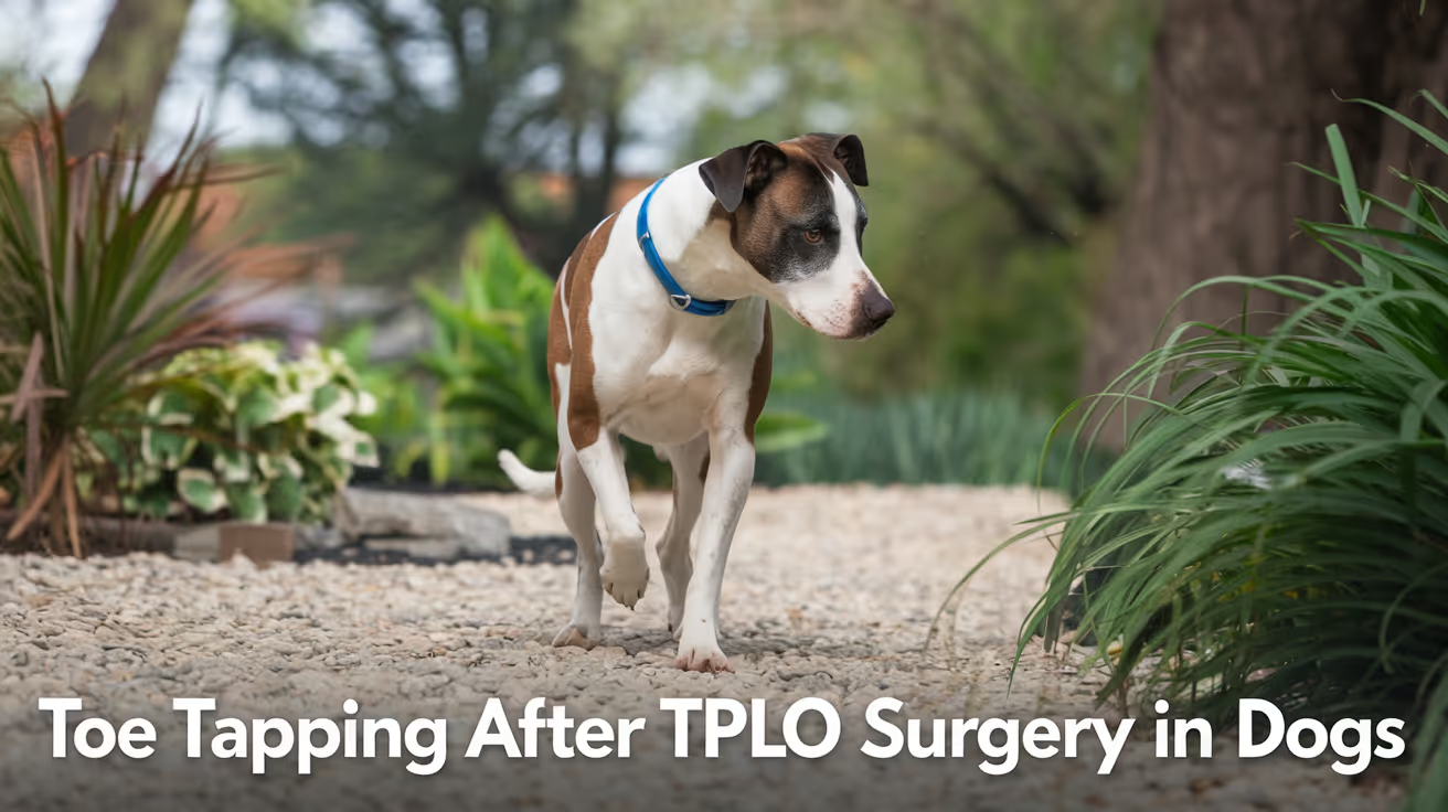
Dog Toe Tapping After TPLO Surgery Explained
Learn why your dog may tap toes after TPLO surgery, what it means, and how to support recovery with expert veterinary advice.
After your dog undergoes TPLO surgery, you might notice unusual behaviors like toe tapping. This can be worrying if you do not understand why it happens or what it means for your dog's recovery. Toe tapping after TPLO surgery is a common sign that your dog is adjusting to changes in limb use and nerve sensation.
This article explains what causes toe tapping after TPLO surgery, how to recognize normal versus concerning signs, and what steps you can take to help your dog heal properly. You will learn about the healing process, pain management, and when to contact your veterinarian for further care.
Why does my dog tap toes after TPLO surgery?
Toe tapping after TPLO surgery often results from nerve irritation or changes in limb sensation. The surgery alters the knee joint mechanics, which can temporarily affect how your dog feels and moves the leg. This tapping is usually a sign your dog is testing the limb and regaining control.
Understanding the cause helps you differentiate normal recovery behavior from signs of complications.
- Nerve sensitivity: Surgery can irritate nerves around the knee, causing your dog to tap toes as a response to altered sensations or mild discomfort.
- Muscle weakness: After TPLO, muscles supporting the leg may be weak, leading to unsteady foot placement and toe tapping during weight shifting.
- Proprioception changes: The dog's sense of limb position may be temporarily impaired, causing tapping as the brain relearns limb control.
- Habitual behavior: Some dogs develop toe tapping as a habit during the recovery phase when adjusting to new limb mechanics.
Most toe tapping is a normal part of the healing process and improves as nerve function and muscle strength return.
How long does toe tapping last after TPLO surgery?
The duration of toe tapping varies depending on the individual dog and the extent of surgery. Typically, toe tapping decreases as healing progresses over weeks to months. Monitoring your dog's behavior helps track recovery.
Knowing the expected timeline can ease your concerns and guide you when to seek veterinary advice.
- Early recovery phase: Toe tapping is most common in the first 2 to 4 weeks post-surgery when nerves and muscles are healing.
- Mid recovery phase: Between 4 to 8 weeks, tapping should reduce as strength and sensation improve with physical therapy.
- Late recovery phase: After 8 weeks, toe tapping usually disappears, indicating good nerve and muscle recovery.
- Persistent tapping: If toe tapping continues beyond 3 months, it may signal complications requiring veterinary evaluation.
Regular follow-ups with your vet ensure your dog's recovery stays on track and toe tapping resolves appropriately.
Is toe tapping after TPLO surgery painful for my dog?
Toe tapping itself is not usually painful but may indicate mild discomfort or nerve irritation. Your dog might tap toes to relieve sensations or test limb stability. Proper pain management during recovery minimizes discomfort and promotes healing.
Recognizing pain signs helps you provide better care and communicate effectively with your veterinarian.
- Mild discomfort: Nerve healing can cause tingling or mild pain, leading to toe tapping as a coping mechanism.
- Pain signs: Excessive licking, whining, or limping along with tapping may indicate pain needing treatment.
- Pain control: Use prescribed pain medications and follow your vet’s instructions to keep your dog comfortable.
- Physical therapy: Gentle exercises improve circulation and reduce nerve irritation, lessening discomfort and tapping.
Always report increased pain or unusual behaviors to your veterinarian for timely intervention.
What should I do if my dog taps toes after TPLO surgery?
If your dog taps toes after TPLO surgery, observe the behavior and support recovery with proper care. Most toe tapping is normal, but you should ensure your dog rests adequately and follows rehabilitation protocols.
Taking the right steps helps your dog regain normal limb function safely and comfortably.
- Monitor behavior: Keep a daily log of toe tapping frequency and any other signs like swelling or limping to share with your vet.
- Follow rehab plan: Adhere to prescribed physical therapy and controlled exercise to strengthen muscles and improve coordination.
- Manage pain: Administer pain medications as directed and watch for signs of discomfort requiring vet attention.
- Protect limb: Prevent jumping or running that could stress the healing knee and worsen symptoms.
Consult your veterinarian if toe tapping worsens or is accompanied by other concerning signs to ensure proper healing.
When should I worry about toe tapping after TPLO surgery?
While toe tapping is often normal, some signs indicate complications requiring veterinary care. Recognizing these signs early prevents worsening problems and supports your dog’s recovery.
Knowing when to seek help ensures your dog receives timely treatment for any issues.
- Increased tapping intensity: Sudden or worsening toe tapping may signal nerve damage or pain needing evaluation.
- Swelling or redness: Signs of infection or inflammation around the surgical site require immediate veterinary attention.
- Persistent limping: If your dog avoids putting weight on the leg for more than a few days, it may indicate complications.
- Behavior changes: Excessive licking, biting at the leg, or signs of distress suggest pain or nerve problems needing assessment.
Contact your veterinarian promptly if you notice these signs to prevent long-term damage.
How can physical therapy help with toe tapping after TPLO surgery?
Physical therapy plays a key role in reducing toe tapping by improving muscle strength, coordination, and nerve function. Tailored exercises help your dog regain normal limb use and reduce abnormal movements.
Incorporating therapy into recovery accelerates healing and enhances your dog’s quality of life.
- Strength building: Targeted exercises increase muscle tone around the knee, stabilizing the joint and reducing tapping.
- Proprioception training: Activities that improve limb position sense help your dog control foot placement better.
- Pain reduction: Gentle massage and stretching relieve nerve irritation contributing to tapping behavior.
- Improved mobility: Controlled movement prevents stiffness and encourages normal gait patterns during recovery.
Work with a veterinary rehabilitation specialist to design a safe and effective therapy plan for your dog.
Conclusion
Toe tapping after TPLO surgery is a common behavior linked to nerve healing, muscle weakness, and changes in limb sensation. It usually decreases over weeks as your dog recovers strength and coordination. Understanding this helps you support your dog’s healing process with patience and proper care.
Always monitor your dog’s behavior closely and follow your veterinarian’s advice on pain management and physical therapy. Promptly report any worsening signs to ensure your dog heals safely and returns to normal activity.
FAQs
Is toe tapping normal after TPLO surgery?
Yes, toe tapping is a normal sign of nerve and muscle recovery after TPLO surgery. It usually improves within a few weeks as your dog regains limb control.
Can toe tapping mean my dog is in pain?
Toe tapping itself is not usually painful but may indicate mild discomfort or nerve irritation. Watch for other pain signs and consult your vet if concerned.
How long should I expect toe tapping to last?
Toe tapping typically lasts 2 to 8 weeks post-surgery and should decrease as your dog heals. Persistent tapping beyond 3 months needs veterinary evaluation.
What can I do to help reduce toe tapping?
Follow your vet’s rehab plan, manage pain with medications, and provide gentle physical therapy to improve strength and nerve function.
When should I contact my veterinarian about toe tapping?
Contact your vet if toe tapping worsens, is accompanied by swelling, persistent limping, or signs of pain to rule out complications.

