

Interrupted vs Continuous Suturing in Cats
Learn the differences between interrupted and continuous suturing in cats, including benefits, risks, and best practices for healing wounds.

This article is for informational purposes only and is not a substitute for professional veterinary advice. Every case is unique, so always consult your veterinarian for guidance specific to your pet.
This content is intended for veterinary professionals for educational purposes. It does not replace clinical judgment or tailored advice. Always rely on your training, expertise, and the specific context of your patients.

Cats are not small dogs. This matters in surgery including in how their wounds are closed. Feline skin is thinner, more mobile, and more prone to self-inflicted wound disruption than canine skin.
These properties influence which suture patterns work best and which materials are best tolerated.
This guide explains the main suture patterns used in feline surgery, what research shows about outcomes, and what owners should expect from each type of closure.
Quick answer: Feline surgery most commonly uses buried continuous intradermal (BCID) closure: sutures placed beneath the skin with no external stitches. BCID has significantly lower dehiscence rates than simple interrupted sutures in cats. No suture removal is needed with absorbable intradermal sutures.
Key takeaways
- BCID closure is preferred in most feline surgeries: significantly lower dehiscence rate than simple interrupted external sutures
- ARSHI Veterinary Letters (2024): BCID had significantly fewer complications including dehiscence than simple interrupted in feline OVH
- Intradermal closure eliminates suture removal stress: absorbable sutures dissolve over 60 to 90 days
- Cats are prone to suture self-trauma: external sutures are accessible to grooming; intradermal sutures are not exposed
- Suture size matters in cats: smaller gauges (4-0 or 5-0) minimize tissue drag and inflammatory reaction in feline skin
- Monofilament sutures are preferred over braided for feline skin to reduce bacterial adherence and tissue drag
Why feline suturing differs from canine
Cats have several anatomical and behavioral characteristics that influence closure technique selection:
Thin, mobile skin: feline dermis is thinner than canine dermis. External sutures can cut through the skin edge more easily, particularly if tension is placed across the closure.
Self-grooming behavior: cats groom surgical incisions relentlessly. An E-collar reduces but does not eliminate this behavior. External sutures that protrude through the skin are at higher risk of being chewed or pulled out. Intradermal sutures that sit below the surface are inherently less accessible.
Smaller incisions: many feline procedures (neuters, spays, lumpectomies) involve relatively short incisions where intradermal placement is technically feasible for most surgeons.
Higher inflammatory response to some materials: cats can show more pronounced tissue reaction to certain suture materials than dogs, particularly braided multifilament absorbable sutures.
Suture patterns used in cats
Buried continuous intradermal (BCID)
The standard approach for feline elective surgery skin closure.
NCBi (PMC study in cats): BCID eliminates suture removal, decreases scar formation, reduces tissue inflammation and SSI risk by avoiding percutaneous suture tracts, and reduces self-induced trauma.
ARSHI Veterinary Letters (2024): "This study evaluated two suture techniques, BCID and simple interrupted (SI), in 42 female cats using nonabsorbable nylon sutures.
The results indicated a significantly higher incidence of complications, notably dehiscence, in the SI group. Consequently, BCID is recommended because of its lower complication rate and reduced risk of infection from self-trauma."
How it works: absorbable monofilament suture (poliglecaprone 25 or polydioxanone) is placed within the dermal layer in a continuous running pattern. Knots are buried within the tissue. No suture material protrudes through the skin surface. The incision appears as a clean line with no external stitches visible.
Suture material: NCBi (PMC study): "Monofilament or multifilament suture materials have been used intradermally for closure of skin incisions in dogs, cats, and humans, but there are no studies comparing absorbable and nonabsorbable monofilament suture material for a continuous intradermal closure in small animals."
The comparison of absorbable vs. nonabsorbable for intradermal closure in cats showed both are usable, but absorbable materials have the advantage of not requiring removal.
Simple interrupted
Each suture is a separate unit placed through full skin thickness and tied externally. Standard for skin closure in many species.
SustainableVet: "Simple interrupted sutures: Provide strong closure and allow precise wound edge alignment, commonly used in feline neuters."
NCBi (PMC study on intradermal closure): "Common interrupted external suture patterns in the skin include the simple interrupted and cruciate patterns.
These patterns allow more precise approximation of the skin edges along with more security than continuous buried suture patterns."
Disadvantages in cats: ARSHI (2024) found significantly higher dehiscence rates in cats compared to BCID. External sutures are accessible to grooming. They require a suture removal appointment at 10 to 14 days stressful for cats and owners.
When appropriate: wounds that cannot be closed intradermally (highly contaminated wounds, wounds under significant tension, locations where intradermal technique is not technically feasible), or where the surgeon prefers external closure for monitoring.
Cruciate sutures
A variation of interrupted sutures where each suture crosses in an X pattern. Distributes tension over a wider area; reduces skin edge inversion. Used in higher-tension skin closures in cats.
Ford interlocking (locking continuous)
A continuous pattern where each bite is locked. Used primarily for body wall and fascia closure rather than skin. Provides the speed advantage of a continuous pattern with improved security.
Suture material selection in cats
Gauge
SustainableVet: "Suture size selection: Smaller sizes (4-0 or 5-0) are preferred for delicate feline skin to minimize trauma and promote fine healing."
Smaller gauge sutures cause less tissue drag on passage through feline dermis and produce smaller suture tracts if external sutures are used.
Monofilament vs. braided
SustainableVet: "Monofilament sutures: These have smooth surfaces that reduce tissue drag and lower infection risk compared to braided sutures."
Braided multifilament sutures have a higher surface area for bacterial colonization and more tissue drag. In thin feline skin, monofilament materials are strongly preferred for both skin and subcutaneous layers.
Absorbable materials for internal layers
SustainableVet: "Absorbable sutures: Materials like polydioxanone (PDS) or poliglecaprone (Monocryl) dissolve over time, ideal for internal tissue layers to avoid suture removal stress."
Typical feline surgery layered closure:
- Body wall/linea alba (if abdominal): absorbable, monofilament, continuous
- Subcutaneous layer: absorbable, monofilament, continuous (closes dead space)
- Skin: intradermal, absorbable monofilament 4-0 or 5-0 (no external sutures)
What owners should know
No external sutures visible: with intradermal closure, the incision should appear as a smooth line. This is correct and not a sign that closure was incomplete.
E-collar is still required: even without external sutures, cats can traumatize a healing wound through licking. The E-collar protects the intradermal closure from grooming during the initial healing phase.
No suture removal appointment: absorbable intradermal sutures dissolve without requiring a procedure. If nonabsorbable intradermal sutures were used, your vet will advise on removal.
What to monitor: a small amount of bruising or mild swelling in the first 2 to 3 days is normal. Increasing redness after day 3, any discharge, or opening of the incision line requires veterinary assessment.
For suturing in dogs for comparison, see interrupted vs. continuous suturing in dogs. For tissue adhesives as an alternative, see tissue adhesives in dog and cat surgery.
Frequently asked questions
Why does my cat have no visible stitches after surgery?
Intradermal closure places sutures beneath the skin surface. The incision is closed; the sutures are simply not visible from outside. This is intentional and provides better outcomes in cats than external sutures.
Does my cat still need an E-collar if there are no external stitches?
Yes. Cats can traumatize a healing wound through licking even without external sutures to grab. The E-collar is required throughout the initial healing phase regardless of closure technique.
When will the stitches dissolve?
Absorbable intradermal sutures typically dissolve over 60 to 90 days depending on the material. The dissolution is internal you will not see suture material emerge or fall out.
My cat's incision opened slightly at one end. What should I do?
Contact the vet same day. Minor dehiscence at a closure end is the most common feline complication. Small gaps may close conservatively; larger openings may need reclosure.
Can my cat's incision be closed with tissue glue instead of sutures?
Tissue glue suits very short, low-tension wounds in cats but not longer incisions like spays. Ask your vet whether glue or sutures are more appropriate for the specific wound.
Can staples be used to close cat skin instead of sutures?
Staples are occasionally used in cats but require removal at 10 to 14 days and cause more tissue reaction than fine monofilament sutures. For most elective feline surgery, intradermal sutures are preferred.
Resources
- ARSHI Veterinary Letters. Comparison of Buried Continuous Intradermal and Simple Interrupted Suture Patterns for Skin Closure in Feline Ovariohysterectomy. journal.ipb.ac.id
- NCBi PMC. Comparison of Absorbable and Nonabsorbable Sutures for Intradermal Skin Closure in Cats. ncbi.nlm.nih.gov
- ScienceDirect. Comparison of Continuous Intradermal with Simple Interrupted Suture Pattern in Dogs. sciencedirect.com
- Academia. Comparison of Absorbable and Nonabsorbable Sutures for Intradermal Skin Closure in Cats. academia.edu

Get a Free Poster
Enhance your workspace with a high-quality radiographs reference poster, designed for veterinary professionals. This free physical poster will be shipped directly to you—just fill out the form to request your copy.






Taking Great TPLO Radiographs
Click Below to Watch Live Video Demos


We'll send you a Free Wall Poster with all the steps
Step #1
Getting Ready
Ensuring a clean surgical field starts with proper skin preparation. This video demonstrates the best practices for:
- Shaving the patient – Achieving a close, even shave while minimizing skin irritation
- The Dirty Scrub – The initial skin prep step to remove surface debris and reduce bacterial load before the sterile scrub.
Following these techniques helps reduce infection risk and improve surgical outcomes. Watch the video to see how it’s done effectively!


Step #2
Reduce Your Risks
Many surgeons are shocked to find out that their patients are not protected from biofilms and resistant bacteria when they use saline and post-op antibiotics.
That’s Where Simini Comes In.
Why leave these risks and unmanaged? Just apply Simini Protect Lavage for one minute. Biofilms and resistant bacteria can be removed, and you can reduce two significant sources of infection.
Step #3
Take the Course
Preventing surgical infections is critical for patient safety and successful outcomes. This course covers:
- Aseptic techniques – Best practices to maintain a sterile field.
- Skin prep & draping – Proper methods to minimize contamination.
- Antibiotic stewardship – When and how to use perioperative antibiotics effectively.
Stay up to date with the latest evidence-based protocols. Click the link to start learning and earn CE credits!

Things to know

Cruciate Sutures in Dog and Cat Skin Closure
Learn how cruciate sutures help close dog and cat skin wounds effectively with step-by-step guidance and care tips.
The cruciate suture pattern is simply a simple interrupted suture that crosses back over itself in an X before the knot is tied. That additional crossing pass is what makes it behave differently under tension.
It is one of the more commonly used skin closure patterns in small animal surgery and one of the least explained to owners.
Quick answer: A cruciate suture is an X-shaped interrupted suture pattern where the needle makes two crossing passes across the wound before tying. Published research (JAVMA, 2016, 60 skin specimens from 30 Beagles) found cruciate sutures placed significantly faster than simple interrupted sutures while achieving equivalent tensile strength at failure. Cruciate patterns are preferred when tissues are fragile, sutures are pulling out, or faster closure time is a priority. Both cruciate and simple interrupted patterns significantly outperform intradermal sutures in tensile strength.
Key takeaways
- Cruciate sutures form an X-shape, passing the needle twice across the wound before tying.
- Placement is significantly faster than simple interrupted sutures, per published JAVMA research.
- Tensile strength is equivalent to simple interrupted sutures at failure.
- Preferred over simple interrupted when tissue is fragile and sutures are pulling out with each bite.
- Both patterns outperform intradermal sutures in tensile strength under tension loading.
- Used at 4 to 6 mm spacing, same as simple interrupted, placed 4 to 6 mm from the wound edge.
What a cruciate suture is
A cruciate suture is an interrupted suture pattern. Like simple interrupted, it is placed individually and tied independently failure of one suture does not open the entire wound.
The technique difference from simple interrupted:
- The needle enters the skin 4 to 6 mm from the wound edge on side A
- Crosses the wound and exits 4 to 6 mm from the edge on side B
- Instead of tying here, the needle re-enters side B, crossing back over the wound
- Exits on side A again, offset from the first entry
- The two ends are tied producing an X-shaped crossing over the wound surface
The crossing configuration is what distributes tension differently from simple interrupted. Each arm of the X engages the tissue on a slightly different vector.
Published evidence: JAVMA 2016 study
The most directly relevant published data comes from a JAVMA 2016 study by Kieves et al. (Iowa State University), comparing four closure patterns on 60 skin specimens from 30 Beagles:
Patterns compared: simple interrupted, cruciate, intradermal, subdermal
Key findings:
Placement time: "Mean suture placement time for the cruciate pattern was significantly less than that for other patterns." Cruciate was faster than all three other patterns.
Tensile strength: "Mean tensile strength at skin-edge separation and suture-line failure for the simple interrupted and cruciate patterns were significantly higher than those for the intradermal and subdermal patterns."
Simple interrupted vs. cruciate: no significant difference in tensile strength at failure. The two patterns are mechanically equivalent.
Mode of failure: simple interrupted failed by suture breakage; cruciate, intradermal, and subdermal failed by tissue failure (the suture held; the tissue gave out).
Wound apposition: no significant difference among all four patterns.
Clinical implication: the cruciate pattern closes wounds faster than simple interrupted while providing equivalent mechanical strength. In fragile tissue where the suture-to-tissue interface is the weak point, cruciate distributes that interface differently which is why Veterinary Surgery Online specifically recommends it when tissue is fragile: "If tissues are fragile and suture pulls out easily, use cruciate or horizontal mattress pattern instead of simple interrupted for less stress on each bite."
When cruciate sutures are preferred
Fragile or friable tissue: when the tissue is thin, inflamed, or poor quality, simple interrupted sutures cut through at the entry point. The cruciate distributes the bite force differently, reducing focal stress.
Speed priority: in high-volume settings or time-sensitive cases (very young patients, high anesthetic risk), the faster placement time of cruciate sutures has a practical advantage.
Moderate tension with no mattress pattern: for wounds with moderate tension that do not require the full tension-relieving capacity of a horizontal or vertical mattress, the cruciate provides additional holding strength over simple interrupted without the complexity of a mattress pattern.
Where tissue is moving: joints, limb skin, and areas subject to post-operative movement. The X configuration provides slightly more surface contact with the tissue.
For how cruciate sutures compare to appositional patterns in high-tension contexts, see appositional vs everting closure patterns.
When simple interrupted is preferred over cruciate
Irregular or curved wounds: simple interrupted sutures give more individual control over tension at each point. In irregular wounds, cruciate sutures can be harder to place accurately.
Maximum monitoring need: since each cruciate suture involves more tissue and two crossing passes, failure of one is more visually apparent and potentially more complex than a single interrupted suture. In very high-infection-risk wounds, simple interrupted allows each stitch site to be individually assessed.
When exact edge apposition is critical: the offset crossing passes of a cruciate suture make precise edge alignment slightly more demanding than simple interrupted.
For how cruciate sutures fit within the full skin closure method comparison, see cruciate and other skin closure methods compared.
Comparing key skin closure patterns
Data from JAVMA 2016 (Kieves et al.) and Veterinary Surgery Online.
For the full tension-relieving pattern comparison including how cruciate compares to mattress sutures, see tension-relieving patterns alongside cruciate sutures.
Suture material for cruciate patterns
The same materials used for simple interrupted skin closure are used for cruciate:
- Nylon (Ethilon): most common external skin suture; low tissue reaction; requires removal at 10 to 14 days
- Prolene (polypropylene): similar to nylon; even lower tissue reaction; good for high-movement areas
- Monocryl 4-0: if absorbable cruciate closure is desired to avoid a removal visit
Size: 3-0 for medium dogs; 2-0 for large dogs; 4-0 for cats and small dogs. The same sizing guidance as for simple interrupted applies.
Removal
Cruciate sutures are removed the same way as simple interrupted sutures: scissors cut under one arm of the X, and the suture is pulled through in one motion. The crossing pass does not complicate removal. Timing: 10 to 14 days, per standard skin closure guidance.
For suture removal timing guidance in dogs, see suture removal timing for cruciate sutures in dogs. For cats, see suture removal timing for cruciate sutures in cats.
Frequently asked questions
My dog has X-shaped stitches. Is that a cruciate pattern?
Yes. The visible X crossing the wound surface is the distinguishing feature of a cruciate suture pattern. Each X is tied individually.
Are cruciate sutures stronger than regular stitches?
Published JAVMA data shows no significant difference in tensile strength between cruciate and simple interrupted at failure. Cruciate sutures place faster and distribute their bite force differently which matters in fragile tissue but they are not inherently stronger than simple interrupted.
Can cruciate sutures be used anywhere on the body?
Yes, in most locations. They are particularly useful on the trunk, dorsum, and limb skin in dogs. In cats, they are used in similar situations, though fine-gauge sutures (4-0) are important given feline skin fragility. Intradermal closure is often preferred in cats for cosmetic procedures, but cruciate is appropriate where external sutures are indicated.
The cruciate suture earns its place in small animal surgery primarily through speed placing faster than every other pattern in the JAVMA study while achieving identical tensile strength to simple interrupted. When tissue is fragile enough that simple interrupted sutures are pulling out with each bite, the different bite geometry of cruciate sutures addresses that clinical problem directly.
Resources
- JAVMA 2016 (Kieves et al.). Comparison of Tensile Strength Among Simple Interrupted, Cruciate, Intradermal, and Subdermal Suture Patterns for Incision Closure in Ex Vivo Canine Skin Specimens. pubmed.ncbi.nlm.nih.gov
- Veterinary Surgery Online. Wound Closure Continued. vetsurgeryonline.com
- University of Minnesota Large Animal Surgery. Suturing Skin. open.lib.umn.edu
Closure Protocol for Neuter Surgery in Dogs
Learn the detailed closure protocol for neuter surgery in dogs to ensure safe healing and prevent complications.
Dog neuter surgery is one of the most common veterinary procedures in the US. The surgical technique itself takes minutes once you know what you are doing. But the closure decisions which approach, which layers, which materials matter significantly for recovery.
Understanding what your vet did and why helps you care for your dog appropriately during healing and recognize when something needs attention.
Quick answer: Dog neuter closure depends on the incision approach. The prescrotal approach uses a three-layer closure: subcutaneous tissue, subcutaneous fat, and skin. The scrotal approach typically does not require full closure the scrotal skin is inverted or left partially open, and only a single dartos fascia suture may be placed. Absorbable sutures are standard for internal layers; skin may use absorbable or non-absorbable depending on surgeon preference.
Key takeaways
- Two main approaches exist: prescrotal (standard) and scrotal, each with different closure requirements.
- Prescrotal approach requires three-layer closure: tunica/subcutaneous layers and skin.
- Scrotal approach does not require full suturing the scrotum is inverted or left open.
- Absorbable monofilament sutures (Monocryl or PDS) are standard for internal layers.
- Intradermal closure is commonly used for skin to reduce self-trauma and eliminate removal visits.
- Scrotal hematoma is the most common complication and is often related to technique or over-activity post-op.
Two approaches, two different closure decisions
Prescrotal approach (traditional)
The incision is made in the skin just in front of the scrotum (the prescrotal area). The testicles are pushed forward through this incision one at a time.
Why prescrotal:
- Standard in most private practice settings
- Allows a clean, anatomically clear incision that is away from the scrotal skin
- Required for cryptorchid dogs where retained testicles need to be located
Prescrotal closure three layers:
- Tunica vaginalis or subcutaneous fascia (where opened): closed with absorbable suture
- Subcutaneous fat: closed with 2-0 or 3-0 absorbable suture (Monocryl or Vicryl) in a continuous or interrupted pattern
- Skin: closed with intradermal absorbable suture, interrupted nylon, or skin staples
Today's Veterinary Practice notes: "Prescrotal incisions for dogs should always be closed by appropriate tissue apposition with buried absorbable suture."
The University of Edinburgh (CNR Good Practice Guide) confirms: "With the pre-scrotal approach, a standard three-layer closure should be performed."
Scrotal approach (increasingly adopted)
The incision is made directly on the scrotum over each testicle. Two small incisions are made one per testicle rather than one prescrotal incision.
Why scrotal:
- Shorter surgical time
- Less suture material needed
- Reduced scrotal hematoma risk
- No risk of accidental urethral injury
- Lower complication rate reported vs. prescrotal in published studies
Scrotal closure minimal:
Clinician's Brief (2016) states explicitly: "Do not attempt to fully close the dartos fascia, intradermal, and/or cutaneous layers of the incision with sutures, as this can lead to discomfort, seroma formation, self-trauma, and postoperative complications."
For the scrotal approach, the scrotum is inverted and left to heal by second intention. A single interrupted suture may be placed in the dartos fascia to reduce the size of the opening. Small amounts of drainage from the scrotal wound in the first 24 to 48 hours are normal.
For comparison with the neuter closure protocol in cats, see neuter closure in cats for comparison.
Suture materials for prescrotal closure
A published randomized controlled trial (PMC11047773) comparing smooth monofilament suture to barbed suture in canine spay/neuter three-layer continuous closure found barbed suture reduced average closure time from 6.5 minutes to 4.9 minutes while maintaining equivalent wound outcomes. Barbed sutures are increasingly used in high-volume spay/neuter clinics for this reason.
For the general closure protocol context that this procedure follows, see general surgical closure protocol.
Closed vs open castration technique
Within either incision approach, the surgeon also chooses between closed and open castration technique:
Closed castration: the parietal tunica vaginalis is left intact. The fascial layers are stripped from the spermatic cord to exteriorize the testicle without opening the tunic. The cord is ligated and transected. Less risk of scrotal hematoma because the tunic containing the cord is never opened.
Open castration: the parietal tunica is incised to expose the vas deferens and vessels separately. Preferred for larger dogs where a single mass ligature might be inadequate. Requires closure of the tunica if opened.
DVM360 documented closed castration using 2-0 Vicryl for the spermatic cord ligature. Both techniques are acceptable; most surgeons use closed castration as their default.
Layered closure principles applied here
Neuter surgery closure follows the same principles as all layered closure in small animal surgery. For the underlying framework, see layered technique applied in neuter closure.
For suture removal timing after neuter surgery, see suture removal after neuter surgery. For a closure checklist applicable to this procedure, see checklist for neuter closure.
Post-operative care for owners
What is normal after neuter closure:
- Mild swelling in the scrotal area for 2 to 5 days
- Slight redness at the skin edge for 3 to 5 days
- Small amount of bloody drainage from the scrotal approach wound in the first 24 hours
- Some licking attempts at the incision area
What needs a vet call:
- Scrotal swelling growing larger after the first 48 hours (possible hematoma)
- Yellow or green discharge
- Wound opening
- Dog unable to urinate or straining
E-collar is non-negotiable. Self-trauma to a neuter incision can cause wound dehiscence within hours. Even dogs that have never licked wounds before will target a fresh incision. Keep it on until your vet clears removal.
Activity restriction:
- Leash walks only for 10 to 14 days
- No running, jumping, or rough play
- No access to water (swimming, puddles) until the wound is healed
Frequently asked questions
My dog had the scrotal approach and the wound looks open. Is that normal?
Yes, if your vet used the scrotal approach. The small openings are intentionally left to heal on their own. Some discharge and minor swelling are expected. Monitor daily and contact your vet if swelling is increasing after day 3, if the discharge becomes yellow or foul-smelling, or if the dog is in obvious discomfort.
When can my dog go back to full activity after neuter surgery?
Ten to 14 days of restriction is standard. After the prescrotal approach, full internal healing takes longer than external skin healing. Early activity is the most common cause of scrotal hematoma and incision complications after dog neuter surgery.
Can my dog get wet after neuter surgery?
No, until the wound is fully healed and your vet clears it. Water softens the suture line, promotes bacterial growth, and can cause wound breakdown. This includes puddles on walks keep your dog away from wet surfaces during the restriction period.
Dog neuter closure looks simple because the surgery is routine. The technique behind it the approach selection, the tissue layers closed, the suture material matched to each layer reflects the same precision applied to any surgical wound. When the closure is done correctly and the dog rests properly, neuter surgery heals quickly and without complication.
Resources
- Today's Veterinary Practice. Update on Orchiectomy Techniques for Dogs and Cats. todaysveterinarypractice.com
- Clinician's Brief. Scrotal Approach to Canine Orchiectomy: Step-by-Step Guide. cliniciansbrief.com
- DVM360. Scrotal Castration versus Prescrotal Castration in Dogs. dvm360.com
- PMC. Randomized trial comparing barbed suture vs smooth monofilament in canine closure. 2024. ncbi.nlm.nih.gov
Closing Fascial Layers in Veterinary Surgery
Learn the best practices for closing fascial layers in veterinary surgery to ensure strong healing and reduce complications.
Fascia is the connective tissue that wraps, separates, and supports muscles and organs. It does not get much attention as a tissue type, but it is the primary holding layer in most veterinary wound closures.
When the linea alba is sutured after abdominal surgery, what is actually being closed is fascia. When a joint capsule is repaired after orthopedic surgery, fascia is doing the structural work. Getting fascial closure wrong leads to hernia, dehiscence, or implant failure complications that require return surgery.
Quick answer: Fascial closure uses absorbable monofilament sutures (PDS or Vicryl, size 0 to 3-0 depending on patient size) placed in a simple continuous or interrupted pattern that incorporates adequate tissue bites (4 to 10 mm). The key principle is that sutures must engage the fascia itself, not just adjacent muscle or fat. Fascia is the holding layer it has the tensile strength muscle belly and fat tissue lack.
Key takeaways
- Fascia is the primary tensile-holding layer in most abdominal and orthopedic wound closures.
- Sutures must engage the fascia itself, not surrounding muscle belly or fat tissue.
- PDS is the preferred material for fascial closure because of its long strength retention profile.
- Simple continuous pattern is the standard for most fascial closures in small animals.
- Interrupted pattern is used when contamination or poor tissue quality raises continuous failure risk.
- Incisional hernia is the most serious complication of inadequate fascial closure.
What fascia is and why it matters
Fascia is a dense, fibrous connective tissue made primarily of collagen. Unlike muscle, fat, or loose connective tissue, fascia resists tension. When sutures are placed in it, they hold.
In veterinary surgery, the most commonly encountered fascial structures requiring closure:
Understanding which fascial structure is being closed helps explain why suture selection and bite size are procedure-specific.
The critical rule: sutures must engage fascia
Veterian Key (Surgery of the Abdominal Cavity) is explicit: "Because the holding layer of abdominal incisions consists of fascia rather than muscle, dehiscence is common if the rectus fascia is not incorporated in sutures."
This is the most common technical error in fascial closure: placing sutures in the muscle belly adjacent to the fascia rather than through the fascia itself. The consequence is predictable the sutures pull through the non-holding tissue and the wound fails.
Technical guidance:
- Incorporate 4 to 10 mm of fascia in each bite
- Ensure the needle actually passes through the white fibrous tissue, not the adjacent muscle
- In the linea alba, the European Hernia Society recommends a 5 mm suture bite-to-stitch interval as optimal for small animals (supported by published feline cadaver research)
For how fascial closure fits within muscle layer closure, see muscle layers closed alongside fascia.
Suture materials for fascial closure
Why PDS is preferred:
Fascial healing is slow. The linea alba regains meaningful tensile strength over 4 to 6 weeks. PDS maintains strength through that entire period before losing tensile capacity. Vicryl's faster absorption may leave the closure unsupported before full healing occurs in larger patients.
For how suture material selection affects fascial closure outcomes, see suture selection for fascial closure.
Suture patterns for fascial closure
Simple continuous (standard)
The preferred pattern for most fascial closures in small animals. Published data on canine linea alba closure (Rosin and Crowe, 530 coeliotomies) found a complication rate of just 0.19% with simple continuous technique.
Properties:
- Distributes tension evenly along the entire closure length
- Faster than interrupted
- Relies on two secure end knots for integrity
Simple interrupted
Used when the risk of continuous pattern failure is elevated:
- Contaminated or infected wounds
- Poor tissue quality from chronic steroid use, malnutrition, or disease
- Cases where post-operative activity compliance is uncertain (some surgeons use 2-0 suture with interrupted pattern in these cases, per WCVM guidance)
Mattress patterns (tension-relieving)
Horizontal or vertical mattress sutures distribute tension over a wider tissue area and are used when standard bite size is insufficient to achieve tension-free apposition.
For a full discussion of tension-relieving applications, see tension relief during fascial closure.
Fascial closure in specific contexts
Abdominal surgery
The linea alba is the only fascial structure requiring closure for a standard ventral midline celiotomy. The muscle bellies of the rectus abdominis are not closed only the fibrous sheath (the fascia) is incorporated.
Bite size: 4 to 10 mm of fascia per bite. Bites placed every 4 to 8 mm apart. Suture should be taut but not strangulating.
Orthopedic surgery
Joint capsule closure is the functional equivalent of fascial closure for orthopedic procedures. The joint capsule is dense fibrous tissue that must be closed securely to restore joint stability and prevent fluid leakage.
PDS or Vicryl in interrupted or continuous patterns are used. The suture must engage the capsule tissue, not the surrounding soft tissue.
High-tension wounds
When the fascial edges are difficult to appose without excessive tension, a far-near-near-far (tension-relieving) pattern or pre-placed sutures may be used to bring edges into contact before final closure.
For how high-tension closures are managed at the fascial level, see high-tension closure techniques for fascia.
Complications of inadequate fascial closure
Incisional hernia: the most serious consequence. Abdominal contents push through the fascial gap. Visible as a soft, reducible bulge at the incision line. Requires surgical repair.
Wound dehiscence: fascial failure at depth causes the wound to separate, potentially opening the abdominal cavity to contamination.
Joint instability: inadequate joint capsule closure allows abnormal joint movement, effusion, and impaired healing after orthopedic procedures.
Seroma at fascial level: when fascial edges do not appose, fluid accumulates in the potential space between them, even if subcutaneous closure above has been performed.
For how fascial closure integrates into the full layered technique, see fascial layer within layered closure.
Frequently asked questions
Can my dog develop a hernia even though the skin closure looks fine?
Yes. The skin heals independently of the fascial layer. A dog can have a healed skin incision with a fascial hernia forming beneath it. New soft swelling at an incision site weeks after skin suture removal should be evaluated by your vet, as it may represent an early incisional hernia.
What does "incorporating fascia" mean in practical terms for a surgeon?
The surgeon can see the difference between white fibrous fascial tissue and the adjacent reddish muscle belly or yellow fat. The needle must visibly pass through the white tissue on both sides of the incision. When it does, the suture has the holding power it needs. When it does not, the suture will eventually fail under tension.
Is fascial closure more important in large dogs than small ones?
It is equally important in all patients, but the consequences of failure are proportionally more significant in large dogs. A large dog's body weight applies more tension to the fascial repair. Large breeds are also more active during recovery, further stressing the closure. This is why suture size scales up with patient size.
Fascial closure is where wound integrity is actually built. The skin may heal on its own given enough time, but the fascial layer does not. When fascia is properly engaged with the right material and an adequate tissue bite, the wound has a structural foundation that holds through the entire healing period. When it is not, the foundation is absent and the consequences appear weeks later.
Resources
- Veterian Key. Surgery of the Abdominal Cavity. veteriankey.com
- WCVM University of Saskatchewan. Lab 6 Part 4: Incision Closure. wcvm.usask.ca
- Veterinary Evidence. Choice of Suture Pattern for Linea Alba Closure. veterinaryevidence.org
- Veterian Key. Suturing Techniques and Common Surgical Procedures. veteriankey.com
Closure Protocol for Tumor Excision in Cats
Learn the detailed closure protocol for tumor excision in cats, including surgical steps, suture choices, and post-op care for optimal healing.
Feline tumor excision creates the same fundamental closure challenge as in dogs the defect left by the tumor must be closed without compromising the margins that determined the cure rate. But the closure is performed on thinner, less elastic feline skin with fewer reconstruction options than in large-breed dogs.
Understanding how closure decisions are made helps owners interpret what the vet describes after surgery and what to expect during recovery.
Quick answer: Feline tumor excision closure follows the same layered sequence as dogs (deep margin if fascia was included, subcutaneous, skin) but with feline-specific modifications: 4-0 sutures throughout, intradermal Monocryl preferred for skin (feline skin tolerates external sutures poorly), and skin flaps used when primary tension-free closure is not achievable. Injection-site sarcomas in cats require particularly wide margins (often involving en bloc muscle removal) and frequently require reconstructive closure.
Key takeaways
- Feline skin is thinner and less elastic than canine skin, limiting primary closure options after wide-margin excision.
- 4-0 sutures throughout (vs. 3-0 in most medium dogs) feline tissue requires finer suture material.
- Intradermal Monocryl 4-0 is the preferred feline skin closure after tumor excision.
- Injection-site sarcomas (FISS) require the widest margins in feline oncological surgery and most commonly require reconstructive closure.
- Drain placement is particularly important in cats after wide excision, as dead space management is harder with thin subcutaneous tissue.
- Histopathology result timing (typically 5 to 10 business days) means re-excision discussion happens while the primary wound is still healing.
Feline tumor types and their closure implications
Injection-site sarcomas deserve specific mention. WSAVA/VIN (Soft Tissue Sarcoma 2016): "The aim of curative-intent surgery is to widely excise the primary tumor (3 cm wide and 3 cm or a fascial plane deep) and achieve negative histopathological margins."
For FISS, achieving these margins at the interscapular region frequently involves removing the trapezius muscle, spinous processes, or portions of scapula leaving a deep muscular defect that requires multi-layer closure and often a flap for the skin component.
Deep layer closure: when fascia or muscle is included
When the deep margin of excision includes the fascia below the tumor (standard for any tumor with invasion concern), the fascial defect is closed before subcutaneous work.
Pattern: simple interrupted or continuous absorbableMaterial: PDS 2-0 to 3-0
For deep muscular defects after FISS excision: the deep layers are closed in sequence (deep muscle to superficial muscle), using PDS 0 to 2-0. If the defect is too large for primary muscle closure, a mesh or mobilized flap may be used.
Subcutaneous closure and dead space
Feline subcutaneous tissue is thin and provides limited substance for suture purchase compared to dogs. Dead space management is therefore particularly challenging after wide-margin excision.
Options:
- Simple continuous absorbable (Monocryl or Vicryl 3-0 to 4-0) for the subcutaneous layer
- Walking sutures to anchor dermis to fascia when a large dead space pocket exists
- Drain placement (Penrose or closed suction) when suturing alone cannot eliminate the cavity
VCAhospitals (Penrose drain discharge instructions): "A Penrose drain is a latex tube placed into a wound with one or two ends exiting the skin, allowing fluids to drain. In most cases, the drain will exit from a new incision site, not the primary wound site."
For how dead space management relates to the broader closure strategy, see dead space management after cat tumor excision.
Skin closure
Intradermal Monocryl 4-0: the preferred method
Feline skin tolerates external sutures more poorly than canine skin. Suture-track irritation, self-trauma, and suture-mark scarring are more pronounced and more rapidly established in cats.
For routine feline tumor excision closure where tension is manageable, intradermal Monocryl 4-0 is preferred:
- No external material for the cat to lick or chew
- No removal visit required
- Finer, less visible healed scar
When primary closure is under tension
When the wound edges cannot be approximated without tension, the options match those in dogs:
Undermining: releasing skin from underlying subcutaneous tissue and fascia to mobilize it toward the defect. In cats, undermining must be carefully limited thin feline skin can be devascularized more easily than dog skin if undermining is too extensive.
Walking sutures: dermis-to-fascia advancement. Useful for moderate-sized trunk defects. Standard material: 3-0 PDS or Biosyn.
Tension-relieving sutures: horizontal mattress or vertical mattress, used with 3-0 nylon or Prolene.
Skin flap: when primary closure is not achievable. ACVS: "Closure of the defect from the excised mast cell with a small skin flap from adjacent skin near the base of the ear." Skin flaps are more commonly needed in cats than equivalent-weight dogs because feline skin has less laxity.
High-tension locations: face, ears, distal limbs
These locations have minimal adjacent skin available for advancement and carry the highest tension risk. Closure at these sites frequently requires staged reconstruction or accepting second intention healing over small residual defects.
Cosmetic closure after feline tumor excision
For tumor excision in cosmetically sensitive locations (face, visible lateral body), intradermal closure and fine suture sizes minimize visible scarring. For the cosmetic closure approach in cats, see cosmetic closure for feline tumor sites.
Histopathology and re-excision
The excised tissue is sent for histopathology, with results typically returned within 5 to 10 business days. Three possible results:
- Complete margins: no tumor cells at the inked edges. Local recurrence risk is low. No further surgery needed unless the tumor biology warrants adjuvant therapy.
- Close margins: tumor cells within 1 to 2 mm of the edge but not at the ink. Recurrence risk is elevated. Discussion of re-excision or radiation.
- Incomplete margins: tumor cells at the inked edge. Re-excision is recommended when possible.
The re-excision decision is made while the primary wound is in the active healing phase (typically at 7 to 14 days post-surgery). The entire scar track is included in the re-excision specimen meaning closure of the second wound is more complex than the first.
For how tumor excision closure compares in dogs, see tumor excision closure comparison in dogs.
Post-operative monitoring in cats
Cats hide signs of pain and discomfort more effectively than dogs. Behavioral changes (reduced appetite, hiding, abnormal posture) are often the first indicators of complications rather than obvious wound signs.
Specific monitoring for cats after tumor excision:
- Check the wound twice daily for redness, discharge, swelling, or separation
- Cats often groom the wound site even with an E-collar if the collar fits poorly verify fit at each check
- If a drain is present, monitor drainage output and color daily
- Watch for systemic signs (reduced appetite, lethargy, fever) that may precede local wound signs
For the closure checklist applicable to feline tumor excision, see closure checklist for feline tumor excision.
Frequently asked questions
What makes injection-site sarcomas so different to close?
FISS requires the widest margins in feline oncological surgery typically 3 to 5 cm laterally and removing one to two fascial planes deep, sometimes including muscle and portions of bone. The resulting defect is far larger relative to the cat's body than equivalent surgery in a dog. Closure almost always requires reconstruction, and staged surgery (debulking followed by definitive reconstruction) may be necessary.
My cat had a mass removed and the vet said they got "clean margins." Does that mean the cancer is gone?
Clean histopathological margins mean no tumor cells were identified at the edges of the tissue submitted for analysis. For many feline tumors, clean margins significantly reduce local recurrence risk. However, "clean margins" does not address the risk of metastatic spread, which depends on tumor type, grade, and whether staging (chest radiographs, lymph node assessment) was performed. Discuss the complete picture with your vet.
How long will my cat's E-collar need to stay on after tumor surgery?
Minimum until suture removal (10 to 14 days for external sutures). With intradermal closure, the E-collar remains recommended for 10 to 14 days post-surgery not for suture removal purposes, but because the incision site can still be disrupted by licking before it has adequate surface healing. Your vet will advise the specific duration based on wound appearance at the recheck.
Feline tumor excision closure is constrained by the cat's limited skin laxity, the finer tissue that tolerates suture placement, and the requirement not to compromise the margins that determine whether the tumor is fully removed. Meeting all three constraints simultaneously adequate margins, tension-free closure, appropriate suture technique is the challenge that distinguishes feline tumor surgery from routine wound closure.
Resources
- ACVS. Mast Cell Tumors. acvs.org
- VIN (WSAVA 2016). Soft Tissue Sarcoma in Dogs and Cats. vin.com
- VCA Animal Hospitals. Penrose Drain Discharge Instructions for Dogs. vcahospitals.com
- Today's Veterinary Practice. Fundamentals of Surgical Oncology in Small Animals. todaysveterinarypractice.com
Closing Bite Wounds in Dogs: Expert Guide
Learn how to safely close bite wounds in dogs with expert tips on treatment, healing, and prevention of infection.
Bite wounds look smaller than they are. The surface puncture from a canine tooth may be half an inch wide, but the tissue damage underneath can extend several inches in every direction.
That's what makes bite wounds one of the more complex wound types in veterinary surgery. Whether and how to close them depends on contamination level, location, time since injury, and how much tissue was crushed.
Quick answer: All dog bite wounds are considered contaminated and require veterinary evaluation. Fresh, minimally contaminated wounds may be closed primarily after thorough lavage and debridement. Heavily contaminated, infected, or complex wounds are typically left open or treated with delayed closure. Antibiotics are routinely used. Most dogs require sedation or anesthesia for proper wound assessment and treatment.
Key takeaways
- All bite wounds are contaminated regardless of how small they appear on the surface.
- Immediate primary closure is appropriate only for clean, fresh wounds after debridement.
- Delayed primary closure is used for contaminated or infected wounds once the tissue stabilizes.
- Drains are often placed in closed bite wounds to prevent seroma and abscess formation.
- Antibiotics are typically prescribed to control or prevent infection in all bite wound cases.
- Surface size is misleading: puncture wounds can hide serious underlying damage to muscle, fascia, or organs.
Why bite wounds are different from other lacerations
A dog bite delivers two forces simultaneously: penetration and crushing. The teeth pierce the skin while the jaw compresses surrounding tissue. This creates tissue damage that extends far beyond the visible wound margins.
VCA Animal Hospitals explains: "Wounds that appear to be minor on the surface can be deceptive and may be life-threatening, depending on the location of the injury."
The American Animal Hospital Association (AAHA) emphasizes: "A clean fresh wound can be closed primarily, after appropriate lavage, debridement, and assessment of the potential need for a drain."
The key word is clean. Most bite wounds are not clean when they arrive.
When vets choose immediate (primary) closure
Primary closure means suturing the wound shut at the initial visit. This is appropriate when:
- The wound is less than 6 to 8 hours old
- The wound has been thoroughly lavaged and debrided
- There is minimal contamination (no gross debris, necrotic tissue, or obvious infection)
- The wound is in a cosmetically significant area (face) where open healing causes more problems than closure risk
Even primary closures in bite wounds are typically performed with drains placed to allow fluid escape. This reduces the risk of seroma and abscess formation.
For how drains interact with closure, see drains used in bite wound closure.
When vets delay closure
Delayed primary closure (waiting 3 to 5 days before suturing) is used when:
- The wound is heavily contaminated with debris, saliva, or infected material
- Tissue viability is uncertain after the initial crushing injury
- Infection is already established at presentation
- The wound is older than 6 to 8 hours with no prior treatment
The Merck Veterinary Manual states: "The time between initial debridement and final closure varies according to the extent of contamination or infection. Minimally contaminated wounds may be closed after 24 to 72 hours. Longer periods may be required for heavily infected wounds."
During the delay, the wound is treated as an open wound: cleaned, debrided, and bandaged daily. Once the tissue looks healthy and viable, closure is performed.
For broader guidance on the delayed closure decision, see delayed closure often used for bite wounds.
The closure procedure step by step
When the wound is ready for closure, your veterinarian will follow this sequence:
- Clip and clean the wound margins to create a sterile operating field
- Irrigate the wound under pressure with sterile saline to flush remaining bacteria
- Debride any remaining necrotic or non-viable tissue
- Assess depth: check for involvement of muscle, fascia, body cavities, or bones
- Place a drain if dead space is present or infection risk is high
- Close in layers: suture deep tissue first, then subcutaneous tissue, then skin
- Skin closure using interrupted or simple sutures or staples
For bite wounds on the head or face, the same process applies, but cosmetic outcome is a higher priority and skin flaps may be used to cover defects.
Infection risk in bite wound closure
Bite wounds carry significant infection risk from the bacteria in the biting animal's mouth. Common organisms include Pasteurella, Staphylococcus, Streptococcus, and various anaerobes.
VCA Animal Hospitals states: "Left untreated, the bacteria in an infected bite wound will cause a localized abscess or more generalized cellulitis that spreads through the surrounding area."
In rare cases, deep bite wounds can cause:
- Septic arthritis if near a joint
- Osteomyelitis if penetrating bone
- Pyothorax if penetrating the chest
Any of these requires hospitalization and intensive treatment beyond routine closure.
For how infection risk varies with closure technique, see infection risk in bite wound closure.
Owner care after bite wound closure
Your vet will provide specific instructions, but standard care after bite wound closure includes:
- Keep the E-collar on at all times to prevent licking, which rapidly introduces bacteria
- Limit activity to leash walks only during healing
- Check the wound twice daily for redness, swelling, discharge, or odor
- Complete the full antibiotic course even if the wound looks fine early
- Do not soak or bathe the wound area until the vet clears it
- Attend all scheduled rechecks so drain removal or suture removal can be performed safely
For how bite wound closure compares to cat bite wound management, see bite wound closure in cats for comparison. For how bite wounds are classified as contaminated wounds, see bite wounds as contaminated wounds.
Frequently asked questions
My dog was bitten but the wounds look small. Does it still need vet care?
Yes, always. Small puncture wounds from canine teeth close over quickly on the surface while bacteria are already tracking through the underlying tissue. VCA notes that minor-appearing wounds can be life-threatening depending on location. All bite wounds warrant prompt veterinary evaluation.
Will my dog need to be anesthetized to treat the wound?
Most bite wounds require at minimum heavy sedation for proper assessment. Complete wound exploration, irrigation, debridement, and closure all require the dog to be still and comfortable. Your vet will recommend the appropriate level of anesthesia for your dog's specific wounds.
How long does healing take after bite wound closure?
Uncomplicated closed bite wounds typically heal in 10 to 14 days. Wounds managed open, with delayed closure, or complicated by infection may take 3 to 6 weeks or longer. Your vet will monitor healing at recheck appointments.
Bite wounds demand more respect than their surface appearance suggests. The crush injury, contamination, and hidden tissue damage make them fundamentally different from simple lacerations. Early evaluation, thorough debridement, and appropriate closure timing are what keep a manageable wound from becoming a serious infection.
Resources
- VCA Animal Hospitals. Bite Wounds in Dogs. vcahospitals.com
- Merck Veterinary Manual. Management of Specific Wounds in Small Animals. merckvetmanual.com
- AAHA. Bite Wound Treatment Refresher. aaha.org
- Merck Veterinary Manual. Initial Wound Management in Small Animals. merckvetmanual.com

Closure Protocol for Orthopedic Incisions in Dogs
Learn the best closure protocol for orthopedic incisions in dogs to ensure optimal healing and reduce complications.
Orthopedic incisions differ from soft tissue incisions in several important ways. They are placed over joints or bone, are subject to movement-related tension from the moment the dog wakes from anesthesia, and the underlying surgical work bone cuts, implant placement, or joint reconstruction depends on the structural integrity of the closure above it to remain protected during healing.
A closure failure over an orthopedic site is not just a wound problem. It is a potential pathway to implant infection, joint sepsis, or loss of the surgical repair itself.
Quick answer: Orthopedic closure in dogs proceeds in layers: joint capsule (if opened), deep fascial layer, subcutaneous tissue, and skin. Joint capsule closure uses strong absorbable monofilament (PDS 0 to 2-0) in interrupted or continuous pattern this layer restores joint integrity. Fascia and deep tissue use PDS or Biosyn. Subcutaneous layer uses Monocryl or Vicryl. Skin uses interrupted nylon, Prolene, or staples. All external sutures are removed at 10 to 14 days. Activity restriction through this period is mandatory.
Key takeaways
- Joint capsule closure is the most critical layer in joint surgeries it restores synovial seal and joint stability.
- PDS (polydioxanone) is the standard for joint capsule and deep fascial closure due to long strength retention.
- Movement-related tension makes orthopedic incisions higher risk for dehiscence than abdominal incisions.
- Staples are commonly used for skin closure in orthopedic cases because they are fast and tolerate some movement.
- Activity restriction through 10 to 14 days is more critical in orthopedic than soft tissue cases.
- Infection at an orthopedic site can reach implants or joint space, making it far harder to treat.
How orthopedic incisions differ from soft tissue
Location over bone or joint: the incision must be closed with enough tension resistance to withstand the dog's movement and weight-bearing. Each time the dog moves or bears weight, the closure is under dynamic load.
Underlying implants: many orthopedic procedures involve plates, screws, pins, or prosthetic components. These implants cannot mount an immune response if bacteria reach them through a closure failure, infection becomes extremely difficult to resolve without implant removal.
Joint space exposure: procedures that open the joint capsule (articular fracture repair, joint replacement, arthroscopy conversion to open) create direct communication between the skin surface and the joint space during surgery. Closure must restore this barrier completely.
For how layered closure principles apply in this context, see layered closure in orthopedic incisions.
Layer 1: Joint capsule (where opened)
Why this is the most critical closure
The joint capsule contains synovial fluid and forms the sealed environment in which the joint functions. When opened for surgery, it must be closed in a way that:
- Restores the synovial seal (prevents joint fluid leakage)
- Maintains mechanical stability of the joint
- Does not constrict joint movement
Pattern: simple interrupted or simple continuous, depending on capsule length and surgeon preference. Interrupted sutures allow individual adjustment and do not create a single point of failure if one suture is compromised.
Material: PDS 0 to 2-0 (monofilament absorbable, long-duration strength). Biosyn is an alternative with a similar absorption profile.
Bite depth: full-thickness bites through the capsule wall to ensure structural engagement. The capsule is relatively thin but tough partial-thickness bites do not hold reliably under joint motion.
Layer 2: Deep fascia and muscle fascia
After joint capsule closure (if applicable), the deep fascial layers overlying the surgical site are closed.
Pattern: simple continuous or interrupted, depending on the length and complexity of the fascial incision.
Material: PDS 0 to 2-0, matched to patient size and tissue thickness. The same considerations as abdominal fascial closure apply the material must retain strength through 4 to 6 weeks while the fascia heals.
Key principle: suture bites must engage the fascial layer, not just the muscle belly above it. Muscle tissue is not load-bearing in this context it tears through under the repetitive load of a walking dog.
For the fascial closure technique and why it matters for strength, see fascial layer closure in orthopedic context.
Layer 3: Subcutaneous tissue
Subcutaneous closure in orthopedic cases serves the same function as in other surgeries: eliminate dead space below the skin to prevent seroma formation and reduce infection risk.
Pattern: simple continuous absorbable.
Material: Monocryl 2-0 to 3-0, or Vicryl of equivalent size. Monocryl is preferred in high-infection-risk cases (such as revision surgeries or patients with prior infections).
Key principle: close subcutaneous tissue in a separate step from deep fascia. In orthopedic cases with significant soft tissue dissection, there may be substantial dead space that requires careful attention.
Layer 4: Skin
Skin closure in orthopedic surgery has two specific considerations not shared with all soft tissue procedures:
Movement pressure: the limb moves constantly during recovery. Even with strict activity restriction, the dog will shift weight, turn, and reposition. Skin closure must tolerate this without loosening.
E-collar compliance: dogs recovering from orthopedic surgery may not tolerate an E-collar if it interferes with how they position the limb. Assess this before selecting a skin closure method.
Common choices for orthopedic skin closure:
For how suture removal timing applies to orthopedic skin sutures, see suture removal timing after orthopedic closure. For the full TPLO closure protocol specifically, see TPLO-specific closure protocol.
Infection risk in orthopedic closure
Orthopedic surgical site infection (SSI) carries consequences beyond a soft tissue wound infection. Bacteria that reach the implant surface form a biofilm that resists both host immune response and antibiotic penetration.
Risk factors for orthopedic SSI:
- Implant presence (plates, screws, prosthetics)
- Extended surgery time
- Contaminated environment or revision surgery
- Poor tissue handling during closure
- Inadequate dead space elimination
Closure practices that reduce infection risk:
- Monofilament materials in all buried layers
- Thorough subcutaneous dead space closure
- Minimal suture material consistent with adequate strength
- Intradermal or interrupted skin closure that minimizes external licking targets
For infection risk and closure technique in the broader context, see infection risk factors in closure.
Post-operative monitoring for orthopedic incisions
The first 72 hours: the highest risk period for acute complications. Watch for:
- Excessive swelling around the incision (beyond normal post-operative swelling)
- Wound discharge
- Behavioral changes suggesting pain (not eating, not moving, excessive vocalization)
Days 3 to 10: wound is in the active healing phase. Watch for:
- Sutures remaining intact and wound edges apposed
- Gradual reduction in swelling
- No signs of fever or systemic illness
Day 10 to 14: recheck and suture removal visit. The vet will assess wound healing before removing sutures. In high-tension or movement-affected incisions, some sutures may be left until day 14 even if the wound looks healed at day 10.
For the post-operative monitoring checklist that applies to orthopedic incisions, see post-operative monitoring after orthopedic closure.
Frequently asked questions
My dog had TPLO surgery and the vet used staples on the skin. Is that standard?
Yes. Staples are commonly used for orthopedic skin closure because they are fast to place and tolerate some movement better than fine interrupted sutures. Published research confirms equivalent healing outcomes to sutures for straight incisions. The staple remover visit at 10 to 14 days is a brief, low-stress procedure.
Why is activity restriction so important after orthopedic closure?
Every time the dog runs, jumps, or moves abruptly, the closure over the joint or bone takes dynamic load. Before the tissues have healed, this force can pull sutures through tissue, open the wound, or disrupt the deeper joint capsule repair. Strict leash-only activity for the first 10 to 14 days protects the closure while it heals.
The wound looks healed at day 10 but the vet wants to wait until day 14. Why?
Orthopedic incisions are under more mechanical stress than abdominal incisions. Even when the surface looks healed, the deep fascial and joint capsule layers are still in the early repair phase. The vet is being appropriately cautious about premature suture removal in a high-stress closure location.
Orthopedic closure is the most consequence-laden closure in small animal surgery. A failure does not just mean a wound opens it means the surgical repair underneath is exposed, potentially contaminated, and at risk. The layer-by-layer protocol exists to build redundancy into the closure so that no single layer bears all the risk.
Resources
- Clinician's Brief. How to Fine-Tune Suture Choices for Today's Veterinarian. cliniciansbrief.com
- VCA Animal Hospitals. Care of Surgical Incisions in Dogs. vcahospitals.com
- Veterian Key. Selection of Suture Materials, Suture Patterns, and Drains for Wound Closure. veteriankey.com

Suture Removal Timing in Cats
Learn when and how to safely remove sutures in cats to ensure proper healing and avoid complications.
Cats are not cooperative surgical patients. They lick their wounds, hide signs of discomfort, and will often remove sutures themselves long before the scheduled recheck. This makes the two-week post-surgical period more demanding for owners than it is for dogs.
Knowing when sutures should come out and why the timing matters helps you recognize both early and late removal risks before they become complications.
Quick answer: Most external cat sutures (nylon, Prolene, staples) are removed 10 to 14 days after surgery, once the skin has healed sufficiently to hold without mechanical support. VCA confirms: "If your cat's incision has non-dissolving sutures, staples, or stent sutures, they are usually removed 10 to 14 days after surgery." Factors that extend timing include age, immune status, high-tension wounds, and poor nutrition. Intradermal absorbable sutures dissolve on their own and require no removal.
Key takeaways
- The standard removal window is 10 to 14 days for external non-absorbable sutures in healthy adult cats.
- Intradermal absorbable sutures require no removal visit they dissolve internally over 60 to 120 days.
- Cats heal slightly faster superficially than large breed dogs, but their thin skin tolerates suture marks poorly.
- Age, immune status, and steroid use can slow healing and may require sutures to stay in longer.
- Sutures removed before day 10 risk wound dehiscence in most cats.
- Sutures left beyond 14 to 16 days risk suture-track irritation and permanent suture marks in feline skin.
The healing timeline in cats
Skin wounds in cats heal through the same three phases as dogs:
Inflammation (days 0 to 4): redness, mild swelling, warmth. The wound is fragile and cannot hold without sutures.
Repair / proliferation (days 4 to 14): fibroblasts lay down collagen. Tensile strength rebuilds progressively. By day 10 to 12, most cat wounds have adequate strength for suture removal.
Maturation (day 21 onward): collagen reorganization continues. The scar matures over weeks to months, long after suture removal.
VCA Animal Hospitals (Care of Surgical Incisions in Cats): "If your cat's incision has non-dissolving sutures, staples, or stent sutures, they are usually removed 10 to 14 days after surgery, depending on the type of surgery performed."
The 10-to-14-day window corresponds to the end of the active repair phase when the wound has adequate intrinsic strength but before suture-track complications become a significant risk.
Why timing matters in cats specifically
Feline skin is thin and delicate
Cat skin tears more readily at suture entry points than dog skin. Sutures left significantly beyond 14 days create pronounced suture marks that can be permanent. This matters most in visible areas (face, lateral thorax) and in cats with fine-coated or white fur where scars are visible.
Catwatch (Cornell Feline Health Center newsletter): "For sutures closed with non-absorbable suture or with staples, you will usually be instructed to bring your cat in for suture removal 10 to 14 days after surgery."
Cats lick wounds aggressively
A cat that is allowed to lick its incision may remove sutures within hours. The licking itself introduces oral bacteria to the wound and mechanically disrupts the healing surface. If sutures are self-removed early, the wound must be assessed by a vet immediately it may need re-closure or open management depending on how much healing has occurred.
Reduced owner compliance with E-collar
Cats tolerate E-collars poorly. Some owners remove the collar early. This significantly increases the risk of suture self-removal and wound trauma. If your cat cannot tolerate a standard E-collar, discuss alternatives (inflatable collar, surgical suit) with your vet before the recheck.
Factors that change removal timing
Quora/veterinary consensus: "Some cats heal slower (older, diabetic, on corticosteroids, or immunocompromised). These may need sutures left longer or closer monitoring."
For how the same timing factors apply in dogs for comparison, see suture removal timing in dogs.
Signs the wound is ready for removal
Your vet will assess the wound at the scheduled recheck. Signs of readiness:
- Skin edges fully apposed with no visible gap
- No redness extending beyond the immediate wound margin
- No discharge, or only a small amount of dried crust at suture sites
- Wound feels firm and dry when gently palpated
- Cat is not showing pain or guarding when the wound area is touched
If any of these findings are absent, the vet may reschedule the removal or opt for partial removal (taking alternate sutures) to assess wound integrity.
What happens if sutures are left too long
Feline skin begins to epithelialize down the suture tract after approximately 10 to 14 days. The longer non-absorbable sutures stay in, the more pronounced this track becomes. Consequences of delayed removal:
- Suture-track irritation: the tissue around each suture becomes red, raised, and itchy which increases licking behavior
- Epithelialized suture tracks: permanent narrow channels at each suture site (the crosshatch pattern)
- Suture-track infection: bacteria colonize the partially epithelialized tract
- Embedded sutures: skin grows over the knot, requiring local anesthetic and minor surgery to retrieve
For context on how these complications are classified as common closure errors, see suture timing errors in closure.
Absorbable intradermal sutures: no removal required
Many vets use intradermal (subcuticular) absorbable sutures for cat spays, tumor removals, and other elective procedures. These sutures run horizontally within the dermis and dissolve over 60 to 120 days.
Owner expectations:
- No external suture material is visible
- No removal visit is required
- A faint linear ridge may be palpable under the skin for several weeks this is the suture material and is normal
- The scar is finer and produces no suture marks
For full intradermal technique detail in cats, see intradermal closure details in cats. For the cat spay-specific closure protocol using intradermal sutures, see intradermal sutures in cat spay closure.
The removal procedure in cats
Most cats tolerate suture removal well with gentle restraint, though anxious cats may need mild sedation.
What happens:
- The vet or technician clips or wipes any dried crust from the suture site
- Small scissors or suture scissors cut under the knot at one arm
- The suture is pulled through in one smooth motion
- The wound is assessed for any separation or signs of incomplete healing
For cats, the removal should be done gently and quickly. Prolonged restraint causes stress that can interfere with subsequent wound assessment.
VCA: "Most skin stitches or sutures are removed 7 to 14 days after the operation; the actual time depends on the type of surgery performed. Your veterinarian will tell you if and when your cat should return for suture removal."
Frequently asked questions
My cat's spay was done with intradermal sutures. Is a recheck visit still needed?
Yes, even without external sutures, the vet needs to assess the wound at 10 to 14 days. They check that the incision is fully closed, the subcutaneous closure is intact, and there are no early signs of seroma or infection. No suture removal is needed, but wound assessment is still important.
My cat removed some of her sutures on day 8. What should I do?
Contact your vet the same day. At day 8, the wound is in the active repair phase but has not yet reached full suture-independent strength in most cats. If several sutures are missing, the wound may need reassessment and possible re-closure. Bring the cat in rather than waiting until the scheduled recheck.
Is it safe to remove sutures a couple of days late?
A day or two past the 14-day mark is generally acceptable. Quora/veterinary consensus: "Day 16 is generally safe and often fine; many veterinarians remove external sutures between 10 and 14 days but a few extra days usually do not harm healing." Focus on wound appearance rather than the calendar alone.
Suture removal timing in cats balances two risks: removing too early (wound reopens) and leaving too long (suture marks, tract infection). The 10-to-14-day standard represents the window where most cat wounds have enough intrinsic strength to hold while the suture-track clock has not yet run out. What confirms readiness is the wound, not the date.
Resources
- VCA Animal Hospitals. Care of Surgical Incisions in Cats. vcahospitals.com
- VCA Animal Hospitals. Post-Operative Instructions in Cats. vcahospitals.com
- Catwatch (Cornell Feline Health Center). All About Incisions. catwatchnewsletter.com
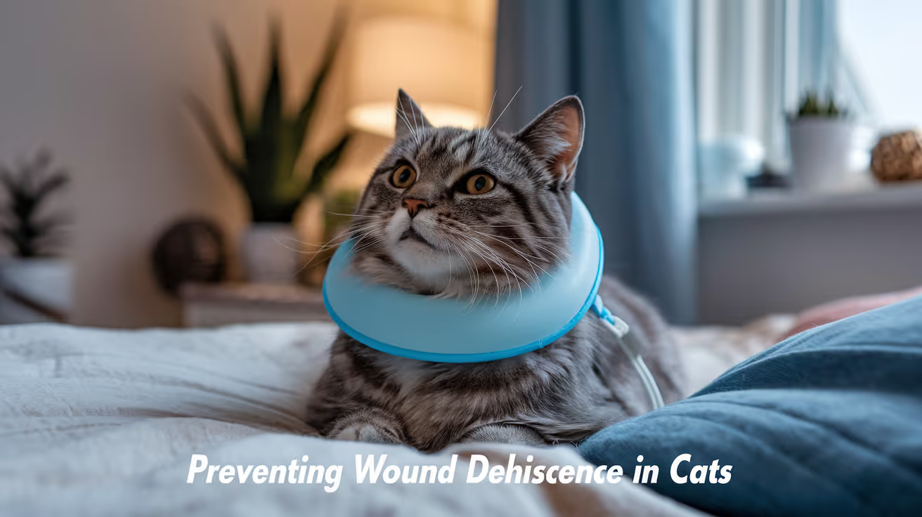
Preventing Dehiscence in Cat Surgical Wounds
Learn effective strategies to prevent dehiscence in cat surgical wounds and ensure faster, safer healing for your feline friend.
A cat's surgical wound can look perfectly healed on the surface while still being at serious risk of opening.
Cat skin heals more slowly and with less tensile strength than dog skin at the same point in recovery. Research has measured sutured cat wounds at only half the breaking strength of equivalently sutured dog wounds at seven days post-surgery.
That single fact changes how seriously you need to take activity restriction, E-collar use, and monitoring in cats.
Quick answer: Dehiscence is the breakdown and reopening of a surgically closed wound. Cats are at higher dehiscence risk than dogs because their skin heals more slowly, has fewer cutaneous blood vessels, and builds tensile strength more gradually. The most common causes in cats are licking, excessive activity, and infection. Prevention requires consistent E-collar or recovery suit use for the full post-operative period, strict activity restriction, and daily wound monitoring.
Key takeaways
- Cat sutured wounds are half as strong as dog wounds at 7 days: This is measured fact, not an approximation. The implication for activity restriction and suture-removal timing is significant.
- Licking is the single most common cause of dehiscence in cats: Cats are meticulous groomers and extremely persistent in accessing wounds.
- Dehiscence can happen even when the surface looks healed: Internal layers may still be vulnerable when the skin surface appears closed.
- Infection significantly raises dehiscence risk: Bacterial enzymes degrade suture material and surrounding tissue, undermining wound integrity.
- Recovery suits are often better tolerated than cones in cats: But they must fully cover the wound without creating pressure.
- Wound reopening is a surgical emergency: Do not wait to contact your vet if a sutured wound separates.
Why cats are at higher dehiscence risk than dogs
This is not widely appreciated by cat owners, and even some general veterinary guidance treats cats and dogs as equivalent in terms of wound care.
They are not equivalent. Research comparing cutaneous wound healing in cats and dogs found several key differences.
Cats have fewer cutaneous perforating vessels: Their trunk skin has a lower density of blood vessels than dog skin. Lower vessel density means lower tissue perfusion, and lower perfusion means slower initial healing.
Breaking strength builds more slowly: In the landmark comparison study by Bohling et al., sutured wounds in cats reached only half the breaking strength of equivalently sutured dog wounds by day seven. This means a cat's wound is substantially more vulnerable to disruption at the point when many owners assume healing is well advanced.
Cats produce less granulation tissue: In open wounds healing by secondary intention, cats generate significantly less granulation tissue than dogs, and what forms distributes differently. This affects the speed of wound closure and the structural quality of repaired tissue.
The clinical implication: Suture removal timing, activity restriction duration, and monitoring intensity all need to account for the fact that a cat's wound at ten days post-surgery is not as strong as a dog's wound would be at the same point.
What is wound dehiscence?
Dehiscence is the breakdown and separation of a surgically closed wound. The sutured edges pull apart, exposing the underlying tissue.
It can be partial, where only a portion of the wound reopens, or complete, where the entire incision separates. In abdominal surgeries, complete dehiscence carries the risk of evisceration, where internal organs protrude through the opening. This is a surgical emergency.
Dehiscence differs from normal surface healing variation. A small amount of scabbing, minor surface irregularity, or a small clear serum pocket near the incision is not dehiscence. Dehiscence is when the edges of the wound visibly separate and the wound is no longer closed.
The most common causes in cats
Licking and chewing
In most cases, dehiscence in cats results from the cat accessing the wound. A cat that licks an incision does several things simultaneously: it physically disrupts forming tissue, applies abrasive mechanical force to fragile sutures, and continuously introduces oral bacteria.
Even a brief, unobserved licking session can undo days of healing. Cats are highly motivated and creative in reaching wounds, including angles that seem impossible.
For a clear understanding of spay incision complications in cats, including how licking specifically contributes to spay incision dehiscence and what the signs look like, that guide covers the spay-specific incision risk in detail.
The E-collar or recovery suit must be in place at all times, including overnight, until the veterinarian confirms healing at the recheck appointment.
Excessive activity
Cats recover faster than most owners expect and will attempt to resume normal activity, including jumping, climbing, and running, within days of major surgery.
Every jump landing puts mechanical stress on an abdominal incision. Every climbing session extends and flexes the tissue around a flank incision. These forces work directly against the tensile strength that is slowly building across the wound.
Strict confinement for the full post-operative restriction period is not optional. A room with no high surfaces, no furniture to jump onto, and no access to stairs is the appropriate recovery environment.
Infection
Bacteria produce enzymes, including collagenase, that actively degrade the suture material and the collagen-based tissue being rebuilt around the wound. An infected incision is far more likely to dehisce than a clean one.
For understanding what happens when wounds fail to close properly and how wounds that have dehisced are subsequently managed, that guide covers secondary intention healing in detail, including the management pathway for wounds that can no longer be sutured closed.
Recognizing early infection signs, warmth, redness, discharge, and odor, and contacting your vet immediately prevents the progression from localized infection to wound breakdown.
Tension on the closure
Surgeons aim to close wounds with appropriate but not excessive tension. However, swelling, weight gain, or movement at an inopportune time can increase tension on sutures beyond what the healing tissue can withstand.
Keeping cats at their normal weight during recovery and restricting movement minimizes the mechanical forces working against the closure.
Suture-related factors
Sutures placed too close to the wound edge, suture material that degrades faster than expected in an inflamed wound, or knots that fail under tension can all contribute to dehiscence. These are surgical factors outside the owner's control, but they underscore why post-operative care at home is so important as a compensating measure.
Recognizing dehiscence early
Early signs:
- The incision line appears to be gapping or separating
- One or more sutures appear missing or have pulled through the skin
- Increased discharge from a point along the incision
- The wound surface looks open rather than closed
- Visible tissue beneath the skin surface
Emergency signs requiring immediate veterinary contact:
- Visible internal tissue or organs through the wound opening
- Any abdominal incision that separates more than a few millimeters
- Rapid deterioration: your cat becoming distressed, limp, or unresponsive
If you notice any wound separation, cover the area loosely with a clean, damp cloth and take your cat to a veterinarian immediately. Do not attempt to clean or close the wound yourself.
Prevention: what owners can control
E-collar and recovery suit
The E-collar must fit correctly. It needs to extend past the tip of your cat's nose by at least two inches. Shorter cones allow a flexible cat to reach the wound.
Recovery suits, when properly fitted, cover the wound without applying pressure to it. They allow cats to eat, drink, and move their faces normally, which many cats tolerate better than rigid cones. Confirm the suit completely covers the wound site and cannot be removed by the cat.
Use whichever works in practice. The one that actually stays on is the right one.
Strict confinement
Confine your cat to a room with no high surfaces for the full activity restriction period recommended by your veterinarian. This is typically ten to fourteen days for routine surgeries and longer for more complex procedures.
Block access to couches, beds, windowsills, and cat trees. A bathroom, laundry room, or large dog crate with food, water, and a litter box at floor level works well.
Daily wound monitoring
Check the incision morning and evening. Look for:
- Redness that is spreading beyond day three
- Discharge that is increasing or changing from clear to yellow or green
- Any visible separation of the wound edges
- Heat at the wound site persisting beyond the first few days
For distinguishing infection from wound breakdown during monitoring, that guide covers the visual signs of each clearly, with a day-by-day comparison of what is normal versus what needs veterinary attention.
Follow-up appointments
Do not skip the post-operative recheck. For most routine feline surgeries, this occurs at ten to fourteen days.
Given that cat wound tensile strength is only 50% of dogs at day seven, the ten to fourteen day mark is when the vet assesses whether the wound has reached sufficient strength for suture removal. Removing sutures too early in a cat is a meaningful dehiscence risk.
If you are uncertain whether healing is progressing normally between your cat's discharge and the recheck appointment, contact your vet and describe what you are seeing. A photo sent by phone can often resolve the question without requiring a visit.
If dehiscence has already occurred
When a wound reopens, the approach depends on the degree of dehiscence and the state of the underlying tissue.
Minor partial dehiscence with healthy tissue: The vet may clean the wound and allow it to heal by secondary intention, or re-suture after debridement.
For context on how dehiscence prevention differs between dogs and cats, the dog-specific guide covers the canine risk factors and prevention strategies side by side, which is useful for households with both species recovering from surgery.
Significant dehiscence with infected or necrotic tissue: Debridement is required before re-closure. Open wound management with daily cleaning and dressing changes may be needed for days to weeks before re-suturing.
Abdominal dehiscence with evisceration: Immediate surgery. Cover the protruding tissue with a clean, moistened cloth during transport. Do not attempt to push tissue back.
Frequently asked questions
How long should I keep the E-collar on my cat after surgery?
Until your veterinarian confirms at the recheck appointment that the wound is sufficiently healed. For most routine feline surgeries, this is ten to fourteen days. Given that cat wound strength builds more slowly than in dogs, it is better to err on the side of leaving it on longer rather than shorter.
My cat removed the cone. How do I prevent this?
Try a properly fitted soft recovery collar, which is often tolerated better by cats than rigid plastic cones. Alternatively, a recovery suit is effective for abdominal wounds. If your cat is genuinely distressed by all physical barriers, ask your vet whether a short-term mild sedative for the recovery period is appropriate.
The wound looks closed but a small section seems soft. Is that dehiscence?
A small seroma (fluid pocket) can develop near a healing incision and feel soft without being dehiscence. However, any area where you can see a gap between wound edges, even a small one, warrants a same-day call to your vet. Describe what you are seeing and follow their guidance.
Can I clean a dehisced wound at home?
No. A reopened surgical wound, particularly an abdominal incision, requires veterinary assessment before any home management. Home cleaning of a dehisced wound risks introducing bacteria to an already compromised area. Contact your vet immediately.
Cats require more careful post-operative wound management than most owners realize, primarily because their skin simply takes longer to heal to a safe tensile strength. The prevention of dehiscence in cats comes down to three consistent actions: keeping the E-collar or recovery suit on at all times, enforcing strict activity restriction, and monitoring the wound daily. If any of these lapses, the risk increases substantially.
Resources
The following sources were used as reference and background for this article:
- Bohling, M.W. et al. (2004). Cutaneous wound healing in the cat: a macroscopic description and comparison with cutaneous wound healing in the dog. Veterinary Surgery. pubmed.ncbi.nlm.nih.gov
- Claeys, S. (2016). Dehiscence. In Complications in Small Animal Surgery. Wiley. onlinelibrary.wiley.com
- MSPCA-Angell. Wound Dehiscence: Causes, Prevention, Management. mspca.org
- Catwatch Newsletter. All About Incisions. catwatchnewsletter.com
- Veterian Key. Wound Healing. veteriankey.com
- WSAVA. Notions, Potions or Lotions? Acute Wound Management in Dogs and Cats. vin.com
Mattress Sutures in Small Animal Surgery
Learn about mattress sutures in small animal surgery, their types, uses, and benefits for effective wound closure in pets.
Some wounds cannot be closed with a simple interrupted suture. The tension would pull the suture straight through the skin edge, the tissue would strangulate, or the wound edges would invert rather than appose.
Mattress sutures exist for these situations. They take a larger, deeper bite of tissue than simple interrupted sutures, distributing tension over a greater area and allowing closure that would otherwise fail.
Quick answer: Mattress sutures are everting suture patterns used in high-tension wounds, fragile skin, or areas where simple interrupted sutures risk tissue cut-through or strangulation. Horizontal mattress sutures distribute tension parallel to the wound over 2 to 3 cm. Vertical mattress sutures take deep and superficial bites to evert wound edges and close dead space simultaneously. Both types are removed at 10 to 14 days if non-absorbable, or earlier if used as temporary tension-relieving sutures before final closure.
Key takeaways
- Horizontal mattress sutures distribute tension parallel to the wound over a wider tissue bite.
- Vertical mattress sutures take a far-near-near-far path to evert edges and relieve deep tension.
- Both are everting patterns they turn wound edges slightly outward, which is preferable to inversion for skin healing.
- Mattress sutures are indicated for high-tension wounds, fragile skin, or areas where simple interrupted patterns would fail.
- Temporary use is common: used to achieve initial apposition, then removed after final sutures are in place.
- Over-tightening causes tissue necrosis the most important technical error to avoid.
What mattress sutures are
Mattress sutures are a category of interrupted suture patterns distinguished by a wider and deeper tissue bite than simple interrupted sutures. Both the horizontal and vertical variants belong to the everting pattern classification they turn wound edges slightly outward rather than aligning them flush.
This slight eversion is beneficial for skin wound healing. University of Melbourne Virtual Vet Surgery explains: "Slight eversion of the skin edges is preferable to inversion in respect to wound healing." Inverted edges trap epithelium beneath the wound, while everted edges allow direct surface apposition and proper epithelial migration.
For how mattress sutures fit within the broader classification of appositional and everting closure patterns, see mattress sutures as everting patterns.
Horizontal mattress suture
How it is placed
- Needle enters skin on the far side of the wound, 5 to 8 mm from the edge
- Needle passes across the wound and exits on the near side
- Needle re-enters the near side at the same depth, 5 to 10 mm along the wound from the first bite
- Needle crosses back across the wound and exits on the far side
- The two ends are tied, creating a "U" shape that bridges the wound with a suture running parallel to it on each side
Properties
- Distributes tension over 2 to 3 cm of tissue rather than 4 to 8 mm
- Provides moderate wound edge eversion
- Lower risk of tissue strangulation than vertical mattress when properly placed
- Does not close deep dead space (takes bites in the superficial dermis only)
Pronorth Medical notes: "The advantages of horizontal mattress sutures include robust closure and reduced risk of tissue strangulation. This technique is particularly preferred in scenarios where wounds are subjected to high tension, such as in the areas around joints."
When horizontal mattress is the right choice:
- Joint-adjacent wounds under significant tension
- Wounds in areas with thick skin (trunk, neck, lateral thigh)
- Pre-suturing before mass excision to stretch skin before removal
- Temporary tension-relieving before apposition-pattern final closure
Stent use
In high-tension wounds, horizontal mattress sutures can cut into skin if the suture presses directly on the surface. MSPCA-Angell notes that stents (tubing or pads placed under the suture) prevent this. The stent distributes the suture's pressure over a broader skin surface area.
Vertical mattress suture
How it is placed
- Needle enters far from the wound edge (5 to 8 mm), passes deep through the dermis, exits on the far side
- Needle returns from the far side, taking a superficial bite very close to the wound edge (1 to 2 mm)
- Needle crosses back close to the wound edge on the near side and exits
- Ends are tied, creating a deep and superficial anchor on each side of the wound
Properties
- Distributes tension over both deep and superficial tissue in the same suture
- Stronger wound edge eversion than horizontal mattress
- Simultaneously closes some dead space through the deep bite component
- Higher risk of tissue strangulation if over-tightened
MSPCA-Angell (Atlas of Small Animal Wound Management, Pavletic, 4th ed.): "In the central zone of tension, vertical mattress sutures are used to further protect the incision from wound dehiscence." 2-0 suture is typically used for vertical mattress placements due to the mechanical forces involved.
When vertical mattress is the right choice:
- High-tension wounds in areas with thin skin (sighthound breeds, geriatric dogs on steroids)
- Wounds where dead space closure and skin tension relief are both needed
- Central "tension zone" within a longer wound where the tension is greatest
- Reconstructive procedures after large mass excision
For high-tension wound management using mattress sutures in specific clinical scenarios, see mattress sutures in high-tension wounds.
Horizontal vs vertical: a direct comparison
Temporary mattress sutures
University of Minnesota (Large Animal Surgery notes) describes a common technique: mattress sutures can be placed temporarily to achieve wound apposition, then simple interrupted sutures placed between them in the now-apposed wound, then the mattress sutures removed after 3 to 4 days once the wound edges are stable.
This technique allows the final wound closure to be appositional (better cosmetic outcome) while using mattress sutures to manage the initial tension before the tissue settles.
For how mattress sutures relate to other tension-relieving options used in high-tension closures, see tension relief techniques for high-tension wounds.
Suture material and removal
Material choices:
- Non-absorbable monofilament (nylon, Prolene): most common for skin mattress sutures; size 2-0 to 3-0
- Must be removed at 10 to 14 days for most wounds
- Horizontal mattress sutures left in place longer than 7 days risk producing suture marks
When to use absorbable material for mattress sutures:
- When suture removal may be difficult (uncooperative patient, remote location)
- Buried vertical mattress patterns (more technical)
For how suture size is selected for mattress patterns alongside other considerations, see suture size for mattress patterns.
Aftercare for wounds with mattress sutures
What to watch for:
- Suture marks (linear skin indentations parallel to the suture): appear if sutures are too tight or left in too long
- Tissue necrosis under the suture (pale or gray skin): indicates over-tightening; contact vet same day
- Wound opening between sutures: normal healing space or early sign of tension overcoming the closure
What to avoid:
- Any activity that increases wound tension (running, jumping, stretching)
- Moisture to the incision before suture removal
- Allowing the dog or cat to lick even brief licking can dislodge or contaminate a tight mattress suture
For where mattress sutures fit within the complete range of skin closure methods, see mattress sutures within skin closure options.
Frequently asked questions
My dog has what looks like two parallel rows of sutures along the incision. Are those mattress sutures?
Possibly. Horizontal mattress sutures create a visible "U" shape with both legs of the suture parallel to the incision on either side of the wound. If the sutures appear as pairs running parallel to the wound rather than individual crossing stitches, they are likely horizontal mattress sutures.
Are mattress sutures more painful than simple interrupted sutures?
Mattress sutures take a larger bite of tissue, which means there is more suture material in the wound. They do not necessarily cause more pain during healing, but they can cause more discomfort during removal if swelling has occurred around the suture loops. Ask your vet about appropriate pain management for the post-operative period.
What happens if mattress sutures are left in too long?
Suture marks (permanent indentations in the healed skin) form when non-absorbable sutures are left in beyond 7 to 10 days. The skin epithelializes down the suture tract, leaving a visible line. Remove on schedule or sooner if wound tension has resolved.
Mattress sutures exist for the wounds that simple interrupted sutures cannot close without failure. Horizontal patterns for wide tension distribution, vertical patterns for simultaneous dead space and skin closure each has a specific indication. Used correctly and removed on schedule, they allow wounds to heal that would otherwise dehisce under the forces working against them.
Resources
- MSPCA-Angell. Incisional Tension Relief: Simple Intraoperative Options (Pavletic, Atlas of Small Animal Wound Management, 4th ed.). mspca.org
- University of Minnesota. Suturing: Skin Closure (Large Animal Surgery). open.lib.umn.edu
- Veterian Key. Suturing Techniques and Common Surgical Procedures. veteriankey.com
- Pronorth Medical. Choosing Horizontal vs Vertical Mattress Sutures. pronorthmed.ca

Tension-Relieving Sutures in Veterinary Surgery
Learn about tension-relieving sutures in veterinary surgery, their types, uses, benefits, and care for better healing in pets.
When wound edges cannot be brought together without significant pulling force, the problem is tension. Standard interrupted sutures placed across a high-tension wound concentrate that force at each suture entry point creating ischemia at the tissue-suture interface and dramatically increasing the risk of sutures cutting through and the wound opening.
Tension-relieving sutures work differently. They distribute that force across a wider area, or recruit more tissue to share the load, so no single point bears enough force to fail.
Quick answer: Tension-relieving sutures are patterns specifically designed to distribute wound tension across more tissue and a wider surface area than standard interrupted sutures. The main types used in veterinary surgery are: horizontal mattress (broad tension distribution, parallel to wound), vertical mattress (deep plus superficial bite for edge eversion), near-far-far-near (appositional and tension-relieving, stays in until healed), and walking sutures (advances skin subcutaneously before surface closure). Each addresses tension at a different structural level.
Key takeaways
- Horizontal mattress sutures spread tension across 8 to 10 mm from the wound edge, reducing cut-through risk.
- Vertical mattress sutures provide deep tissue purchase plus edge eversion ideal in high-tension zones.
- Near-far-far-near (NFFN) is both cosmetic and tension-relieving, staying in through full healing.
- Temporary mattress sutures can be placed first, then removed after 3 to 4 days once appositional sutures are secured.
- Stent sutures use tubing or pads under the knot to prevent large sutures from cutting through fragile skin.
- Walking sutures address tension at the subcutaneous level before skin closure begins.
Why wound tension causes problems
Tension at the wound edge reduces blood supply to the tissue between the suture and the wound margin. Without adequate perfusion, that tissue cannot heal, mount an immune response, or resist bacteria.
The consequences of uncorrected wound tension:
- Sutures cut through skin (when tension exceeds the tissue's tolerance at the suture entry point)
- Wound dehiscence (when sutures fail or the tissue gives out)
- Tissue necrosis at the wound margins
- Delayed healing
- Infection in poorly perfused tissue
VCA (Care of Surgical Incisions in Dogs): "If the surgical procedure involved tissue loss, the incision may be under a lot of tension. Excessive tension across an incision line may cause the wound to gape open and delay healing. To minimize tension on the incision line, your veterinarian may use a special tension-relieving suture pattern, or a type of skin suture called a stent suture."
For how tension directly contributes to high-tension wound closure failure, see high-tension wound closure using tension-relieving sutures.
Horizontal mattress sutures
The horizontal mattress pattern is placed parallel to the wound edge. Each stitch enters the skin, crosses the wound, re-enters on the opposite side, returns parallel to the wound, and exits the skin. The result is a U-shaped loop lying flat across the wound surface.
Veterian Key: "This type of suture can be used in areas of tension as the pressure exerted by the horizontal sutures is spread evenly over a broad area, which reduces the likelihood of tearing through the tissue edges. The action is: holding the needle with needle holders, insert the needle approximately 8 to 10 mm away from the edge of the incision on the far side."
Properties:
- Spreads tension over 8 to 10 mm from the wound edge on each side
- Can be used as a temporary stay stitch to approximate wound edges while interrupted or intradermal sutures are placed
- Can remain in place for several days after the primary closure if tension persists
- Risk of suture marks if left beyond 7 days
University of Minnesota (Large Animal Surgery): "If tension is greater than can be managed with a NFFN suture, vertical and horizontal mattress sutures may be placed temporarily to relieve tension. After 3 to 4 days, the mattress sutures can be removed, leaving just the appositional pattern."
For how mattress sutures are used as the primary tension pattern in small animal surgery, see mattress sutures in small animal surgery.
Vertical mattress sutures
The vertical mattress pattern takes a deep bite far from the wound edge (the "far" component), crosses to the opposite side, returns with a shallower bite close to the wound edge (the "near" component). The loop is vertical crossing the wound at two depths.
Properties:
- Provides strong tissue purchase deep to the skin holds against tension at the fascial level
- Everts wound edges (turns edges outward) counters the tendency of high-tension wounds to invert
- Reduces dead space just below the skin edge by drawing deep tissue into the closure
- More likely to cause suture marks than horizontal mattress if left beyond 10 days
Best for:
- High-tension wounds where deep tissue purchase is needed
- Areas where wound inversion is a problem (e.g., over joints, areas with thick overlying muscle)
- Skin over orthopedic surgical sites
Near-far-far-near (NFFN) sutures
The NFFN pattern is a tension-relieving suture that is also appositional meaning it holds wound edges together while distributing tension, rather than merely providing mechanical support from the outside.
University of Minnesota (Large Animal Surgery, Suturing Skin chapter): "A near-far-far-near type pattern is cosmetic as well as tension relieving. As it is an appositional pattern and will stay in until the tissue is healed, it should not be bigger than 2-0 or 0 in non-bovine patients."
Pattern sequence:
- Near entry close to the wound edge on one side
- Far exit exits the skin far from the wound edge on the opposite side
- Re-enters far re-enters far from the wound on the same side it just exited
- Near exit exits close to the wound edge on the original side
The alternating near-far bites distribute tension while the pattern itself holds wound edge apposition. This makes it more cosmetically acceptable than a horizontal mattress and appropriate for wounds where the suture will be the primary closure rather than a temporary adjunct.
For the clinical comparison of NFFN and walking suture approaches to tension, see NFFN pattern in high-tension wound context.
Stent sutures
Stent sutures are not a pattern they are a modification applied to any external suture in high-tension situations. A piece of rubber tubing, surgical sponge, or button is threaded under the knot of a mattress or interrupted suture.
Why stents are used:
- In thin-skinned or fragile patients, a large-gauge suture alone can cut through skin at the knot
- The stent distributes the contact area of the knot across a wider skin surface
- Allows use of stronger suture material (size 0 to 1) without the cut-through risk
VCA: "Your veterinarian may use... a type of skin suture called a stent suture, which involves the addition of some tubing or a button to the skin layer."
University of Minnesota (Large Animal Surgery): "Larger suture (1-2) can be used for the mattress sutures. Stents (tubing or pads) are often used to prevent the larger suture from cutting through the skin."
Walking sutures: the subcutaneous approach
While the above patterns address tension at the skin surface, walking sutures address it before skin closure begins by advancing the dermis toward the defect at the subcutaneous level.
For full detail on the walking suture technique, see walking sutures for large defects in large breed dogs.
Choosing between tension-relieving options
Suture material for tension-relieving patterns
University of Minnesota: In large animals, mattress sutures use "larger suture (1 to 2)" while the appositional pattern uses "2-0 or 0."
For dogs: horizontal and vertical mattress sutures typically use the same material as other skin sutures but at a size one step larger than the primary closure so if the wound would normally close with 3-0, tension sutures might use 2-0. Monofilament non-absorbable (nylon, Prolene) is preferred to minimize bacterial adhesion. PDS is used when an absorbable material is needed in a tension-relieving position.
For how tissue type and patient factors such as obesity affect closure technique selection, see tissue type and closure technique selection. In obese patients where tissue fragility increases cut-through risk, stent sutures are especially relevant; see tension management in obese dogs.
Frequently asked questions
My dog had large mattress sutures placed around a smaller standard closure. Why two types?
Your vet used the mattress sutures as tension relief to protect the primary appositional closure. The mattress sutures bear the bulk of the wound tension, reducing the load on the finer interrupted or intradermal sutures that produce the actual skin edge contact. In some cases, the mattress sutures are removed after 3 to 4 days, leaving only the appositional closure; in others, they stay through the full healing period.
Are tension-relieving sutures removed at the same time as regular sutures?
Usually around the same time (10 to 14 days), but staggered removal is common for walking sutures and some mattress patterns. In cases with significant tension, the vet may leave some tension-relieving sutures beyond 14 days if the wound is not yet stable. Ask your vet specifically about the removal plan for each suture type placed.
Do tension-relieving sutures hurt more than regular sutures?
Not typically, though mattress sutures do involve deeper tissue bites that may cause more post-operative soreness than fine interrupted skin sutures. Pain management after surgery covers this. The tenderness is usually at its peak for 24 to 48 hours, then diminishes as the wound stabilizes.
The purpose of every tension-relieving technique is the same: ensure that no single point in the closure bears enough force to fail. Whether that means spreading force across the surface with a horizontal mattress, anchoring it deep with a vertical mattress, distributing it along the appositional line with NFFN, or advancing the skin before surface closure with walking sutures, the outcome goal is uniform: a closure that holds while the tissue heals.
Resources
- Veterian Key. Suturing Techniques and Common Surgical Procedures. veteriankey.com
- University of Minnesota. Suturing Skin (Large Animal Surgery Supplemental Notes). open.lib.umn.edu
- VCA Animal Hospitals. Care of Surgical Incisions in Dogs. vcahospitals.com
- WSAVA 2007 (VIN). Reconstructive Surgery. vin.com
Tissue Adhesives in Dog and Cat Surgery
Explore the use, benefits, and safety of tissue adhesives in dog and cat surgery for better healing and less pain.
Tissue adhesives surgical glues are a legitimate wound closure tool in veterinary surgery, but not a universal replacement for sutures.
Knowing when they are appropriate, when they are not, and what the evidence shows helps both clinicians and owners make informed decisions.
Quick answer: Cyanoacrylate adhesives suit short, low-tension skin closures, especially port-site incisions and minor lacerations with subcutaneous sutures already placed. Tissue adhesive alone is insufficient for high-tension or contaminated wounds.
Key takeaways
- Cyanoacrylate is the most common tissue adhesive, polymerizing on contact with tissue to form a flexible, bacteriostatic film
- Tissue adhesive alone is insufficient for longer or high-tension incisions: subcutaneous sutures must first take the mechanical load
- PMC study: intradermal suture had better cosmetic, clinical, and ultrasonographic outcomes than tissue glue in dogs
- No dehiscence occurred with tissue glue in the PMC study when subcutaneous sutures were placed first
- Longer-chain cyanoacrylates (butyl, octyl) cause less tissue inflammation than shorter chains and are preferred for veterinary use
- Application is significantly faster: 2 minutes 16 seconds vs 15 minutes 37 seconds for intradermal suture in one PMC study
What tissue adhesives are
SustainableVet: "Tissue adhesives are special glues used to close wounds or surgical incisions in animals. They form a strong bond that holds the skin edges together while the tissue heals underneath.
These adhesives are made from biocompatible materials safe for pets."
SustainableVet: "Cyanoacrylate adhesives are widely used due to their fast bonding and strong hold suitable for skin closure in pets."
How cyanoacrylate works: when the adhesive contacts tissue moisture, it undergoes rapid polymerization a chemical reaction that turns the liquid monomer into a solid polymer film in seconds. This film bonds the wound edges mechanically and creates a flexible seal.
Formulations: cyanoacrylates differ by the length of the carbon chain attached to the ester group.
NCBi (cyanoacrylate study): "Longer carbon chain cyanoacrylates (butyl and octyl) promote a less intense inflammatory reaction, with greater collagen deposition and greater tensile strength, requiring less time for healing."
Short-chain cyanoacrylates (methyl, ethyl) cause more tissue inflammation. They are used in hardware and household applications but not appropriate for veterinary surgery.
Medical-grade butyl and octyl cyanoacrylate formulations (e.g., Vetbond, Dermabond equivalents) are the appropriate products for clinical use.
Evidence from comparative studies
PMC study: tissue glue vs. intradermal suture vs. staples in dogs
PMC (wound healing study in dogs): "Glue had a less favorable outcome; intradermal suture was the best, though not significantly better than staples, which were applied easier and in significantly less time."
The study also reported: "No dehiscence occurred to the incisions closed with tissue glue.
PMC notes that subcutaneous sutures placed first may have prevented dehiscence: some studies report increased dehiscence when tissue glue is used alone without subcutaneous support."
The timing: stapling took a median of 21 seconds, tissue glue took 2 minutes 16 seconds, and intradermal suture took 15 minutes 37 seconds.
Feline ovariohysterectomy: tissue glue vs. sutures
PMC (feline closure study): "No statistically significant difference in wound dehiscence" between tissue glue and simple interrupted sutures in cats held in a semi-free environment compared to confined animals.
This suggests that tissue glue, when applied appropriately to properly supported incisions, can achieve comparable dehiscence rates to external sutures though the cosmetic and healing outcomes still favor intradermal suture.
Human meta-analysis (low-tension face/neck wounds)
NCBi (meta-analysis): "CTAs offered better cosmetic outcomes by Wound Registry Scale at less than 1 month. The cosmetic outcomes were comparable between 1 and 3 months.
Sutures exhibited superior cosmetic outcomes compared to CTAs at 3 to 12 months."
The early cosmetic advantage of tissue glue in low-tension wounds reverses over time. For wounds where long-term cosmesis matters, sutures remain the stronger option.
Laparoscopic port-site incisions in dogs
Wiley (canine laparoscopic ovariectomy study): evaluated n-butyl-cyanoacrylate for port-site incision closure. These are very short, low-tension incisions an ideal application for tissue glue.
High owner satisfaction and good cosmetic outcomes were reported for this specific indication.
When tissue adhesive is appropriate
Tissue adhesive performs best and is safest in the following scenarios:
Short, low-tension skin closures where the wound edges naturally appose without tension. Port-site incisions from laparoscopy are the clearest indication.
Elective closures with subcutaneous support: PMC data confirm that tissue glue over subcutaneous sutures (which take the mechanical load) produces reliable wound closure.
Minor lacerations: clean, fresh, low-tension skin lacerations where suture placement would cause additional tissue trauma.
Reduced-stress procedures: in cats or fractious animals where suture removal would require sedation, absorbable sutures or tissue glue that do not require removal are preferred.
When tissue adhesive is not appropriate
High-tension wounds: any closure that relies on the adhesive to resist significant mechanical pull the glue will fail. Subcutaneous sutures must take the load.
Contaminated or infected wounds: tissue adhesive seals the wound surface and traps contamination beneath the film. It should never be used on wounds with debris, bacteria, or exudate.
Deep wounds with significant dead space: dead space beneath tissue adhesive creates a seroma risk and an environment for bacterial proliferation.
Joints or high-motion locations: repeated motion breaks the adhesive bond. Incisions over joints or in high-flexion/extension areas require suture support.
Long incisions: the mechanical strength of tissue adhesive is limited. Longer incisions need suture support regardless of tension level.
SustainableVet: "Tissue adhesives are suitable for superficial skin closures but cannot replace sutures for deep or high-tension wounds requiring stronger support."
Application technique
Wound preparation: the wound edges must be clean, dry, and free of blood and fluids. Tissue adhesive does not bond well to wet surfaces or across contamination.
Edge approximation: manually appose the wound edges before applying the adhesive. The adhesive holds the edges in place; it does not pull them together.
Application: apply a thin, even layer along the wound edge. Do not apply deep into the wound itself only to the skin surface.
Wait for polymerization: typically 30 to 60 seconds. Do not allow the patient to disturb the site during this period.
Do not bandage tightly over adhesive: tight bandaging can lift the adhesive film before it has fully cured.
What owners should know
No suture removal: tissue adhesive dissolves as the wound heals, typically within 5 to 10 days. No removal appointment is needed.
Keep dry: moisture degrades cyanoacrylate bonds. Keep the wound dry for at least 5 days after application.
E-collar: even without external sutures, licking can degrade and lift the adhesive film. An E-collar may still be required depending on wound location.
The adhesive may crack or peel as it dissolves: this is normal. The wound beneath should be healing; the adhesive is no longer holding edges if the incision has adequately healed.
For suture patterns as an alternative, see interrupted vs. continuous suturing in dogs. For feline-specific suture considerations, see interrupted vs. continuous suturing in cats.
For site preparation that precedes closure, see surgical site preparation in dogs.
Frequently asked questions
Can tissue glue be used to close my dog's spay incision?
Not as the primary skin closure. Spay incisions require subcutaneous sutures first to close dead space and reduce tension. Tissue glue may then be applied over the skin surface; ask your vet.
Is tissue glue safe if my cat licks it?
Cyanoacrylate tissue adhesives are generally considered low-toxicity if small amounts are ingested. However, licking degrades the adhesive bond and risks premature wound opening. An E-collar is still recommended to protect the closure.
How long does tissue adhesive take to dissolve?
Medical-grade cyanoacrylate typically dissolves over 5 to 10 days as wound healing progresses. The adhesive does not need to be removed.
What if the wound opens after tissue adhesive is applied?
Contact the vet same day. A wound that reopens after glue closure may need suture reclosure. Do not reapply tissue adhesive over a wound that has separated proper wound assessment first.
Can tissue adhesive cause an allergic reaction in my pet?
Medical-grade butyl and octyl cyanoacrylate are biocompatible. NCBi: longer-chain formulations cause "a less intense inflammatory reaction." True allergic reactions are uncommon. Unusual swelling or discharge should be assessed.
Does tissue adhesive have any antibacterial properties?
Yes, minor. Cyanoacrylate polymerization has some bacteriostatic effect. This is minimal and should not be relied on for contaminated wounds; tissue adhesive is not a substitute for debridement or antiseptic lavage.
Resources
- PMC. Evaluation of Incisional Wound Healing in Dogs after Closure with Staples or Tissue Glue and Comparison to Intradermal Suture Pattern. ncbi.nlm.nih.gov
- NCBi. Cyanoacrylate Adhesives for Cutaneous Wound Closure. ncbi.nlm.nih.gov
- NCBi. Cyanoacrylate Tissue Adhesives Compared With Sutures on Facial and Neck Wounds: A Meta-analysis. ncbi.nlm.nih.gov
- Wiley. Efficacy of n-butyl-cyanoacrylate Tissue Adhesive for Closure of Canine Laparoscopic Ovariectomy Port Site Incisions. onlinelibrary.wiley.com
Choosing Suture Material for Cat Surgery
Learn how to choose the right suture material for cat surgery, including types, uses, and tips for best healing outcomes.
Cats are not small dogs. Their tissue is more delicate, their healing is faster, and their skin is thinner and more reactive to foreign material than most canine breeds.
Suture selection in cats follows the same basic logic as in dogs match the material's properties to the tissue's needs but with specific adjustments for feline physiology. The materials that work best in cats tend toward lower tissue reaction, faster absorption, and finer gauge than their dog equivalents.
Quick answer: In cats, PDS (polydioxanone) is preferred for deep structural layers like the linea alba; Monocryl (poliglecaprone 25) is preferred for subcutaneous and subcuticular closure due to lower tissue reaction than PDS in feline tissue; nylon or absorbable monofilament (3-0 or 4-0) for skin. Suture sizes in cats run 3-0 to 4-0 for most layers. Clinician's Brief notes Monocryl "may induce less tissue reaction than PDS, especially in cats."
Key takeaways
- Monocryl is the preferred subcutaneous suture in cats due to lower tissue reaction than PDS in feline tissue.
- PDS remains the standard for linea alba because subcutaneous absorption speed of Monocryl is too fast for fascial support.
- 3-0 and 4-0 are the standard sizes for most feline surgical layers.
- Vicryl (braided multifilament) causes more tissue reaction than monofilament alternatives in cats.
- Tissue adhesive is a practical and effective skin closure option for many feline procedures.
- Suture removal is needed only for non-absorbable skin sutures most feline surgery uses absorbable materials throughout.
Why cat suture selection differs from dogs
Three feline-specific factors drive material selection:
1. Thinner, more reactive skin. Cat dermis is more sensitive to suture-related inflammation. Materials that cause more tissue reaction (braided multifilament, natural gut) produce more visible swelling, licking behavior, and risk of suture track infection.
2. Faster tissue healing. Cats' subcutaneous and skin layers heal in 7 to 10 days rather than 10 to 14 days. This allows faster-absorbing materials to be appropriate choices at layers where dogs might need longer retention.
3. Smaller tissue volumes. Cats' smaller bodies require finer gauges. A 2-0 suture that is appropriate in a 20 kg dog would cause unnecessary tissue damage in a 4 kg cat.
Suture material guide by tissue layer
Abdominal wall / linea alba
Best choice: PDS (polydioxanone) 2-0 to 3-0
The linea alba is the structural layer of abdominal closure. It heals slowly. Monocryl loses tensile strength too quickly for fascial support: Clinician's Brief states Monocryl "is a poor choice for tissue that is slow to regain tensile strength, such as fascia (linea alba) or tendons."
PDS retains approximately 70% of its tensile strength at 2 weeks and 50% at 4 to 6 weeks appropriate for feline fascial healing timelines.
Published feline cadaver research (Frontiers in Veterinary Science, 2019) supports using 3-0 PDS with a 5 mm suture bite-to-stitch interval for feline linea alba closure.
For how abdominal wall closure works in the context of spay surgery, see absorbable vs non-absorbable decision.
Subcutaneous tissue
Best choice: Monocryl (poliglecaprone 25) 3-0 to 4-0
For subcutaneous closure in cats, Monocryl outperforms Vicryl and PDS on the tissue reaction criterion. Clinician's Brief (Suture Choice for Today's Veterinarian): "Poliglecaprone 25 may induce less tissue reaction than polydioxanone especially in cats."
Monocryl is absorbed by 90 to 119 days, which exceeds the healing timeline by a comfortable margin.
WSAVA 2015 conference notes Biosyn (glycomer 631) as comparable to Monocryl: "Biosyn is relatively rapidly absorbed with approximately 50% loss of tensile strength at 2 to 3 weeks and is completely absorbed at 90 to 110 days. When compared with Vicryl, Biosyn is stronger and has greater knot security." Biosyn is an appropriate alternative where Monocryl is unavailable.
For how monofilament vs. multifilament affects tissue reaction in this layer, see monofilament vs multifilament decision. For suture material selection in dogs as a comparison, see suture material selection in dogs.
Subcuticular / intradermal skin
Best choice: Monocryl 4-0
For intradermal closure in cats, Monocryl provides:
- Low tissue drag (smooth passage through delicate feline dermis)
- Adequate tensile strength through the 7 to 10 day feline skin healing period
- Dissolution timeline that exceeds healing without requiring removal
A PMC study comparing absorbable and non-absorbable sutures for intradermal skin closure in cats found that monofilament materials cause "less tissue drag and induce less inflammation than multifilament sutures."
For intradermal closure technique in cats specifically, see needle selection alongside material.
Skin (external interrupted sutures)
Options:
Tissue adhesive (n-butyl cyanoacrylate) is widely used in feline skin closure, particularly after scrotal and prescrotal neuter incisions. It provides waterproof closure, requires no needle passage through delicate skin, and eliminates removal visits.
Suture size selection in cats
Veterinary Practice News guidance: "Suture size should be the smallest size that will adequately hold the tissue." In cats this is almost always 3-0 or 4-0 for standard soft tissue layers.
For how suture size decisions are made alongside material selection, see suture size alongside material.
Materials to avoid in cats
Surgical gut (catgut): natural material with unpredictable absorption rate and high inflammatory response in cats. Largely replaced by synthetic materials; not recommended.
Silk: multifilament non-absorbable; highest tissue reaction of any suture material; associated with sinus tract formation; not appropriate for feline surgery.
Chromic gut: improved over plain gut but still variable absorption and more inflammatory than synthetic options.
Vicryl for subcutaneous closure: higher tissue reaction than Monocryl in cats; suitable but not the preferred choice where Monocryl is available.
What suture choice means for your cat's recovery
If your cat had absorbable sutures throughout:
- No removal visit is needed
- You may feel a slight ridge under the incision for 2 to 4 weeks (suture material dissolving)
- No suture material should emerge through the skin in normal healing
If your cat had non-absorbable skin sutures:
- A scheduled removal visit at 10 to 14 days is required
- Do not attempt to remove sutures at home incorrect removal can open the wound
- Use an E-collar to prevent licking, which can loosen sutures or introduce bacteria
Signs of suture reaction (contact your vet):
- Swelling that increases rather than decreases after day 5
- A small firm lump forming along the incision line
- Discharge at a suture site
- Persistent licking at a specific point on the incision
Frequently asked questions
Why did my vet use a different suture for my cat than they used on my dog?
Cats have more reactive skin and a faster healing timeline than most dogs. The materials optimized for feline tissue (particularly Monocryl for subcutaneous closure) differ from the broader range used in dogs. This is an intentional species-specific selection.
My cat keeps licking one point on her incision but it looks fine. Should I be worried?
A cat persistently targeting one spot often indicates localized irritation possibly a suture knot that is slightly prominent, early suture reaction, or mild discomfort at that site. Keep the E-collar on and contact your vet. They may want to assess the site to rule out early dehiscence or reaction.
Do tissue adhesives work as well as sutures in cats?
For thin-skinned areas, low-tension incisions, and cats prone to licking and suture removal, tissue adhesive performs comparably to sutures and eliminates the need for needle passage through delicate feline skin. It is not appropriate for high-tension wounds or areas requiring layered closure.
Suture selection in cats is not a trivial detail. The thin, reactive feline dermis, faster healing timeline, and smaller tissue volumes all argue for the same general direction: the finest gauge that provides adequate strength, in the material with the lowest tissue reaction profile, using absorbable materials wherever removal creates compliance challenges. Monocryl and PDS together cover the vast majority of feline surgical closure needs.
Resources
- Clinician's Brief. How to Fine-Tune Suture Choices for Today's Veterinarian. cliniciansbrief.com
- WSAVA 2015 (VIN). So Many Pieces of String: How to Choose Appropriate Suture Material. vin.com
- Frontiers in Veterinary Science. Optimal Suture Bite Size for Closure of Feline Linea Alba. PMC, 2019. ncbi.nlm.nih.gov
- Veterinary Practice News. What Suture Size Should I Use? veterinarypracticenews.com

