
Protecting
Pets, People & Planet
Join a group of veterinarians leveraging the latest technologies to deliver excellent care to their patients while being a responsible and positive force for their local and global communities.
100% secure. We do not share your information
Recent Articles
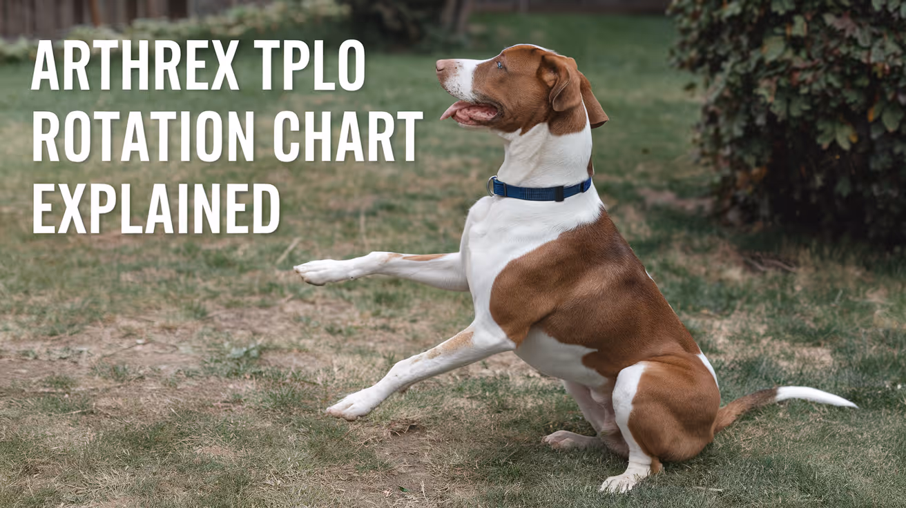
Arthrex TPLO Rotation Chart Explained
Learn how to read and use the Arthrex TPLO rotation chart for precise tibial plateau leveling osteotomy surgery in dogs.
Performing a tibial plateau leveling osteotomy (TPLO) is a common surgical procedure to treat cranial cruciate ligament injuries in dogs. The Arthrex TPLO rotation chart is a vital tool that helps veterinary surgeons determine the exact amount of tibial rotation needed during surgery. Understanding this chart ensures accurate bone alignment and improves surgical outcomes.
This article explains the Arthrex TPLO rotation chart in detail. You will learn how to interpret the chart, why it matters for your pet’s surgery, and how it guides surgeons in achieving the correct tibial plateau angle. This knowledge helps pet owners understand the surgical process better and supports informed discussions with your veterinary surgeon.
What is the Arthrex TPLO rotation chart?
The Arthrex TPLO rotation chart is a reference guide used during TPLO surgery. It shows the degrees of rotation needed to achieve a target tibial plateau angle based on the preoperative measurement. This chart helps surgeons plan and execute the bone cut and rotation precisely.
Using this chart reduces guesswork and improves the accuracy of the surgical correction. It is designed specifically for the Arthrex TPLO surgical system, which includes specialized plates and instruments.
- Rotation guidance: The chart provides exact degrees of tibial rotation required to reach the desired postoperative tibial plateau angle, ensuring surgical precision.
- Preoperative planning: It uses the measured preoperative tibial plateau angle to determine how much rotation is necessary during surgery.
- Standardization tool: The chart standardizes the surgical approach, reducing variability between different surgeons and cases.
- Integration with Arthrex system: It is designed to work with Arthrex-specific implants and instruments for seamless surgical workflow.
Understanding this chart is essential for surgeons performing TPLO with Arthrex equipment and benefits pet owners by improving surgical success rates.
How does the Arthrex TPLO rotation chart improve surgical accuracy?
Accurate rotation of the tibial plateau is critical to restore normal joint mechanics after TPLO surgery. The rotation chart helps surgeons avoid under- or over-rotation, which can lead to poor outcomes or complications.
By providing a clear reference, the chart minimizes errors during the procedure. It also supports consistent results across different patients and surgeons.
- Precise angle correction: The chart ensures the tibial plateau angle is corrected to the target, improving joint stability post-surgery.
- Reduces complications: Proper rotation lowers risks of implant failure, arthritis progression, and abnormal gait after surgery.
- Improves recovery: Accurate alignment supports better healing and faster return to normal activity for dogs.
- Supports training: The chart helps less experienced surgeons perform TPLO with confidence and accuracy.
Using the Arthrex TPLO rotation chart is a key step in achieving the best surgical outcomes for dogs undergoing cruciate ligament repair.
What measurements are needed before using the Arthrex TPLO rotation chart?
Before referencing the rotation chart, the surgeon must measure the tibial plateau angle on preoperative radiographs. This angle indicates how sloped the tibial plateau is, which influences the degree of rotation needed.
Accurate measurement is essential for the chart to provide correct rotation values. The process involves specific radiographic positioning and angle calculation techniques.
- Radiographic positioning: Proper lateral X-rays of the stifle joint are required to visualize the tibial plateau accurately.
- Angle measurement: The tibial plateau angle is measured using anatomical landmarks on the radiograph, typically with digital tools.
- Recording values: The measured angle is noted and used as the input for the rotation chart to find the corresponding rotation degree.
- Repeatability: Consistent measurement technique ensures reliable data for surgical planning and chart use.
Accurate preoperative measurements are the foundation for effective use of the Arthrex TPLO rotation chart during surgery.
How do surgeons use the Arthrex TPLO rotation chart during surgery?
During TPLO surgery, the surgeon references the rotation chart after measuring the tibial plateau angle. The chart indicates how many degrees to rotate the tibial segment after the osteotomy cut.
This rotation changes the slope of the tibial plateau to a safer angle, reducing strain on the cruciate ligament. The chart guides the surgeon to achieve this precisely.
- Osteotomy planning: The surgeon plans the bone cut location and angle based on the chart’s rotation recommendations.
- Rotation execution: After cutting the tibia, the surgeon rotates the bone segment by the degree indicated on the chart.
- Verification: Intraoperative imaging or jigs may be used to confirm the rotation matches the chart’s guidance.
- Plate fixation: The Arthrex TPLO plate is applied to stabilize the rotated tibia, maintaining the corrected angle during healing.
Following the rotation chart during surgery helps ensure the tibial plateau angle is corrected accurately and consistently.
What are the benefits of using the Arthrex TPLO rotation chart for pet owners?
For pet owners, the Arthrex TPLO rotation chart contributes to safer surgeries and better recovery for dogs with cruciate ligament injuries. It supports precise surgical correction, which improves joint function.
Understanding the chart can help owners feel more confident about the procedure and its outcomes.
- Improved surgical outcomes: Accurate rotation reduces complications and improves joint stability after surgery.
- Faster recovery: Correct alignment supports quicker healing and return to normal activity for pets.
- Reduced arthritis risk: Proper tibial plateau angle correction lowers the chance of arthritis development later.
- Enhanced surgeon confidence: The chart helps surgeons perform the procedure with precision, benefiting your pet’s health.
Knowing that the surgeon uses tools like the Arthrex TPLO rotation chart can reassure owners about the quality of care their pet receives.
Are there limitations or challenges with the Arthrex TPLO rotation chart?
While the Arthrex TPLO rotation chart is a valuable tool, it has some limitations. Surgeons must combine chart data with clinical judgment and experience for best results.
Variations in anatomy or measurement errors can affect the chart’s accuracy. Understanding these challenges helps set realistic expectations.
- Measurement variability: Inaccurate preoperative angle measurement can lead to incorrect rotation recommendations.
- Anatomical differences: Individual dog anatomy may require adjustments beyond the chart’s standardized values.
- Technical skill required: Proper use of the chart depends on surgeon experience and surgical technique.
- Not a standalone tool: The chart should be used alongside other surgical planning methods and intraoperative assessments.
Awareness of these limitations ensures the Arthrex TPLO rotation chart is used effectively as part of comprehensive surgical care.
Conclusion
The Arthrex TPLO rotation chart is an essential tool for veterinary surgeons performing tibial plateau leveling osteotomy in dogs. It provides clear guidance on the degree of tibial rotation needed to correct the tibial plateau angle accurately.
By understanding and using this chart, surgeons can improve surgical precision, reduce complications, and support better recovery for pets. Pet owners benefit from knowing their dog’s surgery is planned with advanced, reliable tools like the Arthrex TPLO rotation chart.
What is the Arthrex TPLO rotation chart used for?
The Arthrex TPLO rotation chart is used to determine the exact degrees of tibial rotation needed during TPLO surgery to achieve the desired tibial plateau angle correction.
How do you measure the tibial plateau angle before surgery?
The tibial plateau angle is measured on a properly positioned lateral radiograph of the stifle using anatomical landmarks and digital tools to ensure accuracy.
Can the Arthrex TPLO rotation chart prevent surgical complications?
Yes, by guiding precise tibial rotation, the chart helps reduce risks like implant failure and arthritis progression after TPLO surgery.
Is the Arthrex TPLO rotation chart suitable for all dog breeds?
The chart is designed for general use but surgeons may adjust rotation based on individual anatomical differences in certain breeds.
Do surgeons use imaging during TPLO surgery with the rotation chart?
Yes, intraoperative imaging or jigs are often used to verify that the tibial rotation matches the chart’s recommendations for accuracy.
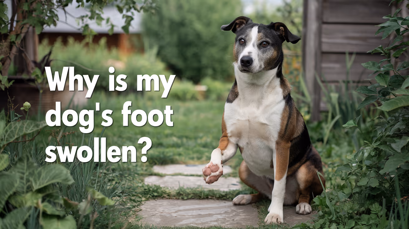
Why Is My Dog's Foot Swollen?
Discover why your dog's foot is swollen, common causes, treatments, and when to see a vet for proper care.
Seeing your dog's foot swollen can be worrying. Swelling in a dog's foot can happen for many reasons, from injuries to infections. Understanding why this happens helps you act quickly and keep your dog comfortable.
This article explains common causes of swollen dog feet, how to spot serious problems, and what treatments work best. You will learn when to treat at home and when to visit a vet for urgent care.
What Causes Swelling in a Dog's Foot?
Swelling in a dog's foot can come from many different problems. It often shows as puffiness, redness, or heat in the paw area. Knowing the cause helps you decide the right care.
Common causes include injuries, infections, allergies, and insect bites. Each cause needs a different approach to treatment.
- Injury or trauma: A cut, sprain, or broken bone can cause swelling due to inflammation and fluid buildup in the foot tissues.
- Infections: Bacterial or fungal infections can cause swelling, redness, and pain, often needing antibiotics or antifungal treatment.
- Allergic reactions: Allergies to plants, chemicals, or insect stings can cause sudden swelling and itching in the foot.
- Foreign objects: Thorns, splinters, or glass stuck in the paw can cause swelling and discomfort until removed.
Identifying the cause early helps prevent complications and speeds healing.
How Can I Tell If My Dog's Foot Swelling Is Serious?
Not all swelling is an emergency, but some signs mean you should see a vet quickly. Serious swelling can affect your dog's ability to walk or cause severe pain.
Look for symptoms like severe limping, open wounds, or signs of infection. These require prompt veterinary care.
- Severe limping or inability to walk: Indicates pain or serious injury needing urgent veterinary evaluation.
- Open wounds or bleeding: Risk of infection and need for cleaning and possibly stitches.
- Fever or lethargy: Signs that infection may have spread and requires medical treatment.
- Rapidly increasing swelling: Could signal an allergic reaction or deep infection needing emergency care.
When in doubt, it is safer to consult your vet to avoid worsening problems.
What Home Treatments Can Help a Swollen Dog Foot?
For mild swelling without serious signs, you can try some home care steps. These help reduce swelling and keep your dog comfortable.
Always watch your dog closely and stop home treatment if symptoms worsen or do not improve.
- Rest and limit activity: Keep your dog from running or jumping to reduce stress on the swollen foot.
- Cold compress application: Apply a cold pack wrapped in cloth for 10-15 minutes to reduce swelling and pain.
- Clean the paw gently: Use warm water to clean dirt or debris, especially if there are small cuts or irritations.
- Prevent licking or chewing: Use an Elizabethan collar if your dog tries to lick or bite the swollen area, which can worsen irritation.
These steps can help minor swelling but do not replace veterinary care for serious cases.
When Should I Take My Dog to the Vet for a Swollen Foot?
Knowing when to seek professional help is important. Some swelling needs medical treatment to avoid complications.
If your dog's swelling is severe, painful, or lasts more than a day or two, a vet visit is necessary. Early treatment can prevent infections or permanent damage.
- Persistent swelling over 48 hours: Indicates that the problem may not resolve without medical intervention.
- Signs of infection: Pus, foul odor, or heat around the swollen area require antibiotics or cleaning by a vet.
- Suspected broken bone or sprain: Needs X-rays and pain management from a veterinary professional.
- Severe allergic reactions: Swelling with difficulty breathing or collapse needs emergency veterinary care immediately.
Your vet can diagnose the cause and recommend the right treatment plan for your dog's recovery.
How Do Vets Diagnose the Cause of a Swollen Dog Foot?
Veterinarians use several methods to find the cause of swelling. A correct diagnosis is key to effective treatment.
They will examine your dog’s foot carefully and may use tests to look deeper into the problem.
- Physical examination: Checking for wounds, foreign objects, and signs of pain or infection in the foot.
- X-rays: Used to detect fractures, bone infections, or foreign bodies inside the paw.
- Skin scrapings or cultures: To identify infections caused by bacteria, fungi, or parasites.
- Blood tests: To check for systemic infection or allergic reactions affecting the swelling.
These tools help your vet create a treatment plan tailored to your dog's needs.
What Treatments Do Vets Use for Swollen Dog Feet?
Treatment depends on the cause of the swelling. Your vet may use medications, procedures, or supportive care to help your dog heal.
Some treatments can be done at home under vet guidance, while others require clinic visits.
- Antibiotics or antifungals: Prescribed to treat infections causing swelling and prevent spread.
- Anti-inflammatory drugs: Help reduce pain and swelling, improving your dog's comfort.
- Wound care and bandaging: Cleaning and protecting open wounds to promote healing and prevent infection.
- Surgery: May be needed to remove foreign objects or repair fractures causing swelling.
Follow your vet’s instructions carefully to ensure the best outcome for your dog.
How Can I Prevent My Dog's Foot from Swelling?
Preventing foot swelling involves protecting your dog from injuries and infections. Regular care and attention can reduce risks.
Simple habits help keep your dog's paws healthy and avoid painful swelling episodes.
- Regular paw inspections: Check your dog's feet daily for cuts, thorns, or swelling to catch problems early.
- Keep nails trimmed: Prevents nails from breaking or causing injury to the foot pads.
- Avoid walking on rough surfaces: Protect paws from sharp objects or hot pavement that can cause injuries.
- Use protective booties: Especially in harsh weather or rough terrain to shield paws from damage.
Good paw care supports your dog’s overall health and comfort.
Conclusion
Swelling in your dog's foot can have many causes, from minor injuries to serious infections. Understanding why your dog's foot is swollen helps you provide the right care quickly.
Always watch for signs of pain, infection, or worsening symptoms. When in doubt, seek veterinary advice to protect your dog's health and comfort. Early treatment can prevent complications and get your dog back on their feet faster.
Why is my dog's foot swollen after walking?
Your dog's foot may swell after walking due to minor injuries, irritation from rough surfaces, or allergic reactions. Rest and paw care usually help reduce swelling quickly.
Can a swollen dog foot heal without a vet?
Mild swelling from minor injuries or irritations can heal at home with rest and care. However, persistent or severe swelling needs veterinary evaluation to avoid complications.
How long does it take for a dog's swollen foot to go down?
Swelling may reduce within a few days with proper care. If swelling lasts more than 48 hours or worsens, consult a vet for treatment.
Is a swollen dog foot painful?
Yes, swelling often causes pain and discomfort. Your dog may limp, lick, or avoid putting weight on the swollen foot.
Can allergies cause a dog's foot to swell?
Yes, allergies to insect bites, plants, or chemicals can cause sudden swelling and itching in a dog's foot, sometimes requiring veterinary treatment.
All Articles

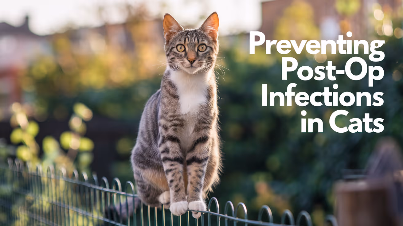
MRSP Prevention Through Proper Asepsis
Learn how proper asepsis prevents MRSP infections in pets with expert veterinary tips and practical steps.
MRSP, or methicillin-resistant Staphylococcus pseudintermedius, is a serious bacterial infection affecting pets, especially dogs. This resistant bacteria can cause skin infections, wounds, and other complications that are hard to treat. Preventing MRSP is crucial to protect your pet’s health and avoid costly treatments.
The best way to prevent MRSP is through proper asepsis, which means keeping everything clean and sterile during veterinary care and at home. This article explains how asepsis works, why it matters, and practical steps you can take to reduce MRSP risks for your pet.
What is MRSP and why is it dangerous?
MRSP is a type of bacteria that resists many common antibiotics. It mainly affects dogs but can also infect cats and other animals. Because it is resistant, infections caused by MRSP are harder to treat and can last longer.
MRSP can spread through contact with infected animals, contaminated surfaces, or improper hygiene. Understanding the dangers helps you appreciate why prevention is vital.
- Antibiotic resistance: MRSP bacteria do not respond to many antibiotics, making infections difficult and expensive to treat effectively.
- Infection risks: MRSP can cause skin infections, wounds, and sometimes more serious problems like surgical site infections.
- Transmission modes: The bacteria spread through direct contact with infected pets or contaminated objects, increasing infection chances.
- Public health concern: Though rare, MRSP can infect humans, especially those with weakened immune systems, highlighting the need for control.
Knowing these risks underlines the importance of strict aseptic techniques to stop MRSP from spreading.
How does proper asepsis prevent MRSP infections?
Asepsis means creating an environment free of harmful bacteria and germs. In veterinary medicine, it involves cleaning, disinfecting, and sterilizing tools, surfaces, and hands to prevent infection.
Proper asepsis breaks the chain of infection by removing or killing MRSP bacteria before they can infect your pet. This is essential during surgeries, wound care, and routine check-ups.
- Barrier protection: Using gloves, gowns, and masks prevents bacteria from spreading between animals and people during treatment.
- Surface disinfection: Cleaning exam tables and equipment removes MRSP bacteria that could infect other pets.
- Hand hygiene: Washing hands thoroughly before and after contact with pets stops bacteria transmission.
- Sterile instruments: Proper sterilization of surgical tools ensures no MRSP bacteria remain to cause infection.
Following these aseptic steps reduces MRSP infection chances and protects both pets and veterinary staff.
What are the key aseptic techniques in veterinary clinics?
Veterinary clinics use several aseptic methods to prevent MRSP and other infections. These techniques are standard practice and critical for safe pet care.
Understanding these methods helps pet owners recognize the importance of asepsis and support infection control efforts.
- Handwashing protocols: Staff wash hands with soap and water or use alcohol-based sanitizers before and after each patient contact.
- Use of personal protective equipment: Gloves, gowns, and masks protect both animals and staff from bacterial spread.
- Instrument sterilization: Autoclaving or chemical sterilization kills all bacteria on surgical tools before use.
- Environmental cleaning: Regular disinfection of floors, cages, and surfaces removes MRSP bacteria from the clinic environment.
These practices create a safer space for pets and reduce the risk of MRSP outbreaks in clinics.
How can pet owners maintain asepsis at home?
Preventing MRSP is not only a clinic responsibility. Pet owners play a big role in maintaining asepsis at home, especially if their pet has wounds or infections.
Simple hygiene steps can stop MRSP bacteria from spreading within your home and protect your pet’s recovery.
- Clean wound care: Always wash your hands and use clean materials when treating your pet’s wounds to avoid introducing bacteria.
- Disinfect pet items: Regularly clean bedding, collars, and toys with pet-safe disinfectants to reduce bacterial buildup.
- Limit contact: Keep infected pets separated from others to prevent MRSP spread within multi-pet households.
- Follow veterinary advice: Use prescribed antibiotics exactly as directed and complete the full course to prevent resistance.
By practicing good asepsis at home, you help your pet heal faster and reduce MRSP transmission risks.
What cleaning products are effective against MRSP?
Not all cleaning products kill MRSP bacteria. Choosing the right disinfectants is important to ensure proper asepsis.
Veterinarians recommend specific products that are proven to eliminate MRSP on surfaces and instruments.
- Chlorhexidine solutions: This antiseptic is effective for skin cleaning and surface disinfection against MRSP bacteria.
- Quaternary ammonium compounds: Commonly used disinfectants that kill MRSP on hard surfaces when used properly.
- Alcohol-based sanitizers: Hand sanitizers with at least 60% alcohol effectively kill MRSP on skin and hands.
- Hydrogen peroxide cleaners: These can disinfect surfaces but must be used carefully to avoid damage to materials.
Always follow product instructions and veterinary recommendations to ensure safe and effective MRSP control.
What are the signs of MRSP infection in pets?
Recognizing MRSP infection early helps get prompt treatment and prevents spread. Pets with MRSP may show signs similar to other infections but often do not respond well to standard antibiotics.
Knowing these signs helps you seek veterinary care quickly.
- Skin lesions: Red, swollen, or pus-filled sores that do not heal or worsen despite treatment may indicate MRSP infection.
- Persistent wounds: Cuts or surgical sites that stay inflamed or discharge fluid could be infected with resistant bacteria.
- Itching and discomfort: Pets may scratch or lick infected areas excessively due to irritation from MRSP.
- Fever and lethargy: In severe cases, systemic signs like fever and low energy may appear, signaling serious infection.
If you notice these symptoms, contact your veterinarian promptly for diagnosis and appropriate care.
Conclusion
MRSP is a challenging infection due to its antibiotic resistance, but proper asepsis can prevent it effectively. Both veterinary clinics and pet owners must follow strict hygiene and cleaning protocols to stop MRSP spread.
By understanding MRSP risks, practicing aseptic techniques, and recognizing infection signs early, you can protect your pet’s health and support successful treatment outcomes.
FAQs
How long can MRSP survive on surfaces?
MRSP can survive on surfaces for days to weeks, making regular cleaning and disinfection essential to prevent infection spread in homes and clinics.
Can humans get MRSP from pets?
Humans can rarely get MRSP, especially those with weak immune systems. Good hygiene reduces this risk significantly.
Is MRSP infection always visible on pets?
Not always. Some pets carry MRSP without symptoms but can still spread bacteria to others.
What should I do if my pet has MRSP?
Follow your veterinarian’s treatment plan carefully, maintain strict hygiene, and keep your pet isolated from others until cleared.
Can MRSP be cured?
Yes, with proper antibiotic treatment and aseptic care, most MRSP infections can be managed successfully, though they may take longer to heal.

Cruciate Sutures in Dog and Cat Skin Closure
Learn how cruciate sutures help close dog and cat skin wounds effectively with step-by-step guidance and care tips.
When your dog or cat needs skin closure after surgery or injury, choosing the right suture technique is crucial. Cruciate sutures are a popular method for closing skin wounds in dogs and cats because they provide strong wound support and promote healing. Understanding how cruciate sutures work can help you better care for your pet’s recovery.
This article explains what cruciate sutures are, how they differ from other suture patterns, and why veterinarians often prefer them for skin closure in small animals. You will also learn about the materials used, the suturing process, and aftercare tips to ensure your pet heals well.
What are cruciate sutures in dog and cat skin closure?
Cruciate sutures are a type of interrupted suture pattern shaped like a cross or X. They are designed to hold the edges of a skin wound firmly together while distributing tension evenly across the wound. This helps reduce the risk of wound dehiscence, where the skin pulls apart after surgery or injury.
These sutures are especially useful in areas where the skin moves a lot, such as joints or limbs, because they provide extra strength compared to simple interrupted sutures. The pattern also allows for good blood flow and healing by minimizing tissue strangulation.
- Cross-shaped pattern: Cruciate sutures form an X shape over the wound, providing balanced tension and secure closure for dog and cat skin wounds.
- Interrupted technique: Each suture is tied separately, so if one fails, the rest still hold the wound edges together, improving safety during healing.
- Strong wound support: The pattern distributes tension evenly, reducing pressure on any single point and lowering the chance of wound opening.
- Good for mobile areas: Cruciate sutures work well on joints and limbs where skin stretches and moves frequently, helping maintain closure.
Overall, cruciate sutures are a reliable choice for skin closure in dogs and cats, especially when strength and flexibility are needed to support healing.
How do cruciate sutures compare to other skin closure techniques?
There are many suture patterns used in veterinary skin closure, including simple interrupted, continuous, and mattress sutures. Cruciate sutures offer unique benefits compared to these methods, making them a preferred option in many cases.
Simple interrupted sutures are easy to place but may not distribute tension as well. Continuous sutures are faster but risk wound opening if the suture breaks. Mattress sutures provide strong tension relief but can cause more tissue damage. Cruciate sutures balance strength, security, and tissue preservation.
- Better tension distribution: Cruciate sutures spread tension evenly across the wound, unlike simple interrupted sutures that focus tension at individual points.
- Reduced risk of wound failure: Because each suture is independent, cruciate sutures maintain closure even if one knot loosens or breaks.
- Less tissue strangulation: Compared to mattress sutures, cruciate sutures minimize pressure on skin edges, promoting better blood flow and healing.
- Moderate placement time: Cruciate sutures take longer than simple interrupted but less time than complex patterns, balancing efficiency and effectiveness.
Choosing cruciate sutures depends on the wound location, size, and expected movement. They often provide the best combination of strength and healing support for dog and cat skin wounds.
What suture materials are best for cruciate skin closure in pets?
The choice of suture material affects healing, infection risk, and comfort for your pet. For cruciate sutures in dog and cat skin closure, veterinarians select materials that are strong, absorbable or non-absorbable, and cause minimal tissue reaction.
Absorbable sutures dissolve over time, reducing the need for removal, while non-absorbable sutures require removal but may offer longer-lasting support. The suture size also matters; smaller sizes reduce tissue trauma but must be strong enough to hold the wound.
- Absorbable options: Materials like poliglecaprone (Monocryl) or polyglycolic acid (Dexon) dissolve safely, ideal for internal layers or pets that resist suture removal.
- Non-absorbable options: Nylon or polypropylene sutures provide durable skin closure but require removal after healing to avoid irritation.
- Suture size choice: Sizes 3-0 to 4-0 are common for dog and cat skin, balancing strength and minimal tissue damage.
- Monofilament preferred: Single-strand sutures reduce infection risk and drag less through tissue compared to braided sutures.
Your veterinarian will select the best suture material based on your pet’s wound type, location, and healing needs to optimize recovery.
How is the cruciate suture technique performed step-by-step?
Performing cruciate sutures requires precision and care to ensure proper wound closure. The technique involves placing stitches in a cross pattern that securely holds the skin edges together without excessive tension.
Veterinarians follow a systematic approach to place each suture, tie knots correctly, and space sutures evenly. This promotes good healing and reduces complications like scarring or infection.
- Prepare the wound: Clean and debride the wound edges to remove debris and promote healthy tissue healing before suturing.
- Insert needle first pass: Pass the needle through one side of the skin edge about 5 mm from the wound margin, entering perpendicular to the skin surface.
- Cross to opposite side: Bring the needle across the wound and exit the opposite skin edge at a similar distance, forming the first diagonal of the X.
- Complete the X pattern: Reverse the needle direction to pass through the skin edges again, crossing the first suture line to form the cruciate or X shape.
After placing the suture, the knot is tied securely but not too tight to avoid strangulating the tissue. Sutures are spaced evenly along the wound, typically 5-10 mm apart, depending on wound size and location.
What are the benefits and risks of using cruciate sutures in pets?
Cruciate sutures offer many advantages for dog and cat skin closure but also carry some risks. Understanding these helps you recognize why your veterinarian may choose this method and what to watch for during healing.
Benefits include strong wound support, reduced tension, and good healing outcomes. Risks involve potential suture reactions, infection, or improper technique leading to wound complications.
- Strong wound closure: Cruciate sutures provide excellent mechanical support, reducing the chance of wound reopening in active pets.
- Even tension distribution: The pattern minimizes localized pressure, promoting better blood flow and faster healing of the skin edges.
- Lower infection risk: Using monofilament sutures and proper technique reduces bacterial colonization compared to other patterns.
- Possible suture reaction: Some pets may develop mild inflammation or irritation around suture sites, requiring monitoring and care.
Overall, cruciate sutures are a reliable choice for skin closure in dogs and cats, with benefits outweighing risks when placed correctly and cared for properly.
How should you care for your pet’s cruciate sutures after surgery?
Proper aftercare is essential to ensure your pet’s cruciate sutures heal well without complications. You will need to keep the wound clean, prevent your pet from licking or biting the sutures, and watch for signs of infection or wound problems.
Following your veterinarian’s instructions and scheduling suture removal if needed will help your pet recover quickly and comfortably.
- Keep wound clean and dry: Avoid bathing or wetting the sutured area until the veterinarian approves to prevent infection and suture loosening.
- Prevent licking or chewing: Use an Elizabethan collar or other protective devices to stop your pet from disturbing the sutures and causing wound damage.
- Monitor for infection signs: Watch for redness, swelling, discharge, or foul odor around the sutures and contact your vet if these occur.
- Follow suture removal schedule: Non-absorbable sutures usually need removal 10-14 days after surgery; keep appointments to avoid complications.
By providing careful wound care and following veterinary advice, you help your dog or cat heal safely and comfortably after cruciate suture skin closure.
Conclusion
Cruciate sutures are a strong and effective method for closing skin wounds in dogs and cats. Their cross-shaped pattern provides balanced tension and secure closure, making them ideal for active pets and mobile skin areas.
Choosing the right suture material, performing the technique carefully, and providing proper aftercare are key to successful healing. Understanding cruciate sutures helps you support your pet’s recovery and recognize when veterinary care is needed.
What is the main advantage of cruciate sutures over simple interrupted sutures?
Cruciate sutures distribute tension more evenly across the wound, reducing pressure points and providing stronger, more secure skin closure than simple interrupted sutures.
Can cruciate sutures be used on all types of skin wounds in dogs and cats?
They are suitable for most skin wounds, especially where strength and movement resistance are needed, but very small or delicate wounds may require different suture patterns.
How long do cruciate sutures stay in a dog or cat before removal?
Non-absorbable cruciate sutures are usually removed 10 to 14 days after placement, depending on the wound healing progress and veterinarian’s advice.
Are absorbable sutures effective for cruciate skin closure?
Yes, absorbable sutures can be used for cruciate patterns, especially for pets that may not tolerate suture removal, but they must maintain strength long enough for healing.
What signs indicate a problem with cruciate sutures after surgery?
Signs include redness, swelling, discharge, wound opening, or excessive licking, which require prompt veterinary evaluation to prevent complications.
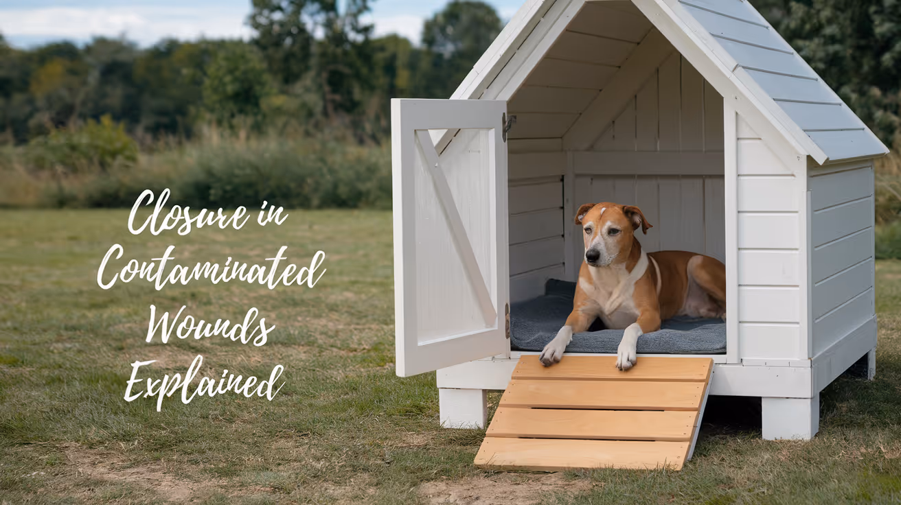
Closure in Contaminated or Dirty Wounds in Dogs
Learn how to safely manage closure in contaminated or dirty wounds in dogs to prevent infection and promote healing.
Introduction
Contaminated or dirty wounds in dogs pose a significant challenge in veterinary care. These wounds carry a high risk of infection and complications if not managed properly.
Proper closure techniques are essential to promote healing and prevent further issues. This article explains how to approach closure in such wounds safely and effectively.
What is closure in contaminated or dirty wounds in dogs?
Closure refers to the process of sealing a wound to allow healing. In contaminated or dirty wounds, closure must balance infection control with tissue repair.
These wounds often contain bacteria, debris, or dead tissue, increasing the risk of infection if closed improperly.
- Definition of closure: The act of bringing wound edges together to promote tissue healing and restore skin integrity in dogs.
- Contaminated wound meaning: A wound exposed to bacteria or foreign material that increases infection risk if closed too early.
- Dirty wound characteristics: Wounds with dead tissue, pus, or heavy contamination requiring special care before closure.
- Importance of closure timing: Closing too soon can trap bacteria, while delayed closure may prolong healing and increase scarring.
Understanding these basics helps in deciding the best closure method for each wound.
When should closure be performed on contaminated wounds in dogs?
Timing is critical in closing contaminated wounds. Immediate closure is often unsafe due to infection risk.
Veterinarians usually wait until the wound is clean and healthy before closing it to reduce complications.
- Early closure risks: Closing a contaminated wound immediately can trap bacteria, leading to abscesses or systemic infection.
- Delayed closure benefits: Waiting 3 to 5 days allows infection control and tissue assessment before sealing the wound.
- Signs for closure readiness: Healthy granulation tissue, absence of pus, and reduced swelling indicate the wound is ready.
- Use of staged closure: Some wounds require multiple cleaning sessions before final closure to ensure safety.
Proper timing reduces infection risk and improves healing outcomes.
What are the common closure techniques for dirty wounds in dogs?
Several closure methods exist, each suited to different wound conditions. Choosing the right technique depends on contamination level and tissue health.
Techniques range from leaving wounds open to various suturing methods after cleaning.
- Secondary intention healing: Leaving the wound open to heal naturally when contamination is high or tissue loss is extensive.
- Delayed primary closure: Cleaning the wound first, then closing it surgically after infection control, usually within 3 to 5 days.
- Primary closure: Immediate suturing for clean wounds with minimal contamination and good tissue viability.
- Use of drains: Placing drains to remove fluid and prevent abscess formation in deeper or heavily contaminated wounds.
Each method aims to balance infection control with optimal healing.
How should you prepare a contaminated wound before closure in dogs?
Preparation is key to successful closure. Cleaning and debridement remove bacteria and dead tissue that impair healing.
Proper wound preparation reduces infection risk and creates a healthy environment for tissue repair.
- Thorough cleaning: Use sterile saline or antiseptic solutions to flush out debris and bacteria from the wound.
- Debridement importance: Removing dead or damaged tissue prevents bacterial growth and promotes healthy granulation.
- Antibiotic use: Systemic or topical antibiotics may be prescribed to control infection before closure.
- Assessing tissue viability: Only healthy, well-perfused tissue should be closed to ensure proper healing.
Following these steps prepares the wound for safer closure and better recovery.
What are the risks of improper closure in contaminated wounds in dogs?
Closing contaminated wounds incorrectly can lead to serious complications. Understanding these risks helps avoid mistakes.
Proper technique and timing are essential to prevent worsening infection and promote healing.
- Infection development: Trapping bacteria inside the wound can cause abscesses, cellulitis, or systemic illness.
- Delayed healing: Infection and tissue death slow down the repair process, prolonging recovery.
- Wound dehiscence: Poor closure can cause the wound to reopen, requiring additional surgery.
- Scarring and dysfunction: Improper healing may result in excessive scar tissue, affecting skin flexibility and appearance.
Recognizing these risks emphasizes the need for careful wound management and closure decisions.
How can you care for a dog’s wound after closure?
Post-closure care is vital to ensure healing and prevent infection. Owners must follow veterinary instructions closely.
Proper wound care supports tissue repair and reduces complications.
- Keep wound clean: Avoid dirt and moisture contact to prevent new contamination during healing.
- Monitor for infection: Watch for redness, swelling, discharge, or odor and report concerns promptly.
- Restrict activity: Limit movement to prevent stress on the wound and avoid reopening.
- Follow medication plan: Administer prescribed antibiotics or pain relief as directed by the veterinarian.
Good aftercare improves healing speed and reduces the chance of complications.
Conclusion
Closure in contaminated or dirty wounds in dogs requires careful timing, cleaning, and technique to prevent infection and promote healing.
Understanding when and how to close these wounds helps ensure the best outcome for your dog’s recovery and comfort.
FAQs
Can all contaminated wounds in dogs be closed immediately?
No, most contaminated wounds require cleaning and a delay before closure to reduce infection risk and ensure healthy tissue repair.
What is delayed primary closure in dog wounds?
Delayed primary closure involves cleaning the wound first and closing it surgically after a few days when infection is controlled and tissue is healthy.
How often should a dog’s wound be cleaned before closure?
Wounds should be cleaned daily or as directed by a veterinarian until they show healthy granulation tissue and are free of infection signs.
Are antibiotics always needed for contaminated wound closure?
Antibiotics are commonly prescribed to control infection but depend on wound severity and veterinarian assessment.
What signs indicate a wound infection after closure?
Signs include redness, swelling, heat, discharge, foul odor, pain, or the wound reopening, requiring prompt veterinary attention.

Closure Protocol for Laparotomy in Cats
Learn the detailed closure protocol for laparotomy in cats, including step-by-step surgical techniques and post-op care.
What is the closure protocol for laparotomy in cats?
Laparotomy in cats is a common surgical procedure involving an incision into the abdominal cavity. Proper closure of the incision is crucial to ensure healing, prevent infection, and avoid complications like hernias.
The closure protocol for laparotomy in cats involves layered suturing techniques, choice of suture materials, and careful tissue handling. This article explains the detailed steps and considerations for closing a cat’s laparotomy incision effectively.
- Layered closure approach: The abdominal wall is closed in layers including the peritoneum, muscle, and subcutaneous tissue to provide strength and reduce dead space.
- Suture material choice: Absorbable monofilament sutures like polydioxanone (PDS) are preferred for internal layers to minimize tissue reaction and maintain tensile strength.
- Skin closure methods: Skin can be closed with non-absorbable sutures, staples, or tissue glue depending on surgeon preference and wound location.
- Gentle tissue handling: Minimizing trauma during closure reduces inflammation and promotes faster healing.
Following this protocol helps reduce post-operative complications and supports optimal recovery for cats undergoing laparotomy.
Why is layered closure important in feline laparotomy?
Layered closure is essential because the abdominal wall consists of multiple tissue layers, each with different healing properties. Closing each layer separately restores the abdominal wall’s strength and function.
Failing to close layers properly can lead to complications such as herniation, wound dehiscence, or infection. Layered closure distributes tension evenly across the incision site.
- Peritoneal closure benefits: Closing the peritoneum prevents abdominal contents from contacting the muscle and subcutaneous layers, reducing infection risk.
- Muscle layer strength: Suturing the muscle layer restores abdominal wall integrity and supports internal organs.
- Subcutaneous tissue role: Closing subcutaneous fat reduces dead space where fluid can accumulate, lowering seroma risk.
- Skin layer protection: Proper skin closure protects underlying tissues from external contaminants and aids cosmetic healing.
Each layer plays a vital role in healing, making layered closure a standard practice in feline laparotomy surgeries.
What suture materials are best for laparotomy closure in cats?
Choosing the right suture material is critical for successful laparotomy closure. The material must provide adequate strength, cause minimal tissue reaction, and maintain integrity during healing.
Absorbable sutures are generally preferred for internal layers, while skin closure may use non-absorbable sutures or alternatives depending on the case.
- Polydioxanone (PDS): A slowly absorbable monofilament suture ideal for muscle and peritoneal layers due to prolonged tensile strength.
- Polyglactin 910 (Vicryl): A braided absorbable suture suitable for subcutaneous tissue but may cause more tissue reaction than monofilaments.
- Nylon or polypropylene: Non-absorbable monofilament sutures commonly used for skin closure because they resist infection and maintain wound edge apposition.
- Tissue glue option: Cyanoacrylate-based glues can be used for skin closure in some cases, offering quick sealing and reduced suture removal stress.
Proper suture selection tailored to each tissue layer promotes healing and reduces complications in feline laparotomy closures.
How should the abdominal wall be closed during feline laparotomy?
Closing the abdominal wall requires precise technique to restore strength and prevent complications. The surgeon must suture each layer carefully with appropriate tension and spacing.
The peritoneum, muscle, and fascia are typically closed together or separately depending on surgeon preference. The goal is to achieve a secure, tension-free closure.
- Peritoneal closure technique: Use simple continuous or interrupted sutures with absorbable material to close the peritoneum without excessive tension.
- Muscle and fascia closure: Incorporate the external rectus sheath and muscle fibers with strong, evenly spaced sutures to restore abdominal wall integrity.
- Suture spacing guidelines: Sutures should be placed 5-10 mm apart and 5-10 mm from the incision edge to distribute tension evenly.
- Avoiding excessive tension: Over-tightening sutures can cause tissue ischemia and increase risk of dehiscence; aim for snug but not constrictive closure.
Following these techniques ensures a durable abdominal wall closure that supports healing and reduces postoperative complications.
What are the best practices for skin closure after feline laparotomy?
Skin closure is the final step in laparotomy and protects the underlying tissues. Choosing the right method and technique affects healing time, infection risk, and cosmetic outcome.
Options include sutures, staples, or tissue adhesives. The choice depends on wound location, surgeon preference, and patient factors.
- Interrupted sutures: Provide precise wound edge apposition and allow removal of individual sutures if infection occurs.
- Continuous sutures: Faster to place and distribute tension evenly but risk wound opening if one part fails.
- Staples: Quick to apply and reduce surgery time but may cause more skin irritation and require removal.
- Tissue glue: Useful for small, clean incisions offering waterproof closure and no need for suture removal.
Proper skin closure technique reduces infection risk and supports faster healing after feline laparotomy.
How should post-operative care be managed after laparotomy closure in cats?
Post-operative care is vital to ensure the laparotomy incision heals well and complications are minimized. Monitoring and supportive care help detect problems early.
Owners and veterinarians must follow specific guidelines to protect the surgical site and promote recovery.
- Incision monitoring: Check daily for redness, swelling, discharge, or opening that may indicate infection or dehiscence.
- Activity restriction: Limit jumping, running, and rough play for 10-14 days to prevent stress on the incision.
- E-collar use: Prevent the cat from licking or biting the incision to avoid contamination and damage.
- Follow-up visits: Schedule veterinary checks to assess healing progress and remove skin sutures or staples as needed.
Careful post-operative management supports successful healing after laparotomy closure in cats.
What complications can occur if laparotomy closure is improper in cats?
Improper closure of the laparotomy incision can lead to serious complications that affect the cat’s health and recovery. Recognizing these risks highlights the importance of following the closure protocol.
Complications may require additional treatment or surgery and prolong healing time.
- Wound dehiscence: Partial or complete opening of the incision due to poor suturing or excessive tension can expose internal organs.
- Herniation risk: Failure to close the abdominal wall properly can cause abdominal contents to protrude through the incision site.
- Infection development: Inadequate aseptic technique or closure can lead to bacterial contamination and wound infection.
- Seroma formation: Dead space left in subcutaneous tissue can accumulate fluid, causing swelling and delaying healing.
Following the recommended closure protocol minimizes these risks and improves surgical outcomes for cats.
Conclusion
Closure protocol for laparotomy in cats is a critical part of surgical success. Proper layered closure, suture selection, and careful technique help restore abdominal wall integrity and promote healing.
Post-operative care and monitoring further support recovery and reduce complications. Following these guidelines ensures the best outcomes for cats undergoing laparotomy procedures.
What suture pattern is recommended for closing the abdominal muscle layer?
The simple continuous or interrupted suture pattern with absorbable monofilament material is recommended to provide strength and even tension distribution in the muscle layer.
How long does it take for a laparotomy incision in cats to heal?
Typically, the incision heals externally within 10-14 days, but full internal healing of the abdominal wall can take 4-6 weeks depending on the cat’s health and care.
Can tissue glue be used instead of sutures for skin closure?
Yes, tissue glue is suitable for small, clean incisions and offers quick closure without the need for suture removal, but it is not recommended for large or high-tension wounds.
When should sutures or staples be removed after laparotomy?
Skin sutures or staples are usually removed 10-14 days post-surgery once the skin edges have healed sufficiently to maintain closure without support.
What signs indicate a post-operative complication at the incision site?
Signs include redness, swelling, discharge, wound opening, excessive pain, or fever. These require prompt veterinary evaluation to prevent worsening complications.
Closure Protocol for Tumor Excision in Dogs
Learn the detailed closure protocol for tumor excision in dogs to ensure proper healing and reduce complications after surgery.
Tumor excision in dogs is a common surgical procedure that requires careful closure to promote healing and prevent complications. Proper closure protocol is essential to minimize infection risk, reduce scarring, and ensure the best outcome for your pet.
This article explains the closure protocol for tumor excision in dogs, including step-by-step techniques, materials used, and postoperative care. You will learn how veterinarians close surgical wounds after tumor removal and what you should expect during recovery.
What is the closure protocol for tumor excision in dogs?
The closure protocol for tumor excision in dogs involves several steps to close the surgical wound securely and promote healing. It includes layered closure of tissues, selection of appropriate suture materials, and techniques to reduce tension on the skin.
Following a strict closure protocol helps prevent wound dehiscence, infection, and excessive scarring after tumor removal surgery.
- Layered closure technique: Closing the wound in multiple layers including muscle, subcutaneous tissue, and skin to provide strength and reduce dead space.
- Suture material choice: Using absorbable sutures for deeper layers and non-absorbable or absorbable sutures for skin depending on healing needs.
- Tension reduction methods: Employing techniques such as undermining skin edges or using tension-relieving sutures to avoid wound stress.
- Aseptic handling: Maintaining sterile conditions during closure to minimize infection risk.
Proper closure protocol is critical for a successful recovery after tumor excision in dogs.
Why is layered closure important after tumor excision in dogs?
Layered closure is important because it restores the normal anatomy of the surgical site and provides mechanical strength to the wound. Each tissue layer has different healing properties and functions, so closing them separately improves outcomes.
Failing to close layers properly can lead to complications like seroma formation, wound breakdown, or delayed healing.
- Muscle layer closure: Re-approximates muscle fibers to restore function and reduce dead space where fluid can accumulate.
- Subcutaneous tissue closure: Supports skin edges and reduces tension on the skin closure line.
- Skin closure: Protects the wound from external contaminants and allows epithelial healing.
- Dead space elimination: Layered closure prevents pockets where blood or fluid can collect, reducing infection risk.
Layered closure ensures the surgical site heals efficiently and reduces postoperative complications.
What suture materials are best for closing tumor excision wounds in dogs?
Choosing the right suture material is essential for wound strength and healing. Different layers require different suture types based on tissue properties and healing time.
Veterinarians select suture materials that balance strength, absorption rate, and tissue reaction to optimize healing after tumor excision.
- Absorbable sutures: Used for muscle and subcutaneous layers to avoid the need for removal and support healing over weeks.
- Non-absorbable sutures: Sometimes used for skin closure when prolonged wound support is needed, requiring later removal.
- Monofilament sutures: Preferred to reduce tissue drag and lower infection risk compared to braided sutures.
- Suture size selection: Smaller sizes reduce tissue trauma but must be strong enough to hold tissues securely.
Proper suture choice helps maintain wound integrity and promotes smooth healing after tumor removal.
How do veterinarians reduce tension on the skin during closure?
Reducing tension on the skin edges during closure is crucial to prevent wound dehiscence and improve cosmetic results. Several techniques help distribute tension evenly and protect the wound.
These methods allow the skin to heal without excessive stress that can cause tearing or delayed healing.
- Undermining skin edges: Separating skin from underlying tissues to allow easier approximation without tension.
- Tension-relieving sutures: Placing deep sutures that offload stress from the skin closure line.
- Use of skin staples or adhesive strips: Supplementing sutures to distribute tension and support the wound.
- Proper incision planning: Designing incisions along skin tension lines to minimize stress during closure.
These techniques improve wound strength and reduce complications after tumor excision.
What postoperative care is needed after tumor excision closure in dogs?
After closure, proper postoperative care is vital to support healing and detect complications early. Owners must follow veterinary instructions closely to ensure the best recovery.
Good care helps prevent infection, wound opening, and discomfort for your dog.
- Wound monitoring: Regularly check the incision for redness, swelling, discharge, or opening to catch problems early.
- Prevent licking or chewing: Use an Elizabethan collar or other devices to stop the dog from disturbing the wound.
- Limit activity: Restrict running, jumping, or rough play to avoid stress on the healing wound.
- Follow medication schedule: Administer prescribed antibiotics or pain medications exactly as directed by the veterinarian.
Careful postoperative management supports healing and reduces the risk of complications after tumor excision.
What complications can occur if closure protocol is not followed?
Ignoring proper closure protocol can lead to several complications that affect your dog's recovery and health. Understanding these risks highlights the importance of meticulous surgical technique.
Prompt veterinary attention is needed if complications arise after tumor excision.
- Wound dehiscence: The surgical site may reopen if closure is weak or under tension, exposing tissues to infection.
- Infection risk: Poor aseptic technique or dead space can lead to bacterial infection requiring additional treatment.
- Seroma or hematoma formation: Fluid accumulation under the skin can delay healing and cause discomfort.
- Excessive scarring: Improper closure can result in large or unsightly scars affecting skin function and appearance.
Following a strict closure protocol minimizes these risks and promotes a smooth recovery.
How long does it take for a dog to heal after tumor excision closure?
The healing time after tumor excision closure varies depending on the tumor size, location, and the dog's overall health. Generally, skin wounds heal within 10 to 14 days, but deeper tissues take longer.
Understanding the healing timeline helps owners provide proper care and know when to seek veterinary advice.
- Initial healing phase: The first 3 to 5 days involve inflammation and early tissue repair with swelling and redness expected.
- Skin suture removal: Usually occurs 10 to 14 days after surgery once the skin has healed sufficiently.
- Complete healing: Deeper tissues like muscle and subcutaneous layers may take 4 to 6 weeks to fully recover.
- Activity restriction duration: Dogs should have limited activity for at least 2 weeks to protect the wound during healing.
Following veterinary instructions during the healing period ensures the best outcome after tumor excision closure.
Conclusion
The closure protocol for tumor excision in dogs is a critical part of the surgical process that ensures proper healing and reduces complications. It involves layered closure, careful suture selection, tension reduction, and strict aseptic technique.
Owners play an important role in postoperative care by monitoring the wound, preventing self-trauma, and following veterinary advice. Understanding this protocol helps you support your dog's recovery and achieve the best surgical outcome.
FAQs
How soon can my dog go home after tumor excision surgery?
Most dogs can go home the same day or the day after surgery once they are stable and pain is controlled. Your veterinarian will provide specific discharge instructions.
When should I remove my dog's skin sutures after tumor excision?
Skin sutures are typically removed 10 to 14 days after surgery, depending on healing progress. Your vet will advise the best timing during follow-up visits.
Can my dog get an infection after tumor excision closure?
Yes, infection is possible if the wound is contaminated or closure is poor. Watch for redness, swelling, or discharge and contact your vet if signs appear.
Is it normal for my dog’s incision to be swollen after surgery?
Mild swelling and redness are normal in the first few days after surgery. Excessive swelling or heat may indicate infection and should be evaluated by a vet.
What should I do if my dog licks or chews the surgical site?
Use an Elizabethan collar or protective clothing to prevent licking or chewing, which can cause wound damage and infection. Contact your vet if the wound is disturbed.
Surgical Site Infection Prevention in Cats
Learn effective surgical site infection prevention in cats with expert tips on hygiene, antibiotics, and wound care to keep your cat safe.
Surgical site infections (SSIs) in cats are a serious concern that can complicate recovery and cause pain or illness. Preventing these infections is crucial for your cat’s health after surgery. Understanding how to reduce the risk of SSIs helps you protect your cat and support healing.
This article explains surgical site infection prevention in cats. You will learn about hygiene practices, antibiotic use, wound care, and monitoring to keep your cat safe from infections after surgery.
What causes surgical site infections in cats?
Surgical site infections occur when bacteria enter the wound during or after surgery. These bacteria multiply and cause inflammation, redness, swelling, and sometimes pus. Knowing the causes helps you prevent infections effectively.
Common sources of infection include the cat’s skin, the surgical environment, and even the surgical team. Understanding these factors is key to reducing risk.
- Skin bacteria: Cats naturally have bacteria on their skin that can enter the surgical wound if not properly cleaned before surgery.
- Environmental contamination: Unclean surgical tools or surfaces can introduce bacteria into the wound during the procedure.
- Improper surgical technique: Poor handling or long surgery times increase the risk of bacteria entering the wound.
- Postoperative care lapses: If the wound is not kept clean and dry after surgery, bacteria can infect the site.
By controlling these causes, you can lower the chance of your cat developing an SSI.
How can hygiene prevent surgical site infections in cats?
Hygiene is the first line of defense against infections. Both the surgical team and the pet owner play roles in maintaining cleanliness before, during, and after surgery.
Proper hygiene reduces bacteria around the surgical site and prevents contamination. This includes skin preparation, surgical environment cleanliness, and wound care hygiene.
- Pre-surgical skin prep: Shaving and disinfecting the surgical area removes hair and bacteria, reducing infection risk.
- Sterile surgical tools: Using sterilized instruments ensures no bacteria are introduced during surgery.
- Clean surgical environment: Operating rooms should be sanitized regularly to minimize bacterial presence.
- Owner wound care: Keeping the wound clean and dry at home prevents bacteria from infecting the site.
Maintaining strict hygiene protocols is essential for preventing SSIs in cats.
When should antibiotics be used to prevent surgical site infections in cats?
Antibiotics can help prevent infections but should be used carefully to avoid resistance. Your veterinarian decides when antibiotics are necessary based on the surgery type and infection risk.
Not all surgeries require antibiotics. They are most useful in high-risk cases or when the wound is contaminated.
- Prophylactic antibiotics: Given before surgery to prevent infection in high-risk procedures or immunocompromised cats.
- Therapeutic antibiotics: Used after surgery if signs of infection appear to treat existing bacteria.
- Avoid unnecessary use: Overusing antibiotics can cause resistance and harm your cat’s health.
- Follow vet instructions: Always give antibiotics exactly as prescribed to ensure effectiveness.
Proper antibiotic use supports infection prevention without causing harm.
What wound care practices help prevent infections in cats?
After surgery, careful wound care is vital to prevent bacteria from entering the site. You must monitor the wound and keep it protected during healing.
Good wound care reduces swelling, irritation, and contamination, which lowers infection risk.
- Keep wound dry: Moisture promotes bacterial growth, so avoid bathing or wetting the wound area.
- Prevent licking or scratching: Use an Elizabethan collar to stop your cat from disturbing the wound.
- Clean gently: Follow vet advice on cleaning with mild antiseptics if needed, avoiding harsh chemicals.
- Watch for signs: Redness, swelling, or discharge may indicate infection and need prompt vet attention.
Consistent wound care helps your cat heal faster and stay infection-free.
How does surgical technique affect infection risk in cats?
The skill and methods used during surgery impact the chance of infection. Surgeons must follow best practices to minimize tissue damage and contamination.
Good surgical technique reduces wound exposure and speeds healing, lowering infection chances.
- Minimize tissue trauma: Gentle handling preserves blood flow and immune response at the wound site.
- Use sterile gloves and instruments: Prevents bacteria transfer during surgery.
- Limit surgery time: Shorter procedures reduce exposure to environmental bacteria.
- Proper wound closure: Secure sutures or staples protect the site from bacteria entering.
Experienced surgeons following strict protocols help ensure safer outcomes for your cat.
What signs indicate a surgical site infection in cats?
Recognizing infection signs early allows quick treatment to prevent complications. You should check the surgical site daily during recovery.
Common signs include redness, swelling, and discharge. Knowing these helps you act promptly.
- Redness and warmth: The area around the wound may look redder and feel warm due to inflammation.
- Swelling or lumps: Infection causes tissue swelling or raised bumps near the incision.
- Pus or discharge: Yellow, green, or foul-smelling fluid leaking from the wound signals infection.
- Behavior changes: Your cat may lick the wound excessively, show pain, or have reduced appetite.
If you notice these signs, contact your veterinarian immediately for evaluation and treatment.
Conclusion
Preventing surgical site infections in cats requires careful attention before, during, and after surgery. Hygiene, proper antibiotic use, skilled surgical technique, and diligent wound care all play vital roles.
By understanding infection causes and watching for warning signs, you can help your cat recover safely and comfortably. Always follow your veterinarian’s advice to protect your cat’s health after surgery.
What is the best way to prepare a cat’s skin before surgery?
Shaving the surgical area and cleaning it with an antiseptic solution removes hair and bacteria, reducing infection risk during surgery.
How long should antibiotics be given after surgery in cats?
Antibiotic duration depends on the surgery and vet’s advice, usually ranging from a single dose before surgery to several days after if infection risk is high.
Can a cat’s licking cause a surgical site infection?
Yes, licking can introduce bacteria and irritate the wound, increasing infection risk. Using an Elizabethan collar helps prevent this behavior.
When should I contact the vet about my cat’s surgical wound?
Contact your vet if you notice redness, swelling, discharge, foul odor, or if your cat shows pain or lethargy during recovery.
Are surgical site infections common in cats?
SSIs are relatively uncommon with proper care but can occur. Following hygiene and wound care guidelines greatly reduces the risk.

Closure Considerations in Geriatric Dogs and Cats
Learn essential closure considerations for geriatric dogs and cats to ensure safe, effective surgical outcomes and recovery.
Older dogs and cats often require surgeries due to age-related health issues. However, closing surgical wounds in geriatric pets needs special care to avoid complications. Closure considerations in geriatric dogs and cats are critical for safe healing and reducing risks.
This article explains key closure techniques, materials, and precautions for older pets. You will learn how to manage fragile skin, delayed healing, and other age-related factors to improve surgical outcomes in your senior dog or cat.
What are the main challenges in closing wounds in geriatric dogs and cats?
Older pets have unique challenges that affect wound closure. Their skin is thinner and less elastic, which can cause sutures to tear through. Healing is slower due to reduced blood flow and immune function. These factors increase the risk of wound dehiscence and infection.
Understanding these challenges helps veterinarians choose the best closure methods and materials for geriatric patients.
- Fragile skin: Geriatric pets have thinner, less elastic skin that tears easily, requiring gentle handling and careful suture placement.
- Delayed healing: Reduced blood flow and immune response slow tissue repair, increasing infection and dehiscence risk.
- Comorbidities: Chronic diseases like diabetes or kidney issues impair healing and affect anesthesia and recovery.
- Reduced collagen: Lower collagen production weakens tissue strength, making wound closure less secure.
These challenges demand tailored closure techniques and close postoperative monitoring to ensure successful healing in older dogs and cats.
Which suture materials are best for geriatric pets?
Choosing the right suture material is vital for wound strength and minimizing tissue reaction. Absorbable sutures reduce the need for removal, which is helpful for older pets who may be stressed by repeated handling. Monofilament sutures cause less tissue drag and inflammation.
Material choice depends on wound location, tension, and expected healing time.
- Monofilament absorbable sutures: Materials like poliglecaprone cause minimal tissue reaction and maintain strength during slow healing.
- Non-absorbable monofilaments: Nylon or polypropylene are good for skin closure but require removal, which may stress geriatric pets.
- Delayed absorption sutures: Polydioxanone offers prolonged support, ideal for slow-healing tissues in older animals.
- Minimal tissue reaction: Choosing sutures that reduce inflammation helps prevent complications in fragile geriatric skin.
Veterinarians often prefer monofilament absorbable sutures for internal layers and may combine with non-absorbable for skin, balancing strength and patient comfort.
How should incision closure techniques be adapted for older dogs and cats?
Older pets benefit from modified closure techniques that reduce tension and protect fragile skin. Layered closure supports deeper tissues and distributes stress evenly. Avoiding tight sutures prevents skin tearing and necrosis.
Using appropriate needle size and spacing also helps minimize trauma.
- Layered closure: Closing muscle, subcutaneous tissue, and skin separately reduces tension on the skin and improves wound strength.
- Wide suture spacing: Placing sutures farther apart decreases skin tearing risk in thin geriatric skin.
- Use of tension-relieving sutures: Techniques like horizontal mattress sutures help distribute tension and protect wound edges.
- Gentle tissue handling: Minimizing trauma during closure preserves blood supply and promotes healing.
Adapting closure techniques to the pet’s age and skin condition helps prevent complications and supports better recovery.
What postoperative care is essential for wound healing in geriatric pets?
After surgery, geriatric dogs and cats need careful monitoring to detect early signs of complications. Protecting the wound from licking and trauma is critical. Nutrition and hydration also influence healing quality.
Owners should follow veterinary instructions closely and report any concerns promptly.
- Wound monitoring: Regularly check for redness, swelling, discharge, or opening to catch infection or dehiscence early.
- Prevent licking: Use Elizabethan collars or bandages to stop pets from disturbing the incision site.
- Proper nutrition: Balanced diets rich in protein and vitamins support tissue repair and immune function.
- Hydration maintenance: Adequate fluids improve circulation and help deliver nutrients to healing tissues.
Good postoperative care reduces complications and speeds recovery in older pets.
When should surgical drains be considered in geriatric patients?
Surgical drains help remove fluid buildup that can delay healing or cause infection. In geriatric pets, drains may be useful when dead space or excessive fluid accumulation is expected. However, they require careful management to avoid additional risks.
Deciding on drain use depends on surgery type and patient condition.
- Dead space reduction: Drains prevent fluid accumulation in large surgical cavities that impair healing.
- Infection control: Removing fluid reduces bacterial growth risk in compromised older immune systems.
- Drain type selection: Closed suction drains minimize contamination compared to open drains.
- Close monitoring: Drains require daily inspection and timely removal to prevent complications.
Drains can be beneficial but must be used judiciously in geriatric patients with strict postoperative care.
How do comorbidities affect closure decisions in older dogs and cats?
Many older pets have chronic diseases that impact wound healing and anesthesia risks. Conditions like diabetes, kidney disease, or heart problems require tailored surgical and closure plans. These comorbidities can delay healing and increase infection risk.
Veterinarians must consider these factors when planning closure and postoperative care.
- Diabetes impact: High blood sugar impairs immune function and collagen formation, slowing wound healing.
- Kidney disease effects: Reduced toxin clearance affects tissue repair and anesthesia tolerance.
- Cardiac conditions: Poor circulation decreases oxygen delivery to healing tissues, risking necrosis.
- Medication interactions: Drugs for chronic diseases may affect clotting or immune response, influencing closure choices.
Accounting for comorbidities helps optimize closure techniques and improve surgical outcomes in geriatric pets.
Conclusion
Closure considerations in geriatric dogs and cats are essential to ensure safe and effective surgical healing. Fragile skin, delayed healing, and comorbidities require careful suture selection, adapted techniques, and close postoperative care.
By understanding these factors, veterinarians and pet owners can work together to support recovery and improve quality of life for older pets after surgery.
What suture patterns are best for fragile geriatric skin?
Simple interrupted and horizontal mattress sutures are preferred for fragile skin as they distribute tension evenly and reduce the risk of suture pull-through and skin tearing.
How long does wound healing take in older dogs and cats?
Healing in geriatric pets can take 1.5 to 2 times longer than in younger animals, often requiring 14 to 21 days or more depending on health and wound type.
Can older pets tolerate general anesthesia for surgery?
Older pets can tolerate anesthesia with proper preoperative assessment, monitoring, and tailored protocols to minimize risks related to age and comorbidities.
Are staples better than sutures for closing geriatric skin?
Staples may cause more skin trauma and are less flexible than sutures, so sutures are generally preferred for delicate geriatric skin closure.
When should a veterinarian remove sutures in a senior pet?
Sutures in geriatric pets are usually removed 10 to 14 days post-surgery, but timing depends on healing progress and the pet’s overall health status.

Closure Strategy in Emergency Surgery
Learn about closure strategies in emergency surgery, including techniques, materials, risks, and best practices for optimal healing.
Emergency surgery often requires rapid and effective wound closure to prevent complications and promote healing. Closure strategy in emergency surgery involves choosing the right techniques and materials to ensure the surgical site heals properly despite urgent conditions.
This article explains the key factors in closure strategy during emergency surgery. You will learn about different closure methods, materials used, risk management, and how to optimize outcomes for your pet or patient.
What is closure strategy in emergency surgery?
Closure strategy refers to the plan and techniques used to close surgical wounds after emergency operations. It is critical because emergency surgeries often involve contaminated or complex wounds that need special care.
Choosing the right closure method helps reduce infection risk and supports faster healing. The strategy depends on wound type, location, and patient condition.
- Definition of closure strategy: It is the selection of methods and materials to close surgical wounds effectively after emergency procedures.
- Importance in emergencies: Proper closure prevents infection, fluid leakage, and supports tissue repair under urgent conditions.
- Factors influencing choice: Wound contamination, tissue damage, patient stability, and surgery type guide closure decisions.
- Goal of closure: To restore tissue integrity, minimize complications, and promote rapid healing.
Understanding closure strategy helps surgeons make informed decisions during emergencies to improve patient outcomes.
What are common closure techniques used in emergency surgery?
Several closure techniques are used depending on wound characteristics and urgency. Each technique has advantages and limitations in emergency settings.
Surgeons select from primary, delayed primary, or secondary closure based on contamination and tissue condition.
- Primary closure: Immediate suturing of a clean wound to promote quick healing and reduce infection risk.
- Delayed primary closure: Wound is initially left open to monitor for infection, then closed after a few days.
- Secondary closure: Wound heals naturally without suturing, used for heavily contaminated wounds.
- Stapling and adhesive use: Staples or tissue adhesives may be used for rapid closure in accessible wounds.
Choosing the right technique balances speed and safety to optimize healing in emergency surgery.
Which materials are best for wound closure in emergencies?
Material choice impacts wound strength, infection risk, and healing speed. Emergency surgery often requires materials that are strong, sterile, and easy to use quickly.
Sutures, staples, and adhesives each have roles depending on wound type and location.
- Absorbable sutures: Used internally to avoid removal, they dissolve as tissue heals, reducing follow-up procedures.
- Non-absorbable sutures: Provide long-term support for skin closure but require removal after healing.
- Staples: Allow fast closure of large wounds but may cause more tissue trauma than sutures.
- Tissue adhesives: Useful for small, clean wounds; they reduce closure time and minimize scarring.
Material selection depends on wound size, location, and contamination level to ensure effective closure.
How do surgeons manage infection risk during closure?
Infection is a major concern in emergency surgery due to contaminated wounds and urgent conditions. Surgeons use several strategies to reduce infection risk during closure.
Proper wound cleaning, antibiotic use, and closure timing are key factors.
- Thorough wound cleaning: Removing debris and bacteria before closure reduces infection chances significantly.
- Use of antibiotics: Prophylactic antibiotics help prevent bacterial growth in high-risk wounds.
- Delayed closure: Leaving the wound open initially allows monitoring and reduces infection risk in contaminated wounds.
- Aseptic technique: Strict sterile procedures during closure minimize introduction of new bacteria.
Managing infection risk is essential to successful healing after emergency surgery.
What complications can arise from poor closure strategy?
Poor closure strategy can lead to serious complications that delay healing and increase patient risk. Understanding these helps prioritize proper closure techniques.
Complications include infection, wound dehiscence, and delayed healing.
- Infection development: Improper closure can trap bacteria, leading to abscesses or systemic infection.
- Wound dehiscence: Failure of the wound edges to stay closed causes reopening and additional surgery.
- Delayed healing: Incorrect materials or techniques slow tissue repair and increase scarring.
- Hernia formation: In abdominal surgeries, poor closure may cause internal organs to protrude through the wound.
Recognizing these risks guides surgeons to use effective closure strategies to avoid complications.
How can closure strategy be optimized for better outcomes?
Optimizing closure strategy involves careful planning, technique selection, and patient monitoring. This improves healing and reduces complications.
Surgeons must consider wound type, patient health, and available materials.
- Individualized approach: Tailoring closure methods to each wound and patient condition improves success rates.
- Use of advanced materials: Incorporating antimicrobial sutures or adhesives can reduce infection risk.
- Postoperative care: Monitoring and wound care after closure supports healing and early complication detection.
- Surgeon experience: Skilled technique and decision-making are critical for optimal closure outcomes.
Continuous evaluation and adaptation of closure strategy ensure the best possible recovery after emergency surgery.
What role does patient condition play in closure strategy?
Patient factors such as age, immune status, and comorbidities affect wound healing and closure choices. Surgeons must assess these before deciding on closure methods.
Patients with poor health may need more cautious approaches to avoid complications.
- Immune system status: Immunocompromised patients have higher infection risk, influencing closure timing and materials.
- Chronic diseases: Conditions like diabetes impair healing and require careful closure planning.
- Age considerations: Older patients may have thinner skin and slower healing, affecting suture choice.
- Nutritional status: Malnourished patients heal poorly, so closure strategy may include enhanced postoperative care.
Evaluating patient condition helps customize closure strategy to support safe and effective healing.
Conclusion
Closure strategy in emergency surgery is a vital component that affects healing and complication rates. Selecting the appropriate technique and materials based on wound and patient factors ensures better outcomes.
Understanding infection control, managing risks, and tailoring closure to individual needs help surgeons provide safe, effective care during emergencies. Proper closure strategy supports faster recovery and reduces the chance of serious complications.
FAQs
What is the difference between primary and delayed closure?
Primary closure involves immediate suturing of a clean wound, while delayed closure leaves the wound open initially to monitor for infection before closing later.
Can staples be used for all emergency wounds?
Staples are best for large, accessible wounds but may not suit delicate or contaminated wounds where sutures or adhesives are preferred.
How do absorbable sutures benefit emergency surgery?
Absorbable sutures dissolve over time, eliminating the need for removal and reducing follow-up visits, which is helpful in emergency cases.
Why is infection control critical in closure strategy?
Infections can cause wound breakdown and systemic illness; controlling infection during closure prevents complications and promotes healing.
How does patient health affect closure decisions?
Patients with poor immune function or chronic diseases may require delayed closure or special materials to reduce risks and support healing.

Asepsis During Soft Tissue Surgery in Dogs
Learn essential asepsis techniques during soft tissue surgery in dogs to prevent infections and promote healing.
Soft tissue surgery in dogs is a common veterinary procedure that requires strict asepsis to prevent infections. Maintaining a sterile environment is crucial for successful healing and reducing complications. Understanding asepsis during these surgeries helps pet owners and veterinary staff ensure the best outcomes for their dogs.
This article explains what asepsis means in soft tissue surgery, why it is important, and how it is maintained. You will learn about the key steps and tools used to keep the surgical area clean and safe for your dog.
What is asepsis in soft tissue surgery for dogs?
Asepsis refers to the absence of harmful bacteria, viruses, and other microorganisms during surgery. In soft tissue surgery, this means creating and maintaining a sterile field to avoid infections. It involves careful preparation of the surgical site, instruments, and personnel.
Proper asepsis reduces the risk of post-surgical infections, which can cause pain, delayed healing, and serious health problems for dogs.
- Sterile environment: Asepsis ensures the surgical area is free from microbes that could infect the wound and cause complications.
- Infection prevention: It helps prevent bacteria from entering the body during surgery, reducing the risk of wound infections.
- Improved healing: Maintaining asepsis promotes faster and better healing by minimizing inflammation and tissue damage.
- Safety for staff and pets: Aseptic techniques protect both veterinary staff and dogs from cross-contamination and disease transmission.
Understanding asepsis is the first step toward safe and effective soft tissue surgery in dogs.
Why is asepsis critical during soft tissue surgery in dogs?
Soft tissue surgery involves cutting or manipulating skin, muscles, or organs. These tissues are vulnerable to infection if exposed to bacteria. Asepsis is critical to keep these tissues clean and safe.
Infections can lead to serious complications such as abscesses, delayed healing, or systemic illness. Preventing infection through asepsis improves surgical success and dog recovery.
- Reduces infection risk: Asepsis lowers the chance of bacteria entering the surgical wound, preventing infections.
- Minimizes complications: Infections can cause swelling, pain, and longer recovery times, which asepsis helps avoid.
- Protects vital organs: Soft tissue surgery often involves organs; asepsis prevents harmful microbes from causing organ infections.
- Supports antibiotic effectiveness: Proper asepsis reduces the need for antibiotics and helps them work better when needed.
Maintaining asepsis is essential for the health and comfort of dogs undergoing soft tissue surgery.
How is the surgical site prepared to maintain asepsis?
Preparing the surgical site is a key step in asepsis. It involves cleaning and disinfecting the area where the surgery will occur. This reduces the number of microbes on the skin and fur.
The preparation process must be thorough to prevent contamination during surgery. It includes clipping hair, washing the skin, and applying antiseptics.
- Hair clipping: Removing hair around the surgical site reduces places where bacteria can hide and contaminate the wound.
- Skin cleaning: Washing the area with antiseptic soap removes dirt and reduces microbes on the skin surface.
- Antiseptic application: Applying solutions like chlorhexidine or povidone-iodine kills remaining bacteria and protects the site.
- Drying the area: Ensuring the skin is dry after antiseptic application prevents bacterial growth and helps maintain sterility.
Proper site preparation is vital to create a clean field for surgery and reduce infection risks.
What role do surgical instruments and gloves play in asepsis?
Surgical instruments and gloves must be sterile to prevent introducing bacteria into the surgical site. Sterilization kills all microbes on these tools before use.
Gloves protect both the dog and surgical team by creating a barrier against contamination. Using sterile instruments and gloves is a fundamental part of aseptic technique.
- Instrument sterilization: Autoclaving or chemical sterilization removes all bacteria and spores from surgical tools before use.
- Sterile gloves: Wearing sterile gloves prevents bacteria from the hands contaminating the surgical wound.
- Single-use materials: Using disposable gloves and instruments when possible reduces infection risks from reuse.
- Instrument handling: Proper handling avoids touching non-sterile surfaces, maintaining instrument sterility throughout surgery.
Careful use of sterile instruments and gloves is essential to keep the surgery safe and infection-free.
How do surgical drapes and gowns contribute to asepsis?
Surgical drapes and gowns create physical barriers that protect the sterile field from contamination. They cover the dog’s body and the surgical team’s clothing to prevent bacteria spread.
These barriers reduce the risk of microbes entering the surgical site from surrounding areas or personnel.
- Surgical drapes: Drapes cover the dog’s body except for the surgical site, isolating the area from bacteria on the skin or fur.
- Surgical gowns: Wearing sterile gowns protects the surgical team’s clothes and skin from contaminating the sterile field.
- Barrier protection: Drapes and gowns block bacteria and fluids, maintaining a clean environment around the surgery.
- Proper placement: Correctly positioning drapes and wearing gowns ensures maximum protection and reduces contamination risks.
Using drapes and gowns properly helps maintain asepsis and supports successful surgery outcomes.
What are the best practices for maintaining asepsis during surgery?
Maintaining asepsis during surgery requires strict attention to detail and following protocols. This includes hand hygiene, limiting movement, and monitoring the sterile field.
Following best practices helps prevent accidental contamination and protects the dog’s health.
- Hand hygiene: Thorough scrubbing of hands and arms before surgery removes bacteria and reduces contamination risks.
- Minimal movement: Limiting unnecessary movement in the operating room reduces airborne bacteria and contamination chances.
- Monitoring sterile field: Constantly checking that sterile areas remain uncontaminated helps catch and correct breaches quickly.
- Proper waste disposal: Removing used instruments and materials safely prevents cross-contamination during surgery.
Strict adherence to aseptic protocols during surgery ensures the best protection against infections.
How can pet owners support asepsis during their dog’s surgery?
Pet owners play a role in supporting asepsis by following veterinary instructions before and after surgery. This helps reduce infection risks and promotes healing.
Understanding what to expect and how to care for the surgical site is important for every dog owner.
- Pre-surgery instructions: Following fasting and hygiene guidelines helps prepare the dog for a clean surgery.
- Post-surgery care: Keeping the wound clean and dry prevents infections and supports healing.
- Monitoring signs: Watching for redness, swelling, or discharge helps detect infections early for prompt treatment.
- Follow-up visits: Attending scheduled check-ups allows the vet to assess healing and address any concerns.
Active involvement by pet owners enhances the success of asepsis and the overall surgery outcome.
Conclusion
Asepsis during soft tissue surgery in dogs is essential to prevent infections and ensure smooth healing. It involves careful preparation, sterile instruments, and strict protocols to maintain a clean surgical environment.
Understanding and supporting aseptic techniques helps protect your dog’s health and improves surgical success. Always follow your veterinarian’s guidance before and after surgery to keep your pet safe.
What antiseptics are commonly used in dog soft tissue surgery?
Chlorhexidine and povidone-iodine are the most common antiseptics used to clean the surgical site. They effectively kill bacteria and reduce infection risk during surgery.
How long should surgical instruments be sterilized before use?
Instruments are typically sterilized using an autoclave for 15 to 30 minutes at high temperature and pressure to ensure all microbes are destroyed before surgery.
Can asepsis prevent all infections during dog surgery?
Asepsis greatly reduces infection risk but cannot guarantee zero infections. Other factors like the dog’s health and wound care also affect infection rates.
Is it safe to reuse surgical gloves during dog surgery?
No, surgical gloves are single-use only. Reusing gloves increases contamination risk and compromises asepsis during surgery.
How soon can a dog resume normal activity after soft tissue surgery?
Dogs usually need restricted activity for 7 to 14 days post-surgery to allow healing and prevent wound complications. Follow your vet’s advice closely.

Asepsis During Soft Tissue Surgery in Cats
Learn essential asepsis techniques during soft tissue surgery in cats to prevent infections and ensure safe recovery.
Soft tissue surgery in cats requires strict asepsis to prevent infections and promote healing. Asepsis means keeping the surgical area and instruments free from harmful germs. Without proper aseptic techniques, cats risk serious complications after surgery.
This article explains the key steps to maintain asepsis during soft tissue surgery in cats. You will learn how to prepare the surgical site, handle instruments, and care for your cat after surgery to reduce infection risks.
What is asepsis in soft tissue surgery for cats?
Asepsis is the practice of preventing contamination by microorganisms during surgery. It involves methods to keep the surgical field, tools, and environment clean and germ-free. This is crucial in soft tissue surgery because cats’ skin and tissues can easily become infected.
Maintaining asepsis protects your cat from infections that can delay healing or cause serious illness. It also helps the surgeon work safely and effectively.
- Definition of asepsis: Asepsis means preventing germs from entering the surgical wound to avoid infection and complications.
- Importance in cats: Cats have delicate tissues that heal best when free from bacteria and other microbes.
- Soft tissue surgery risks: Procedures like spays, tumor removals, and wound repairs need sterile conditions to prevent contamination.
- Goal of asepsis: The aim is to create a clean surgical environment that supports safe healing and reduces post-surgical problems.
Understanding asepsis helps pet owners appreciate the care needed during surgery and why strict protocols are followed.
How do veterinarians prepare the surgical site on cats?
Preparing the surgical site is a critical step in asepsis. It involves cleaning and disinfecting the area where the surgery will take place. This reduces the number of germs on the cat’s skin and lowers infection risk.
Veterinarians follow careful steps to ensure the site is sterile before making any incisions.
- Shaving the fur: Removing hair around the surgery site prevents bacteria trapped in fur from entering the wound.
- Cleaning with antiseptics: Solutions like chlorhexidine or povidone-iodine are used to kill germs on the skin.
- Repeated scrubbing: The area is scrubbed multiple times to ensure thorough disinfection before surgery.
- Maintaining sterility: After preparation, the site is covered with sterile drapes to keep it clean during the procedure.
Proper site preparation is essential to reduce infection and improve surgical outcomes in cats.
What aseptic techniques are used with surgical instruments?
Instruments must be sterile to avoid introducing bacteria into the cat’s body. Veterinarians use several methods to ensure tools are safe for surgery.
Handling and sterilizing instruments correctly is a key part of asepsis during soft tissue surgery.
- Autoclaving instruments: Using high-pressure steam sterilizes tools by killing all microbes, including spores.
- Using sterile packs: Instruments are kept in sealed sterile packs until surgery to prevent contamination.
- Wearing sterile gloves: Surgeons wear gloves to avoid transferring germs from their hands to instruments or the surgical site.
- Proper instrument handling: Tools are only touched with sterile gloves and placed on sterile surfaces during surgery.
These techniques ensure instruments remain germ-free and safe for use in cat surgeries.
How is the surgical environment controlled to maintain asepsis?
The surgery room must be clean and controlled to reduce airborne germs and contamination. This includes the room setup, air quality, and staff behavior.
Maintaining a sterile environment supports asepsis and protects the cat during surgery.
- Clean operating room: The room is cleaned and disinfected before and after each surgery to remove germs.
- Air filtration systems: Some clinics use filters to reduce airborne bacteria and dust particles.
- Restricted access: Only essential staff enter the room to limit contamination risks.
- Proper attire: Surgical staff wear gowns, masks, and caps to prevent shedding skin and hair into the sterile field.
Controlling the environment helps keep the surgical area free from infection-causing microbes.
What post-surgical care supports asepsis in cats?
After surgery, proper wound care is vital to prevent infection and promote healing. Owners must follow veterinary instructions closely to maintain asepsis at home.
Good post-surgical care reduces complications and helps your cat recover safely.
- Keeping the wound clean: Avoid touching or contaminating the surgical site to prevent germs from entering.
- Using prescribed medications: Antibiotics or antiseptics may be given to control infection risks.
- Preventing licking or scratching: Using an Elizabethan collar stops cats from disturbing the wound and introducing bacteria.
- Monitoring for signs of infection: Watch for redness, swelling, discharge, or pain and report concerns to your vet promptly.
Following these steps ensures your cat’s surgical site remains aseptic during healing.
What complications can arise from poor asepsis in cat surgery?
Failing to maintain asepsis can lead to infections and other serious problems. Understanding these risks highlights why strict aseptic protocols are necessary.
Recognizing complications early helps get timely treatment and improves outcomes.
- Surgical site infection: Bacteria can cause redness, swelling, pus, and delayed healing at the incision.
- Abscess formation: Infections may lead to painful pus-filled pockets requiring drainage and antibiotics.
- Systemic infection: Bacteria can spread through the bloodstream causing fever and illness.
- Delayed recovery: Infections prolong healing time and may require additional surgeries or treatments.
Maintaining asepsis is the best way to avoid these complications and keep your cat safe during surgery.
How can pet owners support asepsis during their cat’s surgery?
Pet owners play an important role in supporting asepsis before, during, and after surgery. Cooperation with veterinary instructions helps ensure the best care.
Being informed and prepared can reduce stress and improve your cat’s surgical experience.
- Follow pre-surgery instructions: Owners should keep cats clean and fasted as advised to reduce infection risks.
- Provide a clean recovery area: A quiet, clean space helps prevent contamination during healing.
- Administer medications properly: Giving antibiotics or pain relief as prescribed supports aseptic healing.
- Attend follow-up visits: Regular check-ups allow vets to monitor wound healing and catch infections early.
Active owner involvement complements veterinary aseptic techniques for a safe surgical outcome.
Conclusion
Asepsis during soft tissue surgery in cats is essential to prevent infections and ensure smooth healing. It involves careful preparation of the surgical site, sterile instruments, and a controlled environment.
Pet owners must also support asepsis by following veterinary care instructions before and after surgery. Together, these steps protect your cat and promote a healthy recovery.
What antiseptics are best for preparing a cat’s surgical site?
Chlorhexidine and povidone-iodine are commonly used antiseptics because they effectively kill bacteria and are safe for cat skin when used properly.
How long should a cat fast before soft tissue surgery?
Typically, cats should fast for 8 to 12 hours before surgery to reduce anesthesia risks, but always follow your veterinarian’s specific instructions.
Can I bathe my cat before surgery to help with asepsis?
Bathing may be recommended if the cat is very dirty, but only do so if advised by your vet to avoid skin irritation before surgery.
What signs indicate a surgical site infection in cats?
Look for redness, swelling, warmth, discharge, foul odor, or increased pain at the incision site, and contact your vet if these appear.
Is it safe to use human antiseptics on my cat’s surgical wound?
No, human antiseptics can be toxic to cats; always use products prescribed or recommended by your veterinarian for wound care.

Surgical Asepsis in Small Animal Surgery
Learn essential surgical asepsis practices in small animal surgery to prevent infections and ensure safe outcomes for your pet.
Surgical asepsis is critical in small animal surgery to prevent infections and promote healing. It involves strict techniques to keep the surgical area and instruments free from harmful microbes. Understanding surgical asepsis helps pet owners appreciate the care taken during operations and the importance of post-operative care.
This article explains surgical asepsis in small animal surgery, covering key practices, equipment, and protocols. You will learn how veterinarians maintain a sterile environment, why it matters, and how you can support your pet’s recovery.
What is surgical asepsis in small animal surgery?
Surgical asepsis means creating and maintaining a sterile field during surgery. It prevents microorganisms from entering the body through surgical wounds. This practice reduces the risk of infections that can complicate healing and cause serious illness.
In small animal surgery, asepsis includes sterilizing instruments, preparing the surgical site, and using protective barriers. It is a standard part of veterinary care to ensure safe and successful procedures.
- Sterile field creation: Surgical asepsis involves setting up a clean area where only sterilized items and personnel can touch, preventing contamination during surgery.
- Instrument sterilization: All surgical tools are sterilized using autoclaves or chemical methods to kill bacteria, viruses, and spores before use on animals.
- Skin preparation: The animal’s skin is cleaned and disinfected with antiseptics to remove microbes before making any incisions.
- Barrier techniques: Surgeons wear sterile gloves, gowns, masks, and caps to block germs from their skin or breath reaching the surgical site.
These steps collectively maintain a microbe-free environment essential for safe surgery in pets.
Why is surgical asepsis important in small animal surgery?
Maintaining surgical asepsis is vital to prevent post-surgical infections. Infections can delay healing, cause pain, and sometimes threaten the animal’s life. They may also increase treatment costs and require additional surgeries.
Proper aseptic technique protects both the patient and the veterinary team. It ensures the surgery is effective and reduces complications that affect recovery.
- Infection prevention: Asepsis stops harmful bacteria from entering wounds, lowering the chance of surgical site infections that complicate healing.
- Improved healing: Clean surgical environments promote faster tissue repair and reduce inflammation after surgery.
- Reduced antibiotic use: Preventing infections decreases the need for antibiotics, helping avoid resistance and side effects.
- Patient safety: Aseptic practices protect animals from life-threatening infections and improve overall surgical outcomes.
Understanding the importance of asepsis helps pet owners follow post-op care instructions carefully.
How do veterinarians prepare for surgical asepsis?
Veterinarians follow strict protocols before surgery to ensure asepsis. Preparation starts with sterilizing instruments and continues through patient and staff readiness. Every step is designed to minimize contamination risks.
Preparation includes cleaning the operating room, sterilizing equipment, and preparing the animal and surgical team.
- Operating room cleaning: The surgical area is thoroughly cleaned and disinfected before each procedure to remove dust and microbes.
- Instrument sterilization: Surgical tools are sterilized using steam autoclaves or chemical sterilants to ensure they are free from pathogens.
- Patient preparation: The animal’s fur is clipped, and the skin is scrubbed with antiseptic solutions to reduce skin bacteria.
- Staff preparation: Surgeons and assistants scrub their hands and arms with antimicrobial soap and wear sterile gowns, gloves, and masks.
These preparations create a controlled environment that supports safe surgery.
What equipment is essential for surgical asepsis in small animal surgery?
Several key pieces of equipment help maintain asepsis during small animal surgery. These tools and materials are designed to prevent contamination and protect both patient and staff.
Using the right equipment is crucial to uphold sterile conditions throughout the surgical procedure.
- Autoclave sterilizer: This machine uses high-pressure steam to sterilize surgical instruments and materials, eliminating all microbes effectively.
- Sterile drapes and gowns: These barriers cover the patient and surgical team to prevent germs from spreading to the surgical site.
- Antiseptic solutions: Chemicals like chlorhexidine or povidone-iodine are used to clean skin and instruments, killing bacteria on contact.
- Surgical gloves and masks: Wearing sterile gloves and masks reduces the risk of transmitting bacteria from hands or breath to the surgical field.
Proper use and maintenance of this equipment are essential for successful aseptic surgery.
How is the surgical site prepared for asepsis in small animal surgery?
Preparing the surgical site on the animal is a critical step to reduce infection risk. It involves removing hair, cleaning the skin, and applying antiseptics to kill surface microbes.
This preparation helps create a clean area where the surgeon can operate safely without introducing bacteria into the body.
- Hair removal: The fur around the surgical site is clipped closely to remove dirt and bacteria trapped in hair.
- Skin cleaning: The area is washed with soap and water to remove oils and debris before antiseptic application.
- Antiseptic application: Solutions like chlorhexidine or iodine are applied in multiple scrubs to kill remaining bacteria on the skin.
- Drying and draping: The skin is dried, and sterile drapes are placed around the site to isolate it from non-sterile areas.
These steps reduce microbial load and protect the surgical wound from contamination.
What post-operative care supports surgical asepsis in small animal surgery?
After surgery, maintaining asepsis continues to be important to prevent infections during healing. Proper wound care and monitoring help ensure the surgical site stays clean and recovers well.
Pet owners play a key role in following veterinary instructions to support aseptic healing at home.
- Wound monitoring: Regularly check the surgical site for redness, swelling, or discharge that may indicate infection.
- Bandage care: Keep bandages clean and dry, changing them as directed to protect the wound from dirt and bacteria.
- Prevent licking or biting: Use an Elizabethan collar or other devices to stop the pet from disturbing the surgical site and introducing germs.
- Follow medication instructions: Administer prescribed antibiotics or pain medications exactly as directed to support healing and prevent infection.
Good post-operative care helps maintain asepsis and promotes a smooth recovery for your pet.
Conclusion
Surgical asepsis in small animal surgery is essential to prevent infections and ensure successful outcomes. It involves careful preparation, sterile techniques, and proper post-operative care to keep the surgical site free from harmful microbes.
Understanding these practices helps pet owners appreciate the importance of asepsis and follow instructions that support their pet’s healing. Maintaining surgical asepsis protects your pet’s health and improves recovery after surgery.
What are the main steps of surgical asepsis in small animal surgery?
The main steps include sterilizing instruments, preparing the surgical site, creating a sterile field, and using protective barriers like gloves and gowns to prevent contamination.
How do veterinarians sterilize surgical instruments?
Veterinarians use autoclaves that apply high-pressure steam or chemical sterilants to kill all microorganisms on surgical instruments before use.
Why is hair removal important before surgery?
Removing hair reduces bacteria and dirt trapped in fur, lowering the risk of infection when the skin is incised during surgery.
What should I watch for in my pet’s surgical wound after surgery?
Look for signs of infection such as redness, swelling, heat, discharge, or foul odor, and contact your vet if these appear.
Can surgical asepsis prevent all infections in small animal surgery?
While asepsis greatly reduces infection risk, it cannot eliminate it completely; careful monitoring and post-op care remain essential.
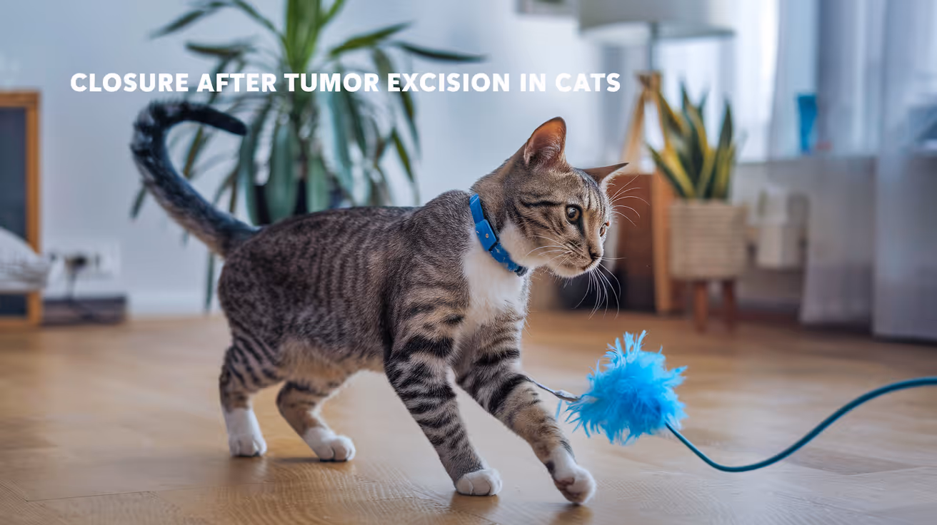
Closure Protocol for Tumor Excision in Cats
Learn the detailed closure protocol for tumor excision in cats, including surgical steps, suture choices, and post-op care for optimal healing.
Tumor excision in cats requires careful closure to ensure proper healing and reduce complications. The closure protocol involves selecting the right sutures, closing tissue layers correctly, and managing post-operative care effectively.
This article explains the step-by-step closure process after tumor removal in cats. You will learn about suture materials, techniques, and aftercare to help your cat recover safely and comfortably.
What is the importance of proper closure after tumor excision in cats?
Proper closure after tumor excision is critical to prevent infection, minimize scarring, and promote fast healing. Cats have delicate skin and tissues that require gentle handling during closure.
Incorrect closure can lead to wound dehiscence, delayed healing, or recurrence of the tumor. Understanding the closure protocol helps ensure the best surgical outcome for your cat.
- Prevents infection: Closing the wound properly seals the surgical site, reducing bacterial entry and lowering infection risk.
- Supports tissue healing: Correct alignment of tissue layers allows natural healing processes to occur efficiently without tension.
- Reduces scarring: Careful suturing minimizes tissue trauma and scarring, improving cosmetic results.
- Prevents wound opening: Secure closure prevents the wound from reopening, which can cause complications and require additional surgery.
Following a strict closure protocol is essential for your cat’s recovery and long-term health after tumor excision.
What suture materials are best for closing tumor excision wounds in cats?
Choosing the right suture material is important for wound strength and healing. Different materials have specific properties suited for various tissue layers in cats.
Veterinarians typically select absorbable sutures for internal layers and non-absorbable or absorbable sutures for skin closure depending on the case.
- Absorbable sutures: Materials like polydioxanone (PDS) or poliglecaprone (Monocryl) dissolve over time, ideal for internal tissue layers to avoid suture removal stress.
- Non-absorbable sutures: Nylon or polypropylene sutures provide strong skin closure but require removal 10-14 days post-op to prevent irritation.
- Monofilament sutures: These have smooth surfaces that reduce tissue drag and lower infection risk compared to braided sutures.
- Suture size selection: Smaller sizes (4-0 or 5-0) are preferred for delicate feline skin to minimize trauma and promote fine healing.
Using appropriate suture materials tailored to tissue type improves healing outcomes and reduces complications.
How should the different tissue layers be closed after tumor excision in cats?
Wound closure after tumor removal involves multiple tissue layers, each requiring specific techniques. Proper layer-by-layer closure restores tissue integrity and function.
Common layers include the deep fascia, subcutaneous tissue, and skin. Each layer has distinct closure needs to ensure strength and healing.
- Deep fascia closure: Use absorbable sutures in a simple interrupted or continuous pattern to reapproximate muscle and fascia layers securely.
- Subcutaneous tissue closure: Close with absorbable sutures in a simple continuous pattern to reduce dead space and support skin edges.
- Skin closure: Use non-absorbable or absorbable sutures in interrupted or cruciate patterns to align skin edges precisely and minimize tension.
- Consider tension relief: Use tension-relieving sutures or skin flaps if the wound edges are tight to prevent dehiscence.
Following this layered closure approach ensures a strong, well-vascularized wound that heals efficiently.
What are the best surgical techniques for skin closure in cats after tumor excision?
Skin closure technique affects wound healing quality and cosmetic appearance. Cats have thin, fragile skin that requires gentle handling and precise suturing.
Choosing the right technique depends on wound size, location, and tension. Proper technique reduces complications like suture reactions or wound opening.
- Simple interrupted sutures: Provide strong, adjustable closure with good edge eversion, suitable for most skin wounds in cats.
- Cruciate sutures: Crossed pattern sutures that distribute tension evenly and reduce skin edge inversion, improving healing.
- Subcuticular sutures: Placed just under the skin surface, these absorbable sutures offer cosmetic closure without external stitches.
- Staples or glue: Occasionally used for small wounds but less common in cats due to skin fragility and movement.
Using the correct skin closure technique tailored to the wound promotes faster healing and reduces scarring.
How should post-operative care be managed after tumor excision closure in cats?
Post-operative care is vital to protect the surgical site and support healing after tumor excision closure. Proper care reduces infection risk and promotes comfort.
Owners must follow veterinary instructions carefully to ensure the best recovery for their cat.
- Prevent licking or scratching: Use an Elizabethan collar to stop the cat from disturbing sutures and causing wound damage.
- Keep the wound clean and dry: Avoid bathing or wetting the area until sutures are removed and healing is confirmed.
- Monitor for complications: Watch for redness, swelling, discharge, or opening of the wound and report concerns to the vet immediately.
- Administer medications: Follow prescribed antibiotics or pain medications exactly as directed to control infection and discomfort.
Careful post-op management helps your cat heal quickly and reduces the chance of complications after tumor excision closure.
What complications can occur from improper closure after tumor excision in cats?
Improper closure of tumor excision wounds can lead to several complications that affect healing and the cat’s health. Recognizing these risks highlights the importance of following closure protocols.
Complications can range from mild to severe and may require additional treatment or surgery.
- Wound dehiscence: The surgical site reopens due to poor suture technique or excessive tension, delaying healing and increasing infection risk.
- Infection: Improper closure allows bacteria to enter the wound, causing redness, swelling, pain, and possible abscess formation.
- Seroma or hematoma: Fluid or blood accumulation under the skin occurs if dead space is not closed properly, causing swelling and discomfort.
- Excessive scarring: Poor tissue alignment or tension can lead to thick, raised scars that affect skin flexibility and appearance.
Following the correct closure protocol minimizes these risks and ensures a smooth recovery for your cat.
Conclusion
Closure protocol for tumor excision in cats is a critical part of surgery that ensures proper healing and reduces complications. Selecting the right sutures, closing tissue layers carefully, and managing post-op care are essential steps.
By understanding and following these closure guidelines, you can help your cat recover safely and comfortably after tumor removal. Always consult your veterinarian for personalized advice and support throughout the healing process.
FAQs
How long do sutures stay in after tumor excision in cats?
Skin sutures typically stay in place for 10 to 14 days. Absorbable sutures inside the tissue dissolve on their own and do not require removal.
Can I bathe my cat after tumor excision surgery?
Bathing should be avoided until the wound is fully healed and sutures are removed to prevent infection and wound opening.
What signs indicate infection at the surgical site?
Look for redness, swelling, heat, discharge, foul odor, or increased pain around the wound. Contact your vet if these occur.
Is an Elizabethan collar necessary after tumor excision?
Yes, it prevents your cat from licking or scratching the wound, which can cause damage or infection during healing.
When should I contact the vet after tumor excision closure?
Contact your vet immediately if the wound opens, bleeds excessively, shows signs of infection, or if your cat is lethargic or in pain.
Sustainable Vet | Copyright ©2026 | All Rights Reserved

